Waking Up Silent Code: Modern Methods for Activating Cryptic Prokaryotic Gene Clusters in Drug Discovery
This article provides a comprehensive overview of contemporary strategies for activating cryptic biosynthetic gene clusters (BGCs) in prokaryotes, with a focus on applications in natural product discovery and drug development.
Waking Up Silent Code: Modern Methods for Activating Cryptic Prokaryotic Gene Clusters in Drug Discovery
Abstract
This article provides a comprehensive overview of contemporary strategies for activating cryptic biosynthetic gene clusters (BGCs) in prokaryotes, with a focus on applications in natural product discovery and drug development. It covers the foundational principles of genome mining, details a suite of genetic, chemical, and synthetic biology methods for cluster activation, addresses common troubleshooting and optimization challenges, and outlines validation and comparative frameworks for evaluating success. Tailored for researchers, scientists, and drug development professionals, this guide synthesizes the latest advances—including CRISPR-based tools like ACTIMOT and ribosome engineering—to equip the scientific community with practical knowledge for unlocking the vast hidden biosynthetic potential of bacteria.
The Hidden Treasure: Understanding Cryptic Gene Clusters and Their Potential
Defining Cryptic Biosynthetic Gene Clusters (BGCs) in Prokaryotes
Frequently Asked Questions (FAQs)
1. What is the fundamental difference between 'cryptic' and 'silent' BGCs? The terms are often used interchangeably, but a precise definition is crucial for clear scientific communication. A silent biosynthetic gene cluster refers specifically to a BGC that is not expressed under standard laboratory conditions [1]. In contrast, a cryptic biosynthetic gene cluster describes a BGC for which the encoded natural product is hidden or unknown [1]. This can occur in two main scenarios: either a natural product has been observed but its cognate BGC has not been identified, or a BGC has been expressed but its predicted product cannot be detected under laboratory conditions [1].
2. Why is activating cryptic BGCs important for drug discovery? Microbial natural products underpin the majority of clinically used antibiotics [1]. Genome sequencing has revealed that prolific producers, like filamentous actinobacteria, typically harbor 20 to 50 natural product BGCs, but express very few under standard lab conditions [1] [2]. This vast reservoir of unexpressed biochemical diversity represents a promising source for novel therapeutic leads, potentially ushering in a new era of antibiotic discovery to combat antimicrobial resistance [1] [3].
3. What are the first experimental steps when a cryptic BGC is identified bioinformatically? After identifying a BGC of interest, the initial step is often to attempt to induce its expression in the native host. A common and straightforward first approach is to manipulate culture conditions (the OSMAC approach), which can include varying media composition, aeration, or the addition of chemical elicitors [4] [5]. Simultaneously, you should analyze the BGC's genetic structure for pathway-specific regulators that can be genetically manipulated, such as by promoter replacement [4].
4. When should I consider heterologous expression for a cryptic BGC? Heterologous expression is a powerful strategy when the native producer is difficult to cultivate genetically, when specific activation in the native host fails, or when you need to simplify the genetic background for easier product detection [2]. This involves transferring the entire BGC into a well-characterized and genetically tractable host like Streptomyces coelicolor or S. lividans [2] [5]. The main disadvantage is that it can be technically challenging to clone large BGCs, and the biosynthetic machinery may not function optimally in a foreign cellular environment [2].
Troubleshooting Guides
Problem: BGC Shows No Expression Under Standard Lab Conditions
Potential Causes and Solutions:
Cause 1: Repressive regulation.
- Solution: Identify and disrupt genes encoding transcriptional repressors within or outside the BGC. Alternatively, introduce chemical inducers. For example, in Burkholderia thailandensis, the global repressor MftR controls several BGCs, and its inhibition by urate can activate these clusters [6].
- Protocol: To test for urate induction, cultivate the producer strain in a suitable medium supplemented with a physiologically relevant concentration of urate (e.g., 200 µM to 5 mM). Use RNA sequencing or a reporter gene system to monitor BGC expression [6].
Cause 2: Lack of the necessary environmental trigger.
- Solution: Employ co-cultivation. Cultivate the producer strain in the presence of another microbe to simulate ecological interactions.
- Protocol:
- Select an interaction partner (e.g., from a library of actinomycetes).
- Co-culture the strains on solid agar, either in direct contact or separated by a membrane (to determine if physical contact is required).
- Use HPLC-MS or imaging mass spectrometry to compare the metabolome of the co-culture with monoculture controls [4].
Cause 3: Incompatible culture medium.
- Solution: Systematically alter cultivation parameters using the OSMAC (One Strain Many Compounds) approach.
- Protocol: Ferment the same strain in at least 5-10 different media with varying carbon/nitrogen sources, pH, and salinity. Extract metabolites after different incubation periods and analyze by HPLC-MS to detect condition-specific metabolites [4] [5].
Problem: BGC is Expressed, but No Expected Product is Detected
Potential Causes and Solutions:
Cause 1: The product is truly novel and falls outside standard detection parameters.
- Solution: Use advanced, untargeted metabolomics.
- Protocol: Perform high-resolution LC-MS/MS analysis of the culture extract. Use computational tools like molecular networking (e.g., with GNPS) to visualize the entire metabolome and identify unique ions that are upregulated when the BGC is activated. This can help prioritize unknown features for isolation [7].
Cause 2: The cluster requires specific precursors or cofactors not present in the medium.
- Solution: Supplement the medium with predicted precursors.
- Protocol: Based on the bioinformatic prediction of the natural product class (e.g., PKS, NRPS), supplement the fermentation medium with potential starter units or amino acid precursors. For example, feed isotope-labeled precursors (e.g., ¹³C-acetate) and use isotope-guided fractionation to track the cryptic metabolite [4].
Problem: Native Producer is Uncultivable or Genetically Intractable
Potential Causes and Solutions:
- Cause: Inability to manipulate the host or achieve adequate growth.
- Solution:
- Ribosome Engineering: Isolate spontaneous antibiotic-resistant mutants. This is a genetics-light approach that alters cellular physiology.
- Protocol: Plate a dense cell suspension on agar containing a sub-lethal concentration of streptomycin (to target ribosomal protein S12) or rifampicin (to target RNA polymerase). Screen resistant mutants for enhanced or new antibiotic production [5].
- Heterologous Expression: Clone the entire BGC into a model host.
- Protocol: This is a multi-step process: (a) Clone the large BGC into a bacterial artificial chromosome (BAC) or use transformation-associated recombination (TAR) in yeast. (b) Introduce the assembled construct into a heterologous host such as S. lividans or S. albus via intergeneric conjugation. (c) Screen exconjugants for production of the target compound [2] [4].
- Ribosome Engineering: Isolate spontaneous antibiotic-resistant mutants. This is a genetics-light approach that alters cellular physiology.
- Solution:
Experimental Protocols for Key Methodologies
Protocol 1: Ribosome Engineering via Streptomycin Selection
This protocol outlines a method to generate antibiotic-overproducing mutants in Streptomyces by inducing ribosomal mutations [5].
- Culture Preparation: Grow the target Streptomyces strain in a rich liquid medium until late-exponential phase.
- Mutant Selection: Plate a concentrated suspension of spores or mycelium onto agar plates containing streptomycin at a concentration of 2-10 µg/mL (this must be pre-determined as the minimal concentration that inhibits growth).
- Isolation: Incubate plates until spontaneous resistant mutants form colonies (typically 3-7 days).
- Fermentation and Screening: Inoculate the mutant strains into production media and cultivate alongside the wild-type strain.
- Analysis: Extract metabolites from culture broths and analyze via HPLC-MS or bioassay against a sensitive indicator strain to identify mutants with activated or enhanced antibiotic production.
- Validation: Confirm the mutation by sequencing the rpsL gene (encoding ribosomal protein S12), where a common Lys-88 to Glu or Arg mutation is often responsible [5].
Protocol 2: Reporter-Guided Mutant Selection (RGMS)
This protocol uses a fluorescent or antibiotic resistance reporter to screen for mutants where a cryptic BGC has been activated [2].
- Reporter Construction: Fuse the promoter of a key biosynthetic gene from the target BGC to a reporter gene (e.g., gfp for fluorescence or neo for kanamycin resistance) on a plasmid.
- Strain Engineering: Introduce the reporter construct into the wild-type producer strain.
- Mutagenesis: Create a random mutant library of the engineered reporter strain using UV mutagenesis or transposon (Tn) insertion.
- Mutant Selection: Screen or select for mutants based on the reporter signal. For Tn mutagenesis, the insertion site can be easily mapped to identify the inactivated gene responsible for activation.
- Metabolite Analysis: Ferment the selected mutants and analyze their metabolome to discover the cryptic natural product(s) produced by the activated BGC [2].
Research Reagent Solutions
Table 1: Essential Reagents for Cryptic BGC Activation Research
| Reagent / Tool | Function / Application | Key Considerations |
|---|---|---|
| antiSMASH [1] [8] | Bioinformatic tool for BGC identification and annotation. | The primary tool for initial BGC discovery; results should be manually curated. |
| Streptomycin / Rifampicin [5] | Antibiotics for ribosome engineering selection. | Use sub-inhibitory concentrations to select for spontaneous resistant mutants with altered metabolism. |
| Urate (Sodium Salt) [6] | Chemical inducer for MftR-regulated BGCs in Burkholderia. | A physiologically relevant signaling molecule encountered during host infection. |
| HDAC Inhibitors (e.g., Suberoylanilide hydroxamic acid) [4] | Epigenetic modifiers to alter chromatin structure and activate silent genes in fungi. | Can lead to global metabolic changes, not just activation of a single target cluster. |
| Heterologous Hosts (e.g., S. coelicolor, S. lividans) [2] [5] | Genetically tractable platform strains for BGC expression. | Choose a host with a minimized native metabolome to reduce background interference. |
| Bacterial Artificial Chromosome (BAC) Vectors [2] | Cloning system for large DNA fragments (>100 kb). | Essential for capturing and transferring entire BGCs for heterologous expression. |
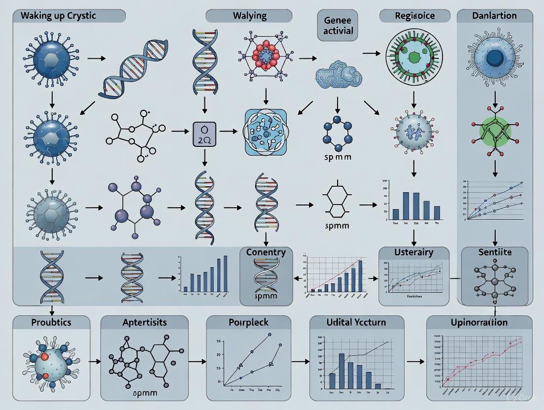
Visualization of Workflows and Relationships
Diagram 1: BGC Classification and Experimental Strategy
Diagram 2: Cryptic BGC Activation Workflow
In the genomes of prokaryotes, and most famously in gifted actinomycetes, lies a vast, untapped treasure trove of potential new natural products, including novel antibiotics. Bioinformatic analyses of sequenced microbial genomes routinely reveal a remarkably large number of biosynthetic gene clusters (BGCs)—sets of genes responsible for the synthesis of a natural product—for which the corresponding metabolites are unknown [4] [9]. These are known as orphan clusters. A significant subset of these clusters are "silent" or "cryptic," meaning they are not expressed, or are expressed only at very low levels, under standard laboratory growth conditions [4] [10].
The silent nature of these BGCs presents a major challenge and opportunity for natural product discovery. It is estimated that silent clusters outnumber the constitutively active ones by a factor of 5–10, suggesting a hidden realm of microbial chemistry waiting to be discovered [9]. Unlocking these clusters is critical for accessing new therapeutic leads and for understanding microbial chemical ecology [9].
FAQs: Understanding Silent Gene Clusters
Q1: What is the fundamental genomic reason why a gene cluster is "silent"? A1: A gene cluster is classified as silent when its genes are not transcribed, or are transcribed at very low levels, under typical laboratory fermentation conditions. This is not due to a defect in the DNA sequence but because the chemical or environmental signals necessary for triggering the pathway are absent in the lab setting [4]. The cluster is essentially in a state of transcriptional repression.
Q2: Beyond the absence of a trigger, what are the specific molecular mechanisms enforcing this silence? A2: Research has identified several key regulatory mechanisms that can silence a BGC:
- Lack of Cluster-Specific Activation: Many BGCs contain genes for pathway-specific transcription factors. In the absence of the required inducing signal, these activators are not produced, keeping the entire cluster off [4].
- Epigenetic Silencing: In eukaryotic microbes like fungi, DNA can be packaged into a closed chromatin structure through modifications like histone deacetylation. This structure makes the DNA physically inaccessible to the transcription machinery, effectively silencing the genes within that region [4].
- Global Regulatory Control: Master regulatory proteins, such as LaeA in fungi, control the expression of many secondary metabolite gene clusters simultaneously. Deletion or disruption of such global regulators can lead to the silencing of multiple BGCs [4].
- Ribosomal and Transcriptional Fidelity: In bacteria, the efficiency of transcription and translation can globally impact secondary metabolism. Mutations in ribosomal proteins or RNA polymerase can surprisingly lead to the activation of silent clusters, indicating that the native state of these machineries can contribute to their repression [4].
Q3: Are there bioinformatic tools to identify silent clusters? A3: Yes. The primary method is genome mining. Sequencing a microbial genome allows researchers to use bioinformatic tools (e.g., antiSMASH) to scan for hallmark genes of secondary metabolism, such as polyketide synthases (PKSs) and nonribosomal peptide synthetases (NRPSs). Any identified BGC for which no product is detected under laboratory growth conditions is a candidate silent or orphan cluster [4] [11].
Q4: What is the evolutionary advantage for a microbe to maintain silent gene clusters? A4: It is generally accepted that secondary metabolites provide a biological advantage in response to the environment, for instance, to compete against other organisms or to respond to specific stresses [4] [9]. Maintaining silent clusters allows a microbe to retain a large "chemical arsenal" that can be deployed only when needed, which is likely more energy-efficient than constitutively producing all possible compounds. The clusters are often evolutionarily conserved and can be horizontally transferred, facilitating the spread of these adaptive functions [10].
Troubleshooting Guide: Diagnosing and Overcoming Silence
When your experiment to express a silent BGC fails, follow this guide to diagnose and address the problem.
Common Failure Modes and Corrective Actions
| Problem Area | Specific Failure Mode | Recommended Corrective Action |
|---|---|---|
| Genetic Manipulation | Failed promoter replacement or heterologous expression. | Optimize transformation protocol; use CRISPR-Cas9 for more precise editing; verify promoter strength and compatibility in the host [9]. |
| Culture Conditions | Standard lab media do not induce the cluster. | Employ the OSMAC (One Strain-Many Compounds) approach: systematically vary media composition, aeration, and culture vessel type [4]. |
| Environmental Cues | Missing biological or chemical interactions. | Use co-culture: cultivate the producer strain with other microorganisms to simulate natural competition and interaction [4]. |
| Epigenetic Block | Closed chromatin structure (in eukaryotes). | Cultivate the strain in the presence of histone deacetylase (HDAC) inhibitors (e.g., suberoylanilide hydroxamic acid) to open chromatin [4]. |
| Regulatory Complexity | The cluster is under complex, multi-layer repression. | Combine strategies: use "ribosome engineering" (select for antibiotic-resistant mutants) to introduce global regulatory changes while also optimizing culture conditions [4]. |
Advanced Experimental Protocols
Protocol 1: Heterologous Expression of a Silent BGC
- Principle: Clone the entire silent BGC and express it in a genetically tractable host (e.g., Streptomyces coelicolor or Aspergillus oryzae).
- Methodology:
- Identify and clone the target BGC, often using BAC (Bacterial Artificial Chromosome) or TAR (Transformation-Associated Recombination) cloning.
- Introduce the cloned cluster into a suitable heterologous host via transformation.
- If the native promoters are weak, replace them with strong, inducible promoters functional in the host.
- Screen for metabolite production in the heterologous host through LC-MS or biological activity assays [4].
Protocol 2: Promoter Replacement via CRISPR-Cas9
- Principle: Precisely replace the native promoter of a silent BGC with a strong, constitutive promoter to drive expression.
- Methodology:
- Design a CRISPR guide RNA (gRNA) to target the sequence immediately upstream of the BGC's first gene.
- Construct a donor DNA template containing the new promoter.
- Co-transform the gRNA, Cas9 protein, and donor DNA into the producer strain.
- Screen for successful promoter replacement via antibiotic selection or PCR [9].
Protocol 3: High-Throughput Elicitor Screening (HiTES)
- Principle: Identify small molecule "elicitors" that naturally induce the silent cluster.
- Methodology:
- Integrate a reporter gene (e.g., GFP, lacZ) into the silent BGC to provide an expression read-out.
- Expose the reporter strain to libraries of small molecules, such as supernatants from other microbes.
- Use high-throughput screening (e.g., fluorescence activation) to identify molecules that induce the reporter.
- Isolate and characterize the inducing molecule, then use it to activate the cluster in the wild-type strain [9].
Key Signaling Pathways and Regulatory Logic
The decision to activate a silent gene cluster often integrates multiple environmental and internal signals. The following diagram illustrates the core regulatory logic and major pathways that control the expression of silent biosynthetic gene clusters.
The Scientist's Toolkit: Key Research Reagents
| Research Reagent | Function in Waking Up Silent Clusters |
|---|---|
| HDAC Inhibitors (e.g., Suberoylanilide hydroxamic acid) | Blocks histone deacetylase activity, leading to an open chromatin configuration and activation of epigenetically silenced clusters in fungi [4]. |
| DNMT Inhibitors (e.g., 5-Azacytidine) | Inhibits DNA methyltransferases, preventing DNA methylation and potentially reactivating genes silenced by this mechanism [4]. |
| CRISPR-Cas9 System | Enables precise genome editing for promoter replacement, activation, or deletion of repressors within silent BGCs in genetically tractable organisms [9]. |
| Inducible Promoters (e.g., tetO, PtipA) | Used in genetic constructs to place the expression of a cluster-specific transcription factor or the entire BGC under external control (e.g., by adding an antibiotic) [4] [9]. |
| Antibiotics for Ribosome Engineering (e.g., Streptomycin, Rifampicin) | Selection on sub-inhibitory concentrations yields mutants with altered ribosomal protein S12 or RNA polymerase, leading to pleiotropic activation of silent metabolism [4]. |
The explosion of microbial genome sequencing has revealed a hidden treasure trove of biosynthetic gene clusters (BGCs) – sets of genes responsible for producing natural products like antibiotics, antifungals, and other bioactive compounds. In prolific secondary metabolite producers, these silent or cryptic BGCs outnumber the constitutively active ones by a factor of 5-10, representing an immense, largely untapped resource for drug discovery [9]. Bioinformatics genome mining, defined as the computational analysis of nucleotide sequence data based on the comparison and recognition of conserved patterns, provides the key to unlocking this potential [12]. This technical support center addresses the practical challenges researchers face when using antiSMASH and other genome mining tools to identify hidden BGCs, providing troubleshooting guidance and methodological frameworks to advance cryptic gene cluster research.
Frequently Asked Questions (FAQs)
What is genome mining and what can it discover? Genome mining involves analyzing genomes with specialized algorithms to find BGCs that encode diverse natural products. These include polyketides (PKs), non-ribosomal peptides (NRPSs), ribosomally synthesized and post-translationally modified peptides (RiPPs), terpenes, saccharides, and alkaloids [12]. Each class has distinct biosynthetic machinery and potential pharmaceutical applications, from antibiotics like erythromycin to immunosuppressants like cyclosporine [12].
Why should I use antiSMASH over other tools? antiSMASH (antibiotics and Secondary Metabolite Analysis Shell) is one of the most comprehensive tools for identifying and annotating secondary metabolite gene clusters across both bacterial and fungal genomes [13]. It provides integrated analysis of multiple BGC types, comparative genomics features against the MIBiG database, and predictive structural biology insights that make it particularly valuable for initial genome surveys [14] [13].
Can antiSMASH detect all types of gene clusters? No. antiSMASH specifically targets secondary metabolite BGCs and does not detect clusters involved in primary metabolism, such as those for fatty acid or cofactor biosynthesis [15]. If you believe a true secondary metabolite BGC is escaping detection, the antiSMASH developers encourage researchers to contact them to add the necessary detection models [15].
What input formats does antiSMASH support? antiSMASH accepts both annotated genomes in GenBank or EMBL format and unannotated genome sequences in FASTA format. For FASTA inputs, it offers integrated gene prediction using Prodigal or GlimmerHMM, though using a dedicated annotation pipeline like RAST first typically yields higher-quality results [15].
How are antiSMASH results organized and interpreted? antiSMASH results are organized by regions containing detected gene clusters. The visualization shows genes color-coded by function (biosynthetic genes in red, transport-related in blue, regulatory in green) [14]. Key elements include protoclusters (core biosynthetic machinery with neighborhoods) and candidate clusters (which may contain multiple protoclusters and are useful for identifying hybrid systems) [16]. The tool also provides comparisons with known clusters in the MIBiG database to help characterize novelty [16].
Troubleshooting Common antiSMASH Issues
Input File Problems
Error: "All records skipped" or "All input records smaller than minimum length"
- Causes: Records shorter than the 1,000 nucleotide default minimum length [17] or records containing no gene features when submitting GBK files to the web service [17].
- Solutions:
- Ensure at least one record exceeds the minimum length requirement
- Run genefinding tools prior to submission and verify genes are annotated as
CDSfeatures (notgenefeatures, which don't contain sufficient information) [17] - Adjust the
--minlengthparameter in the stand-alone version to suit your input data [17]
Error: "Multiple CDS features have the same name for mapping"
- Cause: Duplicate identifiers in CDS features, which may cause misleading results [17].
- Solution: Locate features with the duplicated identifier provided in the error message and either remove exact duplicates or change identifiers to be unique [17].
Error: "Record contains no sequence information"
- Cause: Valid record identifiers and annotations are present but no actual sequence data is included [17].
- Solution: Remove records lacking sequence information or include the missing sequence data in the input file [17].
General formatting errors in GenBank files
- Common Issues: Incorrectly formatted LOCUS lines, unusual character sets, or missing record terminators [17].
- Solutions:
- Ensure LOCUS lines are correctly formatted with all required fields
- Remove non-ASCII or non-UTF-8 characters
- Verify GBK files contain the record terminator
//at the end of each record [17]
Result Interpretation Challenges
Unexpectedly large gene clusters spanning multiple systems
- Cause: antiSMASH uses conservative border detection, sometimes including extra upstream/downstream genes rather than risk excluding key biosynthetic elements [15].
- Interpretation Guidance: Carefully examine comparative gene cluster analysis results (available in the "Homologous gene clusters" tab) to determine whether a large cluster represents a true hybrid or multiple separate clusters located close together [15].
Low similarity to known clusters in MIBiG
- Interpretation: This potentially indicates novel chemistry. The MIBiG comparison completion score is calculated as: Completion = (Number of genes with BLAST hits in both antiSMASH predicted region and MIBiG region) / (Number of all genes in the MIBiG curated region) [16].
- Recommended Action: Proceed with cluster characterization through additional analytical tools (e.g., PRISM, BAGEL, RODEO) depending on the BGC type [13].
Missing domain predictions or structural information
- Potential Causes:
- Running antiSMASH without all analysis modules enabled
- Low sequence similarity to characterized systems
- Novel biosynthetic mechanisms
- Solutions:
- Ensure all relevant prediction options are selected when configuring analysis
- Use complementary tools like PRISM for structural prediction of nonribosomal peptides, type I/II polyketides, and RiPPs [13]
- Consult specialized tools for specific BGC classes (e.g., BAGEL for bacteriocins and RiPPs, RODEO for RiPP precursor prediction) [13]
Experimental Protocols & Workflows
Standard Genome Mining Pipeline for BGC Discovery
Step 1: Sample Collection and DNA Extraction
- Extract high-quality genomic DNA from microbial cultures or environmental samples
- For metagenomic approaches, co-assemble reads from similar sample types using MEGAHIT with
--presets meta-sensitivefor improved BGC recovery [18]
Step 2: Genome Assembly and Quality Assessment
- Assemble quality-controlled reads using metaSPAdes (for individual samples) or MEGAHIT (for co-assembly) [18]
- Filter contigs (<1500 bp) using seqtk to reduce computational burden in downstream analysis [18]
- Assess assembly completeness and contamination with CheckM/CheckM2, retaining MAGs with ≥50% completeness and ≤10% contamination [18]
Step 3: BGC Prediction with antiSMASH
- Run antiSMASH with comprehensive parameters:
-taxon bacteria -genefinding-tool prodigal -cb-knownclusters -cc-mibig -cb-general -cb-subclusters -fullhmmer[18] - For bacterial genomes, use the "Bacteria" taxonomic classification option [19]
- Process results to extract key information: number of BGCs, types (NRPS, PKS, RiPPs, etc.), and locations
Step 4: Cluster Analysis and Prioritization
- Use BiG-SCAPE to analyze BGC families and classify antiSMASH outputs into gene cluster families with parameters:
--cutoffs 0.3 --include_singletons --mode auto[18] - Extract nucleotide sequences of promising BGCs from antiSMASH GBK outputs for further analysis [18]
- Query predicted compounds against specialized databases (MIBiG) using cheminformatic tools [19]
Step 5: Experimental Validation
- Select activation strategy based on genetic tractability of host (heterologous expression, promoter engineering, elicitor screening)
- For genetically amenable strains, consider CRISPR-Cas9 for promoter replacement [9]
- For non-model organisms, implement HiTES (high-throughput elicitor screening) to identify small molecule inducers [9]
Specialized Workflow for NRPS/PKS Clusters
Domain Analysis and Substrate Prediction
- For NRPS clusters: Identify adenylation (A), condensation (C), and peptidyl carrier protein (PCP) domains [14]
- For PKS clusters: Identify ketosynthase (KS), acyltransferase (AT), and ketoreductase (KR) domains [14]
- Predict A-domain specificities using NRPSPredictor2 and Minowa methods [14]
- Predict PKS AT-domain specificities using signature sequence analysis and pHMMs [14]
Structure Prediction and Analysis
- Generate predicted core structures by combining domain analysis results [14]
- Extract SMILES representations from antiSMASH results for chemical analysis and database queries [19]
- Important: Recognize that predictions represent core structures only - final molecules often undergo additional modifications [14]
Research Reagent Solutions
Table 1: Essential Bioinformatics Tools for BGC Discovery and Analysis
| Tool Name | Primary Function | Specific Applications | Reference |
|---|---|---|---|
| antiSMASH | BGC identification & annotation | Comprehensive detection of secondary metabolite BGCs; comparative analysis | [19] [13] |
| PRISM | Structural prediction of NPs | Prediction of NRPs, type I/II PKS, and RiPP chemical structures | [13] |
| BAGEL4 | RiPP & bacteriocin mining | Identification of ribosomally synthesized and post-translationally modified peptides | [13] |
| ARTS | Antibiotic target prediction | Genome mining based on antibiotic resistance targets for novel compound discovery | [13] |
| BiG-SCAPE | BGC classification & networking | Analysis of BGC families across multiple genomes; gene cluster family classification | [13] |
| RODEO | RiPP precursor prediction | Identification of biosynthetic gene clusters and prediction of RiPP precursor peptides | [13] |
| RiPPER | RiPP genome mining | Exploration of thioamidated ribosomal peptides in Actinobacteria | [13] |
Table 2: Key Databases and Resources
| Resource | Content Type | Applications in BGC Research | |
|---|---|---|---|
| MIBiG | Curated BGC database | Comparison of predicted clusters to experimentally characterized gene clusters | [19] [16] |
| NCBI SRA | Sequencing data repository | Access to raw sequencing data for metagenomic and transcriptomic analysis | [18] |
| GTDB | Taxonomic database | Standardized taxonomic classification of microbial genomes | [18] |
Advanced Applications: Activating Silent BGCs
CRISPR-Cas9 Promoter Insertion
- Replace native promoters of silent BGCs with constitutive or inducible variants in genetically tractable organisms [9]
- Particularly valuable for Streptomyces and other actinomycetes with complex genetics [9]
- Enables targeted activation of specific silent clusters for product characterization
High-Throughput Elicitor Screening (HiTES)
- Insert reporter genes (e.g., GFP) into silent BGCs to monitor expression [9]
- Screen libraries of small molecules to identify potential inducers of silent clusters [9]
- Effective for non-model organisms where genetic manipulation is challenging
Reporter-Guided Mutant Selection (RGMS)
- Combine genome-wide mutagenesis with reporter systems to select for hyperproducing mutants [9]
- Provides insights into regulatory networks controlling BGC expression
- Identifies indirect regulators that may silence multiple BGCs simultaneously
One Strain Many Compounds (OSMAC) Approach
- Systematic variation of cultivation parameters (media, temperature, aeration)
- Co-culture with potential microbial interactors to simulate ecological interactions
- Metatranscriptomic analysis to link BGC expression to specific growth conditions [18]
The end of the Golden Age of antibiotic discovery in the 1970s, marked by frequent rediscovery of known compounds, led to a critical gap in the antibiotic development pipeline [1]. However, the genomics revolution has revealed a vast, unexplored reservoir of potential novel drugs. Genome sequencing has shown that a single strain of filamentous Actinobacteria typically harbors 20 to 50 natural product biosynthetic gene clusters (BGCs), yet expresses very few under standard laboratory conditions [1]. This disparity highlights a universe of "hidden" biochemical diversity, with one study of 830 actinobacterial genomes identifying over 11,000 natural product BGCs representing more than 4,000 distinct chemical families [1]. Accessing these cryptic or silent clusters is now a primary imperative for modern drug discovery, offering a path to usher in a second Golden Era and combat the growing threat of antimicrobial resistance.
Defining the Terminology
Inconsistent use of the terms "cryptic" and "silent" has created confusion in the field. To ensure clarity:
- Cryptic describes BGCs and/or natural products that are hidden or unknown. This applies when a natural product has been observed but its cognate BGC has not been identified (Unknown Knowns), or when a BGC is expressed but its predicted product cannot be observed (Known Unknowns) [1].
- Silent describes BGCs that are not expressed under given laboratory conditions. Their expression may be induced by specific environmental cues or genetic manipulation [1].
Troubleshooting Guides & FAQs
This section addresses common experimental challenges in the activation and characterization of cryptic gene clusters.
FAQ 1: Why is my bacterial strain not producing the expected novel compound under standard laboratory culture conditions?
Answer: This is a fundamental challenge in the field. Your BGC of interest is likely silent under the conditions you are using. Standard laboratory media and conditions often do not replicate the precise environmental or physiological signals required to trigger the expression of these clusters [1]. Furthermore, the compound itself may be cryptic, meaning it is produced at levels below the detection limit of your analytical methods or is modified in a way that masks its detection.
FAQ 2: After confirming expression of the target BGC via RT-qPCR, I cannot detect the compound. What could be the cause?
Answer: This scenario points to a cryptic product. The issue likely lies downstream of gene expression. Possible explanations include:
- Post-translational Regulation: The biosynthetic enzymes may not be active.
- Rapid Degradation: The compound is produced but is chemically unstable or is being degraded by other enzymes in the culture.
- Sequestration: The compound is being bound to cellular components or exported and adsorbed to the culture vessel.
- Incorrect Structural Prediction: Bioinformatic tools may have mispredicted the final chemical structure of the compound, leading you to look for the wrong molecule [1].
FAQ 3: What is the most efficient strategy to prioritize which cryptic BGCs to investigate from genome mining data?
Answer: Prioritization should be based on a combination of bioinformatic and strategic factors:
- Novelty: Focus on BGCs that show low similarity to clusters encoding known compounds using tools like antiSMASH and the MIBiG database [1].
- Taxonomic Origin: Prioritize BGCs from under-explored or unique microbial genera isolated from novel environments.
- Presence of Resistance Genes: Co-localization of putative self-resistance genes can indicate biological activity and a specific cellular target.
- Evolutionary Mining: Use algorithms like EvoMining, which can identify BGCs that have been missed by standard prediction tools, potentially revealing entirely novel chemistries [1].
Troubleshooting Guide: No Detectable Product After Heterologous Expression
Problem: After cloning and expressing a target BGC in a heterologous host (e.g., Streptomyces coelicolor), no expected compound is detected.
Resolution: Follow this systematic troubleshooting process, adapted from general laboratory principles [20]:
Table: Troubleshooting Heterologous Expression
| Step | Possible Cause | Experimentation & Solution |
|---|---|---|
| 1. Identify Problem | Heterologous expression fails to produce compound. | Confirm the problem is specific to the compound, not host growth. |
| 2. List Explanations | - Cloning/Sequence Error- Poor Transcription- Poor Translation- Incompatible Host Physiology- Post-biosynthetic Modification | List all possible causes from start (DNA) to finish (compound). |
| 3. Collect Data | - Controls: Check growth of positive control host.- Storage: Verify integrity of genetic constructs.- Procedure: Review cloning and culture protocols. | Collect data on the easiest explanations first [20]. |
| 4. Eliminate & Experiment | - Sequence: Re-sequence the cloned cluster to check for errors.- Transcription: Use RT-qPCR to confirm mRNA is present.- Translation: Use Western blot (if antibodies are available) to check for enzyme production.- Host: Test different heterologous hosts or cultivation conditions. | Design experiments to test remaining explanations [20]. |
| 5. Identify Cause | The cause is the one remaining explanation after all others are eliminated. | Based on experimental results, implement a fix (e.g., re-clone, change host, optimize culture conditions) [20]. |
Experimental Protocols for Activation and Characterization
Protocol 1: OSMAC-Based Activation of Silent BGCs
The One Strain Many Compounds (OSMAC) approach is a fundamental method to induce the expression of silent BGCs by varying culture conditions [1].
Methodology:
- Media Variation: Cultivate the producer strain in a panel of 5-10 different liquid and solid media (e.g., R2A, ISP2, SFM, Xylose-Tryptone). Viate carbon and nitrogen sources.
- Physical Parameters: Incubate parallel cultures at different temperatures (e.g., 20°C, 28°C, 30°C, 37°C) and agitation speeds.
- Co-cultivation: Introduce another microbial strain (bacterial or fungal) onto the same agar plate or in a divided co-culture system to simulate microbial competition.
- Chemical Elicitors: Supplement media with sub-inhibitory concentrations of potential elicitors such as heavy metals, histone deacetylase inhibitors (e.g., sodium butyrate), or antibiotics.
- Extraction and Analysis: After 3-7 days of incubation, extract culture broths and mycelia with organic solvents like ethyl acetate or methanol-dichloromethane (1:1) [21]. Analyze extracts using HPLC-MS and compare chromatographic profiles across conditions to identify unique metabolites.
Protocol 2: Extraction and Bioactivity-Guided Fractionation
This protocol outlines the process for extracting and isolating a bioactive compound from a microbial culture.
Methodology:
- Extraction: Separate the culture broth from the biomass via centrifugation or filtration. Extract the broth separately with a polar solvent (e.g., ethyl acetate) and the biomass with a more non-polar solvent (e.g., dichloromethane/methanol 1:1) to cover a wide range of compound polarities [21].
- Concentration: Combine the organic extracts and concentrate them to dryness under reduced pressure using a rotary evaporator.
- Initial Phytochemical Screening: Perform preliminary thin-layer chromatography (TLC) on the crude extract, spraying with specific reagents (e.g., anisaldehyde for sugars, Dragendorff's for alkaloids) or viewing under UV light to gain initial information on the chemistry [21].
- Bioactivity-Guided Fractionation:
- Use preparative TLC or column chromatography (e.g., silica gel, Sephadex LH-20) to separate the crude extract into fractions.
- Test all fractions for the desired bioactivity (e.g., antimicrobial, anticancer).
- Take the active fraction and subject it to further, higher-resolution separation using techniques like HPLC.
- Repeat the cycle of separation and bioassay until a pure, active compound is isolated [21].
- Characterization: Identify the structure of the pure compound using spectroscopic methods including NMR (1D and 2D), high-resolution mass spectrometry (HRMS), and Fourier Transform Infrared Spectroscopy (FTIR) [21].
Research Reagent Solutions
Table: Essential Reagents for Cryptic Gene Cluster Research
| Reagent / Material | Function / Application |
|---|---|
| antiSMASH Software | The primary bioinformatic tool for the genomic identification and annotation of Biosynthetic Gene Clusters (BGCs) [1]. |
| Ethyl Acetate | A medium-polarity solvent optimally used for the extraction of a broad range of medium-polarity bioactive compounds from culture broth [21]. |
| Dichloromethane-Methanol (1:1) | A versatile, relatively non-polar solvent mixture used for the efficient extraction of compounds from microbial biomass [21]. |
| Silica Gel (for Column Chromatography) | A standard stationary phase for the primary fractionation of crude natural product extracts based on compound polarity [21]. |
| Sephadex LH-20 | A size-exclusion chromatography medium used for de-salting and fractionating extracts, particularly useful for separating compounds from pigments. |
| MIBiG Database | A curated database of known BGCs, used as a reference for comparing and prioritizing newly identified clusters based on novelty [1]. |
Experimental Workflows and Pathways
Diagram 1: Cryptic Cluster Research Workflow
Diagram 2: Bioactivity-Guided Fractionation
The Activation Toolkit: Genetic, Environmental, and Synthetic Biology Strategies
A significant challenge in modern natural product discovery is the prevalence of cryptic or silent biosynthetic gene clusters (BGCs) in prokaryotic genomes. Single Streptomyces genomes have been found to encode 25-50 BGCs, approximately 90% of which remain silent under standard laboratory cultivation conditions [22]. These clusters have the potential to encode novel antibiotics, anticancer agents, and other pharmaceuticals, but their silent nature poses a major barrier to discovery. Within the broader thesis on methods for awakening cryptic prokaryotic gene clusters, this technical support guide focuses specifically on in situ activation approaches—namely promoter engineering and transcription factor manipulation—to activate these silent genetic treasures within their native hosts.
Technical FAQs and Troubleshooting Guides
FAQ 1: What are the primary molecular mechanisms that keep a gene cluster silent?
Silent BGCs are typically transcriptionally repressed through complex regulatory networks. The key mechanisms include:
- Lack of Inducer Signal: Many clusters require specific environmental or chemical elicitors that are not present in standard lab media [23].
- Repressive Chromatin State: In some cases, DNA may be in a transcriptionally inactive conformation.
- Absence of Specific Activators: Essential pathway-specific transcription factors may not be expressed under test conditions.
- Presence of Specific Repressors: Clusters may be actively silenced by dedicated repressor proteins that bind to promoter regions and block transcription [22].
FAQ 2: When should I choose in situ activation over heterologous expression?
In situ activation is generally preferable when:
- The native host has complex or unknown growth requirements that are difficult to replicate in a heterologous system.
- The gene cluster is extremely large (>100 kb), making cloning and manipulation challenging.
- The biosynthetic pathway relies on host-specific primary metabolism or cofactors.
- You have limited knowledge of cluster boundaries.
Heterologous expression is advantageous when the native host is uncultivatable, grows extremely slowly, or lacks genetic tools [22].
Troubleshooting Guide 1: Low Activation Efficiency After Promoter Replacement
| Symptom | Possible Cause | Solution |
|---|---|---|
| No detectable expression | Incorrect promoter integration | Verify integration via PCR and sequencing |
| New promoter is too weak | Use a stronger constitutive promoter (e.g., ermE*P) | |
| Critical regulatory elements missing | Include native 5' UTR and RBS with the new promoter | |
| Low expression level | Suboptimal promoter strength | Test a library of promoters with varying strengths |
| Metabolic burden on host | Use inducible promoter to control expression timing | |
| Toxic effects on host | Constitutive expression of toxic proteins | Switch to a tightly regulated inducible system |
Troubleshooting Guide 2: Transcription Factor Manipulation Yields No Product
| Symptom | Possible Cause | Solution |
|---|---|---|
| No activation after TF overexpression | TF requires post-translational activation | Co-express potential modifying enzymes; add suspected effector molecules |
| TF is not the master regulator | Identify and co-express additional pathway-specific regulators | |
| Partial activation only | Insufficient TF expression level | Increase TF gene dosage; use stronger promoters |
| Multiple TFs regulate the cluster | Identify and manipulate all relevant regulators in the network | |
| Unpredictable results | TF has pleiotropic effects | Use cluster-specific TF mutants to avoid global regulation |
Core Methodologies and Experimental Protocols
Protocol 1: Promoter Replacement via CRISPR-Cas9
This protocol enables targeted replacement of native promoters with strong constitutive or inducible variants in the native host [22].
Materials Required:
- CRISPR-Cas9 system (plasmid expressing Cas9 and guide RNA)
- Donor DNA containing new promoter with homology arms (60-80 bp)
- Host-specific transformation materials
- Selection antibiotics
Step-by-Step Workflow:
Design gRNA: Select a 20-nucleotide guide RNA sequence immediately upstream of the native promoter region of the target gene.
Construct Donor DNA: Synthesize a linear donor DNA fragment containing:
- Strong promoter (e.g., ermEP, *kasO*p, or rpsLp)
- Appropriate 5' UTR and RBS sequences
- 60-80 bp homology arms flanking the target site
Transform Host: Co-transform the CRISPR-Cas9 plasmid and donor DNA into the native host.
Screen and Validate: Screen for successful recombinants via antibiotic selection and verify by colony PCR and sequencing.
Ferment and Analyze: Cultivate engineered strains under appropriate conditions and analyze metabolite production via LC-MS.
Protocol 2: Transcription Factor Activation Strategies
This protocol outlines multiple approaches to manipulate transcription factors for cluster activation [24] [22].
Materials Required:
- Vectors for TF overexpression (high-copy number plasmids)
- Chemicals for ribosome engineering (antibiotics)
- Potential elicitor molecules
Approach Selection Table:
| Method | Key Advantage | Typical Timeframe | Success Rate |
|---|---|---|---|
| TF Overexpression | Direct activation | 2-3 weeks | Variable (cluster-dependent) |
| Ribosome Engineering | Simple, no genetic manipulation required | 3-4 weeks | Moderate to high |
| Effector Addition | Non-genetic approach | 1-2 weeks | Low to moderate |
| Repressor Deletion | Removes negative regulation | 4-5 weeks | High (when repressor identified) |
Detailed Workflow for TF Overexpression:
Identify Target TF: Use bioinformatics tools to identify pathway-specific regulators within or near the target BGC.
Clone TF Gene: Amplify the TF coding sequence and clone into an expression vector with a strong, constitutive promoter.
Express TF in Native Host: Introduce the construct into the native host and confirm TF expression.
Analyze Metabolite Profile: Use HPLC and LC-MS to compare metabolite profiles of engineered versus wild-type strains.
Research Reagent Solutions
Essential materials and reagents for successful in situ activation experiments:
| Reagent Category | Specific Examples | Function & Application Notes |
|---|---|---|
| Expression Vectors | pIJ10257, pSET152, pKC1139 | Shuttle vectors for TF overexpression and promoter delivery [22] |
| Promoter Libraries | ermEP, *kasO*p, rpsLp, tipAp | Provide a range of expression strengths for fine-tuning |
| CRISPR Systems | pCRISPomyces series | Enable precise genome editing for promoter replacement [22] |
| Ribosome Engineering Antibiotics | Streptomycin, Rifampicin, Gentamicin | Select for ribosomal mutations that activate ppGpp-mediated stress response [23] |
| Chemical Elicitors | N-Acetylglucosamine, Rare Earth Elements | Mimic natural environmental signals to trigger cluster activation |
Signaling Pathways and Workflow Visualizations
Diagram 1: Transcription Factor Activation Mechanism
Diagram 2: Experimental Workflow for In Situ Activation
Quantitative Data and Performance Metrics
Table 1: Performance Comparison of In Situ Activation Methods
| Method | Typical Time Investment (weeks) | Relative Cost | Technical Difficulty | Success Examples |
|---|---|---|---|---|
| Promoter Replacement | 4-6 | $$-$$$ | High | Activation of jadomycin, pikromycin [22] |
| TF Overexpression | 3-5 | $-$$ | Medium | Activation of actinorhodin, streptomycin [23] |
| Repressor Deletion | 5-8 | $-$$ | Medium-High | Activation of scl BGC [22] |
| Ribosome Engineering | 3-4 | $ | Low | Activation of >20 cryptic clusters [23] |
Table 2: Common Constitutive Promoters for Streptomyces
| Promoter | Relative Strength | Origin | Key Features & Applications |
|---|---|---|---|
| ermE*P | High | Saccharopolyspora erythraea | Very strong, constitutive; ideal for strong overexpression [22] |
| rpsLp (XylR) | Medium-High | Mutant ribosomal protein S12 | Strong, constitutive; linked to ribosome engineering [23] |
| tipAp | Inducible | S. lividans | Thiostrepton-inducible; tight regulation when needed |
| kasO*p | Medium | S. coelicolor | Medium strength, constitutive; balanced expression |
In the field of natural product research, a significant challenge is that the vast majority of biosynthetic gene clusters (BGCs) in prokaryotes are silent or cryptic, meaning they are not expressed under standard laboratory conditions [9]. Heterologous expression—cloning and expressing BGCs in genetically tractable model hosts—has emerged as a powerful strategy to "wake up" these cryptic clusters. This approach bypasses the genetic intractability of native hosts and allows researchers to convert genetic potential into chemical reality, facilitating the discovery of novel compounds with potential pharmaceutical applications [25] [26]. This technical support center provides troubleshooting guides and detailed methodologies to overcome common obstacles in these experiments.
FAQs and Troubleshooting Guides
Q1: Why is my heterologous expression host not producing the expected natural product, even after successful BGC cloning?
This is a common challenge often stemming from issues with cluster recognition, expression, or functionality in the new host environment.
- A1: Potential Causes and Solutions:
- Incorrect Cloning or Missing Regulatory Elements: The cloned fragment might be incomplete or lack native promoters and regulatory genes essential for expression.
- Solution: Verify the integrity of the cloned insert by sequencing and confirm it contains all necessary genes, including potential regulatory elements. Consider using vectors with strong, constitutive, or inducible promoters (e.g., using CRISPR-Cas9 to insert a strong promoter upstream of the BGC) to drive expression [9].
- Incompatibility with the Heterologous Host: The host may lack necessary precursors, post-translational modification machinery, or compatible tRNA pools for efficient synthesis, or it may express proteases that degrade the heterologous enzymes.
- Solution: Screen a panel of different, well-characterized heterologous hosts (e.g., various Streptomyces species, Pseudomonas putida, Bacillus subtilis) to find one that is more compatible [25]. Consider engineering the host to supply limiting precursors or delete problematic proteases.
- Silent Cluster in the New Context: The BGC may remain silent even in the new host due to lack of specific induction signals.
- Solution: Employ strategies like High-throughput Elicitor Screening (HiTES), where a reporter gene is inserted into the BGC and small molecule libraries are screened for inducers of cluster expression [9]. Alternatively, use Reporter-Guided Mutant Selection (RGMS) to select for host mutants that activate the cluster [9].
- Incorrect Cloning or Missing Regulatory Elements: The cloned fragment might be incomplete or lack native promoters and regulatory genes essential for expression.
Q2: What can I do if I encounter difficulties cloning a large BGC (>50 kb) into a vector system?
Large BGCs are fragile and difficult to clone using traditional restriction enzyme-based methods.
- A2: Potential Causes and Solutions:
- Inefficient In Vitro Assembly: Large fragments are prone to breakage and can be difficult to ligate efficiently.
- Solution: Utilize Transformation-Associated Recombination (TAR) cloning in Saccharomyces cerevisiae. This method leverages the high homologous recombination efficiency of yeast to directly capture large genomic fragments (up to 300 kb) into a yeast artificial chromosome (YAC) vector [25]. Ensure the TAR vector contains:
- Homology arms (typically 200-500 bp) specific to the ends of the target BGC.
- A counter-selectable marker (e.g., URA3) to prevent high background from empty vector recircularization via non-homologous end joining [25].
- Appropriate origins of replication and selection markers for your downstream heterologous hosts.
- Solution: Utilize Transformation-Associated Recombination (TAR) cloning in Saccharomyces cerevisiae. This method leverages the high homologous recombination efficiency of yeast to directly capture large genomic fragments (up to 300 kb) into a yeast artificial chromosome (YAC) vector [25]. Ensure the TAR vector contains:
- Inefficient In Vitro Assembly: Large fragments are prone to breakage and can be difficult to ligate efficiently.
Q3: How do I choose the right vector and host system for my heterologous expression experiment?
The choice of vector and host is critical and depends on the source of the BGC and the desired product.
- A3: Selection Guide:
- For Actinobacterial BGCs (e.g., from Streptomyces): Use integration vectors like pCAP01 that contain a ΦC31 attP site for stable chromosomal integration into specific host strains [25].
- For Expression in Bacillus subtilis: Use vectors like pCAPB02, which is designed for integration into the B. subtilis chromosome via double-crossover recombination at the amyE locus, ensuring stable maintenance [25].
- For Gram-Negative Proteobacteria (e.g., Pseudomonas putida): Select shuttle vectors that are stable and can replicate in these hosts [25].
- General Consideration: For very large clusters, Bacterial Artificial Chromosomes (BACs), which can maintain inserts of 150-350 kbp in E. coli, are a valuable tool for initial cloning and storage before shuttling into the expression host [27] [28].
Research Reagent Solutions
The table below summarizes key reagents and vectors used in heterologous expression of BGCs.
Table 1: Essential Research Reagents for Heterologous BGC Expression
| Reagent/Vector Name | Type | Key Features & Function | Compatible Hosts |
|---|---|---|---|
| pCAP01/pCAP03 [25] | Shuttle Vector (YAC) | Contains ΦC31 attP for chromosomal integration; TAR cloning; URA3 for counter-selection (pCAP03). | S. cerevisiae (cloning), E. coli, Streptomyces spp. |
| pCAPB02 [25] | Integration Vector | Contains homology arms for amyE locus; allows stable integration via double-crossover. | S. cerevisiae (cloning), E. coli, Bacillus subtilis |
| BAC (Bacterial Artificial Chromosome) [27] [28] | Cloning Vector | Based on F-plasmid; stable maintenance of very large DNA inserts (150-350 kb). | E. coli |
| TAR Vector System [25] | Cloning Platform | Uses yeast homologous recombination for direct capture of large genomic fragments. | Saccharomyces cerevisiae |
| CRISPR-Cas9 System [9] | Genome Editing Tool | For precise promoter insertion upstream of silent BGCs to activate expression. | Various genetically tractable hosts |
Detailed Experimental Protocols
Protocol for TAR Cloning of a Biosynthetic Gene Cluster
This protocol outlines the method for capturing a large BGC directly from genomic DNA using Transformation-Associated Recombination in yeast [25].
- Principle: The method uses homologous recombination in S. cerevisiae to co-recombine genomic DNA fragments with linearized TAR vectors containing homologous ends, resulting in a circular yeast artificial chromosome containing the entire BGC.
Materials:
- TAR Vector (e.g., pCAP01 or pCAP03 [25])
- Genomic DNA from the source organism (high molecular weight)
- S. cerevisiae strain (e.g., VL6-48N)
- Spheroplasting solutions (sorbitol, zymolyase)
- Selective media (e.g., SD/-Trp, with 5-FOA if using pCAP03)
Step-by-Step Method:
- Design and Prepare TAR Vector:
- Design a TAR vector (e.g., pCAP01 derivative) containing 200-500 bp homology arms that match the 5' and 3' ends of the target BGC.
- Linearize the circular TAR vector using appropriate restriction enzymes to expose the homology arms. Gel-purify the linearized fragment.
Prepare Genomic DNA:
- Extract high-quality, high-molecular-weight genomic DNA from the source organism. Gently fragment the DNA by pipetting or brief sonication to an average size of 50-150 kb.
Prepare Yeast Spheroplasts:
- Grow a culture of the recipient yeast strain to mid-log phase.
- Treat the cells with zymolyase in a sorbitol buffer to enzymatically remove the cell wall, creating spheroplasts.
Co-transformation and Recombination:
- Mix the linearized TAR vector and the fragmented genomic DNA.
- Add the DNA mixture to the prepared yeast spheroplasts in the presence of PEG and calcium ions to facilitate DNA uptake.
- Allow homologous recombination to occur, which circularizes the vector and captures the BGC.
Selection and Screening:
- Plate the transformation mixture onto selective agar plates lacking tryptophan (to select for the TRP1 marker on the vector). If using pCAP03, include 5-FOA to counter-select against vectors that recircularized without an insert.
- Incubate plates for 3-5 days until colonies form.
Validation:
- Pick yeast colonies and screen for the presence of the correct recombinant by colony PCR.
- Isolate the Yeast Artificial Chromosome (YAC) DNA from positive clones and transform it into E. coli for amplification.
- Validate the final plasmid by restriction digest analysis and sequencing across the junctions.
Protocol for Activating Silent BGCs Using CRISPR-Cas9 Promoter Insertion
This protocol describes a genetic approach to activate a silent BGC by inserting a strong, constitutive promoter upstream of its biosynthetic genes [9].
- Principle: The CRISPR-Cas9 system creates a site-specific double-strand break near the target site of the silent BGC. A donor DNA template containing a strong promoter is provided, and the host's repair machinery incorporates it via homologous recombination, leading to constitutive expression of the downstream genes.
Materials:
- CRISPR-Cas9 system (plasmid expressing Cas9 and the sgRNA, or a ribonucleoprotein complex)
- Donor DNA template containing a strong promoter (e.g., ermE*p) flanked by homology arms (~1 kb) matching the sequence upstream and downstream of the Cas9 cut site.
- Standard molecular biology reagents for transformation and selection.
Step-by-Step Method:
- Target Selection and gRNA Design:
- Identify a target site for Cas9 immediately upstream of the core biosynthetic gene (e.g., the NRPS or PKS gene) of the silent BGC.
- Design and synthesize a single-guide RNA (sgRNA) specific to this target site.
Donor DNA Construction:
- Synthesize a linear donor DNA fragment containing a strong, constitutive promoter. This fragment must be flanked by homology arms (typically 500-1000 bp) that are homologous to the regions immediately upstream and downstream of the planned Cas9 cut site.
Delivery and Transformation:
- Introduce the CRISPR-Cas9 system (as DNA or pre-assembled ribonucleoprotein complexes) and the donor DNA fragment into the host organism using the appropriate method (e.g., protoplast transformation, electroporation for actinomycetes).
Selection and Screening:
- Allow the cells to recover and then plate them on medium containing the appropriate antibiotic to select for cells that have incorporated the donor DNA (which should include a selectable marker).
- Screen the resulting colonies for correct promoter insertion using colony PCR and confirm by DNA sequencing.
Metabolite Analysis:
- Ferment the engineered strain alongside the wild-type control under various conditions.
- Analyze the culture extracts using HPLC or LC-MS to detect the production of new secondary metabolites that are specific to the engineered strain.
The table below compares several key strategies for accessing the products of silent biosynthetic gene clusters, extending beyond heterologous expression.
Table 2: Strategies for Activation of Silent Biosynthetic Gene Clusters
| Method | Principle | Key Advantages | Common Challenges |
|---|---|---|---|
| Heterologous Expression [25] | Cloning and expressing BGCs in a genetically tractable model host. | Bypasses host-specific regulation; applicable to uncultured microbes. | Cloning large fragments; host compatibility (precursors, folding). |
| CRISPR-Cas9 Promoter Insertion [9] | Site-specific insertion of a strong promoter to drive BGC expression. | Precise and targeted; can be applied to native hosts. | Requires genetic tractability and efficient transformation. |
| High-Throughput Elicitor Screening (HiTES) [9] | Using a reporter system to screen libraries of small molecules for BGC inducers. | Does not require genetic manipulation; can reveal ecological signals. | Requires construction of a reporter strain; hit molecules may be unknown. |
| Reporter-Guided Mutant Selection (RGMS) [9] | Coupling random mutagenesis with a reporter to select for activating mutants. | Can reveal novel regulatory genes and mechanisms. | Mutations may be complex and difficult to characterize. |
| Ribosome Engineering | Using sub-inhibitory concentrations of antibiotics to perturb cellular physiology and activate secondary metabolism. | Simple to implement; can be highly effective. | Mechanism is indirect and often strain-specific. |
Troubleshooting Guide: FAQs on Ribosome Engineering
FAQ 1: My bacterial strains treated with sub-inhibitory antibiotics are not showing activated cryptic gene clusters. What could be wrong?
- Potential Cause & Solution: The concentration of the inducing antibiotic is critical. Using a concentration that is too high will kill the cells, while a concentration that is too low will not generate the selective pressure needed to induce resistance mutations in ribosomal components.
- Protocol Adjustment: Perform a preliminary experiment to determine the sub-inhibitory concentration of antibiotics like streptomycin, rifampicin, or paromomycin for your specific bacterial strain. This is the highest concentration of antibiotic that does not visibly inhibit growth. Treatment with this concentration can induce mutations in genes like
rpsL(ribosomal protein S12) orrpoB(RNA polymerase β-subunit), leading to pleiotropic effects that activate silent biosynthetic gene clusters (BGCs) [29] [23].
- Protocol Adjustment: Perform a preliminary experiment to determine the sub-inhibitory concentration of antibiotics like streptomycin, rifampicin, or paromomycin for your specific bacterial strain. This is the highest concentration of antibiotic that does not visibly inhibit growth. Treatment with this concentration can induce mutations in genes like
FAQ 2: I am not detecting new secondary metabolites after successful ribosome engineering. What steps should I check?
- Potential Cause & Solution: The activation of a BGC is only the first step. The secondary metabolite may be produced in very low titers, or your fermentation conditions may not be optimal for its production and detection.
- Protocol Adjustment: Review and optimize your fermentation protocol. Consider:
- Extended Incubation: Some metabolites are produced only in late stationary phase. Extend your fermentation time [29].
- Multiple Media: Use a panel of different culture media (e.g., varying carbon/nitrogen sources) to provide different physiological cues that can dramatically affect the yield of the awakened metabolite [29] [23].
- Metabolite Extraction: Ensure your extraction protocol (solvent polarity, pH) is suitable for the chemical class of the expected metabolite.
- Protocol Adjustment: Review and optimize your fermentation protocol. Consider:
FAQ 3: My engineered ribosome strain has a severe growth defect, hindering metabolite production. How can this be mitigated?
- Potential Cause & Solution: Mutations in core translational machinery (e.g.,
rpsL) can impose a fitness cost, slowing growth. This is a common trade-off.- Protocol Adjustment:
- Adaptive Laboratory Evolution: Passage the slow-growing mutant strain sequentially in liquid culture to allow it to adapt and potentially acquire compensatory mutations that restore fitness while maintaining the activated phenotype [29].
- Use of Elicitors: Instead of, or in addition to, ribosome engineering, add chemical elicitors (e.g., rare earth metals, histone deacetylase inhibitors, or sub-inhibitory concentrations of other antibiotics) to the culture. These can activate cryptic clusters without genetically altering the ribosome, potentially avoiding severe growth defects [23].
- Protocol Adjustment:
Core Experimental Protocols
Protocol for Ribosome Engineering via Antibiotic Selection
This protocol is used to generate antibiotic-resistant mutants with altered ribosomes or RNA polymerase, leading to the activation of cryptic biosynthetic gene clusters [29] [23].
Key Research Reagent Solutions:
| Reagent / Material | Function in the Experiment |
|---|---|
| Streptomycin Sulfate | Selective agent inducing mutations primarily in rpsL (ribosomal protein S12) [29]. |
| Rifampicin | Selective agent inducing mutations in rpoB (RNA polymerase β-subunit) [29]. |
| Paromomycin / Gentamicin | Alternative aminoglycoside antibiotics for inducing ribosomal mutations, sometimes with higher efficiency [29]. |
| R2YE Agar Plates | A common complex medium for the cultivation and selection of Streptomyces and other actinomycetes. |
| HT-2 (High-Throughput) Fermentation Media | A set of multiple liquid media with varied compositions used to screen for metabolite production under different conditions [23]. |
Methodology:
- Strain Preparation: Grow your target bacterial strain (e.g., a Streptomyces species) in an appropriate liquid medium to mid-exponential phase.
- Plating and Selection: Spread the cell suspension onto solid agar plates containing a sub-inhibitory concentration of your selected antibiotic (e.g., streptomycin at 1-5 μg/mL for Streptomyces; concentration must be determined empirically).
- Mutant Isolation: Incubate the plates until single colonies form. Resistant mutants will arise spontaneously.
- Purification: Pick and re-streak isolated resistant colonies onto fresh antibiotic plates to purify the mutant strains.
- Fermentation and Screening:
- Inoculate the mutant strains into a set of HT-2 fermentation media.
- Incubate with shaking for an extended period (e.g., 5-14 days).
- Analyze the culture extracts using liquid chromatography-mass spectrometry (LC-MS) and compare the metabolic profiles to the wild-type strain to identify newly produced compounds [29] [23].
Protocol for Analyzing Ribosome Hibernation States
This protocol is used to identify and characterize the formation of hibernating ribosomes (e.g., 100S dimers or Balon-bound ribosomes) under stress conditions, which is crucial for understanding cellular survival mechanisms [30] [31].
Methodology:
- Stress Induction: Subject the bacterial culture to a specific stressor relevant to your study.
- Nutrient Starvation: Harvest cells in stationary phase.
- Cold Shock: Rapidly shift a growing culture to 0-4°C for 30-60 minutes [31].
- Other Stressors: Apply oxidative stress, acid stress, or antibiotic treatment.
- Ribosome Isolation: Lyse the cells gently using a bead beater or lysozyme treatment in a buffer containing Mg²⁺. Clarify the lysate by centrifugation. The ribosomes can be purified from the supernatant via ultracentrifugation through a sucrose cushion.
- Sucrose Density Gradient Centrifugation:
- Layer the ribosome extract on a 10-40% linear sucrose gradient.
- Centrifuge at high speed (e.g., 150,000 x g for 2-4 hours) to separate ribosomal particles based on their mass and shape.
- Fractionation and Analysis:
- Fractionate the gradient and monitor the absorbance at 254 nm.
- Identify the peaks corresponding to 70S monosomes, polysomes, and hibernation complexes (e.g., 100S dimers or 70S particles with bound hibernation factors) [30] [31].
- For identification of specific hibernation factors like Balon, analyze the ribosomal fractions by SDS-PAGE and Western blotting or mass spectrometry [31].
Data Presentation: Efficiency of Ribosome Engineering
Table 1: Activation of Cryptic Gene Clusters via Antibiotic-Induced Ribosome Engineering [29]
| Antibiotic Used | Target Gene / Protein | Example Activated Compound(s) | Reported Fold-Increase in Production |
|---|---|---|---|
| Streptomycin | rpsL / Ribosomal Protein S12 |
Actinorhodin (in S. coelicolor) | Significant activation (from silent) |
| Rifampicin | rpoB / RNA Polymerase β-subunit |
Fredericamycin (in S. somaliensis) | Significant activation (from silent) |
| Paromomycin | rsmG / 16S rRNA methyltransferase |
Toyocamycin, Tetramycin A (in S. diastatochromogenes) | 4.1 to 12.9-fold |
| Gentamicin | Ribosomal Proteins | Antibiotics in S. coelicolor | Enhanced in double/triple mutants |
Table 2: Bacterial Stress Responses and Resulting Ribosome Heterogeneity [30]
| Stress Condition | Bacterial Species | Ribosomal Alteration | Functional Consequence |
|---|---|---|---|
| Antibiotic Stress | E. coli | Formation of 61S ribosomes (lacking bS1, bS21) | Selective translation of leaderless mRNAs |
| Toxin Activation (MazF) | E. coli | 70SΔ43 ribosomes (cleaved 16S rRNA) | Selective translation of leaderless mRNAs |
| Growth Arrest | E. coli | Dimerization into 100S particles | Translational inactivation (hibernation) |
| Cold Shock / Stationary Phase | Psychrobacter urativorans | Binding of Balon protein to A-site | Ribosome hibernation, even on translating ribosomes |
| Zinc Starvation | E. coli | Replacement of Zn-binding L31/L36 with paralogs YkgM/YkgO | Zinc mobilization; maintained translation |
Signaling Pathways and Workflows
Diagram 1: Bacterial Stress Response via Ribosome Remodeling. This workflow illustrates how different environmental stressors lead to specific ribosomal modifications, which in turn drive distinct cellular responses that enhance survival and can activate cryptic metabolic pathways.
Diagram 2: Formation and Role of Specialized Ribosomes. This diagram details the pathways through which specific stresses trigger the formation of specialized ribosomes, which perform distinct translational functions to facilitate rapid adaptation.
Microorganisms, particularly bacteria, are a prolific source of natural products with potential pharmaceutical applications, such as antibiotics and anti-cancer drugs. The blueprints for these molecules are encoded in Biosynthetic Gene Clusters (BGCs) within the bacterial genome [32] [33]. However, a significant challenge in natural product discovery is that many of these BGCs are "cryptic" or "silent," meaning they are not activated under standard laboratory conditions, leaving a vast reservoir of chemical diversity untapped [34] [32].
Focusing on the prolific bacterium Streptomyces, researchers have developed ACTIMOT (Advanced Cas9-mediaTed In vivo MObilization and mulTiplication of BGCs), a groundbreaking CRISPR-Cas9-based method that artificially simulates the natural process of antibiotic resistance gene (ARG) mobilization to activate these cryptic clusters [34] [33]. This technical support center provides a detailed guide to implementing and troubleshooting the ACTIMOT system for unlocking new natural products.
Key Concepts: BGCs and the ACTIMOT Workflow
Prokaryotic Gene Clusters: A Rich Toolbox for Synthetic Biology
In bacteria, genes required for a specific, complex function—such as the synthesis of a natural product—are often grouped together in prokaryotic gene clusters [32] [35]. This organization facilitates the coordinated expression of these genes and allows for the horizontal transfer of the entire function between different bacterial species, a key evolutionary driver [32]. ACTIMOT leverages this natural principle for biotechnological ends.
The ACTIMOT Mechanism
The ACTIMOT system mimics the widespread dissemination of ARGs, which are mobilized by mobile genetic elements [34]. It uses a CRISPR-Cas9-based "release plasmid" (pRel) to make precise double-strand breaks in the chromosome, excising the target BGC. A separate "capture plasmid" (pCap), equipped with a multicopy replicon, then relocates and multiplies the freed BGC [34]. This multiplication creates a gene dosage effect, often sufficient to wake up the cryptic pathway and lead to the production of the encoded natural compound directly in the native strain or in a heterologous host [34] [33].
Visual summary of the core ACTIMOT workflow for activating cryptic gene clusters.
Experimental Protocols
Core ACTIMOT Workflow for BGC Activation
The following protocol outlines the key steps for implementing the ACTIMOT system in Streptomyces.
Step 1: Target Selection and gRNA Design
- Identify Target BGC: Use genome mining software to identify a cryptic BGC of interest in the bacterial chromosome.
- Design gRNA Sequences: Design two single-guide RNAs (sgRNAs) that bind to the flanking regions of the target BGC. The target sites must be adjacent to a 5'-NGG-3' PAM (Protospacer Adjacent Motif) [36].
- Check Specificity: Ensure the 12-nucleotide "seed" region adjacent to the PAM has minimal similarity to other genomic regions to reduce off-target effects [36].
Step 2: Plasmid Construction
- Build the Release Plasmid (pRel): Clone the expression cassettes for Cas9 and the two sgRNAs into a plasmid containing the SG5 Streptomyces replicon.
- Build the Capture Plasmid (pCap): Clone upstream and downstream homologous arms (corresponding to the regions just outside the BGC excision points) into a plasmid containing a multicopy Streptomyces replicon and a bacterial artificial chromosome (BAC). A PAM cassette should be placed between the homologous arms [34].
Step 3: Plasmid Transfer and BGC Mobilization
- Co-transfer Plasmids: Introduce both the pRel and pCap plasmids into the native Streptomyces host strain via conjugation or protoplast transformation.
- Induce Excision and Capture: The expressed Cas9/sgRNA complex from pRel creates double-strand breaks, excising the target BGC. The pCap plasmid then acts as a template for homologous recombination, capturing and circularizing the mobilized BGC [34].
Step 4: Screening and Product Detection
- Select for Exconjugants: Plate transformations on appropriate antibiotics to select for strains that have successfully incorporated the plasmids.
- Screen for Product: Use liquid chromatography-mass spectrometry (LC-MS) to analyze culture extracts of exconjugants for the production of new or enhanced natural products.
- Validate BGC Capture: Isolate plasmid DNA from productive strains and use PCR or sequencing to confirm the successful relocation of the BGC to pCap.
Single-Plasmid ACTIMOT System
An optimized, single-plasmid version of ACTIMOT that combines the essential functions of pRel and pCap has also been developed, which simplifies the genetic manipulation and improves competence for natural product discovery [34].
The Scientist's Toolkit: ACTIMOT Research Reagent Solutions
Table: Essential reagents and components for implementing the ACTIMOT system.
| Reagent/Component | Function in ACTIMOT |
|---|---|
| Release Plasmid (pRel) | Carries CRISPR-Cas9 system and sgRNAs to create double-strand breaks for precise BGC excision from the chromosome [34]. |
| Capture Plasmid (pCap) | Contains homologous arms and multicopy replicon to relocate, circularize, and amplify the excised BGC via homologous recombination [34]. |
| CRISPR-Cas9 System | Provides the "gene scissors" (Cas9 nuclease) and targeting system (sgRNA) for specific DNA cleavage. |
| sgRNAs (Single-Guide RNAs) | Two guide RNAs designed to flank the target BGC; they direct Cas9 to the specific genomic locations for cleavage [34]. |
| Homologous Arms | DNA sequences on pCap identical to regions flanking the target BGC; essential for homologous recombination-based capture [34]. |
| Multicopy Replicon | A high-copy-number origin of replication on pCap that multiplies the captured BGC, enhancing expression via gene dosage [34]. |
| Bacterial Artificial Chromosome (BAC) | Enables stable maintenance of large, captured BGCs (up to 149 kb reported) within the pCap plasmid [34]. |
| PAM Cassette | Plasmid-safe sequence placed between homologous arms on pCap; replaced by the excised BGC during successful capture [34]. |
Troubleshooting Guides and FAQs
Frequently Asked Questions (FAQs)
Q1: What is the main advantage of ACTIMOT over traditional heterologous expression for BGC activation? ACTIMOT performs in vivo autologous mobilization and multiplication of BGCs directly in the native strain, bypassing the need for an intermediate cloning host like E. coli, which is often required in classical strategies. This can be faster and grants access to natural products that might rely on host-specific factors for biosynthesis [34].
Q2: What size of BGCs can be mobilized using ACTIMOT? The system has been successfully used to mobilize BGCs of varying sizes, from 24 kb to as large as 149 kb, demonstrating its capability to handle very large genetic loci [34].
Q3: My target BGC sequence lacks a suitable PAM site for Cas9. What can I do?
- Consider using Cas9 from Streptococcus pyogenes, which can sometimes recognize 'NAG' as an alternative PAM in mammalian cells, though with reduced efficiency [36].
- Alternatively, explore other established nuclease-based gene editing tools like Zinc Finger Nucleases (ZFNs) or TALENs, which do not require a PAM sequence [36].
Q4: Can ACTIMOT be used in bacterial genera other than Streptomyces? While initially developed for Streptomyces, the potential applications of ACTIMOT can be extended to other genetically manipulatable bacteria (e.g., rare Actinobacteria, Proteobacteria, and Firmicutes) by optimizing Cas9 on-target efficiency and using broader compatible genetic elements [34].
Troubleshooting Common Experimental Issues
Table: Common problems, causes, and solutions when using the ACTIMOT system.
| Problem | Possible Causes | Suggested Solutions |
|---|---|---|
| No BGC Excision/Capture | Inefficient sgRNA design or cleavage; low homology in arms; low plasmid transfer efficiency. | Redesign 3-4 different sgRNAs; verify homologous arm sequences; optimize conjugation/transformation protocol [36]. |
| Low Product Yield After Capture | Insufficient gene dosage; BGC requires native host regulators; poor expression in heterologous host. | Ensure pCap uses a strong multicopy replicon; try expression in the native host; use promoter engineering on captured BGC [34]. |
| High Off-Target Activity | sgRNA has similarity to non-target genomic regions; high concentrations of Cas9/sgRNA. | Use bioinformatics to design sgRNAs with maximal specificity; titrate sgRNA and Cas9 amounts; use mutated Cas9 nickase requiring two guides for double-strand breaks [36]. |
| Low Efficiency of Modification | Poor sgRNA or tracrRNA design; general low efficiency of genetic modification in the strain. | Increase the length of the tracrRNA; enrich for modified cells via antibiotic selection or FACS sorting [36]. |
Logical flowchart for diagnosing and resolving common issues with the ACTIMOT protocol.
The application of ACTIMOT has led to the significant expansion of accessible natural products. The table below summarizes some of the key successes reported in the foundational study.
Table: Summary of natural product discoveries enabled by the ACTIMOT system [34].
| Native Strain | Target BGC (TDR) | BGC Size | Key Discoveries | Number of New Compounds |
|---|---|---|---|---|
| S. coelicolor M145 | act (actinorhodin) | 24 kb | Proof-of-concept: Enhanced actinorhodin production via BGC multiplication. | Not Applicable (Known compound) |
| S. avidinii DSM40526 | Sav11 (Two NRPS BGCs) | 48 kb | Avidistatins and Avidilipopeptins; activation of two tail-to-tail NRPSs in heterologous host. | Part of 39 total new compounds |
| S. armeniacus DSM19369 | Sar13 (mop, ladderane-NRPS) | 67 kb | Mobilipeptins with enhanced yield; discovery of easily degraded "transient" final products. | Part of 39 total new compounds |
| S. avidinii DSM40526 | Sav17 (Giant NRPS) | 149 kb | Actimotins, a new family of benzoxazole-containing natural products, revealed in heterologous host. | Part of 39 total new compounds |
| Various | Multiple BGCs | 24-149 kb | Total New Natural Products Discovered via ACTIMOT in the study. | 39 |
Frequently Asked Questions (FAQs) and Troubleshooting Guide
Co-cultivation Strategies
Q1: Why is my co-cultivation experiment not producing new compounds, even with diverse microbial partners?
This is a common challenge. Success depends on creating genuine competitive interactions.
- Potential Cause 1: Lack of Proximity. The microbes are not engaging in close-range or physical contact, which is necessary for some activation mechanisms.
- Solution: Modify your cultivation method. Instead of cultivating in liquid broth, try agar-based co-culture where microbes grow in close proximity, better mimicking natural surface environments. This method has successfully revealed novel metabolites not produced in liquid cultures [37].
- Troubleshooting Protocol: Inoculate the two strains 1-2 cm apart on a solid agar plate and analyze the interaction zone where their metabolomes mix after 3-7 days of incubation.
- Potential Cause 2: Sub-optimal Partner Selection. The chosen partner does not present an ecological threat or competitive challenge to the producer strain.
- Solution: Prioritize partner strains from the same ecological niche (e.g., both from rhizosphere soil) to increase the likelihood of pre-evolved chemical interactions [38]. Consider using fast-growing or known antibiotic-producing strains like Streptomyces species as challengers.
Q2: How can I track which microbe is producing the cryptic metabolite in a co-culture?
This is a key technical hurdle in deconvoluting results.
- Solution 1: Physical Separation. Use a dialysis membrane or plate inserts to separate the two organisms while allowing the diffusion of small molecules. If induction persists, it is likely due to a diffusible signal [39].
- Solution 2: Comparative Metabolomics. Perform a meticulous UPLC-MS analysis of axenic cultures (each strain grown alone) and the co-culture. Use software like MetEx to map the metabolome and pinpoint features unique to or highly upregulated in the co-culture [37] [39].
- Solution 3: Genetic Labeling. If genetically tractable, introduce a reporter gene (e.g., for fluorescence or β-galactosidase) into the promoter region of the target Biosynthetic Gene Cluster (BGC) in the suspected producer strain. The reporter will activate specifically when the BGC is induced during co-culture [9].
Chemical Epigenetics and HiTES
Q3: My HiTES (High-Throughput Elicitor Screening) yielded no hits. What are common pitfalls?
A failed screen can result from the library composition or detection sensitivity.
- Potential Cause 1: Limited Elicitor Library Diversity.
- Potential Cause 2: Insensitive Detection Method.
- Solution: Move beyond growth or pigmentation assays. Implement high-throughput UPLC-MS to create a comparative metabolomic profile of elicited cultures versus controls. This untargeted approach can detect non-pigmented, non-antibacterial cryptic metabolites [39].
- Troubleshooting Protocol: Use robotic liquid handlers to set up 96- or 384-well assays. Extract cultures with methanol and analyze with UPLC-Qtof-MS. Process data with metabolomic software to identify induced ions across the entire dataset.
Q4: How do I validate that an epigenetic modifier is specifically activating a silent BGC?
Confirming the mechanism is crucial for chemical epigenetics.
- Solution 1: Analyze Histone Modification Marks. For fungal cultures, perform chromatin immunoprecipitation (ChIP) assays with antibodies against specific histone marks (e.g., acetylated H3K9, H3K14). Enrichment of these active marks at the promoter of the target BGC after elicitor treatment confirms an epigenetic mechanism [40].
- Solution 2: Monitor Transcript Levels. Use RT-qPCR to measure the mRNA levels of key biosynthetic genes within the BGC before and after treatment with the epigenetic elicitor. A significant increase confirms transcriptional activation [40].
- Solution 3: Genetic Knockout. Genetically inactivate the writer/eraser enzyme targeted by the elicitor (e.g., a histone deacetylase). If the elicitor loses its effect in the mutant strain, it confirms the on-target mechanism [40].
General Experimental Issues
Q5: How can I prioritize which cryptic BGCs to target for elicitation studies?
With numerous BGCs in a single genome, prioritization is essential.
- Solution 1: Bioinformatic Analysis. Use tools like antiSMASH to predict the core scaffold of the potential natural product. Prioritize BGCs with unusual or hybrid architectures (e.g., PKS-NRPS hybrids) that are likely to produce novel chemistry [18].
- Solution 2: Correlation with MGEs. Analyze the genomic context of BGCs. Clusters located near Mobile Genetic Elements (MGEs), such as transposons or phage integration sites, are often "plug-and-play" units that may be more readily inducible and can be correlated with higher transcriptional activity [18].
Q6: My elicited compound is produced in extremely low yields. How can I scale up for purification and characterization?
Low yield is a typical bottleneck in cryptic metabolite discovery.
- Solution 1: Elicitor Dose Optimization. Perform a dose-response curve with the identified elicitor. The optimal concentration for maximum production is often not the highest possible, but a specific sub-inhibitory level [37].
- Example Protocol: Test elicitor concentrations at 15, 30, 60, 90, and 120 μM. Monitor compound production via LC-MS to find the peak yield point [37].
- Solution 2: Cultivation Scale-Up. Transition from microtiter plates to larger solid agar plates or liquid culture volumes, replicating the optimal conditions from your small-scale screen. For agar-based production, scaling to 100-150 plates has successfully yielded milligram quantities for structure elucidation [37].
- Solution 3: Genetic Manipulation. Once the BGC is identified, use CRISPR-Cas9 to replace the native promoter with a strong, constitutive promoter. This can lead to stable, high-level production in the native host [9].
Detailed Protocol: Agar-Based HiTES
This protocol is adapted from a study that discovered the novel burkethyls from Burkholderia plantarii [37].
- Preparation: Dispense liquid media into 96-well microtiter plates.
- Elicitor Addition: Using a robotic liquid handler, add a library of 320+ candidate elicitors (e.g., FDA drug library) to individual wells. Include DMSO-only wells as negative controls.
- Inoculation: Mix each well with a bacterial inoculum containing 1% agar, maintained at 45°C to keep it liquid.
- Solidification and Incubation: Allow the agar to solidify at room temperature and then incubate the plates for 3 days at 30°C.
- Extraction: Add methanol to each well to extract metabolites, and filter the extracts to remove cells and debris.
- Analysis: Analyze filtered extracts by UPLC-Qtof-MS.
- Data Processing: Use metabolomics software (e.g., MetEx) to generate a 3D map plotting metabolite m/z and intensity against the elicitor library, identifying significantly induced features.
Quantitative Data on Elicitor Effectiveness
Table 1: Summary of Elicitor-Induced Metabolite Production
| Elicitor Class | Example Elicitor | Target Microbe | Induced Metabolite(s) | Fold Induction / Yield | Citation |
|---|---|---|---|---|---|
| Tropane Alkaloid | Ipratropium Bromide | Burkholderia plantarii | Burkethyl A & B | 12-15 fold | [37] |
| Antibiotic | Trimethoprim | Burkholderia thailandensis | Malleicyprol | Mechanism elucidated | [39] |
| Co-culture Partner | Streptomyces lividans + Mycolic Acid Bacteria | Streptomyces spp. | Undecylprodigiosin, Actinorhodin | Significant production | [39] |
| Epigenetic Modifier | HDAC Inhibitors | Various Fungi | Not specified | Activates silent BGCs | [38] [40] |
The Scientist's Toolkit: Key Research Reagents
Table 2: Essential Reagents for Elicitor-Based Studies
| Reagent / Tool | Function / Application | Specific Examples |
|---|---|---|
| FDA-Approved Drug Library | A diverse collection of bioavailable small molecules for HiTES to find inducing signals. | Ipratropium bromide, atropine, zolmitriptan [37]. |
| Histone Deacetylase Inhibitors | Chemical epigenetic modifiers that open chromatin structure, activating silent fungal BGCs. | Suberoylanilide hydroxamic acid (SAHA) [40]. |
| UPLC-Qtof-MS with Metabolomics Software | High-throughput analytical platform for detecting and comparing hundreds of metabolomes. | MetEx software; used to detect ~130-170 metabolic features in Burkholderia spp. [37] [39]. |
| antiSMASH | Bioinformatics tool for genome mining to identify and annotate Biosynthetic Gene Clusters (BGCs). | Predicts BGC type (e.g., PKS, NRPS) and core structure [18]. |
| CRISPR-Cas9 System | For precise genome editing to activate BGCs (promoter replacement) or validate elicitor targets. | Replacing a native promoter with a strong constitutive one (e.g., ermEp) [9]. |
Signaling Pathway and Workflow Diagrams
Diagram: Elicitor-Induced BGC Activation Pathways
Diagram: High-Throughput Elicitor Screening Workflow
Overcoming Hurdles: Technical Challenges and Optimization in Cluster Activation
Addressing Plasmid Instability and Cryptic Prokaryotic Promoters
Troubleshooting Guide: Common Plasmid Instability Issues
Q: I am not getting any colonies after transforming my plasmid into E. coli. What could be wrong? A: Several factors can cause this issue. First, verify that the antibiotic in your plate corresponds to the resistance marker on your plasmid. Check the competency of your cells, as old competent cells lose transformation efficiency. Ensure your cloning design doesn't involve toxic elements, and confirm that all buffers in your purification or cloning kits were added correctly, such as ethanol to wash buffers [41] [42].
Q: My bacterial colonies are surrounded by many small colonies. What are these? A: These are satellite colonies. They form when the antibiotic in the agar plate has been depleted or inactivated by the primary, resistant colonies. You can avoid them by not over-incubating your plates. When picking colonies, always select the large, primary colonies and not the smaller satellite colonies [42].
Q: I am getting low plasmid DNA yield from my miniprep. How can I improve it? A: Low yield can stem from several sources. For low-copy plasmids, process a larger amount of cells and scale up the buffers accordingly. Ensure the bacterial pellet is completely resuspended before lysis. For elution, use pre-warmed elution buffer (50°C for plasmids >10 kb) and incubate for 5 minutes to increase yield. Also, harvest cultures during the transition from logarithmic to stationary phase (~12-16 hours) [41].
Q: My plasmid appears to be unstable in E. coli, and I often find mutations. What could be the cause? A: A common cause is the unintended presence of a cryptic prokaryotic promoter within your inserted DNA sequence. This promoter can drive the transcription of toxic genes (e.g., membrane proteins) in bacteria, creating a selective pressure for cells that have acquired inactivating mutations [43] [44]. This is a frequently observed issue with cDNAs from eukaryotes and viruses.
FAQ: Cryptic Prokaryotic Promoters and Solutions
Q: What exactly is a cryptic prokaryotic promoter? A: A cryptic prokaryotic promoter is a DNA sequence, often originating from a non-bacterial source (e.g., mammalian cDNA, viral RNA genome), that is fortuitously recognized by the bacterial transcription machinery. It contains sequences that resemble the -35 and -10 consensus elements of native E. coli promoters, leading to unintended and often deleterious expression of foreign proteins in bacteria [43] [44].
Q: Which types of sequences are known to harbor cryptic promoters? A: Studies have identified efficient cryptic promoters in the cDNA of the Dengue virus (DENV) 5' UTR and the mouse (mdr1a) P-glycoprotein cDNA [43] [44]. This suggests the issue is widespread, particularly with eukaryotic cDNAs and viral genomes.
Q: How can I confirm if my plasmid has a cryptic promoter? A: You can use a reporter assay. Clone the suspect DNA fragment upstream of a promoterless reporter gene (e.g., GFP) in a standard cloning vector (like pUC18). Expression of the reporter in bacteria, in the absence of any known inducer or promoter, indicates the presence of a cryptic promoter [43] [44]. Bioinformatic tools (e.g., BPROM) can also predict potential promoter elements [43].
Q: What are the strategies to overcome instability from cryptic promoters? A: Several approaches can help:
- Mutation of the start codon: If the cryptic promoter drives expression of a toxic protein, mutating the translational start codon (AUG) can abolish protein production while preserving the DNA sequence for later expression in your target system [44].
- Deletion analysis: Identify and remove the minimal promoter region responsible for the transcription [43].
- Use of special bacterial strains: Employ specialized E. coli strains like Stbl2 or NEB Stable, which are designed to reduce recombination and may improve the stability of problematic inserts [42].
- Lower growth temperature: Growing transformed cultures at 30°C instead of 37°C can sometimes improve stability [42] [45].
Experimental Data on Cryptic Promoter Mapping
The table below summarizes key experimental findings from a study that mapped a cryptic promoter in the Dengue virus (DENV) genome [43].
Table 1: Mapping a Cryptic Promoter in DENV cDNA using GFP Reporter Constructs
| Plasmid Construct | Description | GFP Expression in E. coli? | Relative GFP mRNA Level (Log₁₀ copies/µg RNA) |
|---|---|---|---|
| pD2-GFP | Full 5'-170 nt DENV2 cDNA | Yes | 6.6 |
| pΔ50D2-GFP | Deletion of DENV nt 1-50 | Yes | 6.5 |
| pΔ67D2-GFP | Deletion of DENV nt 1-67 | Yes | 6.3 |
| pΔ85D2-GFP | Deletion of DENV nt 1-85 | No | 3.8 |
| pD2-74G-GFP | Mutation in -10 element (TTTTTAAT → TTGTTAAT) | Reduced | 5.7 |
| pD2-74GCG-GFP | Mutation in -10 element (TTTTTAAT → TTGTTGCG) | Further Reduced | 4.6 |
Key Conclusion: The critical region for promoter activity was located between nucleotides 68-86 of the DENV cDNA, and mutations in the predicted -10 element significantly reduced transcription, confirming its functional role [43].
Protocol: Identifying a Cryptic Prokaryotic Promoter
Objective: To experimentally identify and characterize a cryptic bacterial promoter within a DNA fragment of interest.
Materials:
- Your DNA fragment (e.g., cDNA) suspected of causing instability.
- Standard cloning vector (e.g., pUC18) without a promoter.
- Reporter gene (e.g., GFP).
- Competent E. coli (e.g., DH5α).
- LB agar plates with appropriate antibiotic.
- Equipment: UV light, SDS-PAGE, Western blot.
Method:
- Construct Reporter Plasmids: Clone your DNA fragment directly upstream of and in-frame with a promoterless reporter gene (GFP) in the vector. Create control plasmids: one with no insert and one with a truncated/mutated insert.
- Transform and Culture: Transform each plasmid into competent E. coli and plate on selective media. Incubate overnight at 37°C.
- Visual Screening: Examine plates under UV light. Fluorescence indicates expression of the GFP reporter and suggests cryptic promoter activity.
- Protein Analysis: Perform SDS-PAGE and Western blot on bacterial lysates using an antibody against your protein-of-interest or the reporter tag to confirm the expression of the expected fusion protein.
- Mapping the Promoter: Systematically truncate the DNA fragment from the 5' and 3' ends in new reporter constructs to identify the minimal sequence required for expression.
- Bioinformatic Verification: Use promoter prediction software (e.g., SoftBerry's BPROM) on the minimal sequence to identify putative -35 and -10 elements. Validate by introducing point mutations into these elements and repeating the reporter assay [43] [44].
Diagram 1: Cryptic promoter identification workflow.
Research Reagent Solutions
Table 2: Essential Reagents for Investigating Cryptic Promoters and Plasmid Stability
| Reagent / Tool | Function / Application | Examples / Notes |
|---|---|---|
| Reporter Genes | Visualizing unintended gene expression driven by cryptic promoters. | GFP, eGFP, lacZ. Use in promoterless vectors. |
| Specialized E. coli Strains | Improving stability of toxic inserts and reducing recombination. | Stbl2, NEB Stable, MAX Efficiency Stbl2 [43] [42]. |
| Promoter Prediction Software | In silico identification of potential bacterial promoter elements. | SoftBerry BPROM [43]. |
| Low-Copy Number Vectors | Reducing gene dosage and potential toxicity of expressed genes. | pACYC, pSC101 origins. Can slow growth but improve stability. |
| Toxin-Antitoxin (TA) Systems | Stabilizing plasmids in populations without antibiotics via post-segregational killing. | axe/txe, hok/sok, microcin-V [46]. |
| CRISPR-Cas9 Systems | Precise genome editing to refactor problematic gene clusters or insert stabilizing elements. | Used in Streptomyces and other bacteria to activate silent clusters [47]. |
Connecting to Cryptic Gene Cluster Research
The challenge of silencing and instability is not limited to single genes on plasmids; it is a central theme in natural product discovery. The majority of biosynthetic gene clusters (BGCs) in bacteria and fungi are "cryptic" or "silent" under standard lab conditions [48] [47] [49]. The methods used to "awaken" these clusters share conceptual parallels with addressing cryptic promoters in plasmids.
- Promoter Engineering: Just as replacing a cryptic promoter with a silent one can stabilize a plasmid, inserting strong, constitutive promoters upstream of silent BGCs is a proven method to activate them in their native hosts [47].
- Environmental Stimulation: Unintended protein expression from a cryptic promoter is a form of gene activation in a non-native host (bacteria). Similarly, cryptic clusters in their native hosts can be awakened by simulating their natural environment, such as through co-culture with other microbes or by adding small-molecule elicitors [47] [49].
- Genetic Stability for Discovery: Ensuring stable maintenance and expression of refactored BGCs in heterologous hosts is critical for drug development pipelines. Techniques like using TA systems to ensure plasmid retention are directly applicable to this field [46].
Diagram 2: Connecting plasmid instability to cryptic gene cluster research.
Optimizing Cloning and Transfer of Large, Complex BGCs
A vast reservoir of biosynthetic gene clusters (BGCs) in prokaryotic genomes holds immense potential for novel therapeutic discovery, yet over 80% of these clusters are transcriptionally silent "cryptic" BGCs under standard laboratory conditions [50]. This crypticity represents a significant bottleneck in uncovering new bioactive natural products, such as antibiotics and anti-cancer agents [51] [52]. The field of genome mining has revolutionized our ability to identify these clusters, but connecting them to their chemical products requires sophisticated strategies for cluster activation, cloning, and heterologous expression [53] [51]. This technical support center addresses the critical experimental hurdles researchers face when working with large, complex BGCs, providing targeted troubleshooting and methodology to "wake up" these silent genetic elements and access their untapped chemical diversity.
Frequently Asked Questions (FAQs)
Q1: What are the primary strategies for activating cryptic biosynthetic gene clusters? Researchers primarily employ two complementary strategies:
- Homologous Activation: Manipulating the native host's regulatory network to induce cluster expression. This requires identifying the specific transcription factors and environmental cues that control the BGC [54].
- Heterologous Expression: Cloning and transferring the entire BGC into a well-characterized, tractable host bacterium. This bypasses the native host's complex regulation and allows for cluster refactoring and optimization [51] [50].
Q2: Why is direct cloning of large BGCs particularly challenging? Direct cloning is the rate-limiting step in heterologous expression [51]. Challenges include:
- Size and Complexity: Large BGCs (>20 kb) are difficult to amplify and clone without fragmentation.
- Toxicity: Expression of cluster genes in the cloning host (typically E. coli) can be toxic, leading to low transformation efficiency or plasmid instability [55] [42].
- Unstable DNA Sequences: Regions with high GC content, repeats, or secondary structures are prone to recombination and errors during propagation [55].
Q3: My cloning efficiency is low when working with large BGCs. What could be the cause? Low cloning efficiency can stem from multiple factors. The table below outlines common causes and solutions.
Table: Troubleshooting Low Cloning Efficiency with Large BGCs
| Possible Cause | Recommendation |
|---|---|
| Poor Transformation Efficiency | Use electroporation instead of chemical transformation for large inserts (>5 kb). Use high-efficiency competent cells (>1 x 10⁹ CFU/μg) [55]. |
| Toxic Insert | Use a low-copy-number plasmid and tightly regulated, inducible promoters. Try specialized bacterial strains (e.g., Stbl2 for repeats) and grow at lower temperatures (30°C) [55] [42]. |
| Unstable Insert | For unstable DNA with direct repeats, use recombination-deficient competent cells (e.g., with recA mutation) to prevent plasmid recombination [55]. |
| Poor Ligation Efficiency | Optimize insert-to-vector molar ratios (start at 1:2). Ensure DNA is clean and free of contaminants from previous enzymatic reactions [42]. |
| Incorrect Band Extraction | Gel-purify fragments from a well-resolved gel to ensure the correct band is excised, minimizing co-migration of unwanted fragments [55]. |
Q4: How can I improve the heterologous expression of a cloned BGC in a new host? Successful expression depends on host compatibility. Key considerations include:
- Promoter Tuning: Replace native promoters with well-characterized constitutive or inducible promoters functional in the new host [50].
- Multi-Chassis Approach: Express the BGC across several different host organisms (e.g., E. coli, Bacillus subtilis, cyanobacterial strains) to increase the chance of successful production, as precursor availability and cellular machinery vary [50].
- Substrate Feeding: Provide potential biosynthetic precursors in the growth medium [50].
- Post-Translational Modification: Ensure the host can perform necessary modifications, such as phosphopantetheinylation by a 4'-phosphopantetheinyl transferase (PPTase) for non-ribosomal peptide synthetase (NRPS) activation [50].
Advanced Methodologies and Protocols
Protocol: Multi-Chassis Heterologous Expression of BGCs
This protocol outlines a robust strategy for expressing BGCs across diverse bacterial hosts to overcome host-specific limitations [50].
Principle: Utilizing broad-host-range vectors allows for the propagation and expression of a single BGC construct in phylogenetically distinct hosts (e.g., E. coli, B. subtilis, and cyanobacteria), leveraging the unique metabolic capabilities of each chassis.
Diagram: Workflow for Multi-Chassis BGC Expression
Materials:
- Broad-Host-Range Vector: e.g., pMSV series with RSF1010 origin of replication, constitutive (PJ23119, Ptrc) and inducible (Prham) promoters, chloramphenicol/streptomycin resistance markers [50].
- Host Strains: E. coli BAP1, B. subtilis 168, Synechocystis PCC 6803, Anabaena sp. PCC 7120.
- Culture Media: LB for E. coli and B. subtilis; BG11 for cyanobacteria, with appropriate antibiotics.
Procedure:
- BGC Amplification & Cloning: Amplify the target BGC from genomic DNA and clone it into the broad-host-range vector using appropriate methods (e.g., Gibson Assembly, Golden Gate).
- Transformation:
- Transform E. coli via chemical transformation or electroporation.
- Transform B. subtilis via natural competence.
- Transform cyanobacterial strains via triparental mating.
- Selection and Segregation: Plate transformed cells on media with chloramphenicol and streptomycin. For cyanobacteria, perform several rounds of segregation after conjugation to ensure complete segregation of the transformed DNA.
- Expression Analysis: Grow positive clones and induce if using an inducible system. Screen for the target natural product using analytical methods like LC-MS or HPLC.
Protocol: Advanced Cas9-Mediated In vivo Mobilization (ACTIMOT)
For BGCs that are difficult to clone in vitro, the ACTIMOT method provides an innovative in vivo alternative [53].
Principle: ACTIMOT uses CRISPR-Cas9 to precisely excise and mobilize target BGCs directly within bacterial cells, facilitating their multiplication and transfer to heterologous hosts.
Diagram: ACTIMOT Mechanism for BGC Mobilization
Troubleshooting Guides
Troubleshooting No Colonies After Transformation
Table: Diagnosing and Solving "No Colonies" Issues
| Possible Cause | Recommendation |
|---|---|
| Incorrect Antibiotic | Verify the antibiotic in the plate matches the vector's resistance marker [55]. |
| Poor Competent Cells | Check cell competency with a control plasmid (e.g., 0.1 ng pUC19). Use fresh, high-efficiency cells [42]. |
| Toxic Insert | Check the sequence for strong E. coli promoters. Use a low-copy vector, inducible promoter, or a specialized strain like Stbl2 [55]. |
| Excess Ligase | Do not use more than 5 µL of ligation mixture for 50 µL of chemical competent cells, as ligase can inhibit transformation [55]. |
| Inefficient Ligation | Ensure insert DNA has 5' phosphates. Optimize insert:vector ratios and check ligase activity with a control reaction [55] [42]. |
Troubleshooting Incorrect Clones After Screening
Table: Addressing Issues with Clonal Integrity
| Possible Cause | Recommendation |
|---|---|
| Satellite Colonies | Do not over-incubate plates (<16 hrs). Pick large, well-isolated colonies, as small satellite colonies lack the plasmid [55]. |
| Vector Self-Ligation | If using a dephosphorylated vector, ensure the dephosphorylation was efficient and complete. Always gel-purify digested vector [55]. |
| Incomplete Digestion | Gel-purify digested fragments to separate uncut vector. Verify digestion efficiency and use high-quality enzymes to prevent star activity [42]. |
| PCR-Induced Mutations | For inserts generated by PCR, use a high-fidelity polymerase to minimize introducing nucleotide errors [55]. |
| UV-Damaged DNA | Limit exposure to UV light during gel excision. Use long-wavelength UV (360 nm) and minimize exposure time [55]. |
The Scientist's Toolkit: Research Reagent Solutions
Table: Essential Reagents for BGC Cloning and Expression
| Reagent / Tool | Function / Application | Example(s) |
|---|---|---|
| Broad-Host-Range Vectors | Plasmid propagation and expression across diverse bacterial phyla. | pMSV series (RSF1010 ori) for E. coli, B. subtilis, cyanobacteria [50]. |
| Specialized E. coli Strains | Cloning unstable or toxic DNA sequences. | Stbl2 for direct repeats; NEB Stable for difficult constructs [55] [42]. |
| CRISPR-Cas9 Systems | In vivo manipulation and mobilization of BGCs. | ACTIMOT for precise BGC excision and multiplication [53]. |
| Bioinformatics Tools | Predicting BGCs, regulatory elements, and sequence analysis. | antiSMASH for BGC identification [52]; COMMBAT for predicting TF binding sites [54]. |
| High-Fidelity Polymerases | Accurate amplification of large BGC fragments from genomic DNA. | Commercial enzymes designed for long, high-GC templates [42]. |
For researchers aiming to wake up cryptic prokaryotic gene clusters (CGCs), the choice between using the native host or a heterologous system is a critical first step. Cryptic clusters are genetic regions encoding potentially valuable functions, such as the production of novel antibiotics, that are not expressed under standard laboratory conditions [10]. Selecting the appropriate chassis organism can determine the success of your efforts to express and characterize these silent genetic treasures. This technical support center provides actionable troubleshooting guides and FAQs to help you navigate this complex decision and overcome common experimental hurdles.
Host System Comparison Table
The table below summarizes the core characteristics of native versus heterologous host systems to guide your initial selection.
| Feature | Native Host | Heterologous Host |
|---|---|---|
| Regulatory Context | Native regulators present [10] | Foreign, often simplified regulators [10] [56] |
| Gene Expression Balance | Naturally optimized ratios [10] | Requires manual optimization [10] [56] |
| Genetic Tractability | Often low, especially in actinomycetes [56] | Typically high (e.g., E. coli) [56] |
| Growth & Production Speed | Often slow and fastidious [56] | Usually rapid and robust [56] |
| Auxiliary Dependencies | Native machinery present [10] | May be missing essential partners [10] |
| Primary Use Case | Initial cluster discovery and validation | Scalable production and engineering [56] |
Troubleshooting Guides
Guide 1: Poor or No Production in Heterologous Host
Problem: After transferring a gene cluster to a heterologous host, the expected product is not detected.
| Possible Cause | Recommended Solution |
|---|---|
| Toxic Insert | Check the sequence for strong bacterial promoters. Use low-copy number plasmids, tightly regulated inducible promoters, or try a different E. coli strain (e.g., Stbl2 for unstable sequences) [55]. |
| Incompatible Codon Usage | Analyze codon adaptation index (CAI). Use hosts engineered with rare tRNA genes or consider codon optimization for critical genes. |
| Missing Essential Cofactors/Precursors | Analyze the pathway for potential unusual precursors. Supplement the growth medium or consider engineering the host's native metabolism to supply the required building blocks. |
| Incorrect Cluster Boundaries | Re-annotate the cluster using updated bioinformatic tools. Try constructing variants with additional flanking genes that may contain missing regulatory elements. |
| Silent Cluster in Native Host | The cluster may be cryptic in its original genome. Implement cluster "waking" strategies first in the native host, such as deleting repressors or introducing heterologous regulators [10]. |
Guide 2: Low Titer in Native Host
Problem: The native host produces the desired compound, but the titer is too low for practical applications.
| Possible Cause | Recommended Solution |
|---|---|
| Suboptimal Regulation | Engineer the native regulatory network. Delete known repressors or replace the native promoter with a strong, inducible promoter to boost expression [10]. |
| Slow Growth Rate | Optimize fermentation conditions (media, temperature, aeration). Use strain improvement programs, such as adaptive laboratory evolution, to select for faster-growing variants. |
| Genetic Instability | The cluster or its regulators may be lost over generations. Use genomic integration instead of plasmids and monitor culture stability over multiple generations. |
| Feedback Inhibition | The final product or an intermediate may be inhibiting the pathway. Implement a continuous product removal system (e.g., resin extraction) or engineer feedback-resistant enzymes. |
Guide 3: Cloning and Stability Issues with Large Gene Clusters
Problem: Difficulty in cloning large gene clusters into a vector, or the constructed plasmid is unstable in the host.
| Possible Cause | Recommended Solution |
|---|---|
| Unstable DNA Sequence | For DNA with direct repeats or secondary structures, use specialized competent cells (e.g., Stbl2) and lower the incubation temperature (30°C) after transformation [55]. |
| Large Insert Size | For inserts >5 kb, use electroporation instead of chemical transformation for higher efficiency. Consider using bacterial artificial chromosomes (BACs) or cosmic vectors designed for large fragments [55]. |
| UV-Damaged DNA | During gel extraction, limit UV exposure. Use long-wavelength UV (360 nm) and minimize exposure time to prevent DNA damage that can hinder ligation [55]. |
| Incomplete Digestion or Ligation | Always verify digestion completeness by gel electrophoresis. Optimize ligation conditions with controls, and purify vector/insert fragments to remove enzymes or contaminants [55]. |
Frequently Asked Questions (FAQs)
Q1: When should I prioritize a native host for expressing a cryptic gene cluster? Prioritize the native host when the gene cluster is very large (>50 kb), has complex and poorly understood regulation, is suspected to rely on specific host physiology (e.g., sporulation), or requires essential but unidentified cofactors from the native background. The native host provides the full context for initial cluster validation [10].
Q2: What are the key factors when choosing a heterologous host? The choice depends on several factors [56]:
- Phylogenetic Proximity: A host related to the native organism is more likely to have compatible transcription/translation machinery and necessary precursors.
- Genetic Tools: The host must be easily genetically manipulated.
- Growth Properties: Prefer hosts with fast growth, simple media requirements, and established scale-up processes.
- Pathway Compatibility: Ensure the host can supply necessary precursors, energy (ATP), and cofactors (e.g., NADPH) for your pathway.
Q3: How can I "wake up" a cryptic cluster in its native host? Several strategies can be employed [10]:
- Regulatory Engineering: Delete putative repressor genes or introduce strong, inducible promoters upstream of the cluster.
- Co-culture: Cultivate the native host with other microbes that may elicit a stress or competitive response, triggering cluster expression.
- Epigenetic Manipulation: Use histone deacetylase inhibitors (for eukaryotes) or DNA methyltransferase inhibitors to alter the chromatin state and potentially activate silent clusters.
Q4: My heterologous host expresses the cluster proteins but shows no activity. What could be wrong? This points to a post-translational issue. Possible causes include:
- Incorrect Protein Folding: Check for inclusion body formation; use lower induction temperatures or fusion tags to improve solubility.
- Lack of Essential Post-Translational Modifications: Some enzymes require specific glycosylation, phosphorylation, or the addition of prosthetic groups that your host may not provide.
- Incorrect Subcellular Localization: Ensure enzymes are targeted to the correct compartment (e.g., cytoplasm, membrane).
- Imbalanced Enzyme Ratios: The "push-pull" of the pathway may be inefficient. Use modular cloning to systematically tune the expression of each gene [10].
Experimental Protocols
Protocol 1: Refactoring a Cryptic Gene Cluster for Heterologous Expression
Principle: To replace the native, complex regulation of a cryptic cluster with well-defined, synthetic genetic parts that allow for predictable expression in a heterologous host [10].
Methodology:
- Cluster Analysis: Identify all open reading frames (ORFs) within the cluster boundary using bioinformatic tools. Remove or ignore annotated regulatory genes and transposases.
- Parts Design: Design a synthetic construct where each ORF is placed under the control of a strong, orthogonal promoter (e.g., T7, synthetic bacterial promoters) and a strong ribosome binding site (RBS). Place terminators between each gene cassette.
- Assembly: Assemble the refactored cluster step-wise in a suitable vector (e.g., BAC, cosmic) using techniques like Gibson Assembly or Golden Gate Assembly.
- Host Transformation: Introduce the refactored cluster into your chosen heterologous host (e.g., Streptomyces lividans for actinomycete clusters, E. coli for simpler clusters).
- Screening: Induce expression and screen for product formation using HPLC, MS, or a relevant bioassay.
Protocol 2: Rapid Screening for Cluster Expression via Metatranscriptomics
Principle: To identify which cryptic clusters are actively transcribed in a complex microbial community without the need for cultivation, guiding the prioritization of targets [18].
Methodology:
- Sample Collection & RNA Extraction: Collect samples from an environmental source or fermentation. Preserve immediately in RNA-stabilizing reagent. Extract total RNA.
- Metatranscriptomic Library Prep: Deplete ribosomal RNA from the total RNA. Convert the remaining mRNA to a cDNA library suitable for sequencing.
- Sequencing & Bioinformatic Analysis: Sequence the libraries on an Illumina platform. Map the reads to a curated database of BGCs (e.g., using antiSMASH) to quantify the transcriptional activity (reads per kilobase per million, RPKM) of each cluster [18].
- Correlation with MGEs: Cross-reference the highly transcribed BGCs with mobile genetic element (MGE) annotations, as co-location with MGEs is positively correlated with higher transcription and strain competitiveness [18].
Research Reagent Solutions
| Reagent / Material | Function in CGC Research |
|---|---|
| Broad-Host-Range Cosmids/BACs | Vectors for cloning and maintaining large (>30 kb) gene clusters in a variety of bacterial hosts. |
| RecA- E. coli Strains (e.g., Stbl2) | Specialized competent cells for stable propagation of repetitive or unstable DNA sequences, common in gene clusters [55]. |
| Inducible Promoter Systems (IPTG, aTc) | Provide tight, tunable control over gene cluster expression, essential for testing and optimizing production, especially of potentially toxic compounds. |
| antiSMASH Software | The standard bioinformatic tool for identifying and annotating Biosynthetic Gene Clusters in genomic DNA [18]. |
| Gibson or Golden Gate Assembly Master Mixes | Enable seamless, one-pot assembly of multiple DNA fragments, crucial for building refactored gene clusters. |
| HPLC-MS Instrumentation | Used for detecting, quantifying, and characterizing the low-abundance chemical products often generated by newly awakened CGCs. |
Decision Workflow Diagram
This flowchart outlines a logical sequence for deciding on and implementing a chassis selection strategy for cryptic gene cluster research.
Heterologous Expression Workflow Diagram
This diagram details the key steps and decision points in a standard workflow for expressing a gene cluster in a heterologous host.
Maximizing Product Yields through Gene Dosage and Pathway Refactoring
Troubleshooting Guide: Common Experimental Issues
Q1: I am getting few or no transformants during my cloning steps for pathway assembly. What could be the cause?
A: This common issue in bacterial transformation can stem from several sources [57] [58]:
- Suboptimal transformation efficiency: This is often due to improper handling of competent cells. Avoid freeze-thaw cycles, always thaw cells on ice, and do not vortex them. Using an incorrect heat-shock protocol (e.g., wrong temperature or duration) will also drastically reduce efficiency [57] [58].
- Issues with the transforming DNA: The DNA quality, quantity, and size are critical. Ensure the DNA is free of contaminants like phenol or ethanol. For ligation reactions, do not use more than 5 µL for 50 µL of chemically competent cells without purification. Large plasmids inherently transform with lower efficiency [57] [58].
- Toxicity of the cloned DNA/product: If the expressed gene or metabolic pathway is toxic to the host, it will prevent colony formation. Use a tightly regulated inducible promoter, a low-copy-number plasmid, or grow the cells at a lower temperature (e.g., 30°C) to mitigate toxicity [57].
- Incorrect antibiotic selection: Verify that the antibiotic in your plates matches the resistance marker on your vector. Also, check that the antibiotic is not degraded and is used at the correct concentration [57] [58].
Q2: My engineered strain shows poor growth or low protein yield after pathway refactoring. How can I address this?
A: Poor growth or yield can indicate metabolic burden or toxicity [57] [59].
- Metabolic Burden: Expressing heterologous pathways consumes cellular resources. Implement strategies to reduce this burden, such as using lower-copy-number plasmids or integrating genes into the chromosome.
- Toxicity of Intermediates/Products: Some pathway intermediates or final products can be toxic to the host cell. Consider using a more robust chassis organism or engineering efflux pumps to export the toxic compound [60].
- Improper Growth Conditions: Ensure you are using the optimal medium and growth conditions (temperature, aeration) for your specific host. For example, using Terrific Broth (TB) instead of LB for E. coli can increase plasmid yields by 4-7 times [57].
Q3: I have successfully expressed a cryptic gene cluster but see no product formation. What are potential reasons?
A: Activating silent genes requires more than just expression [23].
- Insufficient Precursor Supply: The native metabolic network may not supply enough precursor molecules to your engineered pathway. Perform metabolic remodeling to enhance carbon flux toward the desired precursor. This can be achieved by overexpressing key precursor-generating enzymes or knocking out competing pathways [23] [60].
- Lack of Essential Cofactors or Activating Proteins: The cryptic pathway might require specific cofactors (e.g., SAM) or regulatory proteins that are not present or active under your lab conditions. Supplementing with precursors of these cofactors or co-expressing putative activator genes can help [23].
- Silent Pathway Regulators: The gene cluster may contain its own silent regulatory genes. Use global genetic manipulation techniques like ribosome engineering (introducing rpsL or rpoB mutations) to perturb the cellular state and potentially activate these regulators. These mutations can lead to accumulation of (p)ppGpp, a global regulator of secondary metabolism [23].
Q4: How can I efficiently test multiple gene dosage combinations to optimize my pathway?
A: Systematically testing all possible combinations of promoters/RBSs for multiple genes is impractical. Efficient strategies include [61]:
- Probabilistic Dosage Design: Instead of testing a fixed subset of combinations, assign dosage levels (e.g., promoter strengths) as probabilities. Cells then randomly receive combinations based on these levels, generating less biased data. After each experimental round, outcomes are fed back into the framework to adapt the dosage strategy for the next round, actively optimizing toward the goal [61].
- Modular Pathway Engineering: A common practice in metabolic engineering is to divide a pathway into modules and optimize the expression within each module before balancing the modules together. This reduces the combinatorial complexity [60].
Experimental Protocols for Key Techniques
Protocol 1: Ribosome Engineering for Cryptic Gene Cluster Activation
Principle: Introducing specific mutations in ribosomal protein S12 (rpsL) or RNA polymerase beta subunit (rpoB) can confer resistance to antibiotics like streptomycin or rifampicin. These mutations perturb cellular physiology, often leading to elevated levels of the alarmone ppGpp, a key global regulator that activates silent secondary metabolite biosynthetic gene clusters [23].
Methodology:
- Strain and Culture: Grow your actinomycete or bacterial strain to mid-exponential phase in a suitable liquid medium.
- Mutation Induction:
- Spontaneous Mutants: Plate a high density of cells (e.g., 10^8 to 10^9 CFU) onto agar plates containing a sub-lethal to lethal concentration of an antibiotic (e.g., streptomycin at 50-100 µg/mL or rifampicin at 5-50 µg/mL). Incubate until resistant colonies appear.
- Site-Directed Mutagenesis: For a more targeted approach, introduce known gain-of-function mutations (e.g., rpsL K88E or rpoB S433L) via genetic engineering [23].
- Screening: Pick the resulting antibiotic-resistant colonies and cultivate them in fermentation media. Analyze the metabolic profiles of these mutants (e.g., via HPLC or LC-MS) and compare them to the wild-type strain to identify newly activated compounds [23].
Protocol 2: PROMoter Engineering and RBS Library Construction for Gene Dosage Optimization
Principle: Fine-tuning the expression level of each gene in a pathway is critical for maximizing yield and minimizing metabolic burden. This is achieved by creating libraries of genetic parts with varying strengths [60].
Methodology:
- DNA Design: For each gene in your pathway, design a set of synthetic DNA constructs where the gene is preceded by different promoters (e.g., strong, medium, weak) and/or Ribosome Binding Sites (RBSs) of varying calculated strengths.
- Pathway Assembly: Use advanced cloning techniques (e.g., Gibson Assembly, Golden Gate Shuffling) to assemble these parts combinatorially into your chosen vector(s). This will generate a library of variants with different expression level combinations for the entire pathway.
- Transformation and Screening: Transform the library into your host strain and plate on selective media. Screen the resulting colonies for the desired phenotype (e.g., product formation detected by a colorimetric assay, fluorescence, or via high-throughput analytics) [60].
Quantitative Data on Strain Improvement
Table 1: Yield Improvements Achieved through Metabolic and Ribosome Engineering
| Product | Host Organism | Engineering Strategy | Titer/Yield/Improvement | Key Genetic Modifications |
|---|---|---|---|---|
| Actinorhodin [23] | Streptomyces lividans | Ribosome Engineering | Induced production | rpsL mutation (streptomycin resistance) |
| Antibiotics [23] | Streptomyces coelicolor | Cumulative Drug Resistance | Dramatically activated | Multiple rpsL and rpoB mutations |
| α-Amylase [23] | Bacillus subtilis | Ribosome Engineering | Production improved | rpsL mutation (streptomycin resistance) |
| Erythromycin [23] | Streptomyces albus | Industrial Strain Improvement | Production improved | Introduction of gentamicin resistance |
| L-Lysine [60] | Corynebacterium glutamicum | Metabolic Engineering | 223.4 g/L, Yield: 0.68 g/g glucose | Cofactor & Transporter engineering, Promoter engineering |
| Succinic Acid [60] | E. coli | Modular Pathway Engineering | 153.36 g/L, Productivity: 2.13 g/L/h | High-throughput genome engineering, Codon optimization |
| 3-Hydroxypropionic acid [60] | C. glutamicum | Substrate & Genome Editing | 62.6 g/L, Yield: 0.51 g/g glucose | Genome editing engineering |
The Scientist's Toolkit: Research Reagent Solutions
Table 2: Essential Reagents and Strains for Pathway Refactoring Experiments
| Reagent/Material | Function/Application | Key Considerations |
|---|---|---|
| High-Efficiency Competent Cells (e.g., GB10B, DH5α, BL21) [57] [58] | Cloning and plasmid propagation. | Check transformation efficiency (e.g., >1x10^8 cfu/µg for routine cloning). Use RecA- strains (e.g., DH5α) for stable plasmid propagation. |
| SOC Outgrowth Medium [57] [58] | Recovery of transformed cells after heat-shock/electroporation. | Nutrient-rich medium boosts cell viability and plasmid expression post-transformation. |
| CRISPR-Cas9 System [60] [62] | Precise genome editing for gene knock-ins, knock-outs, and point mutations. | Enables targeted pathway integration into the chromosome and deletion of competing pathways. |
| Vector Systems with Tightly Regulated Promoters (e.g., pLATE, T7/lac) [57] | Controlled expression of potentially toxic genes or pathways. | Minimizes basal expression, allowing cell growth before induction of the metabolic burden. |
| Antibiotics for Selection (Ampicillin, Kanamycin, etc.) [57] [58] | Selection of successfully transformed cells. | Use correct concentration; avoid old or degraded stocks. For ampicillin, consider using the more stable carbenicillin. |
Pathway and Workflow Visualizations
Cryptic Gene Activation Workflow
Metabolic Engineering for Yield Maximization
Measuring Success: Analytical Validation and Comparative Method Analysis
Frequently Asked Questions (FAQs)
Q1: What is the primary difference between targeted and untargeted metabolomics in the context of detecting products from activated gene clusters?
Targeted and untargeted metabolomics serve distinct purposes in product detection. Targeted metabolomics is used to detect a few known metabolites with high sensitivity and precise quantification, making it ideal for validating specific differences between samples after a cryptic gene cluster has been activated and its products are hypothesized [63]. Untargeted metabolomics provides an unbiased, comprehensive profile, capable of detecting hundreds to thousands of known and unknown metabolites simultaneously, resulting in relative quantification. This approach is best for the discovery of significant differences when the expected metabolic products are unknown, a common scenario in initial screens of awakened gene clusters [63] [64].
Q2: Why is LC-MS particularly suitable for detecting metabolites from cryptic prokaryotic gene clusters?
LC-MS (Liquid Chromatography-Mass Spectrometry) is a high-throughput, soft ionization platform that offers extensive coverage of metabolites, which is essential for detecting the novel and diverse compounds often encoded by cryptic gene clusters [65]. Its key advantages include:
- Soft Ionization: Techniques like electrospray ionization (ESI) often produce intact molecular ions, aiding in the initial identification of unknown compounds [65].
- Broad Metabolome Coverage: By using different chromatographic columns (e.g., reverse phase, HILIC) and ionization modes (positive and negative), a wide range of metabolite classes can be detected [64] [65].
- High Resolution and Sensitivity: High-resolution mass spectrometers (HRMS) like Orbitrap or TOF can provide accurate mass measurements, which are crucial for differentiating novel metabolites and determining their elemental composition [65].
Q3: What are the common challenges in sample preparation for LC-MS-based metabolomics, and how can they be addressed?
Sample preparation is critical for reproducible and accurate results. Key challenges and solutions include:
- Challenge: Metabolite Diversity. The vast chemical differences between metabolites make it difficult to extract all compounds efficiently.
- Solution: Employ comprehensive extraction protocols. Liquid-liquid extraction (LLE), solid-liquid extraction (SLE), and solid-phase extraction (SPE) can be tested and optimized for specific sample types. Using multiple solvents or sorbents can improve recovery across different metabolite classes [65].
- Challenge: Metabolite Degradation and Turnover. Enzymatic activities can rapidly alter the metabolome after sample collection.
- Solution: Implement strict quenching and freezing protocols. Samples should be flash-frozen in liquid nitrogen and stored at -80°C. Minimize freeze-thaw cycles. The addition of isotope-labeled internal standards during extraction can help monitor metabolite recovery and degradation [65].
- Challenge: Matrix Interference.
Q4: What are the key data analysis steps following LC-MS data acquisition for metabolite identification?
The workflow typically involves:
- Peak Alignment and Integration: Correcting for retention time shifts and integrating peak areas across all samples [64].
- Statistical Analysis: Using multivariate statistics like Principal Component Analysis (PCA) to identify group differences and select features (potential metabolites) that contribute most to the variance [64] [65].
- Metabolite Annotation: This is a major step for identifying unknown metabolites. Strategies include:
- Pathway Analysis: Annotating identified metabolites to biological pathways (e.g., via KEGG) to understand the functional impact of the activated gene cluster [64].
Troubleshooting Guides
Issue 1: High Variation in QC Samples Leading to Unreliable Data
Problem: Poor reproducibility and high coefficients of variation (CVs) in pooled quality control samples, indicating instability in the LC-MS system or sample preparation.
Possible Causes and Solutions:
- Cause: Inconsistent Instrument Performance.
- Cause: Sample Preparation Variability.
- Solution: Standardize all sample handling protocols meticulously. Use automated pipettes where possible. Ensure all samples undergo the exact same number of freeze-thaw cycles. The use of internal standards added at the beginning of extraction can help track technical variability [65].
- Cause: Contamination or Carry-Over.
- Solution: Include blank samples (pure solvent) in the analytical sequence. If carry-over is detected, increase the wash volume and duration in the LC method. Clean the ion source of the mass spectrometer regularly [65].
Issue 2: Low Number of Metabolites Detected or Poor Coverage
Problem: The total number of metabolite signals detected is lower than expected, limiting the depth of the analysis.
Possible Causes and Solutions:
- Cause: Suboptimal Metabolite Extraction.
- Solution: Re-optimize the extraction protocol for your specific sample matrix (e.g., bacterial cells, culture broth). Test different solvent combinations (e.g., methanol, acetonitrile, water) and extraction methods (e.g., SLE, SPE) to maximize the range of extracted metabolite classes [65].
- Cause: Inadequate LC-MS Method.
- Cause: Low Instrument Sensitivity.
- Solution: Check mass spectrometer performance and ensure ion source parameters are optimized for the broadest possible mass range. Concentrate the sample if metabolite levels are very low [65].
Issue 3: Difficulty Annotating or Identifying Novel Metabolites
Problem: Many significant spectral features remain unknown after database searching, which is common when working with novel products from cryptic gene clusters.
Possible Causes and Solutions:
- Cause: Limited Database Coverage.
- Cause: Insufficient Fragmentation Data.
- Solution: Acquire MS/MS data for all precursor ions, not just the most abundant ones. Use data-dependent acquisition (DDA) or data-independent acquisition (DIA) modes to collect fragmentation spectra for as many features as possible, which is essential for structural elucidation [65].
- Cause: The Metabolites are Truly Novel.
- Solution: For a complete structural identification, purification of the metabolite (e.g., through preparative LC) followed by analysis using orthogonal techniques like NMR spectroscopy is required [66].
Experimental Protocols for Key Experiments
Protocol 1: Comprehensive Metabolite Extraction from Bacterial Cultures
Objective: To efficiently extract a wide range of metabolites from bacterial cells for subsequent LC-MS analysis.
Materials:
- Bacterial cell pellet from culture.
- Pre-cooled methanol (e.g., 80% methanol in water, -80°C).
- Internal standard mixture (e.g., stable isotope-labeled compounds).
- Bead beater or sonicator.
- Centrifuge and microcentrifuge tubes.
Procedure:
- Quenching & Harvesting: Rapidly quench 1 mL of bacterial culture by mixing with cold methanol directly or by fast filtration and immediate flash-freezing in liquid nitrogen.
- Cell Lysis: Resuspend the cell pellet in 1 mL of pre-cooled 80% methanol. Lyse cells by bead beating for 3-5 minutes or via probe sonication on ice.
- Incubation: Vortex the sample thoroughly and incubate at -20°C for 1 hour to precipitate proteins and further extract metabolites.
- Centrifugation: Centrifuge at high speed (e.g., 14,000 x g) for 15 minutes at 4°C.
- Collection: Carefully transfer the supernatant (containing the metabolites) to a new tube.
- Concentration (Optional): Dry the supernatant under a gentle stream of nitrogen gas or in a vacuum concentrator.
- Reconstitution: Reconstitute the dried extract in a volume of LC-MS compatible solvent (e.g., water or initial mobile phase) suitable for your instrument's sensitivity.
- Clearance: Centrifuge again briefly to remove any insoluble debris before transferring to an LC vial for analysis [65].
Protocol 2: Untargeted LC-MS Analysis for Metabolite Discovery
Objective: To acquire comprehensive metabolomic profiles for the discovery of differentially produced metabolites.
LC Conditions:
- Column: For example, HILIC column (e.g., BEH Amide) for polar metabolites AND C18 reversed-phase column for non-polar metabolites [64].
- Mobile Phase: (A) Water with 0.1% formic acid; (B) Acetonitrile with 0.1% formic acid. (Adjust buffer and pH as needed for the column).
- Gradient: Use a linear gradient from 5% A to 95% A over 15-20 minutes.
- Flow Rate: 0.3-0.4 mL/min.
- Temperature: 40°C.
- Injection Volume: 5-10 µL.
MS Conditions:
- Ionization: Electrospray Ionization (ESI).
- Polarity: Both positive and negative ion modes.
- Mass Analyzer: High-resolution mass spectrometer (e.g., Q-TOF, Orbitrap).
- Scan Range: m/z 50-1000.
- Data Acquisition: Data-Dependent Acquisition (DDA) to collect MS/MS spectra for top N precursors, or Data-Independent Acquisition (DIA) for comprehensive fragmentation data [65].
Table 1: Typical Metabolite Identification Statistics from an Untargeted Metabolomics Platform
| Metric | Typical Value / Capacity | Context & Notes |
|---|---|---|
| Database Size | >280,000 metabolites [63] [64] | Integrated from in-house, public, and AI-augmented sources. |
| Signals Detected per Sample | Over 10,000 [64] | Total spectral features; not all are identified. |
| Metabolites Identified per Sample | 1,500 - 3,000 [63] | Number of metabolites typically annotated and reported. |
| Commonly Covered Classes | Amino acids & derivatives (600+), Organic acids & derivatives (400+), Lipids (500+), Nucleotides & derivatives (200+) [64] | Example counts from an in-house database. |
| Quality Control Indicators | >10 different metrics [64] | Includes blanks, pooled QCs, internal standards, reference samples. |
Table 2: Key Strategies for Activating Cryptic Biosynthetic Gene Clusters (BGCs)
| Activation Strategy | Key Principle | Example Techniques / Reagents |
|---|---|---|
| Global Regulator Manipulation | Altering master regulators that control multiple metabolic pathways. | Overexpression or deletion of regulators like LaeA [48]. |
| Cluster-Specific Induction | Directly manipulating the transcription factor within a target BGC. | Overexpression of pathway-specific transcription factors (e.g., AflR) [48]. |
| Epigenetic Manipulation | Using chemicals or genetics to modify chromatin structure and increase DNA accessibility. | HDAC inhibitors (Trichostatin A, SAHA); deletion of chromatin modifiers (HepA, ClrD) [48]. |
| Co-culture | Simulating ecological competition or interaction to induce defense metabolites. | Culturing the producer strain with another bacterial or fungal species [48]. |
| Advanced Genome Mining & Mobilization | Physically manipulating and amplifying BGCs for heterologous expression. | CRISPR-Cas9-based methods like ACTIMOT for BGC mobilization [53]. |
Experimental Workflow and Pathway Diagrams
Workflow from Gene Activation to Metabolite Discovery
Strategies for Waking Up Cryptic Gene Clusters
Research Reagent Solutions
Table 3: Essential Research Reagents and Materials for LC-MS Metabolomics
| Item / Reagent | Function in Experiment |
|---|---|
| LC-MS Grade Solvents (Water, Methanol, Acetonitrile) | Used for metabolite extraction, mobile phases, and sample reconstitution. High purity is critical to minimize background noise and ion suppression [65]. |
| Internal Standards (Stable Isotope-Labeled Compounds) | Added at the start of extraction to monitor technical variability, correct for matrix effects, and evaluate metabolite recovery [65]. |
| Quality Control (QC) Reference Material | A pooled sample from all experimental samples, run repeatedly throughout the analytical sequence to monitor instrument stability and perform data normalization [64] [65]. |
| HDAC Inhibitors (e.g., Trichostatin A, SAHA) | Chemical epigenetics modifiers used in cultivation to activate silent gene clusters by altering chromatin structure [48]. |
| CRISPR-Cas9 System (for ACTIMOT) | Used for precise genome editing to mobilize, amplify, and engineer cryptic BGCs in native or heterologous hosts [53]. |
| antiSMASH Software | A key bioinformatics tool for the in silico prediction and analysis of Biosynthetic Gene Clusters (BGCs) in genomic data [48] [18]. |
Technical Support Center: Troubleshooting Guides and FAQs
This technical support center is designed for researchers employing leading methods to activate cryptic biosynthetic gene clusters (BGCs) in prokaryotes. Below are common experimental challenges and their solutions, framed within the context of modern genome mining and synthetic biology.
Frequently Asked Questions (FAQs)
Q1: What is the fundamental difference between these activation strategies? A1: The core difference lies in their approach to gene cluster activation:
- OSMAC: An empirical, culture-based method that alters the physiological environment (e.g., media composition) to trigger native regulatory pathways [67] [68].
- Ribosome Engineering: A mutagenic approach that introduces mutations in ribosomal or RNA polymerase genes, leading to global transcriptional and translational changes that can derepress silent BGCs.
- ACTIMOT: A targeted genetic tool that uses CRISPR-Cas9 to directly mobilize, multiply, and relocate BGCs into high-copy-number plasmids, creating a gene dosage effect for activation [34].
Q2: My OSMAC experiments are not yielding new metabolites. What should I check? A2: This is a common issue. Focus on the diversity and composition of your culture conditions.
- Verify Media Diversity: Do not rely on 1-2 standard media. The success of OSMAC is based on testing a wide array of nutritional regimes [67]. For example, in the study of Streptomyces globisporus SCSIO LCY30, productive activation was only observed in specific media like N4, Am3, and Am6-1, and not in other conventional media [67].
- Incorporate Adsorber Resins: Add macroporous resins like XAD-16N to your fermentation broth. These resins adsorb metabolites as they are produced, which can prevent feedback inhibition, protect unstable compounds from degradation, and improve yields [67].
- Extend Co-cultivation: If using microbial co-cultivation, ensure the interaction period is sufficient for chemical crosstalk. Many induced metabolites are only detected after several days of co-culture [68].
Q3: The ACTIMOT system seems highly efficient. What are its potential drawbacks? A3: While powerful, ACTIMOT has specific technical hurdles:
- Off-Target Effects: The CRISPR-Cas9 system can cause unintended double-strand breaks in the genome, potentially disrupting vital genes and affecting cell viability. Optimizing the on-target cleavage efficiency is crucial [34].
- Host Compatibility: The technology relies on specific genetic elements (e.g., Streptomyces replicons) and efficient CRISPR-Cas9 function. Its application in non-model or genetically intractable bacteria may require significant optimization and development of compatible genetic parts [34].
- Handling Large BGCs: Although ACTIMOT can mobilize large gene clusters (e.g., 149 kb), the efficiency of capturing and maintaining very large DNA fragments in plasmids can be challenging [34].
Q4: Can these methods be combined? A4: Yes, combination strategies are often more successful than a single approach. A powerful workflow is to:
- Use genome sequencing to identify a strain with numerous cryptic BGCs [67].
- Employ a high-throughput OSMAC screen to quickly identify conditions that activate some of these clusters [67] [68].
- Apply a targeted method like ACTIMOT or Ribosome Engineering to specifically activate the BGCs that remain silent after the OSMAC screen [34].
Troubleshooting Guide
| Problem | Possible Causes | Recommended Solutions |
|---|---|---|
| No metabolite production in OSMAC. | Homogeneous culture conditions; lack of true elicitors [67] [68]. | Systematically vary carbon/nitrogen sources, salinity, pH, and aeration. Introduce biotic elicitors (e.g., co-culture with other actinomycetes or fungi) [68]. Add chemical elicitors or enzyme inhibitors [67]. |
| Low activation efficiency with ACTIMOT. | Off-target Cas9 cleavage; low plasmid mobilization/ capture efficiency [34]. | Re-design sgRNAs to maximize on-target specificity. Verify the functionality of the release (pRel) and capture (pCap) plasmids in a control system. Use high-fidelity Cas9 variants. |
| High cell toxicity or lethality with ACTIMOT/Ribosome Engineering. | Severe off-target effects; mutations in essential genes; dCas9 toxicity [34]. | For ACTIMOT, use engineered dCas9 variants with reduced non-specific binding [34]. For Ribosome Engineering, titrate the antibiotic used for selection to isolate less severe, yet productive, mutants. |
| Inconsistent yields in co-culture. | Unstable microbial interaction; metabolite degradation [68]. | Optimize the partner ratio and incubation time. Use spatial separation (e.g., dual-compartment plates) to slow the interaction and mimic more natural competition. |
| Cannot detect "transient" or unstable metabolites. | Rapid degradation of the bioactive compound during or after biosynthesis. | Use ACTIMOT to amplify the BGC, which can significantly enhance the yield of transient products, making them detectable [34]. Implement rapid sampling and in-situ extraction techniques. |
Quantitative Data Comparison
The following table summarizes key performance metrics and characteristics of the three methods, based on published case studies.
Table 1: Comparative Quantitative Analysis of BGC Activation Methods
| Method | Typical BGCs Activated | Reported Activation Efficiency / Yield Increase | Key Advantages | Key Limitations / Challenges |
|---|---|---|---|---|
| OSMAC [67] [68] | Type II PKS (e.g., angucyclines), Phenazines, NRPS | Activated 3 distinct metabolite families (angucyclines, streptophenazines, dinactin) from a single strain [67]. | Simple, low-cost, scalable; requires no genetic manipulation; high probability of discovering new scaffolds [67] [68]. | Largely empirical and unpredictable; yields can be low; high risk of rediscovering known compounds. |
| Ribosome Engineering | Various, via global cellular response. | Varies significantly by strain and selective agent; can lead to >100-fold increase in specific metabolite production. | Simple (antibiotic selection); can generate diverse mutant libraries; effective in genetically intractable strains. | Random mutagenesis can introduce undesirable traits; requires extensive screening; mechanism is not target-specific. |
| ACTIMOT [34] | Large NRPS, PKS, Hybrid Clusters | 90.9% success rate mobilizing a 67 kb BGC; unlocked 39 previously unknown compounds across 4 classes from diverse Streptomyces [34]. | Highly efficient and targeted; enables activation of very large BGCs (>100 kb); gene dosage effect boosts expression [34]. | Requires genetic tractability and specialized plasmid systems; potential for off-target effects and host toxicity [34]. |
Experimental Protocols
This protocol is adapted from studies on marine-derived actinomycetes [67] [68].
1. Strain Preparation and Pre-culture:
- Obtain the target actinomycete strain (e.g., Streptomyces globisporus SCSIO LCY30) and a suitable elicitor strain (e.g., Tsukamurella pulmonis TP-B0596 for co-cultivation) [68].
- Inoculate each strain into 50 mL of a suitable seed medium (e.g., TSB for Streptomyces) and incubate with shaking (180 rpm) at 28°C for 48 hours.
2. Fermentation Setup (Multiple Conditions):
- Prepare a panel of fermentation media (e.g., N4, Am2ab, Am3, Am6-1, SCAS) [67].
- For monoculture controls: Inoculate 1 L Erlenmeyer flasks containing 250 mL of each medium with 5% (v/v) of the target actinomycete pre-culture.
- For co-culture: Inoculate the same media with both the target and elicitor strains at a 1:1 ratio (e.g., 2.5% v/v each).
- For chemical elicitation: Add chemical elicitors such as suberoyl bis-hydroxamic acid (SBHA) at 50 µM or sodium butyrate at 1 mM to selected flasks after 24 hours of growth [68].
- Add ~2% (w/v) XAD-16N resin to the fermentation broth to adsorb secondary metabolites [67].
- Incubate all flasks at 28°C with shaking at 180 rpm for 7-14 days.
3. Metabolite Extraction and Analysis:
- Post-fermentation, extract the XAD-16N resin and mycelia with methanol.
- Concentrate the methanolic extracts under reduced pressure.
- Analyze the extracts by HPLC-PDA and HPLC-HRESIMS. Compare chromatograms of co-culture/elicited samples with monoculture controls to identify newly induced peaks [67] [68].
- Use bioactivity-guided or MS-guided fractionation to isolate and structurally elucidate the novel compounds.
Detailed Protocol: ACTIMOT for BGC Mobilization and Activation
This protocol is based on the system developed by Xie et al. (2025) [34].
1. Target Selection and Plasmid Design:
- Identify the Target DNA Region (TDR) from the sequenced genome using antiSMASH analysis.
- Design the Release Plasmid (pRel): This plasmid carries a CRISPR-Cas9 system with sgRNAs targeting sequences flanking the TDR, and the SG5 Streptomyces replicon.
- Design the Capture Plasmid (pCap): This plasmid contains a multicopy Streptomyces replicon, a bacterial artificial chromosome (BAC) origin, a PAM cassette, and homologous arms (up to 2 kb) corresponding to the regions just outside the sgRNA cut sites.
2. Protoplast Transformation and Mobilization:
- Prepare protoplasts of the native Streptomyces host strain.
- Co-transform the protoplasts with both pRel and pCap plasmids via PEG-mediated transformation.
- Plate the protoplasts on regeneration plates with appropriate antibiotics and incubate at 30°C until sporulation.
3. Selection and Validation:
- Harvest spores and plate them on selective media to isolate exconjugants where the TDR has been successfully captured into pCap.
- Screen colonies via PCR and sequencing to confirm the correct mobilization and relocation of the TDR.
- Ferment the positive clones and analyze metabolite production via LC-MS. The multiplication of the BGC on the high-copy-number pCap plasmid leads to enhanced expression via a gene dosage effect [34].
Experimental Workflow and Signaling Pathways
Workflow for a Combined OSMAC and ACTIMOT Approach
The following diagram illustrates a strategic research pipeline that integrates broad OSMAC screening with targeted ACTIMOT activation for comprehensive exploration of a microbial strain's biosynthetic potential.
ACTIMOT Molecular Mechanism
This diagram details the core mechanism of the ACTIMOT technology, which mimics the natural dissemination of antibiotic resistance genes to mobilize and multiply target biosynthetic gene clusters.
The Scientist's Toolkit: Essential Research Reagents
Table 2: Key Research Reagent Solutions for BGC Activation
| Item / Reagent | Function / Application | Example Use Case |
|---|---|---|
| XAD-16N Resin | Hydrophobic adsorber resin; added to fermentation broth to capture metabolites, improving yield and stability [67]. | Used in OSMAC fermentation of S. globisporus to optimally capture angucyclines and streptophenazines [67]. |
| Release Plasmid (pRel) | CRISPR-Cas9 plasmid for ACTIMOT; generates double-strand breaks flanking the target BGC to mobilize it from the chromosome [34]. | Contains sgRNAs targeting sites upstream/downstream of a 48 kb NRPS cluster and the SG5 Streptomyces replicon [34]. |
| Capture Plasmid (pCap) | High-copy-number plasmid for ACTIMOT; facilitates the relocation and multiplication of the excised BGC via homologous recombination [34]. | Used to capture a 67 kb 'ladderane-NRPS' BGC, leading to enhanced production of mobilipeptins [34]. |
| AntiSMASH Software | Genome mining platform; identifies and annotates putative biosynthetic gene clusters in a sequenced genome [67]. | Analysis of S. globisporus SCSIO LCY30 revealed 30 putative BGCs, guiding targeted activation efforts [67]. |
| Ribosome-Targeting Antibiotics (e.g., Streptomycin, Rifampicin) | Selective agents for ribosome engineering; induce mutations in ribosomal proteins or RNA polymerase to globally alter gene expression. | Used to select for Streptomyces mutants with deregulated secondary metabolism, activating silent BGCs. |
| Chemical Elicitors (e.g., SBHA, N-Acetylglucosamine) | Epigenetic modifiers or signaling molecules; added to culture to inhibit histone deacetylases or trigger developmental pathways [68]. | Elicitation of actinomycetes with 50 µM SBHA to activate silent PKS and NRPS gene clusters [68]. |
The genomic era has revealed a vast treasure trove of biosynthetic gene clusters (BGCs) in prokaryotic genomes, encoding the blueprints for countless specialized metabolites with potential therapeutic value. However, a significant challenge persists: under typical laboratory conditions, many of these BGCs remain "cryptic" or silent, meaning they are not expressed to produce their corresponding compounds [69]. This silent genetic potential represents both a challenge and an opportunity for natural product discovery. The central question becomes: with thousands of predicted BGCs now documented in databases, how can researchers systematically prioritize the most promising candidates for further investigation? This technical guide outlines established frameworks and troubleshooting approaches for navigating this complex decision-making process.
Frameworks for BGC Prioritization
Several computational and experimental strategies have been developed to help researchers identify BGCs with the highest potential for novel bioactive compound production. The table below summarizes the primary frameworks used in the field.
Table 1: Key Frameworks for Prioritizing Biosynthetic Gene Clusters
| Prioritization Framework | Underlying Principle | Key Indicators | Tools & Databases |
|---|---|---|---|
| Regulator-Guided Prioritization [69] | Certain transcriptional regulator families are strongly associated with antibiotic-producing BGCs. | Presence of SARP, LuxR, TetR, or other specific regulatory genes within or near BGCs. | AntiSMASH, Custom HMMER searches, COMMBAT [54] |
| Resistance-Gene-Guided Mining [70] [71] | BGCs may contain self-resistance genes that protect the host from its own antibiotic. | Co-localization of duplicated resistance genes or unique resistance mechanisms within the BGC. | ARTS (Antibiotic Resistance Target Seeker) [70] |
| Phylogenomics-Guided Discovery [71] | Evolutionary relationships can highlight BGCs in under-explored taxonomic branches. | BGCs located in phylogenetic "gaps" or those that are strain-specific. | Roary, FastTree, BiG-SCAPE [72] [73] |
| Comparative Genomics & Dereplication [73] | Focuses on BGCs that are genetically unique to avoid rediscovering known compounds. | Low similarity to BGCs for characterized compounds in databases like MIBiG. | AntiSMASH, ClusterBlast, MIBiG database [71] |
Troubleshooting Guide: FAQs for Experimental Challenges
FAQ 1: How can I reliably identify BGCs that are truly novel and not duplicates of known compounds?
The Problem: Bioinformatic tools predict numerous BGCs, but many have high similarity to clusters producing already-characterized metabolites, leading to frequent rediscovery.
The Solution:
- Implement Strict Dereplication: Use the ClusterBlast feature in antiSMASH to compare your candidate BGC against the MIBiG database of known clusters [71]. Prioritize BGCs with low similarity scores.
- Explore Sequence Similarity Networks: Utilize tools like BiG-SCAPE to analyze the relationship of your BGC to thousands of others. Target BGCs that fall into "orphan" clusters or new gene cluster families (GCFs) that lack a link to any characterized compound [73].
- Protocol: After antiSMASH analysis, extract the GenBank files of your top BGC candidates. Input them into BiG-SCAPE along with the latest MIBiG database to generate a network. Visually inspect the output to identify candidate BGCs that are distant from large clades of known compounds.
FAQ 2: What should I do when a high-priority BGC remains silent and does not express under standard lab conditions?
The Problem: You have identified a genetically promising BGC, but initial cultivation in the laboratory yields no detectable product.
The Solution:
- Target the Regulatory Machinery: Identify cluster-situated regulators (CSRs) within or near the BGC. Overexpression of these regulators, such as SARP family genes, can directly trigger the expression of the entire cluster [69].
- Employ Advanced TFBS Prediction: Use tools like COMMBAT to identify transcription factor binding sites (TFBSs) within the BGC more accurately. This can reveal the environmental or chemical signals required for activation [54].
- Protocol: Clone the gene encoding the predicted CSR (e.g., a SARP) into an expression plasmid under a strong constitutive promoter. Re-introduce the plasmid into the wild-type host and analyze the metabolome via LC-MS for new compounds. Alternatively, use COMMBAT's web server to scan your BGC's sequence for TFBSs using its integrated context-aware scoring system.
FAQ 3: How can I quickly assess the biological activity and mode of action of a compound from a prioritized BGC?
The Problem: Heterologous expression or activation of a cryptic BGC is successful, but the bioactivity of the new compound is unknown.
The Solution:
- Leverage Resistance Gene Clues: Analyze the BGC for co-localized self-resistance genes. The function of these genes (e.g., a ribosomal protection protein) often directly points to the compound's cellular target and mode of action (e.g., protein synthesis inhibitor) [71].
- Protocol: Annotate all open reading frames in the BGC using antiSMASH and BLAST. Look for genes with predicted functions related to antibiotic resistance, such as efflux pumps, target-modifying enzymes, or antibiotic-inactivating enzymes. The presence of a gene for a variant RNA polymerase subunit, for instance, suggests the compound targets transcription.
FAQ 4: How do I choose the best BGCs from a large-scale genomic analysis of hundreds of bacterial strains?
The Problem: A pangenome or large-scale genome mining study has identified hundreds to thousands of BGCs, making manual prioritization infeasible.
The Solution:
- Apply a Multi-Filter Workflow: Create a systematic pipeline that applies successive filters to narrow down the list.
- Protocol:
- Filter for Completeness: First, remove all BGCs predicted to be partial or truncated.
- Filter for Novelty: Use comparative genomics to remove all BGCs that are highly conserved across most strains (core) and keep those that are strain-specific (cloud) or part of the accessory genome [72].
- Filter for Bioactivity Potential: Finally, prioritize the remaining BGCs based on the presence of high-value markers, such as certain regulatory genes (e.g., small SARPs) or self-resistance genes [69] [71].
The logical workflow for this systematic prioritization is summarized in the following diagram:
Table 2: Key Research Reagents and Computational Tools for BGC Prioritization and Activation
| Category | Tool / Reagent | Specific Function in BGC Research |
|---|---|---|
| Bioinformatics Software | antiSMASH [70] [71] | The primary tool for identifying and annotating BGCs in genomic data. |
| BiG-SCAPE [73] | Groups BGCs into gene cluster families (GCFs) for novelty assessment. | |
| COMMBAT [54] | Improves prediction of transcription factor binding sites (TFBSs) within BGCs to understand regulation. | |
| Databases | MIBiG [71] | A curated repository of known and characterized BGCs, essential for dereplication. |
| antiSMASH DB [71] | A large, searchable database of pre-annotated BGCs from public genomes. | |
| Experimental Systems | SARP Family Regulators [69] | Used as genetic tools (e.g., overexpression) to activate silent type II PKS and NRPS clusters. |
| Self-Resistance Genes [71] | Serve as markers for BGCs with antibiotic activity and can reveal the compound's mode of action. | |
| Methodologies | Heterologous Expression [70] | Refers to expressing a BGC in a model host (e.g., S. coelicolor) to bypass native regulation. |
| CRISPR-Based Activation [74] | Modern gene-editing technique used to directly activate silent BGCs in their native hosts. |
The journey from a silent DNA sequence to a novel therapeutic compound is complex. By integrating the computational prioritization frameworks and experimental troubleshooting strategies outlined in this guide, researchers can systematically navigate the vast genomic landscape and make informed decisions on which BGCs to target. This approach transforms the discovery of novel bioactive natural products from a game of chance into a rational, hypothesis-driven process, ultimately accelerating the development of new medicines to address pressing challenges like antimicrobial resistance.
Assessing Biological Activity and Novelty of Discovered Compounds
This technical support guide addresses the critical challenge of evaluating newly discovered compounds, particularly those sourced from cryptic prokaryotic gene clusters. For researchers in drug development, distinguishing compounds with genuine therapeutic potential from those that are merely structural anomalies is paramount. This process requires a multi-faceted approach, integrating advanced computational predictions with rigorous experimental validation to conclusively demonstrate both biological activity and structural novelty. The following sections provide targeted troubleshooting and methodological support for this complex workflow, framed within the innovative context of waking up silent bacterial gene clusters for drug discovery.
Key Assessment Methodologies
A robust assessment strategy relies on combining computational and empirical techniques. The table below summarizes the core methods used to evaluate compound activity and novelty.
Table 1: Core Methods for Assessing Compound Activity and Novelty
| Method Category | Specific Method | Primary Function | Key Outcome Measures |
|---|---|---|---|
| Computational Screening | Pharmacophore Modeling [75] [76] | Identifies essential structural features for bioactivity | Informacophore definition; Scaffold hopping potential |
| Machine Learning (ML) & AI [75] | Predicts bioactivity and properties from ultra-large libraries | Prioritization of candidates for synthesis | |
| Molecular Docking [75] | Predicts binding affinity and mode to a target protein | Binding energy; Interaction patterns with target | |
| Biological Validation | Biological Functional Assays [75] | Confirms theoretical activity in a biological system | IC50/EC50; Potency; Efficacy |
| High-Content Screening [75] | Provides complex, physiologically relevant data | Phenotypic changes; Mechanism of action insights | |
| Novelty Analysis | Structural Comparison [76] | Compares new compound to known actives | Tanimoto coefficient; Presence of novel scaffold |
| Pharmacophore Fingerprinting [76] | Assesses similarity based on pharmacophoric features | ErG fingerprint similarity (Spharma) |
Troubleshooting Common Experimental Issues
FAQ: How do we confirm that a computationally predicted "hit" is genuinely bioactive?
Issue: A compound shows high predicted binding affinity in silico but fails to show activity in subsequent biological tests.
Solution:
- Validate in a Relevant Biological System: Computational predictions are hypotheses. You must validate them in wet-lab experiments. Begin with target-based functional assays (e.g., enzyme inhibition) to confirm the compound interacts with the intended target [75].
- Progress to Cell-Based Assays: Follow up with cell viability assays or reporter gene assays to confirm activity in a more complex cellular environment [75]. For example, the potent PLK1 inhibitor IIP0943, discovered using a pharmacophore-informed generative model, was validated through both biochemical and cell proliferation assays [76].
- Troubleshoot the Assay Conditions: Ensure the assay conditions (pH, buffer, temperature) are optimal for both the target and the compound. Check the compound's solubility in the assay medium, as precipitation can lead to false negatives.
FAQ: What does it mean for a compound to be "novel," and how is this quantified?
Issue: A newly discovered compound from a cryptic cluster appears structurally similar to a known natural product.
Solution:
- Go Beyond Direct Structural Comparison: Novelty is not just about a new chemical structure. It can also be a known scaffold exhibiting a new mechanism of action or targeting a new protein [75].
- Use "Scaffold Hopping" as a Metric: Employ generative models like TransPharmer that are specifically designed for scaffold elaboration under pharmacophoric constraints. This helps generate compounds that are structurally distinct but pharmaceutically related to known actives [76].
- Quantify with Cheminformatic Tools: Calculate the Tanimoto coefficient using molecular fingerprints (e.g., Morgan fingerprints) to quantitatively measure structural similarity to all known compounds in databases. A low coefficient indicates structural novelty [76].
FAQ: How can we handle the high number of false positives from initial screening?
Issue: Initial screening of compounds, especially from cryptic clusters, yields many hits that are not reproducible or are artifacts.
Solution:
- Implement a Tiered Screening Approach: Do not rely on a single assay. Use a multi-stage process where initial hits from a primary screen (e.g., high-throughput virtual screening) are subjected to more stringent confirmatory secondary assays [75].
- Apply More Stringent Computational Filters: Use additional filters during the virtual screening process to weed out compounds with undesirable properties, such as poor drug-likeness (e.g., violating Lipinski's Rule of Five), potential toxicity, or problematic chemical structures [75].
- Confirm Identity and Purity: Verify the chemical structure of your synthesized or isolated compound using analytical techniques (NMR, LC-MS) to rule out false positives from impurities or degradation products.
Detailed Experimental Protocols
Protocol 1: Pharmacophore-Constrained Generation and Novelty Assessment
This protocol uses tools like TransPharmer to generate novel compounds and assess their novelty [76].
- Define the Target Pharmacophore: Extract the essential pharmacophore features from a known active ligand or the protein's active site. Encode these into a multi-scale, interpretable pharmacophore fingerprint [76].
- Condition the Generative Model: Use the pharmacophore fingerprint as a prompt for a generative pre-training transformer (GPT)-based model to create new molecules that satisfy the pharmacophoric constraints [76].
- Generate and Filter Compounds: Run the model to generate a library of new compounds. Filter them based on drug-likeness and synthetic accessibility.
- Quantify Novelty:
- Calculate the Tanimoto similarity between generated compounds and known active ligands in databases using standard fingerprints (e.g., Morgan fingerprints) [76].
- Analyze the core scaffold of the generated molecules to identify structures not present in known compounds.
- Validate Activity: Synthesize the top novel candidates and test them in biological functional assays to confirm predicted activity [76].
Protocol 2: Functional Validation of Bioactivity in a Therapeutically Relevant Context
This protocol outlines the steps to experimentally validate the bioactivity of a compound targeting a specific protein, as demonstrated in the discovery of PLK1 inhibitors [76].
- In Vitro Kinase Inhibition Assay: Test the purified compound against the target kinase (e.g., PLK1) in a biochemical assay measuring ATP consumption or phosphorylation of a substrate. Determine the half-maximal inhibitory concentration (IC50)
- Selectivity Profiling: Counter-screen the compound against a panel of related kinases (e.g., other Plks like PLK2 and PLK3) to establish selectivity and reduce the potential for off-target effects [76].
- Cellular Efficacy Assay: Evaluate the compound's ability to inhibit cell proliferation in a relevant cancer cell line (e.g., HCT116). Determine the half-maximal effective concentration (EC50) [76].
- Mechanism of Action Studies: Perform Western blotting to assess the compound's effect on pathway-specific biomarkers (e.g., phosphorylation of downstream targets) to confirm the intended mechanism [76].
Essential Signaling Pathways and Workflows
Diagram 1: Compound Discovery and Validation Workflow
Diagram 2: AI-Driven Design-Evaluation Feedback Loop
The Scientist's Toolkit: Key Research Reagents and Solutions
Table 2: Essential Reagents for Compound Assessment Workflows
| Reagent / Material | Function in Assessment | Specific Example / Application |
|---|---|---|
| Ultra-Large Virtual Libraries [75] | Provides billions of "make-on-demand" compounds for virtual screening to identify initial hits. | Enamine (65B compounds), OTAVA (55B compounds). |
| Pharmacophore-Informed Generative Model [76] | AI tool for de novo molecule generation and scaffold hopping under pharmacophoric constraints. | TransPharmer model for generating novel DRD2 and PLK1 ligands. |
| Biological Functional Assay Kits | Empirically tests compound activity in a target-specific biochemical or cell-based system. | Kinase inhibition assays; Cell viability/cytotoxicity assays (e.g., for HCT116 cells). |
| Selectivity Screening Panels | Profiles compounds against related targets to identify off-target effects and confirm specificity. | Kinase profiling panels (e.g., against PLK2, PLK3 to validate PLK1 selectivity). |
| Structure Comparison Software | Quantifies structural novelty by comparing new compounds to databases of known molecules. | Tanimoto coefficient calculation using Morgan fingerprints. |
Conclusion
The systematic activation of cryptic prokaryotic gene clusters represents a paradigm shift in natural product discovery, moving from traditional cultivation to a targeted, genomics-driven approach. The integration of foundational bioinformatics with a diverse methodological toolkit—from in-situ genetic edits to sophisticated heterologous systems—enables researchers to bypass evolutionary silencing mechanisms. As demonstrated by breakthroughs like the ACTIMOT system, which mobilizes BGCs by mimicking antibiotic resistance gene dissemination, these methods are unlocking a previously inaccessible chemical space. Future directions will likely involve the increased automation of cloning and screening, the application of machine learning for BGC prioritization, and the engineering of specialized super-hosts for heterologous expression. For biomedical and clinical research, successfully waking up this silent majority of gene clusters promises a new wave of drug leads to address the growing crises of antibiotic resistance and complex diseases.
References
- 1. Cryptic or Silent? The Known Unknowns ... - PubMed Central [https://pmc.ncbi.nlm.nih.gov/articles/PMC7587438/]
- 2. A Natural Product Chemist's Guide to Unlocking Silent ... [https://pmc.ncbi.nlm.nih.gov/articles/PMC9148385/]
- 3. Gene clusters and other cryptic clues to new antibiotics [https://www.jic.ac.uk/advances/gene-clusters-and-other-cryptic-clues-to-new-antibiotics/]
- 4. Recent advances in awakening silent biosynthetic gene ... [https://pmc.ncbi.nlm.nih.gov/articles/PMC3117463/]
- 5. Recent Advances in Strategies for Activation and Discovery ... [https://www.mdpi.com/2076-2607/8/4/616]
- 6. Global awakening of cryptic biosynthetic gene clusters in ... [https://pmc.ncbi.nlm.nih.gov/articles/PMC5732026/]
- 7. Mining and unearthing hidden biosynthetic potential [https://www.nature.com/articles/s41467-021-24133-5]
- 8. Computational advances in biosynthetic gene cluster ... [https://www.sciencedirect.com/science/article/abs/pii/S0734975025000187]
- 9. Recent advances in activating silent biosynthetic gene ... [https://www.sciencedirect.com/science/article/abs/pii/S1369527418300572]
- 10. Prokaryotic Gene Clusters: A Rich Toolbox for Synthetic Biology [https://pmc.ncbi.nlm.nih.gov/articles/PMC3904232/]
- 11. Chromosomal organization of biosynthetic gene clusters ... [https://www.frontiersin.org/journals/microbiology/articles/10.3389/fmicb.2023.1227206/full]
- 12. Genome Mining in Prokaryotes - The Carpentries Incubator [https://carpentries-incubator.github.io/genome-mining/aio/index.html]
- 13. Genome mining tools [https://actinobase.org/index.php/Genome_mining_tools]
- 14. Results page - antiSMASH Documentation [https://docs.antismash.secondarymetabolites.org/understanding_output/results/]
- 15. FAQ - antiSMASH Documentation [https://docs.antismash.secondarymetabolites.org/faq/]
- 16. AntiSMASH cluster visualization - MicroScope User Doc v3.18.0 [https://microscope.readthedocs.io/en/3.17.5/content/metabolism/domainviewer.html]
- 17. Common issues - antiSMASH Documentation [https://docs.antismash.secondarymetabolites.org/common_issues/]
- 18. PMC Search Update [https://pmc.ncbi.nlm.nih.gov/articles/PMC12392511/]
- 19. Genome Annotation / Secondary metabolite discovery ... [https://training.galaxyproject.org/training-material/topics/genome-annotation/tutorials/secondary-metabolite-discovery/tutorial.html]
- 20. How to be a Better Troubleshooter in Your Laboratory [https://www.goldbio.com/blogs/articles/how-to-be-a-better-troubleshooter?srsltid=AfmBOopfJWRg4Jy395CyFRmMLXZp0EJ5se88CdIFX9NGwwwmGHdm21__]
- 21. Extraction, Isolation and Characterization of Bioactive ... [https://pmc.ncbi.nlm.nih.gov/articles/PMC3218439/]
- 22. Recent Advances in Silent Gene Cluster Activation ... [https://www.frontiersin.org/journals/bioengineering-and-biotechnology/articles/10.3389/fbioe.2021.632230/full]
- 23. Insights into microbial cryptic gene activation and strain ... [https://www.nature.com/articles/ja201682]
- 24. Mechanisms and biotechnological applications of transcription ... [https://pmc.ncbi.nlm.nih.gov/articles/PMC10482752/]
- 25. Direct cloning and heterologous expression of natural ... [https://pmc.ncbi.nlm.nih.gov/articles/PMC6555405/]
- 26. Transcriptional Activation of Biosynthetic Gene Clusters in ... [https://pmc.ncbi.nlm.nih.gov/articles/PMC9335490/]
- 27. Bacterial Artificial Chromosome (BAC) [https://www.genome.gov/genetics-glossary/Bacterial-Artificial-Chromosome-BAC]
- 28. Bacterial artificial chromosome [https://en.wikipedia.org/wiki/Bacterial_artificial_chromosome]
- 29. Recent Advances in Strategies for Activation and Discovery ... [https://pmc.ncbi.nlm.nih.gov/articles/PMC7232178/]
- 30. Bacterial ribosome heterogeneity facilitates rapid response to ... [https://pmc.ncbi.nlm.nih.gov/articles/PMC12186496/]
- 31. A new family of bacterial ribosome hibernation factors [https://www.nature.com/articles/s41586-024-07041-8]
- 32. PROKARYOTIC GENE CLUSTERS: A RICH TOOLBOX ... - NCBI [https://www.ncbi.nlm.nih.gov/books/NBK84463/]
- 33. Activating the hidden pharmaceutical potential of bacteria [https://www.helmholtz-hzi.de/en/media-center/newsroom/news-detail/activating-the-hidden-pharmaceutical-potential-of-bacteria/]
- 34. Activating cryptic biosynthetic gene clusters via ACTIMOT [https://pmc.ncbi.nlm.nih.gov/articles/PMC12173813/]
- 35. Prokaryotic gene clusters: a rich toolbox for synthetic biology [https://pubmed.ncbi.nlm.nih.gov/21154668/]
- 36. CRISPR-Cas9 Troubleshooting | Blog [https://go.zageno.com/blog/crispr-cas9-troubleshooting]
- 37. Discovery of Cryptic Natural Products Using High-Throughput ... [https://pmc.ncbi.nlm.nih.gov/articles/PMC12303134/]
- 38. Co-Cultivation—A Powerful Emerging Tool for Enhancing ... [https://www.mdpi.com/1660-3397/12/2/1043]
- 39. the evolution of high-throughput mass spectrometry in ... [https://pubs.rsc.org/en/content/articlehtml/2025/np/d4np00026a]
- 40. Epigenetics-targeted drugs: current paradigms and future ... [https://www.nature.com/articles/s41392-024-02039-0]
- 41. Troubleshooting Guide for DNA Cleanup and Plasmid ... [https://www.neb.com/en-us/tools-and-resources/troubleshooting-guides/troubleshooting-guide-for-dna-cleanup-and-plasmid-purification?srsltid=AfmBOooqLaHnHsEiyxAMK6SORmMfitYx4WdQXMdQ6D36luUnNNzimIgd]
- 42. Troubleshooting Your Plasmid Cloning Experiment [https://blog.addgene.org/plasmid-cloning-troubleshooting-guide]
- 43. End of Dengue Virus RNA Genome - PMC [https://pmc.ncbi.nlm.nih.gov/articles/PMC3069047/]
- 44. Identification of a Cryptic Bacterial Promoter in Mouse (mdr1a ... [https://journals.plos.org/plosone/article?id=10.1371/journal.pone.0136396]
- 45. Emergence of plasmid stability under non-selective ... [https://www.nature.com/articles/s41467-019-10600-7]
- 46. Two New Plasmid Post-segregational Killing Mechanisms ... [https://pmc.ncbi.nlm.nih.gov/articles/PMC6489366/]
- 47. Recent Advances in Activating Silent Biosynthetic Gene ... [https://pmc.ncbi.nlm.nih.gov/articles/PMC6281788/]
- 48. Toward Awakening Cryptic Secondary Metabolite Gene ... [https://pmc.ncbi.nlm.nih.gov/articles/PMC3703436/]
- 49. Novel antibiotic screening methods to awaken silent or ... [https://www.nature.com/articles/ja201751]
- 50. Multi-chassis expression of cyanobacterial and other ... [https://pmc.ncbi.nlm.nih.gov/articles/PMC12416320/]
- 51. Recent advances in the direct cloning of large natural ... [https://www.sciencedirect.com/science/article/pii/S2667370323000176]
- 52. Genomic insights into biosynthetic gene cluster diversity ... [https://www.nature.com/articles/s41598-025-21523-3]
- 53. Activating cryptic biosynthetic gene clusters via ACTIMOT [https://www.sciencedirect.com/science/article/pii/S2667370325000025]
- 54. Enhanced prediction of expression control in bacterial ... [https://pmc.ncbi.nlm.nih.gov/articles/PMC12527672/]
- 55. Cloning Troubleshooting Guide [https://www.thermofisher.com/us/en/home/life-science/cloning/cloning-learning-center/invitrogen-school-of-molecular-biology/molecular-cloning/cloning/cloning-troubleshooting-guide.html]
- 56. Methods and options for the heterologous production of ... [https://pmc.ncbi.nlm.nih.gov/articles/PMC9896020/]
- 57. Bacterial Transformation Troubleshooting Guide [https://www.thermofisher.com/us/en/home/life-science/cloning/cloning-learning-center/invitrogen-school-of-molecular-biology/molecular-cloning/transformation/bacterial-transformation-troubleshooting-guide.html]
- 58. Troubleshooting: Bacterial Transformation [https://www.goldbio.com/blogs/articles/troubleshooting-bacterial-transformation?srsltid=AfmBOoqYLW9IDVLgqEOX_QQV9wNSveCNXYdbVUKSaVTB0Hvuz1uKUquG]
- 59. How to Refactor Complex Codebases – A Practical Guide ... [https://www.freecodecamp.org/news/how-to-refactor-complex-codebases/]
- 60. Hierarchical metabolic engineering for rewiring cellular ... [https://pmc.ncbi.nlm.nih.gov/articles/PMC12514951/]
- 61. How to more efficiently study complex treatment interactions [https://news.mit.edu/2025/more-efficiently-studying-complex-treatment-interactions-0716]
- 62. Synthetic biology and metabolic engineering paving the way ... [https://pubs.rsc.org/en/content/articlehtml/2025/ya/d5ya00118h]
- 63. Metware Global Metabolite Profiling Servicern - MetwareBio [https://www.metwarebio.com/global-metabolite-profiling/]
- 64. Untargeted Metabolomics Services | LC‑MS/MS Global Profiling & Biomarker Discovery - MetwareBio [https://www.metwarebio.com/untargeted-metabolomics/]
- 65. LC-MS-based metabolomics - PMC [https://pmc.ncbi.nlm.nih.gov/articles/PMC3699692/]
- 66. Securing the Future of NMR Metabolomics Reproducibility [https://pmc.ncbi.nlm.nih.gov/articles/PMC12489887/]
- 67. OSMAC-Based Discovery and Biosynthetic Gene Clusters ... [https://pmc.ncbi.nlm.nih.gov/articles/PMC10817512/]
- 68. Elicitation for activation of the actinomycete genome's cryptic ... [https://pubs.rsc.org/en/content/articlehtml/2023/ra/d2ra08222e]
- 69. Regulatory Genes as Beacons for Discovery and ... [https://pmc.ncbi.nlm.nih.gov/articles/PMC12224297/]
- 70. Detecting and prioritizing biosynthetic gene clusters for bioactive... [https://pmc.ncbi.nlm.nih.gov/articles/PMC6449301/]
- 71. Targeted Large-Scale Genome Mining and Candidate Prioritization for... [https://www.mdpi.com/1660-3397/20/6/398]
- 72. Frontiers | Pan-Genome of the Genus Streptomyces and Prioritization ... [https://www.frontiersin.org/journals/microbiology/articles/10.3389/fmicb.2021.677558/full]
- 73. Genome mining reveals novel biosynthetic gene clusters in ... [https://www.nature.com/articles/s41598-023-47121-9]
- 74. Emerging strategies to enhance microbial natural product– ... [https://www.sciencedirect.com/science/article/pii/S0958166925001132]
- 75. From Patterns to Pills: How Informatics Is Shaping Medicinal ... [https://pmc.ncbi.nlm.nih.gov/articles/PMC12114992/]
- 76. Accelerating discovery of bioactive ligands with ... [https://www.nature.com/articles/s41467-025-56349-0]