Solving PCR Failure: A Comprehensive Troubleshooting Guide for No Product and Low Yield
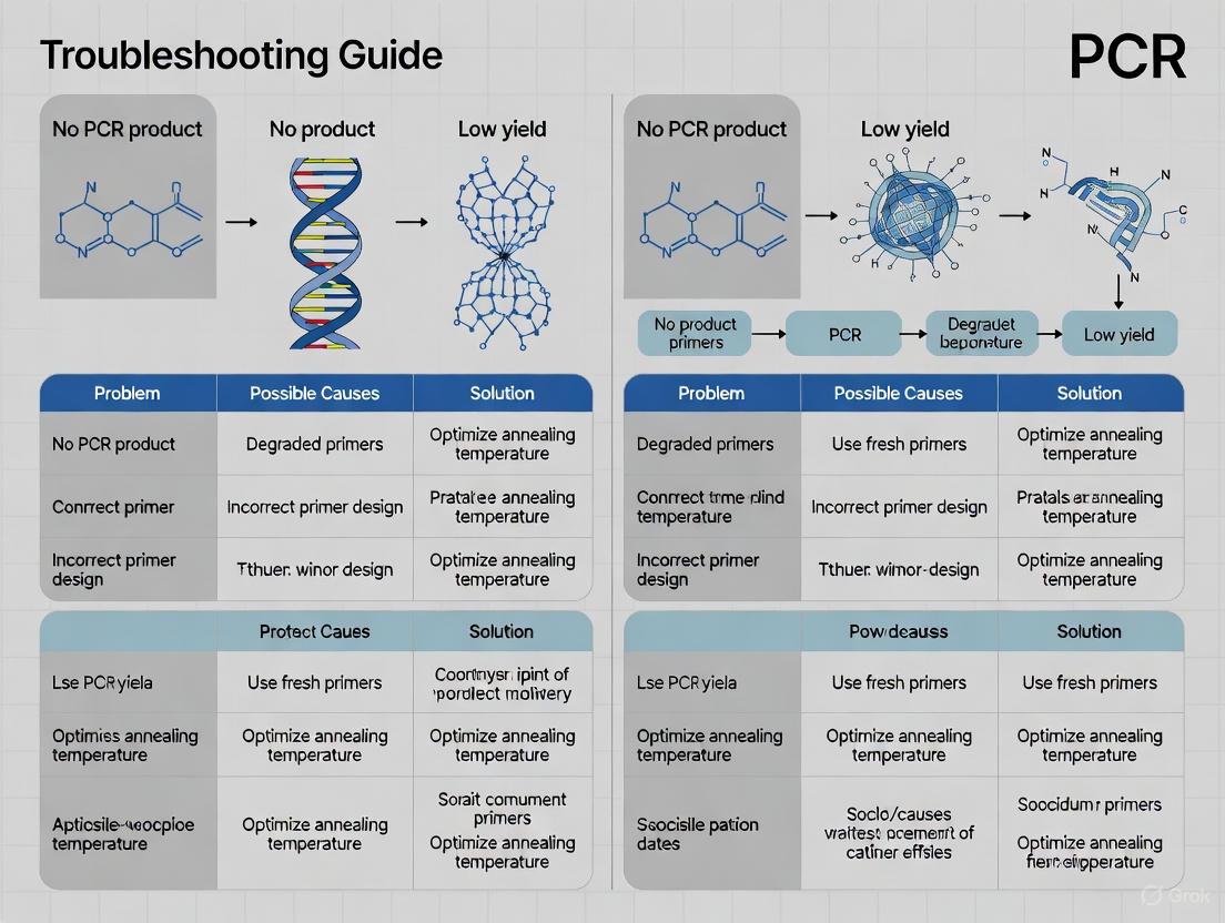
This article provides a systematic framework for researchers, scientists, and drug development professionals to diagnose and resolve the common yet critical issues of no product or low yield in Polymerase...
Solving PCR Failure: A Comprehensive Troubleshooting Guide for No Product and Low Yield
Abstract
This article provides a systematic framework for researchers, scientists, and drug development professionals to diagnose and resolve the common yet critical issues of no product or low yield in Polymerase Chain Reaction (PCR) experiments. Covering foundational principles to advanced validation protocols, the guide details the root causes of PCR failure—from template quality and primer design to reagent integrity and cycling conditions. It further explores specialized methodological adaptations, offers a step-by-step troubleshooting workflow, and emphasizes the importance of rigorous assay validation using current international standards and comparative techniques to ensure reliable, reproducible results in biomedical research and clinical diagnostics.
Understanding the Root Causes of PCR Failure
In polymerase chain reaction (PCR) research, the quality of the starting DNA template is a pivotal factor that can determine the success or failure of an experiment. For researchers and drug development professionals troubleshooting issues of no product or low yield, a thorough investigation of the DNA template is the first and most critical step. Problems related to the template's integrity, purity, and quantity are frequent culprits behind amplification failure. This guide provides a structured, troubleshooting-focused examination of these three aspects, offering specific protocols and solutions to help you reliably obtain robust PCR results.
FAQs and Troubleshooting Guides
How does DNA template integrity affect PCR, and how can I assess it?
DNA integrity refers to the fragment length and structural soundness of your DNA template. Degraded DNA, which is fragmented, will prevent the amplification of your target if the template is broken within the amplicon region.
- Cause and Effect: During PCR, the DNA polymerase can only extend a primer that is bound to a template. If the template strand is physically broken between your two primer binding sites, the polymerase has no continuous strand to follow, and amplification will fail [1]. This is a particular challenge with samples from forensic evidence, ancient tissues, or any source exposed to degrading conditions [1].
- Assessment Method: Traditional methods like agarose gel electrophoresis can provide a visual assessment of degradation (e.g., a smeared band instead of a tight, high-molecular-weight band) [2] [1]. For a more precise, quantitative measurement, advanced techniques like Droplet Digital PCR (ddPCR) can be used. This method can simultaneously detect multiple DNA fragments of different lengths (e.g., 75 bp, 145 bp, and 235 bp) to calculate a degradation ratio and precisely quantify the level of degradation [3] [1].
What are the common PCR inhibitors, and how do I remove them?
Purity concerns the presence of contaminants in your DNA sample that can inhibit the PCR reaction. Even a high-quality, intact DNA template will fail to amplify if inhibitors are present.
- Common Inhibitors: Frequent contaminants include phenol, EDTA, proteinase K, and salts carried over during the DNA extraction process [4] [5]. These substances can directly inhibit the DNA polymerase enzyme or chelate essential co-factors like magnesium ions (Mg²⁺) [2] [6].
- Solutions for Decontamination:
- Ethanol Precipitation: Precipitate and wash your DNA sample with 70% ethanol to remove residual salts and other impurities [4] [5].
- Commercial Cleanup Kits: Use a PCR clean-up or DNA purification kit designed to remove specific contaminants [4] [7].
- Additives: Incorporate additives like Bovine Serum Albumin (BSA) into your PCR mix. BSA can bind to inhibitors and reduce their interference with the polymerase [2].
- Quality Control: Check the purity of your DNA by measuring its absorbance ratios. A pure DNA sample typically has an A260/280 ratio of ~1.8 and an A260/230 ratio between 2.0 and 2.3 [8] [7]. Significant deviations from these values indicate contamination.
What is the optimal amount of DNA template to use?
Using too much or too little template DNA is a common mistake. The optimal quantity depends on the complexity of the DNA source.
- General Guidelines: The table below summarizes recommended starting amounts for a standard 50 µL PCR reaction [6] [5] [7].
| Template Type | Recommended Quantity per 50 µL Reaction |
|---|---|
| Plasmid DNA | 0.01 - 1 ng |
| Genomic DNA | 5 - 50 ng (or up to 1 µg for some applications) |
- Consequences of Improper Quantity:
My PCR shows no product or a very low yield. What should I check first regarding the DNA template?
This is a common problem, and the DNA template is the most likely source. Follow this systematic checklist.
- Verify Template Presence and Quality: First, confirm you have actually added DNA to the reaction. Then, check its concentration and purity using a spectrophotometer and run an agarose gel to assess integrity [2] [4] [7].
- Check for Inhibition: Include a positive control reaction using a template and primers known to work well. If the positive control works but your sample does not, your sample is likely inhibited [5].
- Re-optimize Reaction Conditions: If the template is degraded or inhibited, you may need to adjust your protocol. Consider using a DNA polymerase engineered for high sensitivity and tolerance to inhibitors, or increase the number of PCR cycles to 40 when dealing with very low copy numbers [2] [4].
My PCR results in non-specific products or a smeared band. Could the template be the cause?
Yes, the template can be a direct or indirect cause of this issue.
- Excessive Template Amount: As noted above, too much template DNA is a common cause of non-specific amplification and smeared bands [5].
- Co-amplification of Contaminants: According to one study, smearing can be caused by the gradual accumulation of "amplifiable DNA contaminants" that interact with your specific primers. A highly efficient solution is to switch to a new set of primers with different sequences [2].
- Template-Associated Reaction Conditions: While not a direct property of the template, complex templates (e.g., GC-rich genomes) can require higher denaturation temperatures or additives, and suboptimal conditions for these can lead to smearing [2] [4].
Experimental Protocols for Assessment and Cleanup
● Protocol 1: Assessing DNA Integrity Using Agarose Gel Electrophoresis
This standard protocol provides a visual assessment of your DNA template's quality.
- Prepare Gel: Cast a 0.8% - 1% agarose gel in TAE or TBE buffer, containing a fluorescent nucleic acid stain.
- Prepare Samples: Mix 1-5 µL of your DNA sample with a DNA loading dye.
- Run Gel: Load the samples alongside a DNA ladder (with known fragment sizes) and run the gel at 5-10 V/cm.
- Visualize: Image the gel under UV light.
- Interpret Results: Intact genomic DNA should appear as a single, tight, high-molecular-weight band. A smeared appearance indicates degradation. A sharp, lower band may indicate RNA contamination [2] [4].
● Protocol 2: Cleaning Up DNA Using Ethanol Precipitation
This method effectively removes salts, solvents, and other small contaminants.
- Add Salt and Ethanol: To your DNA sample in an aqueous solution, add 0.1 volumes of 3 M sodium acetate (pH 5.2) and 2.5 volumes of ice-cold 100% ethanol.
- Precipitate: Incubate at -20°C for 30 minutes or overnight to precipitate the DNA.
- Pellet DNA: Centrifuge at >12,000 × g for 15 minutes at 4°C. Carefully decant the supernatant.
- Wash Pellet: Add 500 µL of ice-cold 70% ethanol to the pellet and centrifuge for 5 minutes. Carefully decant the supernatant.
- Dry and Resuspend: Air-dry the pellet for 5-10 minutes and then resuspend it in nuclease-free water or TE buffer (pH 8.0) [4] [5].
Visual Guide: Troubleshooting DNA Template Issues
The following workflow diagram outlines a logical pathway for diagnosing and resolving common DNA template-related PCR failures.
The Scientist's Toolkit: Research Reagent Solutions
The following table details key reagents and kits essential for analyzing and preparing high-quality DNA templates for PCR.
| Item | Function | Example Use Case |
|---|---|---|
| Spectrophotometer / Fluorometer | Accurately measures DNA concentration and assesses purity via absorbance ratios (A260/280, A260/230). | First-line quality control for every DNA sample prior to PCR setup [2] [7]. |
| Droplet Digital PCR (ddPCR) System | Provides absolute quantification of DNA copy number and can assess degradation levels by targeting multiple fragment sizes. | Precisely quantifying template and evaluating integrity in highly degraded forensic or clinical samples [3] [1]. |
| DNA Cleanup Kit (e.g., spin-column based) | Efficiently removes enzymes, salts, primers, and other impurities from DNA samples. | Purifying DNA after extraction or purifying a PCR product before re-amplification [8] [6]. |
| Hot-Start DNA Polymerase | Enzyme engineered to be inactive at room temperature, preventing non-specific amplification and primer-dimer formation during reaction setup. | Essential for improving specificity and yield, especially with low-quality or complex templates [2] [4] [5]. |
| Bovine Serum Albumin (BSA) | PCR additive that binds to common inhibitors, reducing their negative effect on the DNA polymerase. | Added to the PCR mix when analyzing samples prone to contamination, like those from soil or blood [2]. |
FAQs: Resolving Primer-Related PCR Failure
Q1: My PCR reaction shows no product or a very faint band on the gel. My primers have been designed to a complex genomic region. What could be the primary issue?
The most common cause is a lack of primer specificity, leading to inefficient binding. A large-scale study analyzing over 80,000 PCR experiments found that the number of predicted primer-binding sites in the genomic DNA is the most important factor in determining PCR failure. Primers with too many binding sites have a high failure rate. Furthermore, primers with low melting temperatures (Tm) or significant differences in Tm between the forward and reverse primer can prevent simultaneous efficient binding [9] [10] [11].
Q2: I see multiple bands or a smeared product instead of a single clean band. How can primer design cause this?
This is a classic symptom of mispriming due to low specificity or secondary structures. If primers are not unique to your target, they will bind to multiple locations, amplifying non-specific products. Additionally, primers with complementary regions can form primer-dimers (where two primers anneal to each other) or hairpin loops (where a single primer folds back on itself). These structures are preferentially amplified, consuming reagents and creating smeared or multiple bands [12] [2] [13].
Q3: What are the critical parameters I must check when designing a new primer to avoid these problems?
You should verify four key parameters during your in silico design phase. The table below summarizes the optimal values for each.
Table 1: Critical Parameters for Effective Primer Design
| Parameter | Optimal Value / Range | Rationale |
|---|---|---|
| Primer Length | 18–30 nucleotides [10] [11] [13] | Balances specificity (longer) with hybridization efficiency (shorter). |
| Melting Temperature (Tm) | 52–65°C; primers in a pair should be within 5°C of each other [10] [11] [13] | Ensures both primers anneal to the template efficiently at the same temperature. |
| GC Content | 40–60% [11] [13] | Provides sufficient binding strength without promoting non-specific binding. |
| 3'-End Sequence | Avoid runs of 3+ G/Cs; ensure no complementarity between primers [10] [11] | Prevents stable non-specific binding and the formation of primer-dimers. |
Troubleshooting Guide: No Product or Low Yield
Table 2: Troubleshooting Common Primer-Related PCR Failures
| Observation | Possible Primer-Related Cause | Recommended Solution |
|---|---|---|
| No Product | Poor primer design or specificity [12] [14] | Verify primer specificity using tools like NCBI Primer-BLAST. Redesign primers if necessary. |
| Primer Tm is too high or too low [12] [4] | Recalculate Tm using a calculator that accounts for buffer composition. Test an annealing temperature gradient. | |
| Primers form stable secondary structures (hairpins) [11] [13] | Use software to check for self-complementarity. Redesign primers to avoid regions of internal homology. | |
| Low Yield | Primer concentration is too low [12] [11] | Optimize primer concentration, typically between 0.05–1 µM. |
| Primer degradation from multiple freeze-thaw cycles [4] [11] | Aliquot primers after resuspension to avoid repeated freeze-thaw cycles. | |
| Non-Specific Bands / Smearing | Primer annealing temperature is too low [12] [4] | Increase the annealing temperature in 1–2°C increments. |
| Primers bind to multiple genomic sites [9] [2] | Verify primer uniqueness. Increase primer length to enhance specificity. | |
| Primer-dimer formation [2] [13] [15] | Use a hot-start polymerase. Redesign primers to eliminate 3'-end complementarity. |
Experimental Protocol: Annealing Temperature Gradient Optimization
A poorly optimized annealing temperature (Ta) is a major contributor to PCR failure. The following protocol provides a methodology to empirically determine the ideal Ta for any primer pair.
1. Principle The theoretical Tm of a primer is calculated, but the optimal Ta for a specific reaction in a specific buffer must be determined experimentally. Running a gradient PCR allows you to test a range of temperatures simultaneously to find the Ta that provides the highest yield and specificity [10] [4].
2. Materials
- Designed forward and reverse primers
- DNA template
- PCR master mix (including buffer, dNTPs, Mg²⁺, and high-fidelity DNA polymerase)
- Nuclease-free water
- Thermal cycler with gradient functionality
3. Procedure
Step 1: Calculate the Tm for both forward and reverse primers using the formula: Tm = 4(G + C) + 2(A + T) or an online calculator [10] [13].
Step 2: Set up a standard 50 µL PCR reaction mixture as detailed below. If setting up multiple reactions, create a master mix to ensure consistency.
Table 3: PCR Reaction Setup for Ta Optimization
| Component | Final Concentration/Amount | Volume per 50 µL Reaction |
|---|---|---|
| 10X PCR Buffer | 1X | 5 µL |
| dNTP Mix | 200 µM each | 1 µL (from 10 mM stock) |
| MgCl₂ | 1.5 mM (adjust if not in buffer) | Variable (e.g., 0.8 µL of 25 mM stock) |
| Forward Primer | 0.2 µM | 0.5 µL (from 20 µM stock) |
| Reverse Primer | 0.2 µM | 0.5 µL (from 20 µM stock) |
| DNA Template | 1–100 ng (depending on complexity) | Variable |
| DNA Polymerase | 1.25 Units | 0.5 µL |
| Nuclease-free Water | To volume | Q.S. to 50 µL |
Step 3: Program the thermal cycler with a gradient in the annealing step. Set the gradient to span a range of approximately 5°C below to 5°C above the calculated lower Tm of your primer pair [12] [4]. Step 4: Analyze the PCR products using agarose gel electrophoresis. The optimal Ta will produce a single, bright band of the expected size.
4. Workflow Diagram The following diagram illustrates the logical workflow for this optimization procedure.
The Scientist's Toolkit: Research Reagent Solutions
Table 4: Essential Reagents for Overcoming Primer and PCR Challenges
| Reagent / Material | Function / Purpose |
|---|---|
| High-Fidelity DNA Polymerase (e.g., Q5, Phusion) | Reduces sequence errors in the final amplicon, crucial for downstream cloning and sequencing [12] [4]. |
| Hot-Start DNA Polymerase | Prevents polymerase activity at room temperature, minimizing primer-dimer formation and non-specific amplification during reaction setup [12] [2]. |
| PCR Additives (DMSO, Betaine, BSA) | Help denature GC-rich templates, destabilize secondary structures, and overcome inhibition in complex samples [10] [4] [2]. |
| MgCl₂ or MgSO₄ Solution | Cofactor for DNA polymerase; its concentration is critical and often needs optimization to improve yield and specificity [12] [10] [4]. |
| PCR Primer Design Software (e.g., Primer-BLAST, Primer3) | Automated tools to help design primers that meet optimal parameters and check for specificity against genomic databases [10] [11] [16]. |
Polymerase Chain Reaction (PCR) is a foundational technique in molecular biology, yet experiments often fail due to issues with core reaction components. This guide addresses troubleshooting specifically related to DNA polymerase, dNTPs, and Mg2+ concentration—three interlinked factors critical for PCR success. Proper management of these components is essential to overcome the common problem of no product or low yield in research and drug development.
FAQs: Core Reaction Components
1. How does the integrity of DNA polymerase affect PCR yield, and what are the signs of a problem? DNA polymerase can lose activity over time if improperly stored or subjected to multiple freeze-thaw cycles. Signs of compromised polymerase include a complete absence of PCR product, a significant drop in yield compared to previous experiments, or smeared bands on a gel. Using a fresh aliquot of enzyme or a hot-start polymerase, which reduces non-specific amplification during reaction setup, is recommended to mitigate these issues [2] [4] [5].
2. What issues can arise from degraded or unbalanced dNTPs? dNTPs are susceptible to degradation upon repeated freeze-thawing, and unbalanced concentrations of the four nucleotides (dATP, dCTP, dGTP, dTTP) can significantly reduce the fidelity of the polymerase, leading to misincorporation of bases and unexpected mutations in the final product [4] [17] [5]. This can be particularly detrimental for downstream applications like cloning or sequencing.
3. Why is Mg2+ concentration so critical and often need optimization? Mg2+ is a essential cofactor for DNA polymerase activity. It stabilizes the primer-template complex and influences the overall stringency of the reaction [18] [5]. A concentration that is too low can result in no amplification, while a concentration that is too high can promote non-specific binding and the appearance of unwanted bands [2] [19] [17]. Its optimal concentration must be determined empirically because Mg2+ can be chelated by dNTPs, EDTA, or other components in the reaction mix [4] [5].
4. What is the relationship between dNTP and Mg2+ concentrations? There is a critical stoichiometric relationship between dNTPs and Mg2+. Mg2+ binds to dNTPs to form the actual substrate for the polymerase. A general rule is to maintain a 1:2 ratio of the total dNTP concentration to Mg2+ concentration [5]. Therefore, any change in dNTP concentration necessitates a corresponding adjustment to the Mg2+ concentration.
Troubleshooting Tables
Table 1: Optimal Concentration Ranges for Key Components
Use these ranges as a starting point for optimization.
| Component | Typical Optimal Concentration Range | Special Considerations |
|---|---|---|
| DNA Polymerase | 0.5 - 2.0 units per 50 µL reaction [19] | Consult manufacturer's specifications; increase if additives (DMSO) or inhibitors are present [4]. |
| dNTPs (each) | 0.2 - 0.25 mM [19] [5] | Higher concentrations (up to 0.4 mM) may increase yield but can reduce fidelity [19] [5]. |
| Mg2+ | 1.5 - 2.0 mM (for Taq polymerase) [19] | Must be optimized for every primer-template system (range 1-4 mM); affected by dNTP and EDTA concentration [4] [17] [5]. |
Table 2: Troubleshooting No Product/Low Yield
This table links symptoms and causes to specific solutions.
| Symptom | Possible Cause | Recommended Solution |
|---|---|---|
| No amplification | Insufficient DNA polymerase [2] | Increase the amount of enzyme within the recommended range [2] [4]. |
| No amplification | Inactive or degraded dNTPs | Prepare a fresh, balanced dNTP mix from high-quality (99% pure) stocks [17] [5]. |
| No amplification / Low yield | Mg2+ concentration too low [19] [5] | Titrate Mg2+ concentration upward in 0.5 mM increments up to 4 mM [19]. |
| Low yield / Fidelity issues | Unbalanced dNTP concentrations | Ensure equimolar concentrations of all four dNTPs [4] [17]. |
| Non-specific products / Smearing | Mg2+ concentration too high [2] [17] | Titrate Mg2+ concentration downward [19]. |
| Non-specific products | Polymerase activity at room temperature | Use a hot-start DNA polymerase to prevent non-specific priming during setup [2] [4] [17]. |
Experimental Protocols
Protocol 1: Standard Mg2+ Concentration Optimization
This protocol outlines a method to empirically determine the optimal Mg2+ concentration for your PCR assay.
Materials:
- PCR reagents: template DNA, primers, DNA polymerase, 10X reaction buffer (without Mg2+), dNTP mix, nuclease-free water.
- MgCl2 or MgSO4 solution (typically 25 mM or 50 mM).
- Thermal cycler.
Method:
- Prepare a master mix for all common components. Calculate for n+1 reactions, where
nis the number of Mg2+ conditions to test. Per 50 µL reaction, combine:- 5 µL of 10X reaction buffer (without Mg2+)
- 1 µL of forward primer (0.1-1 µM final)
- 1 µL of reverse primer (0.1-1 µM final)
- 1 µL of dNTP mix (0.2 mM each final)
- X µL of template DNA (e.g., 1 ng - 1 µg for genomic)
- 0.5 - 2.0 µL of DNA polymerase (e.g., 1.25 units)
- Nuclease-free water to a final volume of 45 µL (after adding Mg2+).
- Aliquot 45 µL of the master mix into each PCR tube.
- Add a different volume of Mg2+ stock solution to each tube to create a concentration gradient. For a 50 mM MgCl2 stock:
- Tube 1: 1.0 µL → 1.0 mM final
- Tube 2: 1.5 µL → 1.5 mM final
- Tube 3: 2.0 µL → 2.0 mM final
- Tube 4: 2.5 µL → 2.5 mM final
- Tube 5: 3.0 µL → 3.0 mM final
- Tube 6: 4.0 µL → 4.0 mM final [5]
- Run the PCR using your standard cycling conditions.
- Analyze the results by agarose gel electrophoresis. The condition that yields the strongest, correct band with the least background should be selected as optimal.
Protocol 2: Testing dNTP and Polymerase Integrity
This protocol helps diagnose if poor yield is due to degraded dNTPs or a compromised polymerase.
Materials:
- All standard PCR components.
- Fresh, high-quality dNTP aliquot.
- Fresh, high-quality DNA polymerase aliquot.
- Control template and primer set known to work reliably.
Method:
- Set up a series of PCR reactions as follows:
- Reaction A (Test): Uses your current dNTPs and polymerase.
- Reaction B (dNTP Control): Uses your current polymerase but a fresh dNTP aliquot.
- Reaction C (Enzyme Control): Uses your current dNTPs but a fresh polymerase aliquot.
- Reaction D (Positive Control): Uses fresh dNTPs and fresh polymerase.
- Use the same control template, primers, and Mg2+ concentration in all reactions.
- Run the PCR simultaneously.
- Compare the yields. If yield improves only in Reaction B, your dNTPs are likely degraded. If yield improves only in Reaction C, your polymerase may be compromised. If yield is poor in all reactions, the issue likely lies elsewhere (e.g., primer design, template quality) [20] [5].
The Scientist's Toolkit: Research Reagent Solutions
| Item | Function in PCR |
|---|---|
| Hot-Start DNA Polymerase | Remains inactive until a high-temperature activation step, dramatically reducing non-specific amplification and primer-dimer formation during reaction setup [2] [4] [17]. |
| High-Fidelity DNA Polymerase | Possesses proofreading (3'→5' exonuclease) activity to correct misincorporated nucleotides, essential for applications requiring high accuracy like cloning and sequencing [4] [17]. |
| Molecular-Grade dNTPs | High-purity (≥99%) nucleotides ensure efficient and accurate incorporation by the polymerase, minimizing replication errors [5]. |
| MgCl2 or MgSO4 Solution | Provides the essential Mg2+ cofactor. The type (chloride vs. sulfate) can be polymerase-specific [4]. |
| PCR Additives (e.g., BSA, Betaine) | Bovine Serum Albumin (BSA) can bind PCR inhibitors. Betaine can help denature GC-rich templates and destabilize secondary structures, improving amplification efficiency [2] [4]. |
Troubleshooting Workflows and Relationships
PCR Troubleshooting Workflow
Reaction Component Interdependence
In the pursuit of reliable Polymerase Chain Reaction (PCR) results, scientists often contend with two main categories of interference: contaminants and inhibitors. Contaminants are unwanted nucleic acids that lead to false positives or spurious amplification, while inhibitors are substances that reduce PCR efficiency or cause complete amplification failure, potentially resulting in false negatives [21] [22]. Both can compromise data integrity, leading to wasted resources and erroneous conclusions. Understanding these interfering substances is a fundamental component of troubleshooting no product or low yield PCR research. This guide details the common sources of interference and provides proven methodologies for their identification and elimination.
Common PCR Contaminants and Control Strategies
PCR contamination occurs when exogenous DNA is introduced into the reaction, threatening the specificity of the assay. The extreme sensitivity of PCR means even minute, aerosolized amounts of DNA can be amplified.
The most common sources of contamination include:
- Carryover Contamination: Amplified PCR products (amplicons) from previous reactions are the most prevalent contaminant, as they exist in very high concentrations and are perfectly suited for amplification [23].
- Cloned DNA: Plasmid or other cloned DNA previously handled in the laboratory [23].
- Cross-Contamination: Sample-to-sample contamination during processing, particularly in samples requiring extensive manual preparation [23].
- Exogenous DNA: Environmental DNA present on laboratory equipment, in reagents, or introduced during nucleic acid extraction [23].
Identifying Contamination: The Role of Controls
The use of appropriate controls is non-negotiable for diagnosing contamination.
- No-Template Control (NTC): This control contains all reaction components except the DNA template. Amplification in the NTC indicates contamination of one or more reagents or the master mix [24].
- No-RT Control (for RT-PCR): In reverse transcription PCR, this control omits the reverse transcriptase enzyme. Amplification here signals contamination of the RNA preparation with genomic DNA [21].
Interpreting NTC results can provide further clues. If all NTC wells show amplification at similar cycle threshold (Ct) values, a reagent is likely contaminated. If only some NTC wells amplify with varying Ct values, random environmental contamination (e.g., from aerosols) is the probable cause [24].
A Systematic Workflow for Contamination Prevention
The diagram below outlines a logical workflow for preventing PCR contamination, from sample setup to analysis.
Experimental Protocol: Decontamination and UNG Use
A. Surface Decontamination with Bleach
- Prepare Fresh Solution: Dilute sodium hypochlorite to a 10% bleach solution weekly. [24]
- Apply: Spray or wipe down all work surfaces, pipettes, tube racks, and equipment.
- Incubate: Allow the solution to remain on the surface for 10-15 minutes to ensure complete degradation of DNA. [24]
- Rinse: Wipe the area with de-ionized water to remove residual bleach, which can corrode equipment. [24]
B. Using Uracil-N-Glycosylase (UNG) to Prevent Carryover
- Incorporation of dUTP: In the primary PCR, use a dNTP mix where dTTP is replaced with dUTP. All subsequent amplicons will then contain uracil. [24]
- UNG Treatment in Subsequent Reactions: In all future PCR setups, include a master mix containing UNG.
- Incubate: Incubate the reaction mix at room temperature or a mild temperature (e.g., 25-50°C) before PCR cycling. The UNG enzyme will enzymatically cleave any uracil-containing contaminating DNA from previous runs. [24]
- Inactivate and Amplify: The initial high-temperature denaturation step of the PCR cycle (e.g., 95°C) will permanently inactivate the UNG, allowing the new, uracil-free template to amplify without interference. [24]
Common PCR Inhibitors and Mitigation Approaches
PCR inhibitors are substances that prevent the amplification of nucleic acids, even when the target template is present. They typically work by interfering with the DNA polymerase, chelating essential co-factors like Mg²⁺, or damaging the DNA template itself. [22]
Quantitative Data on Inhibition Rates
The table below summarizes inhibition rates across different specimen matrices, as found in a large-scale retrospective study. This data helps laboratories assess the risk associated with different sample types.
Table: Inhibition Rates by Specimen Matrix in Qualitative Real-Time PCR Assays [25]
| Specimen Matrix | Inhibition Rate (%) | Notes |
|---|---|---|
| Overall (post-extraction) | 0.01% | n = 381,093 specimens |
| Overall (pre-extraction) | 0.87% | n = 5,613 specimens |
| Urine | >1% | A known problematic matrix |
| Formalin-Fixed, Paraffin-Embedded (FFPE) Tissue | >1% | Fixative and embedding medium can be inhibitory |
| All other matrices (e.g., swabs, blood, CSF) | ≤1% | Includes nasopharyngeal, blood, stool, etc. |
Inhibitors can originate from the original sample or be introduced during processing and DNA extraction. [22]
- Heme from blood is a potent inhibitor of DNA polymerase. [25]
- Bile Salts and complex polysaccharides found in feces can inhibit PCR. [25]
- Urea present in urine is a common inhibitor. [25]
- Calcium Alginate from certain types of swabs can carry inhibitors. [25]
- Heparin, an anticoagulant found in some blood collection tubes, is a stronger inhibitor than EDTA. [25]
- Formalin used for tissue fixation can damage DNA and introduce inhibitors. [25]
- Laboratory Reagents such as phenol, SDS (ionic detergents), ethanol, and isopropanol can be carried over from extraction protocols and inhibit the reaction. [22] [4]
A Troubleshooting Guide for Inhibition and Low Yield
The following table provides a structured approach to diagnosing and resolving issues related to PCR inhibition and low yield.
Table: Troubleshooting Guide for PCR Inhibition and Low Yield [2] [4] [26]
| Observation | Possible Cause | Recommended Solution |
|---|---|---|
| No Product or Low Yield | Inhibitors in the sample | Further purify the template via alcohol precipitation, drop dialysis, or commercial cleanup kits. [26] |
| Insufficient template quality/quantity | Re-measure DNA concentration and purity (260/280 ratio). Analyze integrity by gel electrophoresis. [2] [4] | |
| Suboptimal cycling conditions | Increase cycle number up to 45. [27] Optimize annealing temperature. [26] Ensure adequate extension time. [4] | |
| Incorrect Mg²⁺ concentration | Optimize Mg²⁺ concentration in 0.2-1 mM increments. [26] | |
| Non-Specific Bands or Smearing | Non-specific priming | Use a hot-start polymerase. [2] [26] Increase the annealing temperature. [4] |
| Primer-dimer formation | Optimize primer design to avoid 3'-end complementarity. Lower primer concentration. [2] | |
| Contamination with amplicons | Follow contamination control workflow (see Section 2.3). Use a new set of primers with a different sequence. [2] |
The Scientist's Toolkit: Key Reagents for Overcoming Interference
This table lists essential reagents and materials that can be employed to prevent or overcome the effects of PCR inhibitors and contaminants.
Table: Essential Research Reagents for Mitigating PCR Interference
| Reagent/Material | Function in Overcoming Interference |
|---|---|
| Bovine Serum Albumin (BSA) | Binds to and neutralizes a wide range of inhibitors, particularly effective for inhibitors in blood and plant tissues. [2] [22] |
| Hot-Start DNA Polymerase | Prevents non-specific amplification and primer-dimer formation by remaining inactive until the initial high-temperature denaturation step. [2] [26] |
| UNG (Uracil-N-Glycosylase) | An enzymatic system to destroy carryover contamination from previous PCR amplifications. [24] |
| PCR Additives (e.g., DMSO, Betaine) | Help denature complex DNA templates (e.g., GC-rich regions) and can destabilize secondary structures, improving yield and specificity. [2] [10] |
| Aerosol-Resistant Filter Tips | Create a physical barrier between the pipette and the liquid, preventing aerosol contamination of reagents and samples. [21] [24] |
| Commercial DNA Cleanup Kits | Designed to remove common inhibitors (e.g., salts, phenols, proteins) from sample extracts, yielding purer template DNA. [26] |
FAQs on PCR Interfering Substances
Q1: My No-Template Control (NTC) shows amplification. What should I do? A1: Immediately discard all reagents, particularly water and the master mix. Decontaminate your workspace and equipment with a 10% bleach solution or UV irradiation. Prepare fresh aliquots of all reagents and repeat the experiment. To prevent recurrence, implement strict physical separation of pre- and post-PCR areas. [21] [24]
Q2: I suspect my sample contains inhibitors. How can I confirm this? A2: The most effective method is a spiking experiment. Add a known amount of a control template (one that amplifies reliably in a clean system) to your investigated reaction mixture. Compare its amplification to the same template amplified in a clean, inhibitor-free reaction. A significant delay or failure in amplification in the sample mixture confirms the presence of inhibitors. [22]
Q3: What is the fastest way to overcome PCR inhibition from a complex sample like blood or stool? A3: The two most immediate strategies are: 1) Use an inhibitor-resistant polymerase. Many modern polymerases are engineered for high tolerance to common inhibitors. 2) Add Bovine Serum Albumin (BSA) to your reaction. BSA is very effective at binding and neutralizing a broad spectrum of inhibitory compounds. [2] [22] [4]
Q4: My PCR used to work but now shows smeared bands. What is wrong? A4: This is a classic sign of accumulating "amplifiable DNA contaminants" in your lab environment, specific to your primer set. The most efficient solution is to switch to a completely new set of primers that target a different sequence and do not interact with the accumulated contaminants. [2]
The precise control of thermal cycler conditions is fundamental to the success of the Polymerase Chain Reaction (PCR). The three core temperature-dependent steps—denaturation, annealing, and extension—must be meticulously optimized to efficiently amplify a specific target DNA sequence [28] [29]. Within the context of troubleshooting no product or low yield in PCR research, suboptimal thermal cycling is a primary suspect. This guide provides researchers and drug development professionals with detailed protocols and troubleshooting frameworks to diagnose and rectify issues related to these critical parameters, thereby restoring amplification efficiency and experimental workflow.
Frequently Asked Questions (FAQs)
1. What are the typical temperature and time ranges for the three main PCR steps?
The table below summarizes the standard parameters. These serve as a starting point and often require optimization based on your specific reaction components [28] [29] [30].
| PCR Step | Purpose | Typical Temperature Range | Typical Time Range |
|---|---|---|---|
| Denaturation | Separates double-stranded DNA into single strands. | 94–98°C | Initial: 1–3 min; Cyclic: 15 sec – 2 min |
| Annealing | Allows primers to bind to complementary sequences on the template DNA. | 45–72°C (Typically 3–5°C below primer Tm) | 15 sec – 2 min |
| Extension | DNA polymerase synthesizes a new DNA strand. | 70–75°C (Optimal for the enzyme used) | 1 min/kb for Taq; 2 min/kb for Pfu |
2. How do I determine the correct annealing temperature for my primers?
The annealing temperature (Ta) is critically dependent on the melting temperature (Tm) of your primers [28].
- Calculate Primer Tm:* Use the formula: Tm = 4(G + C) + 2(A + T). For greater accuracy, use the Nearest Neighbor method via online calculators [28].
- Set Initial Ta:* Start with a temperature 3–5°C below the lowest Tm of your primer pair [28] [4].
- Optimize Experimentally: Use a gradient thermal cycler to test a range of annealing temperatures simultaneously. The optimal Ta provides the strongest specific yield with minimal non-specific products [28] [31].
3. My PCR has no product. Should I adjust the thermal cycles or the conditions?
Focus on conditions first. A lack of product often points to issues with template denaturation or primer annealing [4] [32] [5].
- Increase Initial Denaturation: For complex templates like genomic DNA or GC-rich sequences, extend the initial denaturation to 3-5 minutes, or even up to 10 minutes [28] [5].
- Optimize Annealing Temperature: An overly high Ta prevents primer binding. Use a gradient cycler to find the optimal temperature [4] [32].
- Check Extension Time: Ensure the extension time is sufficient for your amplicon length and polymerase speed (e.g., 1 min/kb for Taq) [28]. If conditions are optimized but yield is still low, then increase the cycle number to 35–40 for low-abundance targets [28] [4].
4. I see non-specific bands (multiple products) in my gel. What thermal cycling adjustments can I make?
Non-specific amplification is typically due to low stringency, allowing primers to bind to incorrect sequences [4] [2].
- Increase Annealing Temperature: This is the most effective step. Raise the temperature in 2–3°C increments to enhance specificity [28] [4] [32].
- Employ a Hot-Start Polymerase: These enzymes are inactive until the initial high-temperature denaturation step, preventing primer-dimer formation and mis-priming during reaction setup [4] [2] [33].
- Reduce Extension Time: Excessively long extension times can allow the polymerase to generate secondary products [32].
5. How does the thermal cycler itself impact my results?
The instrument's performance is crucial [30].
- Temperature Uniformity: Variation across the block can cause wells to amplify with different efficiencies.
- Ramp Rate: The speed at which the cycler transitions between temperatures can affect specificity and overall run time.
- Heated Lid: Prevents condensation and evaporation from the reaction tubes, which is critical for maintaining reaction volume and consistency [30]. Regular calibration and maintenance of the thermal cycler are essential for reproducible results [30].
Troubleshooting Guide: No Product or Low Yield
The following table outlines common causes and solutions for low or absent PCR yield, with a focus on thermal cycling parameters.
| Symptom | Possible Cause | Recommended Solution |
|---|---|---|
| No Product | Incomplete denaturation of template, especially GC-rich DNA. | Increase denaturation temperature (to 98°C) and/or time (up to 3-5 min/cycle) [28] [4]. |
| Annealing temperature is too high. | Lower the annealing temperature in 2–3°C increments. Perform gradient PCR to find the optimal Ta [28] [32]. | |
| Insufficient number of cycles for low-copy-number templates. | Increase cycle number to 40 [28] [4]. | |
| Primer extension time is too short for the amplicon length. | Increase extension time according to polymerase speed (e.g., 1 min/kb for Taq, 2 min/kb for Pfu) [28] [5]. | |
| Low Yield | Poor primer design or degradation. | Redesign primers using dedicated software; check for secondary structures. Use fresh, high-quality primer aliquots [4] [33]. |
| Suboptimal Mg2+ concentration. | Titrate Mg2+ concentration in 0.5 mM increments between 1–4 mM to find the optimum [4] [32] [5]. | |
| Inhibitors present in the template DNA. | Re-purify the template DNA via ethanol precipitation or column purification [4] [32] [5]. | |
| Final extension step is too short, leading to incomplete products. | Implement a final extension step of 5–15 minutes to ensure all products are fully synthesized [28] [5]. |
Systematic Troubleshooting Workflow
The following diagram maps the logical workflow for diagnosing and resolving "no product or low yield" issues.
Diagram: A systematic workflow for troubleshooting no product or low yield in PCR.
Experimental Protocols for Optimization
Protocol 1: Optimizing Annealing Temperature Using a Gradient Thermal Cycler
This is the most critical protocol for enhancing PCR specificity and yield [28] [31].
- Calculate Primers' Tm:* Use an online calculator based on the Nearest Neighbor method [28].
- Set the Gradient Range: Program your thermal cycler's gradient function. Set the highest temperature to 2–3°C above the higher Tm and the lowest to 5–7°C below the lower Tm. This creates a linear temperature gradient across the block [31].
- Prepare the Master Mix: Create a single, homogeneous master mix containing all reaction components (template, primers, polymerase, dNTPs, buffer) and aliquot it equally into the PCR tubes or wells.
- Run the PCR: Execute the standard cycling program with the annealing step using the gradient.
- Analyze Results: Resolve the PCR products on an agarose gel. Identify the well(s) with the strongest specific band and the cleanest background (least smearing or non-specific bands). The temperature for that well is your optimal annealing temperature.
Protocol 2: Titrating Magnesium Ion (Mg2+) Concentration
Mg2+ is a essential cofactor for DNA polymerase, and its concentration can dramatically affect yield and specificity [4] [5].
- Prepare Stock Solutions: Ensure you have a PCR buffer without Mg2+ and a separate MgCl2 or MgSO4 solution (typically 25-50 mM).
- Set Up Reactions: Prepare a series of identical reactions, varying only the concentration of the magnesium salt. A standard range is 1.0 mM to 4.0 mM in 0.5 mM increments.
- Run the PCR: Use the previously determined optimal annealing temperature and standard cycling conditions.
- Analyze Results: Analyze the products by gel electrophoresis. Select the Mg2+ concentration that yields the highest amount of the correct product with the least background.
Annealing Temperature Optimization Logic
Diagram: Decision process for optimizing PCR annealing temperature.
The Scientist's Toolkit: Key Research Reagent Solutions
The following table lists essential reagents and their roles in optimizing thermal cycling and overcoming low yield.
| Reagent | Function in PCR | Optimization Consideration |
|---|---|---|
| Hot-Start DNA Polymerase | Reduces non-specific amplification and primer-dimer formation by remaining inactive until the initial high-temperature denaturation step [4] [2]. | Essential for improving specificity. Choose based on application (e.g., high-fidelity for cloning). |
| Magnesium Salts (MgCl₂/MgSO₄) | Cofactor for DNA polymerase; concentration critically affects primer annealing, enzyme activity, and product specificity [4] [5]. | Must be titrated for each primer-template system. Excess causes non-specific bands; insufficient causes low yield. |
| PCR Additives (e.g., DMSO, Betaine, BSA) | Assist in amplifying difficult templates (e.g., GC-rich regions, secondary structures) by lowering DNA melting temperature or stabilizing enzymes [28] [4] [2]. | Use at appropriate concentrations (e.g., DMSO at 3-10%). Requires re-optimization of annealing temperature. |
| dNTP Mix | The building blocks (dATP, dCTP, dGTP, dTTP) for new DNA strand synthesis [5] [33]. | Use high-quality, nuclease-free dNTPs at balanced equimolar concentrations (typically 0.2 mM each). Unbalanced mixes reduce fidelity. |
| Nuclease-Free Water | The solvent for all reaction components. | Using low-quality water can introduce RNases, DNases, and PCR inhibitors. Always use certified nuclease-free water. |
Advanced PCR Strategies for Challenging Amplifications
Employing Hot-Start PCR to Suppress Non-Specific Amplification
Hot-Start PCR is a specialized molecular biology technique designed to suppress non-specific amplification, a common challenge in conventional PCR that leads to primer-dimers, false-positive results, and reduced yield of the desired amplicon. This method employs various mechanisms to keep the DNA polymerase inactive during reaction setup at room temperature, activating it only at high temperatures during the initial denaturation step. By preventing enzymatic activity during tube preparation, Hot-Start PCR significantly enhances amplification specificity, sensitivity, and reproducibility, making it particularly valuable for diagnostic applications, cloning, and next-generation sequencing library preparation where precision is critical.
How does Hot-ast PCR prevent non-specific amplification?
The Core Problem in Conventional PCR
In conventional PCR, reactions are typically assembled at room temperature or on ice. During this setup, the DNA polymerase retains enzymatic activity. As the thermal cycler ramps up to the initial denaturation temperature (typically 94–98°C), the reaction mixture must pass through the temperature range optimal for primer elongation (around 72°C for Taq polymerase). At these lower temperatures, primers can bind imperfectly to non-target sites on the template DNA (mispriming) or to each other (primer-dimer formation). Any nonspecific products generated at this stage are co-amplified along with the intended target throughout the subsequent cycles, compromising reaction specificity and yield [34].
The Hot-Start Solution
Hot-Start PCR is specifically designed to keep the DNA polymerase inactive until the reaction temperature is high enough to prevent mispriming. The enzyme is only activated after the initial denaturation step, ensuring that the first primer binding events occur at a high, stringent temperature. This approach drastically reduces, and often eliminates, the formation of spurious PCR products [34] [35].
The following diagram illustrates the logical workflow and comparative outcomes of conventional PCR versus Hot-Start PCR:
What are the primary Hot-Start PCR methods?
Several methods have been developed to temporarily inhibit DNA polymerase activity. The table below summarizes the mechanism, advantages, and considerations for the primary Hot-Start technologies.
Table: Comparison of Primary Hot-Start PCR Methods
| Method | Mechanism of Inhibition | Key Features | Activation |
|---|---|---|---|
| Antibody-mediated [34] [36] [35] | An antibody binds to the enzyme's active site, creating a steric block. | High specificity; rapid activation (e.g., 30 sec to 5 min at 95°C); widely used. | High temperature (e.g., 95°C) denatures the antibody, releasing active polymerase. |
| Aptamer-mediated [34] [37] | A short, single-stranded nucleic acid molecule (aptamer) binds reversibly to the polymerase. | Does not require a high-temperature activation step; inhibition is reversible [37]. | Aptamer dissociates at a lower temperature than antibody methods, speeding up protocols [37]. |
| Chemical Modification [36] | Chemical groups (e.g., via anhydrides) covalently modify key amino acids in the enzyme. | Effective inhibition; can be robust. | May require longer high-temperature activation to remove the chemical groups [36]. |
| Hot-Start dNTPs [34] | Deoxynucleotides (dNTPs) have a thermolabile protecting group on the 3'-OH. | Polymerase cannot extend primers until the protecting group is removed. | The initial denaturation step cleaves the protecting group, generating functional dNTPs. |
Advanced developments include double-blocking antibodies that simultaneously inhibit both the 5'→3' polymerase activity and the 5'→3' exonuclease activity of Taq polymerase. This is crucial for qPCR applications, as it prevents not only non-specific amplification but also probe degradation at low temperatures, thereby reducing false-positive signals in pre-mixed master mixes [36].
Troubleshooting Guide: No Product or Low Yield
A frequent concern when implementing Hot-Start PCR is a failure to obtain the desired product or obtaining a low yield. The following table outlines common causes and solutions.
Table: Troubleshooting No Product or Low Yield in Hot-Start PCR
| Possible Cause | Recommended Solution |
|---|---|
| Incomplete Polymerase Activation | Ensure the initial heat activation step is performed at the correct temperature and for the recommended duration (e.g., 95°C for 30 sec to 5 min, depending on the enzyme) [36]. |
| Suboptimal Annealing Temperature | Recalculate primer Tm values and use a gradient thermal cycler to optimize the annealing temperature. Start testing at 3–5°C below the primer's calculated Tm [4] [38]. |
| Insufficient Mg²⁺ Concentration | Mg²⁺ is an essential cofactor. Titrate Mg²⁺ concentration in 0.2–1.0 mM increments, as excessive amounts promote non-specific binding, while insufficient amounts reduce yield [39] [4] [40]. |
| Poor Template Quality or Quantity | Re-purify template DNA to remove inhibitors (e.g., phenol, EDTA, heparin). Evaluate DNA integrity by gel electrophoresis and optimize the amount of input DNA (typically 10 pg–1 µg per 50 µL reaction, depending on complexity) [4] [38]. |
| Inefficient Denaturation | For GC-rich templates (>65% GC), increase the denaturation temperature (e.g., to 98°C) and/or time to ensure complete strand separation [40] [35]. |
FAQs on Hot-Start PCR
Q1: My Hot-Start PCR reaction has no product. What is the first thing I should check? A1: First, verify that you have included the mandatory high-temperature activation step before cycling begins. Consult the manufacturer's instructions for the specific polymerase you are using, as activation times and temperatures can vary. This step is essential to release the polymerase from its inhibited state [4] [35].
Q2: Can I set up Hot-Start PCR reactions at room temperature? A2: Yes, a major advantage of commercial Hot-Start polymerases is that they allow for convenient reaction setup at room temperature without compromising specificity. The enzyme remains inhibited until the first high-temperature denaturation step in the thermal cycler [37] [35].
Q3: When is Hot-Start PCR most critical to use? A3: Hot-Start PCR is highly recommended for multiplex PCR (amplifying multiple targets in one tube), qPCR, PCR from complex templates (like genomic DNA), and any application where high specificity and sensitivity are paramount, such as in clinical diagnostics [34] [35].
Q4: How does Hot-Start PCR improve the detection of low-abundance targets? A4: By preventing the formation of primer-dimers and other non-specific products, Hot-Start PCR reduces background "noise" and ensures that reaction components (dNTPs, enzymes, primers) are dedicated to amplifying the intended target. This increases the assay's sensitivity and improves the limit of detection [37].
Research Reagent Solutions
The following table lists key reagents and their critical functions for a successful Hot-Start PCR experiment.
Table: Essential Reagents for Hot-Start PCR
| Reagent | Function | Optimization Tips |
|---|---|---|
| Hot-Start DNA Polymerase | Catalyzes DNA synthesis; inhibited at low temperatures to prevent mispriming. | Choose based on fidelity, processivity, and target length. Antibody-based Taq is common for routine PCR. |
| Primers | Bind specifically to the target sequence to initiate amplification. | Design primers with a Tm of 55–65°C, GC content of 40–60%, and avoid self-complementarity [41] [39]. |
| Magnesium (Mg²⁺) | Essential cofactor for DNA polymerase activity. | Titrate concentration (typically 1.5–2.5 mM); excess Mg²⁺ reduces fidelity, while too little lowers yield [41] [4]. |
| dNTPs | Building blocks for new DNA strands. | Use balanced equimolar concentrations (e.g., 200 µM each). Unbalanced dNTPs increase error rates [41] [4]. |
| PCR Additives (e.g., DMSO, Betaine) | Assist in amplifying difficult templates like GC-rich sequences. | Use DMSO at 2–10% or Betaine at 1–2 M. Note: Additives can lower the effective Tm of primers [41] [39] [35]. |
Experimental Protocol for Optimization
This protocol provides a systematic method for optimizing a Hot-Start PCR reaction to troubleshoot no product or low yield issues.
Materials
- Hot-Start DNA Polymerase (e.g., antibody-based Taq)
- 10X PCR Buffer (with or without Mg²⁺)
- 25 mM MgCl₂ solution (if required)
- 10 mM dNTP Mix
- Forward and Reverse Primers (10 µM each)
- Template DNA
- Nuclease-free Water
- Thermal Cycler with Gradient Function
Procedure
Master Mix Preparation: On ice, prepare a master mix for n+1 reactions. A typical 50 µL reaction may contain:
- 5.0 µL of 10X PCR Buffer
- 1.0 µL of 10 mM dNTP Mix
- 1.0 µL of Forward Primer (10 µM)
- 1.0 µL of Reverse Primer (10 µM)
- 1.0 µL of Hot-Start DNA Polymerase (e.g., 2.5 U)
- X µL of MgCl₂ (if needed; start at 1.5–2.0 mM final concentration)
- Y µL of Nuclease-free Water to bring the volume to 49 µL
- Total Volume (without template): 49 µL
Aliquot and Add Template: Aliquot 49 µL of the master mix into individual PCR tubes. Add 1 µL of template DNA to each tube (for a no-template control, add 1 µL of water). Mix gently and centrifuge briefly.
Thermal Cycling: Place tubes in the thermal cycler and run the following program, adjusting the annealing temperature for the gradient:
- Step 1 (Activation/Denaturation): 95°C for 2–5 minutes. (1 cycle)
- Step 2 (Denaturation): 94°C for 30 seconds.
- Step 3 (Annealing): Gradient from 50°C to 65°C for 30 seconds. (25–35 cycles)
- Step 4 (Extension): 72°C for 1 minute per 1 kb of product.
- Step 5 (Final Extension): 72°C for 5–10 minutes. (1 cycle)
- Hold: 4°C forever.
Analysis: Analyze the PCR products using agarose gel electrophoresis to identify the cycling condition that produces the highest yield of the specific product with the least background.
By systematically following this guide, researchers can effectively implement and troubleshoot Hot-Start PCR to suppress non-specific amplification, thereby enhancing the reliability and quality of their PCR-based experiments and data.
Implementing Touchdown PCR for Enhanced Specificity
What is Touchdown PCR and how does it improve specificity?
Touchdown PCR is a modified Polymerase Chain Reaction technique designed to enhance the specificity of amplification and reduce the production of non-specific products like primer-dimers [42] [35]. It achieves this by systematically varying the annealing temperature during the initial cycles of the reaction.
The process begins with an annealing temperature set several degrees higher than the calculated melting temperature (Tm) of the primers [42] [35]. Over a series of cycles, this temperature is gradually decreased—typically by 1°C per cycle—until it reaches the optimal, or "touchdown," temperature, which is usually 3–5°C below the lowest primer Tm [4] [42]. This stepwise lowering of the temperature mimics an "airplane landing onto a runway" [42].
This method offers two key advantages for specificity. First, the high initial annealing temperatures are so stringent that only the perfectly matched primer-template pairs, which have the highest complementarity, can form stable complexes and initiate amplification [42] [35]. This selectively enriches the desired amplicon from the very beginning. Second, by the time the cycling conditions become more permissive at lower temperatures, the desired amplicon has a significant head start and will outcompete any non-specific products for reagents, thereby suppressing their formation [42].
When should I consider using Touchdown PCR in my experiments?
You should consider implementing Touchdown PCR in the following common scenarios:
- Presence of nonspecific bands or smearing on an agarose gel after standard PCR [42] [43].
- Formation of primer-dimers, which can occur due to high primer concentration or low annealing temperatures [4] [2].
- When you are using primers with an imperfectly calculated Tm, as the touchdown approach compensates for inaccuracies caused by buffer components or template concentration [42].
- Amplification of long genomic targets, where specificity is critical [44].
- As a general first step in PCR optimization before fine-tuning other parameters [45].
What is a detailed protocol for setting up a Touchdown PCR?
The following protocol can be adapted for most targets. A key prerequisite is calculating the Tm of your primer pair using an appropriate method or software tool [10].
Step-by-Step Methodology:
Reaction Setup: Prepare your PCR master mix on ice, including all standard components: DNA polymerase (preferably a hot-start enzyme), buffer, dNTPs, primers, template DNA, and sterile water [42] [10]. A hot-start polymerase is highly recommended to prevent activity at room temperature and further reduce nonspecific amplification [4] [35] [2].
Thermal Cycling Program: The cycling program consists of two main phases. The example below is based on a primer Tm of 57°C [42].
- Initial Denaturation: One cycle at 95°C for 3 minutes to fully denature the template and activate the hot-start enzyme [42].
- Touchdown Phase (Stage 1): 10 cycles of:
- Denaturation: 95°C for 30 seconds.
- Annealing: Start at 67°C (Tm +10°C) for 45 seconds. The temperature will decrease by 1°C in each subsequent cycle.
- Extension: 72°C for 45 seconds.
- Amplification Phase (Stage 2): 15-20 cycles of:
- Denaturation: 95°C for 30 seconds.
- Annealing: Use the final annealing temperature reached at the end of the touchdown phase (e.g., 57°C) for 45 seconds.
- Extension: 72°C for 45 seconds.
- Final Extension: One cycle at 72°C for 5-15 minutes to ensure all amplicons are fully extended [42] [43].
The workflow for this process is summarized in the diagram below:
What are the key optimization parameters for Touchdown PCR?
Success with Touchdown PCR often requires fine-tuning. The table below summarizes critical parameters you can adjust to improve results.
| Parameter | Optimization Recommendation | Effect |
|---|---|---|
| Initial Annealing Temperature | Set 10–12°C above the calculated Tm [42]. | Maximizes initial stringency to favor specific primer binding. |
| Temperature Decrement | Adjust from 1°C/cycle to 0.5–2°C/cycle [42]. | A slower descent (0.5°C/cycle) can help with difficult templates. |
| Cycles per Temperature | Use 1–3 cycles per temperature step instead of a single cycle [42]. | Allows more amplification at each stringent temperature. |
| Final Annealing Temperature | Set 1–2°C below the calculated Tm [42]. | Ensures efficient amplification in the final cycles. |
| Total Cycle Number | Keep the total number of cycles (touchdown + amplification) below 35 [42]. | Prevents accumulation of nonspecific products from overcycling. |
| Additives | Incorporate DMSO (1-10%) or other enhancers for GC-rich templates [4] [42]. | Helps denature difficult secondary structures in the DNA. |
What are common issues when implementing Touchdown PCR and how can I troubleshoot them?
Even with a solid protocol, you may encounter problems. Here are common issues and their solutions:
Problem: Low yield or no product.
- Solutions: Ensure you are using a sufficient amount of high-quality, pure template DNA [4] [43]. Verify the integrity of your template by gel electrophoresis. Increase the number of cycles in the amplification phase (up to 40 cycles total) [4] [43]. Ensure the final annealing temperature is not too high; it can be set 1–2°C below the calculated Tm [42]. Check for PCR inhibitors in your template and consider diluting or repurifying the DNA [4] [43].
Problem: Nonspecific products are still present.
- Solutions: Increase the starting annealing temperature in the touchdown phase. Shorten the annealing time to minimize nonspecific binding [43]. Use a hot-start DNA polymerase to prevent primer-dimer formation during reaction setup [4] [35] [2]. Reduce the number of cycles to prevent the accumulation of nonspecific amplicons in later cycles [4] [43]. Re-evaluate your primer design for specificity [4] [43].
Problem: Primer-dimer formation.
The Scientist's Toolkit: Essential Reagents and Materials
The following table details key reagents and their functions for a successful Touchdown PCR experiment.
| Reagent/Material | Function & Importance |
|---|---|
| Hot-Start DNA Polymerase | Essential. Remains inactive at room temperature, preventing nonspecific priming and primer-dimer formation during reaction setup [4] [35] [2]. |
| High-Purity Primers | Designed with optimal length (15-30 bp) and GC content (40-60%). Accurate Tm calculation is critical for setting the touchdown parameters [4] [10] [46]. |
| Quality DNA Template | Must be of high integrity and purity, free from inhibitors like phenol, EDTA, or proteins that can reduce amplification efficiency [4] [43]. |
| PCR Additives (e.g., DMSO) | Co-solvents that help denature complex templates, especially GC-rich sequences. Note: they may lower the effective primer Tm, requiring temperature adjustment [4] [42] [35]. |
| Magnesium Ions (Mg²⁺) | A crucial cofactor for DNA polymerase activity. Its concentration must be optimized, as excess Mg²⁺ can reduce fidelity and increase nonspecific amplification [4] [44]. |
| Gradient Thermal Cycler | Highly recommended. Allows for empirical optimization of annealing temperatures across multiple samples simultaneously, which is invaluable for initial setup [4] [46]. |
By integrating Touchdown PCR into your troubleshooting workflow and systematically optimizing the parameters outlined in this guide, you can effectively overcome the common challenges of nonspecific amplification and low yield in your research.
Frequently Asked Questions
1. What makes GC-rich DNA templates difficult to amplify by PCR? GC-rich templates (typically defined as sequences with 60% or greater GC content) are challenging due to the strong hydrogen bonding between guanine and cytosine bases, which have three hydrogen bonds compared to the two in A-T pairs. This makes the DNA strands more thermostable and resistant to denaturation. Furthermore, these regions readily form complex secondary structures, such as hairpins, which can cause the DNA polymerase to stall during amplification [47].
2. How do additives like DMSO, betaine, and BSA help in amplifying these difficult targets? These additives assist through different mechanisms:
- DMSO and Betaine: Work by reducing the formation of secondary structures that inhibit the polymerase, thereby facilitating the denaturation of the GC-rich DNA and increasing the yield of your specific target [47] [4].
- BSA (Bovine Serum Albumin): Helps to overcome PCR inhibition by binding to contaminants that might be present in the reaction, thereby improving the activity of the DNA polymerase [2].
3. Can I use these additives together in a single PCR? It is possible, but it requires careful optimization. Adding multiple additives without a systematic approach can sometimes lead to unexpected inhibition. A more straightforward strategy is to test them individually first to see which one gives the best result for your specific amplicon. Many manufacturers also offer proprietary "GC Enhancer" solutions that are pre-optimized mixtures of such additives [47] [2].
4. What is the typical concentration range for these additives? Common starting concentrations are:
- DMSO: 1-10%
- Betaine: 0.5 M - 1.5 M
- BSA: 0.1-1 μg/μL It is crucial to use a concentration gradient to find the optimal level for your specific reaction, as excessive amounts of any additive can be inhibitory [47] [4] [2].
5. Should I change my PCR cycling conditions when using these additives? Yes, adjustments are often necessary. When using additives that aid denaturation, you might be able to slightly lower the denaturation temperature to preserve polymerase activity over many cycles. Furthermore, because these additives can affect primer binding, you may need to empirically optimize the annealing temperature, potentially using a thermal gradient [47] [4].
Research Reagent Solutions
The following table details key reagents used to overcome challenges in GC-rich PCR.
| Reagent | Function | Mechanism of Action | Typical Working Concentration |
|---|---|---|---|
| DMSO | Additive | Disrupts secondary DNA structures by reducing DNA melting temperature, which helps in denaturing GC-rich regions [47]. | 1-10% [47] [4] |
| Betaine | Additive | Equalizes the contribution of bases to DNA melting temperature, reduces secondary structure formation, and enhances specificity [47] [2]. | 0.5 M - 1.5 M [2] |
| BSA | Additive | Binds to PCR inhibitors carried over from sample preparation (e.g., phenols), thereby stabilizing the DNA polymerase [2]. | 0.1-1 μg/μL [2] |
| MgCl₂ | Cofactor | Essential cofactor for DNA polymerase activity; its concentration is critical and often needs optimization for difficult templates [47] [4]. | 1.0 - 4.0 mM (gradient recommended) [47] |
| High-Fidelity/Hot-Start Polymerase | Enzyme | Specialized polymerases (e.g., Q5, OneTaq) are optimized for difficult amplicons. Hot-start versions prevent non-specific amplification at low temperatures [47] [4]. | As per manufacturer's instructions [47] |
| GC Enhancer | Proprietary Solution | A pre-formulated mixture of additives designed to inhibit secondary structure formation and increase primer stringency for GC-rich targets [47]. | As per manufacturer's instructions [47] |
Experimental Protocols and Data
Protocol 1: Systematic Optimization of Additives for GC-Rich PCR
This protocol provides a method to test the effect of DMSO, betaine, and BSA on your PCR yield.
- Prepare a Master Mix: Create a standard master mix for your PCR, excluding the additives and the DNA polymerase. Include all other components: buffer, dNTPs, primers, template DNA, and water.
- Aliquot the Master Mix: Dispense equal volumes of the master mix into five separate PCR tubes.
- Additives:
- Tube 1 (Control): No additive.
- Tube 2 (DMSO): Add DMSO to a final concentration of 5%.
- Tube 3 (Betaine): Add betaine to a final concentration of 1.0 M.
- Tube 4 (BSA): Add BSA to a final concentration of 0.5 μg/μL.
- Tube 5 (Combination): Add DMSO (3%), betaine (0.8 M), and BSA (0.3 μg/μL) to test a potential synergistic effect.
- Complete the Reaction: Add the DNA polymerase to each tube, mix gently, and briefly centrifuge.
- Thermal Cycling: Run the PCR using your standard protocol for the GC-rich target. It is advisable to use a thermal cycler with a gradient function to simultaneously test different annealing temperatures.
- Analysis: Analyze the results on an agarose gel. Compare the yield and specificity of the PCR product across the different conditions.
Quantitative Effects of Additives on PCR Yield The table below summarizes the potential outcomes you might observe. The impact is highly target-specific, and what works for one amplicon may not work for another [47].
| Additive | Effect on Specific Target Yield | Effect on Non-Specific Background | Notes |
|---|---|---|---|
| DMSO (1-10%) | Moderate to Strong Increase | May Reduce | High concentrations (>10%) can inhibit Taq polymerase [47]. |
| Betaine (0.5-1.5 M) | Strong Increase | Variable | Particularly effective for templates with very high GC content (>80%) [47] [2]. |
| BSA (0.1-1 μg/μL) | Moderate Increase (inhibited reactions) | May Reduce | Most beneficial when template purity is low; may not help with pure plasmid DNA [2]. |
Troubleshooting Guide: No Product or Low Yield with GC-Rich Targets
This workflow outlines a logical sequence of steps to diagnose and resolve issues when amplifying GC-rich DNA sequences.
Adopting Fast PCR Protocols with Highly Processive Enzymes
This technical support center provides targeted troubleshooting guides and FAQs to help researchers resolve the specific challenge of obtaining no product or low yield when adopting Fast PCR protocols with highly processive enzymes.
Frequently Asked Questions (FAQs)
What is the most critical factor to check first when I get no PCR product? First, systematically verify that all essential PCR reagents, including template DNA, primers, DNA polymerase, dNTPs, and Mg2+, were added to the reaction [14]. Confirm the integrity and quality of your template DNA, as degraded or impure DNA (containing residual salts, phenol, or EDTA) is a common cause of failure [4] [2].
My PCR yield is low even with a highly processive enzyme. How can I improve it? Low yield can be due to several factors. Consider increasing the number of PCR cycles (up to 35-45 cycles for low-copy-number templates) and ensure the extension time is sufficient for your amplicon length [4] [48]. Optimize your primer concentrations (typically 0.1-1 µM) and use a manual hot-start by adding the polymerase after the reaction mix has reached the denaturation temperature to prevent non-specific amplification that consumes reagents [4] [48].
How do I prevent non-specific products in a fast, high-processivity PCR? The most effective method is to use a hot-start DNA polymerase, which remains inactive until the high-temperature activation step, thereby preventing primer-dimer formation and mis-priming at low temperatures [4] [2]. Furthermore, optimize the annealing temperature by testing in 1-2°C increments; a temperature that is too low is a common cause of non-specific binding [4] [39].
Why is amplifying GC-rich templates so challenging in Fast PCR, and how can I succeed? GC-rich sequences (>60%) form stable secondary structures and have strong hydrogen bonding, which hinders complete denaturation and primer annealing during fast cycling [49]. Overcome this by using PCR additives like DMSO (1-10%) or betaine (1-2 M), which help denature these stubborn structures, and increase the denaturation temperature or time [4] [41] [49].
Troubleshooting Guide: No Product or Low Yield
The following table outlines common problems, their causes, and solutions specific to protocols using highly processive enzymes.
| Problem & Cause | Recommended Solution |
|---|---|
| Problem: No Amplification | |
| • Poor template DNA quality (degraded, low purity, inhibitors) [4] [2] | • Re-purify template; use 70% ethanol precipitation to remove salts/inhibitors [4].• Evaluate DNA integrity via gel electrophoresis [4]. |
| • Insufficient template quantity [4] | • Increase amount of input DNA.• Use a DNA polymerase with high sensitivity [4]. |
| • Suboptimal primer design or old primers [4] | • Redesign primers using validated software; ensure specificity and correct Tm [4] [14].• Use fresh primer aliquots [4]. |
| • Incorrect annealing temperature [14] | • Optimize annealing temperature using a gradient cycler, typically 3-5°C below primer Tm [4] [39]. |
| Problem: Low Product Yield | |
| • Insufficient number of cycles or extension time [4] [14] | • Increase cycle number to 35-40 for low-copy templates [4].• Prolong extension time according to polymerase speed and amplicon length [4]. |
| • Suboptimal Mg2+ concentration [4] [39] | • Optimize Mg2+ concentration (typically 1.5-2.5 mM); note that EDTA or high dNTPs can chelate Mg2+ [4] [39]. |
| • Enzyme inhibition or insufficient enzyme | • If inhibitors are suspected, dilute template or use polymerases with high inhibitor tolerance [4].• Increase polymerase amount if additives like DMSO are used [4]. |
| Problem: Complex Targets (GC-rich, Long Amplicons) | |
| • Incomplete denaturation of GC-rich templates [4] [49] | • Use a PCR additive (e.g., DMSO, betaine) [4] [49].• Increase denaturation temperature and/or time [4]. |
| • Enzyme not suited for long amplicons | • Use a highly processive DNA polymerase specifically designed for long-range PCR [4].• Reduce annealing/extension temperatures and prolong extension time [4]. |
Research Reagent Solutions
The table below lists key reagents essential for successfully implementing Fast PCR with highly processive enzymes.
| Reagent | Function & Optimization Tips |
|---|---|
| Highly Processive DNA Polymerase | Extends DNA strands rapidly and efficiently, especially for long targets. Offers high affinity for templates and often high tolerance to inhibitors [4]. |
| Hot-Start Polymerase | Prevents non-specific amplification and primer-dimer formation by remaining inactive until a high-temperature activation step [4] [2]. |
| PCR Additives | DMSO (1-10%): Disrupts secondary structures in GC-rich templates [41] [39].Betaine (1-2 M): Homogenizes DNA melting temperatures, beneficial for GC-rich and long templates [39] [49].BSA (e.g., 400 ng/µL): Binds to inhibitors commonly found in biological samples, relieving polymerase inhibition [2] [41]. |
| Magnesium Salts (MgCl₂/MgSO₄) | Essential cofactor for DNA polymerase activity. Concentration must be optimized (often 1.5-2.5 mM); too little causes no yield, too much reduces fidelity and specificity [4] [41] [39]. |
| dNTP Mix | Building blocks for new DNA strands. Use balanced, equimolar concentrations (typically 20-200 µM each) to maintain polymerase fidelity [4] [41]. |
Experimental Protocol for GC-Rich Targets
This detailed protocol is adapted from a study that successfully amplified GC-rich nicotinic acetylcholine receptor subunits [49]. It exemplifies the multi-pronged approach needed for challenging templates.
1. Reagent Setup Prepare a master mix on ice with the following components and concentrations:
- DNA Polymerase: Test a highly processive, proofreading enzyme (e.g., Pfu-based) at a higher-than-standard concentration [49].
- Additives: Include both DMSO (5%) and Betaine (1 M) in the final reaction mix [49].
- Primers: Use primers at a final concentration of 0.5 µM. For GC-rich targets, longer primers (e.g., 25-30 nucleotides) can improve binding [49].
- Mg2+: Start with a concentration of 2.0 mM and optimize from there [39].
- Template: Use 30-100 ng of high-purity genomic DNA.
2. Thermal Cycling Conditions
- Initial Denaturation: 98°C for 2-5 minutes.
- Amplification (35-40 cycles):
- Denaturation: 98°C for 20 seconds.
- Annealing: Use a gradient from 65-72°C for 30 seconds to determine the optimal temperature.
- Extension: 72°C for 1 minute per 1 kb of product length.
- Final Extension: 72°C for 5-10 minutes.
The following workflow diagram illustrates the multi-pronged optimization strategy for challenging PCR targets.
Applying Nested PCR for High-Sensitivity Detection from Low-Input Samples
Nested PCR Troubleshooting FAQs
Why is there no product in my nested PCR, especially from low-concentration samples?
Answer: A lack of PCR product, particularly from low-input or low-concentration samples, is often due to issues with template quality, primer design, or cycling conditions. The following table outlines common causes and their solutions.
| Possible Cause | Recommended Solution |
|---|---|
| Very low initial template concentration [50] | Use a two-step nested PCR approach. The first PCR (e.g., 25 cycles) enriches the target, and the second PCR (e.g., 15 cycles) amplifies the specific nested product, significantly boosting sensitivity [50]. |
| Poor template quality or integrity [4] [51] | Evaluate template DNA integrity by gel electrophoresis. Minimize shearing during isolation and store DNA in nuclease-free water or TE buffer (pH 8.0) [4]. Re-purify template to remove inhibitors [51]. |
| Poor primer design [10] [51] | Verify primers are specific to the target and do not form hairpins or primer-dimers. Ensure the Tm of both outer and inner primer pairs is within 5°C of each other [10]. Use online tools like NCBI Primer-BLAST for design [10]. |
| Insufficient number of cycles [52] | For low-copy-number templates, increase the number of cycles in the first round of PCR, up to 40 cycles [52]. The total cycles in a nested PCR (e.g., 25 + 15) should be optimized to prevent background while ensuring yield [50]. |
| Suboptimal annealing temperature [4] [51] | Recalculate primer Tm and test an annealing temperature gradient, starting at 5°C below the lower Tm of the primer pair [51]. Increase temperature to improve specificity [4]. |
| Presence of PCR inhibitors [4] [52] | Dilute the template to reduce inhibitor concentration. Alternatively, re-purify the template using a PCR clean-up kit or ethanol precipitation [52]. Use DNA polymerases with high processivity, which display higher tolerance to common PCR inhibitors [4]. |
My nested PCR produces nonspecific bands or a smear. How can I improve specificity?
Answer: Nonspecific amplification is a common challenge that can be addressed by optimizing reaction stringency and primer usage.
- Optimize Thermal Cycling Conditions: Increase the annealing temperature in increments of 2°C [52]. Use touchdown PCR, where the initial annealing temperature is set a few degrees above the estimated Tm and is gradually lowered in subsequent cycles. This ensures that only the specific primer-target hybrids are amplified in the early stages [35]. You can also reduce the number of PCR cycles [52].
- Use Hot-Start DNA Polymerases: These enzymes are inactive at room temperature, preventing nonspecific amplification and primer-dimer formation during reaction setup. They are activated only during the initial high-temperature denaturation step [4] [35].
- Check Primer Concentration and Quality: High primer concentrations can promote mispriming. Optimize primer concentrations, typically in the range of 0.1–1 μM [4]. Verify that primers are not complementary to other regions in the template and avoid GC-rich 3' ends [51].
- Ensure Correct Template Amount: Too much template can lead to nonspecific products. Reduce the amount of input DNA by 2–5 fold [52]. For genomic DNA, 1 ng–1 µg per 50 µl reaction is typically sufficient [51].
- Re-amplify with Nested Primers: If the first PCR produces a smear, use a small amount (e.g., a 1:100 dilution) of the primary product as a template for the second round with the nested primers. This will selectively amplify the intended target and often eliminate nonspecific bands [53].
How can I prevent contamination in nested PCR?
Answer: Nested PCR is highly sensitive and prone to contamination from previous PCR products or environmental DNA. Strict laboratory practices are essential [52].
- Physically Separate Work Areas: Establish distinct "pre-PCR" and "post-PCR" areas. The pre-PCR area should be dedicated solely to setting up reaction mixtures, with no PCR products or purified DNA allowed in this space [52].
- Use Dedicated Equipment and Reagents: Use separate sets of pipettes, tips, lab coats, and reagents for pre- and post-PCR work. Never bring items from the post-PCR area back into the pre-PCR area [52].
- Always Include a Negative Control: A control reaction that omits template DNA is crucial for confirming the absence of contamination [10] [52].
- Decontaminate Work Areas: Regularly clean workstations and pipettes with a 10% bleach solution. Leave pipettes under UV light in a laminar flow hood overnight to cross-link any contaminating DNA [52].
Experimental Protocol: Nested PCR for Low-Input Samples
The following workflow and protocol are adapted from studies on characterizing host-associated microbiota from samples with low bacterial DNA concentrations [50].
Step-by-Step Methodology
First PCR – Amplification with Outer Primers
- Reaction Setup: In a sterile PCR tube, combine the following components on ice:
- Template DNA: 1–1000 ng (volume will vary based on concentration) [10].
- Outer Forward and Reverse Primers: 1 µL of each (e.g., 20 µM stock) [10].
- PCR Master Mix: Includes DNA polymerase, dNTPs, Mg²⁺, and reaction buffer. Use a high-processivity or hot-start enzyme for complex templates [4] [35].
- Nuclease-free Water: to the final volume (e.g., 50 µL).
- Thermal Cycling:
- Initial Denaturation: 94–98°C for 2–5 minutes.
- Amplification (25 cycles):
- Denature: 94–98°C for 15–30 seconds.
- Anneal: Use an optimized temperature for the outer primers (typically 3–5°C below their Tm) for 15–30 seconds [4].
- Extend: 72°C for 30–60 seconds/kb.
- Final Extension: 72°C for 5–10 minutes.
- Reaction Setup: In a sterile PCR tube, combine the following components on ice:
Second PCR – Amplification with Nested Primers
- Template Dilution: Dilute the primary PCR product 1:100 to 1:10,000 in nuclease-free water [53]. Using a 1:100 dilution of the first PCR product as the template for the second round is a common and effective starting point [50] [53].
- Reaction Setup: In a new PCR tube, combine:
- Diluted First PCR Product: 1 µL.
- Nested Forward and Reverse Primers: 1 µL of each (e.g., 20 µM stock). These primers are internal to the first amplicon.
- PCR Master Mix.
- Nuclease-free Water: to the final volume.
- Thermal Cycling:
- Initial Denaturation: 94–98°C for 2–5 minutes.
- Amplification (15–20 cycles): Using a similar cycling profile as the first PCR, but with an annealing temperature optimized for the nested primers [50].
- Final Extension: 72°C for 5–10 minutes.
Analysis
The Scientist's Toolkit: Essential Reagents for Nested PCR
| Reagent / Material | Function & Importance |
|---|---|
| High-Quality DNA Polymerase | Essential for efficient amplification. Hot-start versions prevent nonspecific amplification at room temperature. High-processivity enzymes are better for complex templates (GC-rich, long) and tolerate minor impurities [4] [35]. |
| Two Sets of Specific Primers | Outer primers flank the target region. Inner (nested) primers bind within the first amplicon. Both pairs should be designed to have similar Tm and minimal self-complementarity to ensure specific binding in their respective reactions [10] [54]. |
| PCR Additives / Co-solvents | Reagents like DMSO, Betaine, or GC Enhancer help denature difficult templates (e.g., GC-rich sequences or those with secondary structures) by lowering the DNA's melting temperature. This improves yield and specificity [4] [35]. |
| Purified dNTP Mix | Deoxynucleotides (dATP, dCTP, dGTP, dTTP) are the building blocks for new DNA strands. Use an equimolar, high-quality mix to prevent misincorporation of bases, which can lead to sequence errors [4] [14]. |
| Magnesium Salt (MgCl₂/MgSO₄) | Mg²⁺ is a essential cofactor for DNA polymerase activity. Its concentration significantly impacts primer annealing, specificity, and yield. Optimization is often necessary [4] [10]. |
| Nuclease-Free Water and Tubes | Guarantees the reaction is not degraded by environmental nucleases. Thin-walled PCR tubes ensure efficient heat transfer during rapid thermal cycling [10] [35]. |
A Step-by-Step Systematic Troubleshooting Protocol
Frequently Asked Questions (FAQs)
1. My PCR reaction shows no product (a complete failure). What should I check first in my reagents? The first step is to verify that every component was correctly added to the reaction mixture. You should immediately run a positive control with template DNA and primers known to work to confirm all reagents are functional [55]. Also, include a No Template Control (NTC) with sterile water substituted for the DNA template to check for contamination [56]. If the controls behave as expected, the most common reagent-related issues are poor template quality or concentration, inactive or inappropriate DNA polymerase, or problematic primers [4] [57].
2. I have a low yield of my desired PCR product. How can I optimize my reagents to improve amplification? Low yield can often be resolved by optimizing reagent concentrations and quality. First, ensure you are using a sufficient amount of high-quality, pure DNA template [4]. Next, consider using a DNA polymerase known for high sensitivity and ensure you are using an adequate amount of it, especially if your reaction contains additives like DMSO [4]. Optimizing the Mg²⁺ concentration and ensuring balanced dNTP concentrations can also significantly increase product yield [4] [10].
3. My negative control shows amplification, indicating contamination. What is the source and how do I eliminate it? Amplification in your negative control means your reaction has been contaminated, most commonly by PCR products from previous amplifications (carryover contamination) or exogenous DNA in the environment [55]. To address this:
- Decontaminate your workspace and equipment: Clean pipettes and workstations with a 10% bleach solution and UV irradiation [55].
- Use a dedicated pre-PCR area: Establish physically separated work areas for reaction setup (pre-PCR) and product analysis (post-PCR). Never bring equipment or reagents from the post-PCR area back to the pre-PCR area [56] [55].
- Use fresh, aliquoted reagents: Replace your reagents and use fresh aliquots from stock solutions to avoid contaminated working stocks [57] [56].
Reagent Verification and Troubleshooting
Systematically checking each reaction component is crucial when troubleshooting failed PCR. The following table outlines common problems and solutions related to PCR reagents.
| Reagent | Common Issues | Verification & Corrective Actions |
|---|---|---|
| DNA Template | • Poor integrity/Purity: Degraded DNA or contaminants (phenol, EDTA, proteins, salts) inhibit polymerase [4] [55].• Insufficient quantity: Too few copies of the target sequence [4].• Excess quantity: Can lead to non-specific amplification [4]. | • Assess Purity & Integrity: Check DNA via gel electrophoresis (should be a single, high-molecular-weight band) and measure 260/280 nm ratio (~1.8-2.0) [57] [56].• Repurify or Dilute: Precipitate DNA with ethanol to remove inhibitors or dilute the template to reduce inhibitor concentration [4] [55].• Optimize Amount: Use 1 pg–10 ng for plasmid DNA; 1 ng–1 µg for genomic DNA per 50 µl reaction [57]. |
| Primers | • Poor design: Non-specific binding, hairpins, or primer-dimer formation [10].• Insufficient concentration: Low yield [4].• Degraded primers: Due to improper storage or multiple freeze-thaw cycles [4]. | • Verify Design: Use software (e.g., NCBI Primer-BLAST) to check for specificity, secondary structures, and Tm. Ensure 3' ends are not complementary [10].• Check Concentration: Optimize primer concentration, typically between 0.1–1 µM [4] [57].• Use Fresh Aliquots: Store primers in appropriate buffers, avoid repeated freeze-thaw cycles, and use aliquots [4] [56]. |
| DNA Polymerase | • Incorrect type: Standard polymerase vs. hot-start for specificity [4].• Insufficient amount: Low yield, especially with inhibitors or additives [4].• Inactivated enzyme: Improper storage or handling. | • Select Appropriate Enzyme: Use hot-start polymerases to prevent non-specific amplification at low temperatures [4] [2]. For high-fidelity needs, use proofreading enzymes [57].• Optimize Quantity: Increase the amount of polymerase if yield is low, following the manufacturer's guidelines [4]. |
| Mg²⁺ Concentration | • Too low: Can cause no amplification or low yield [4].• Too high: Can promote non-specific products and increase error rate [4] [57]. | • Optimize Systematically: Mg²⁺ is a critical cofactor. Titrate its concentration in 0.2–1 mM increments to find the optimal range for your specific primer-template system [57] [2]. |
| dNTPs | • Unbalanced concentrations: Increases misincorporation and error rate [4] [57].• Degraded dNTPs: After multiple freeze-thaw cycles or improper storage. | • Use Fresh, Balanced Mix: Ensure dATP, dCTP, dGTP, and dTTP are at equimolar concentrations (typically 200 µM of each in the final reaction) [4] [10]. Prepare new aliquots if necessary. |
Control Validation Procedures
Including the correct controls is non-negotiable for robust and interpretable PCR results.
1. No Template Control (NTC)
- Purpose: To detect contamination in any of the PCR reagents or environmental contamination [56].
- Protocol: Prepare a reaction mixture identical to all others, but replace the DNA template with sterile, nuclease-free water [56].
- Interpretation: A clear NTC (no amplification) indicates a clean setup. Any amplification in the NTC invalidates the entire experiment, and the source of contamination must be identified and eliminated [55].
2. Positive Control
- Purpose: To verify that all PCR reagents are functioning correctly and the thermal cycler is programmed properly.
- Protocol: Use a well-characterized template and primer set that is known to amplify reliably under your PCR conditions [55].
- Interpretation: Successful amplification in the positive control confirms the integrity of your core reagents and cycling parameters, allowing you to focus troubleshooting on the experimental samples if they fail [57].
3. Internal Control (or Amplification Control)
- Purpose: To distinguish between true target failure and general PCR inhibition, especially when testing complex samples (e.g., blood, soil).
- Protocol: Amplify a second, non-target sequence (often a housekeeping gene) in the same reaction (multiplex) or in a separate parallel reaction.
- Interpretation: If the internal control amplifies but the target does not, the result is a true negative for the target. If neither amplifies, the sample likely contains PCR inhibitors [2].
Experimental Protocol: Master Mix Setup for Consistency
Using a master mix is a fundamental best practice to minimize pipetting error, reduce sample-to-sample variation, and improve reproducibility [56].
Methodology:
- Thaw all reagents (except the polymerase, if recommended) completely on ice or a cold block [10].
- Calculate the total volume needed for all reactions (n), plus an extra 10% to account for pipetting loss (e.g., for n reactions, calculate volume for n+1).
- In a sterile 1.5 ml microcentrifuge tube, combine the following components for all reactions at once:
- Sterile Nuclease-Free Water
- 10X Reaction Buffer
- dNTP Mix (10 mM total)
- Forward Primer (20 µM)
- Reverse Primer (20 µM)
- DNA Polymerase
- Mix the master mix thoroughly by pipetting up and down gently or by pulsing in a vortex mixer. Avoid creating bubbles.
- Dispense the appropriate volume of the master mix into each individual PCR tube.
- Add the DNA template to each respective tube. Ensure the negative control (NTC) receives water instead of template.
- Cap the tubes and proceed with thermal cycling.
Research Reagent Solutions
This table lists key reagents and materials essential for successful PCR setup and troubleshooting.
| Item | Function |
|---|---|
| High-Fidelity DNA Polymerase | Provides high accuracy in DNA synthesis, reducing error rates for applications like cloning and sequencing [57]. |
| Hot-Start DNA Polymerase | Remains inactive at room temperature, preventing non-specific priming and primer-dimer formation until the initial denaturation step, thereby improving specificity and yield [4] [2]. |
| PCR Additives (e.g., DMSO, Betaine, BSA) | Help amplify difficult templates (e.g., GC-rich sequences) by reducing secondary structures or neutralizing common inhibitors in the sample [4] [10] [2]. |
| dNTP Mix | The building blocks (dATP, dCTP, dGTP, dTTP) for DNA synthesis by the polymerase. Must be fresh and at equimolar concentrations [4] [57]. |
| Magnesium Salt (MgCl₂/MgSO₄) | An essential cofactor for DNA polymerase activity. Its concentration is critical and must be optimized for each primer-template system [4] [57]. |
| Nuclease-Free Water | The solvent for the reaction, guaranteed to be free of nucleases that would degrade the primers or template. |
| Nucleic Acid Purification Kit | For cleaning up and concentrating template DNA to remove contaminants like salts, proteins, or organic inhibitors (e.g., phenol, humic acids) [4] [55]. |
| Dedicated Pre-PCR Workspace | A physically separated area with dedicated equipment (pipettes, tips, lab coats) for setting up PCR reactions to prevent contamination with amplified DNA [56] [55]. |
Optimizing Annealing Temperature Using Gradient PCR
In the context of troubleshooting no product or low yield in PCR research, optimizing the annealing temperature (Ta) is one of the most critical steps for achieving successful amplification. The annealing temperature governs the specificity and efficiency of primer binding to the template DNA. When this temperature is too high, primers may not bind at all, leading to no product. When it is too low, primers may bind non-specifically, resulting in incorrect products or primer-dimers. This guide details how to use the gradient function of thermal cyclers to efficiently determine the optimal annealing temperature, thereby resolving issues of no amplification or low yield.
Core Concepts and Definitions
Annealing Temperature (Ta): The temperature during the PCR cycle at which primers bind to their complementary sequence on the template DNA. The optimal Ta is typically 3–5°C below the calculated melting temperature (Tm) of the primers [28].
Melting Temperature (Tm): The temperature at which 50% of the DNA duplex (the primer and its complementary sequence) dissociates into single strands. It is a theoretical value that serves as the starting point for Ta optimization [28].
Gradient PCR: A technique that allows a single PCR run to be performed with a range of annealing temperatures across different wells of the thermal cycler block. This enables the simultaneous testing of multiple annealing conditions, dramatically accelerating the optimization process [58].
Workflow for Gradient PCR Optimization
The following diagram illustrates the systematic workflow for using gradient PCR to troubleshoot no product or low yield issues.
Determining the Initial Annealing Temperature
The first step in optimization is to calculate the melting temperature (Tm) of your primers. The calculated Tm provides a reference point for setting your initial gradient range. The table below summarizes common methods for Tm calculation [59] [28] [60].
Table 1: Methods for Calculating Primer Melting Temperature (Tm)
| Method | Formula | When to Use |
|---|---|---|
| Basic Rule of Thumb | Tm = 4°C × (G + C) + 2°C × (A + T) |
Quick estimate; for initial guidance only. |
| Salt-Adjusted Formula | Tm = 81.5 + 16.6(log[Na+]) + 0.41(%GC) – 675/primer length |
More accurate; accounts for salt concentration in the buffer. |
| Nearest Neighbor Method | Uses thermodynamic stability of every adjacent dinucleotide pair. | Most accurate; the method used by most online calculators and recommended for Phusion polymerase [59]. |
Once the Tm is calculated, set the gradient on your thermal cycler to test a range from approximately 5°C below to 2°C above the calculated Tm [58]. For example, if your primer Tm is 60°C, a suitable initial gradient would be 55°C to 62°C.
Troubleshooting Guide: FAQs on Low Yield and No Product
This section addresses specific, common problems researchers encounter when optimizing annealing temperature, providing targeted solutions based on the experimental results.
FAQ 1: After a gradient PCR run, I see no product in any of the temperature wells. What should I do next?
A complete lack of product across all temperatures indicates that the problem is likely not related to annealing temperature.
- Confirm Template Quality and Quantity:
- Cause: Degraded template DNA, insufficient template amount, or the presence of PCR inhibitors are common causes [4] [14].
- Solution: Check template integrity by gel electrophoresis. For quantity, use 1–10 ng of plasmid DNA, 10–40 ng of cDNA, or up to 1 µg of genomic DNA [60]. Re-purify the template if you suspect inhibitors like phenol or salts [4].
- Verify Primer Quality and Design:
- Cause: Primers may be degraded, designed incorrectly, or resuspended at the wrong concentration [4].
- Solution: Check primer sequences for specificity. Ensure primers are stored properly in aliquots to avoid freeze-thaw cycles. Confirm the working concentration is typically between 0.1–1 µM [4] [61].
- Check Other Reaction Components:
FAQ 2: I get a clear, specific band at the correct size, but the yield is low. How can I increase it?
If the correct product is formed but the yield is insufficient, fine-tuning the reaction can help.
- Optimize the Identified Annealing Temperature:
- Cause: The chosen Ta from the gradient may be at the high end of the efficiency range.
- Solution: Perform a second, narrower gradient run centered on the temperature that gave the best result, testing in 1°C increments [58].
- Increase Cycle Number or Extension Time:
- Add PCR Enhancers:
FAQ 3: My results show a clear, specific band at some temperatures but a smear or multiple bands at others. What does this mean and how do I fix it?
This is a classic result from a gradient PCR and provides valuable information.
- Interpretation: The smear or multiple bands at lower temperatures indicate non-specific binding, where primers anneal to incorrect sequences. The clear band at higher temperatures indicates specific binding [58] [2].
- Solution:
- Select the Optimal Temperature: Use the highest annealing temperature that still produces a strong, specific band of the expected size. This maximizes stringency and specificity [4] [28].
- Employ a Hot-Start DNA Polymerase: These enzymes are inactive at room temperature, preventing non-specific amplification and primer-dimer formation during reaction setup [4] [2].
- Adjust Mg²⁺ Concentration: High Mg²⁺ concentrations can reduce specificity. Optimize the Mg²⁺ concentration in 0.5 mM increments, as it enhances the stability of primer-template binding [14] [2].
Research Reagent Solutions
The following table lists key reagents and their roles in optimizing PCR annealing temperature and overcoming low yield.
Table 2: Essential Reagents for PCR Optimization
| Reagent | Function in Optimization | Notes and Considerations |
|---|---|---|
| Gradient Thermal Cycler | Allows simultaneous testing of multiple annealing temperatures in a single run. | Crucial for efficient Ta determination. "Better-than-gradient" blocks with separate heating/cooling units offer precise temperature control [28]. |
| High-Fidelity/Hot-Start Polymerase | Reduces non-specific amplification and improves yield by remaining inactive until the initial denaturation step. | Hot-start enzymes are essential for complex templates and multiplex PCR [4]. |
| PCR Enhancers (e.g., DMSO, Betaine) | Help denature GC-rich templates and destabilize secondary structures, improving specificity and yield. | DMSO decreases Tm; lower annealing temperature by 5.5–6.0°C for every 10% DMSO used [59] [4]. |
| Magnesium Salts (MgCl₂/MgSO₄) | Cofactor for DNA polymerase; concentration directly affects primer binding efficiency, specificity, and fidelity. | Optimize concentration (typically 1.5-2.0 mM for Taq). Excess Mg²⁺ can cause non-specific products [4] [60]. |
| dNTP Mix | Building blocks for DNA synthesis. Unbalanced or degraded dNTPs can cause low yield or sequence errors. | Use fresh, equimolar aliquots. A typical final concentration is 50-200 µM each [4] [60]. |
Advanced Optimization: Touchdown PCR
For particularly difficult assays, or if gradient capability is unavailable, Touchdown PCR is an excellent alternative strategy [60].
- Principle: The early cycles begin with an annealing temperature several degrees above the estimated Tm to ensure only highly specific binding occurs. The Ta is then gradually decreased (e.g., by 1°C every one or two cycles) until it reaches the final, calculated Ta.
- Benefit: This method enriches for the desired specific product early in the reaction, which then out-competes non-specific products in later, less stringent cycles.
- Example Protocol:
- Initial Ta: Set to 65°C for 2 cycles.
- Decrease Ta: Reduce by 1°C every 2 cycles: 64°C, 63°C, 62°C...
- Final Cycles: Perform 20-25 cycles at the final, target Ta of 55°C.
Fine-Tuning Mg2+ and dNTP Concentrations for Maximum Efficiency
Troubleshooting FAQs: Resolving No Product or Low Yield in PCR
This guide addresses the critical role of magnesium ions (Mg2+) and deoxynucleoside triphosphates (dNTPs) in polymerase chain reaction (PCR) efficiency. Optimizing these components is essential for overcoming common amplification failures, particularly experiments resulting in no product or low yield.
FAQ 1: Why is Mg2+ concentration so critical for PCR success, and what is its optimal range?
Magnesium ion (Mg2+) is a fundamental cofactor for DNA polymerase activity. It facilitates the formation of the phosphodiester bond between the 3'-OH end of the primer and the phosphate group of the incoming dNTP, enabling DNA strand elongation. Furthermore, Mg2+ helps stabilize the interaction between primers and the DNA template by neutralizing the negative charges on their phosphate backbones [6].
A recent comprehensive meta-analysis of 61 peer-reviewed studies established that the optimal concentration range for MgCl₂ in PCR is typically 1.5–3.0 mM [62]. Within this range, every 0.5 mM increase in MgCl₂ concentration raises the DNA melting temperature by approximately 1.2°C, directly impacting reaction stringency and efficiency [62].
- Concentration too low: Insufficient Mg2+ results in no or poor amplification because the DNA polymerase enzyme lacks the necessary cofactor for catalysis [63] [64].
- Concentration too high: Excessive Mg2+ stabilizes non-specific primer-template interactions, leading to spurious or undesired amplification products and reduced target yield [65] [64]. It can also decrease fidelity by promoting nucleotide misincorporation [4].
FAQ 2: How do dNTPs influence PCR yield and fidelity, and what are the recommended concentrations?
Deoxynucleoside triphosphates (dNTPs)—dATP, dCTP, dGTP, and dTTP—are the fundamental building blocks for new DNA strands [66]. Their concentration is a key determinant of PCR efficiency and accuracy.
The recommended final concentration for each dNTP is generally 200 µM [64]. The four dNTPs should be used in equimolar amounts to ensure balanced base incorporation and minimize errors during DNA synthesis [6] [4].
- Concentration too low: Low dNTP concentrations can lead to incomplete synthesis, low yield, or even complete amplification failure, as the polymerase lacks the necessary substrates [2] [4].
- Concentration too high: While high dNTP concentrations might increase yields for long targets, they can reduce fidelity and inhibit PCR if they exceed optimal levels [6] [64]. Excess dNTPs can also chelate Mg2+ ions, effectively reducing the free Mg2+ available for the polymerase [6].
FAQ 3: How does the interaction between Mg2+ and dNTPs affect PCR, and how should I balance them?
Mg2+ and dNTPs have a direct and crucial relationship in the PCR mix. Mg2+ ions bind to dNTPs to form a productive complex that the DNA polymerase recognizes and incorporates [6]. An imbalance can destabilize the entire reaction.
A key rule is that the concentration of Mg2+ must be higher than the total concentration of dNTPs [65]. This ensures that after binding all available dNTPs, there is still a sufficient concentration of free Mg2+ to act as a cofactor for the DNA polymerase. The total dNTP concentration in a standard reaction (with 200 µM of each dNTP) is 800 µM. Therefore, the Mg2+ concentration, typically in the millimolar range, must be adjusted accordingly.
FAQ 4: What is a systematic protocol to co-optimize Mg2+ and dNTP concentrations?
The following workflow provides a detailed methodology for fine-tuning these components to resolve low-yield issues.
Detailed Co-optimization Protocol:
Establish a Baseline: Begin with a standard 50 µL reaction containing:
Titrate Mg2+ Concentration: Prepare a series of reactions where the MgCl₂ concentration is varied. A typical titration range is from 1.0 mM to 4.0 mM in increments of 0.5 mM [63] [64].
Analyze and Refine: Run the reactions and analyze the products using agarose gel electrophoresis.
Refine dNTP Concentration (if needed): If specificity issues remain after Mg2+ optimization, test a lower range of dNTP concentrations (e.g., 50–100 µM of each dNTP). Lowering dNTPs can enhance fidelity and may require a proportional reduction in Mg2+ to maintain balance [4] [64].
The following tables summarize the key quantitative data and effects of Mg2+ and dNTP concentrations for quick reference.
Table 1: Optimal Ranges and Quantitative Effects
| Component | Optimal Concentration Range | Key Quantitative Relationship | Special Considerations |
|---|---|---|---|
| Mg2+ | 1.5 – 3.0 mM [62] [64] | Every 0.5 mM increase raises DNA melting temp by ~1.2°C [62]. | Genomic DNA templates often require higher concentrations [62]. |
| dNTPs | 200 µM of each dNTP [66] [64] | Total dNTP concentration = 800 µM for all four. | For higher fidelity, use 50-100 µM each [4] [64]. |
Table 2: Troubleshooting Effects of Concentration Imbalances
| Observation | Possible Cause Related to Mg2+/dNTPs | Recommended Solution |
|---|---|---|
| No Amplification | Mg2+ too low [63] [64] | Increase Mg2+ concentration in 0.5 mM increments [63]. |
| Low Yield | dNTPs too low [2]; Mg2+ suboptimal [2] | Ensure dNTPs are at least 200 µM each; titrate Mg2+ [2] [66]. |
| Non-specific Bands/Background Smear | Mg2+ too high [65] [64]; dNTPs too high [6] | Lower Mg2+ concentration; test lower dNTP concentrations (e.g., 50-100 µM) [4]. |
| Poor Fidelity (Sequence Errors) | Unbalanced dNTP concentrations [63] [4]; Excess Mg2+ [4] | Use fresh, equimolar dNTPs; reduce Mg2+ concentration [63] [4]. |
The Scientist's Toolkit: Essential Research Reagent Solutions
The following reagents are critical for systematic optimization of PCR experiments.
Table 3: Essential Reagents for PCR Optimization
| Reagent | Function | Key Considerations |
|---|---|---|
| MgCl₂ Solution | Cofactor for DNA polymerase; stabilizes nucleic acids [6] [65]. | Concentration is template-dependent. Must be optimized for each primer-template system [62]. |
| dNTP Mix | Provides nucleotides (A, T, C, G) for DNA synthesis [66]. | Use ultra-pure, equimolar mixtures. Store at -20°C to prevent freeze-thaw degradation [66]. |
| Hot-Start DNA Polymerase | Reduces non-specific amplification by remaining inactive until high temperatures [35]. | Ideal for complex templates and multiplex PCR. Eliminates mispriming during reaction setup [35]. |
| PCR Additives (e.g., DMSO, BSA, Betaine) | Improves amplification of difficult templates (e.g., GC-rich sequences) [4] [65]. | DMSO helps denature GC-rich structures. BSA can bind inhibitors [4]. May require annealing temperature adjustment [65]. |
| Gradient Thermal Cycler | Allows simultaneous testing of different annealing temperatures across a block [4]. | Crucial for rapid primer annealing temperature optimization without multiple runs [4]. |
Adjusting Cycle Numbers and Extension Times for Long Amplicons
Amplifying long DNA targets (typically >5 kb) presents unique challenges that require adjustments to standard PCR protocols. Success hinges on the coordinated optimization of reaction components, particularly cycle numbers and extension times, alongside the use of specialized enzymes. Failures in long-range PCR often manifest as no product or low yield due to the increased susceptibility of large DNA fragments to damage and the higher probability of polymerase dissociation before synthesis is complete. This guide provides targeted troubleshooting and FAQs to address these specific issues within the broader context of PCR optimization.
Core Concepts and Definitions
- Long Amplicon PCR: The amplification of DNA fragments that are typically longer than 5 kilobases (kb), often extending up to 30-40 kb or more [41] [67].
- Processivity: The number of nucleotides a DNA polymerase can incorporate into a growing DNA strand before dissociating from the template. High processivity is critical for long PCR [68] [41].
- Fidelity: The accuracy of a DNA polymerase in correctly incorporating nucleotides. Polymerases with proofreading (3'→5' exonuclease) activity exhibit higher fidelity, which is essential for reducing errors in long amplicons [41] [67].
- Extension Time: The duration allotted during each PCR cycle for the DNA polymerase to synthesize the new DNA strand. It is directly proportional to the length of the amplicon [69] [28].
- Cycle Number: The number of times the three-step PCR process (denaturation, annealing, extension) is repeated. While more cycles can increase yield, excessive cycling can be detrimental [68] [28].
Troubleshooting Guide: No Product or Low Yield
The following table outlines common causes and solutions for failed or inefficient long-range PCR amplifications.
| Problem Category | Specific Cause | Recommended Solution |
|---|---|---|
| Reaction Components | Non-optimal DNA Polymerase | Use a specialized long-range polymerase mix (e.g., blend of a high-processivity polymerase and a proofreading enzyme) [41] [67]. |
| Insufficient DNA Polymerase | Increase the amount of enzyme, especially if using high denaturation temperatures (>95°C) that may inactivate it over time [68] [28]. | |
| Inadequate Mg2+ Concentration | Optimize Mg2+ concentration, typically between 1 and 5 mM. Excess Mg2+ can reduce fidelity, while too little inhibits activity [69] [67]. | |
| Poor Template Quality/Quantity | Use intact, high-quality DNA. Nicked or damaged DNA prevents full-length amplification [68] [67]. For complex genomes, use 10–500 ng of template [69]. | |
| Thermal Cycling Parameters | Insufficient Extension Time | Increase extension time according to polymerase speed and product length. Use 1 min/kb for standard polymerases or 10-20 sec/kb for fast enzymes [69] [28] [67]. |
| Excessive Denaturation Time/Temperature | Keep denaturation time to a minimum (e.g., 10-30 sec) at 94-98°C to reduce DNA depurination, which fragments the template [69] [67]. | |
| Non-optimal Annealing Temperature | Use primers with a high Tm (>68°C) and optimize annealing temperature via gradient PCR. Consider touchdown PCR to enhance specificity [69] [28]. | |
| Inadequate Number of Cycles | Increase cycle number to 35-40 cycles when amplifying low-copy-number targets or long fragments [68] [28]. |
Logical Troubleshooting Workflow
The diagram below outlines a systematic approach to diagnosing and resolving no product or low yield in long amplicon PCR.
Frequently Asked Questions (FAQs)
How do I calculate the correct extension time for a long amplicon?
Extension time is determined by the length of your amplicon and the synthesis speed of your DNA polymerase. A general guideline is 1 minute per kilobase for conventional polymerases like Taq [28]. However, "fast" enzymes require significantly less time. The table below provides specific recommendations:
| Polymerase Type | Example Enzymes | Extension Time per kb | Notes |
|---|---|---|---|
| Conventional | Standard Taq | 60 seconds | A baseline for standard polymerases [28]. |
| Fast | SpeedSTAR HS, SapphireAmp Fast | 10 seconds | Designed for rapid cycling [69]. |
| High-Performance Long-Range | PrimeSTAR GXL, AccuTaq LA | 15-20 seconds | Contains elongation factors for efficient synthesis [69] [67]. |
Protocol Note: For targets greater than 20 kb, extension times may need to be extended beyond 20 minutes per cycle to ensure complete synthesis [67]. Always include a final extension step of 5-15 minutes to ensure all amplicons are fully elongated [68] [28].
Should I increase the number of PCR cycles for long targets?
Yes, but within limits. For long amplicons or when the template copy number is low (e.g., <10 copies), increasing the cycle number to 35-40 cycles can help achieve a sufficient yield [68] [28]. However, avoid exceeding 45 cycles, as this leads to accumulation of nonspecific products and reaction component depletion, resulting in a plateau effect [28]. If yield remains low after 40 cycles, focus on optimizing other parameters like template quality or enzyme specificity rather than further increasing cycles.
What is the ideal extension temperature for long-range PCR?
A lower extension temperature of 68°C is often preferred over 72°C for long amplicons (>4 kb) [69] [67]. This lower temperature helps maintain polymerase stability and processivity during the longer extension times required, and it also reduces the rate of DNA depurination, a process that damages the template and halts elongation [69]. For shorter fragments (<4 kb) in standard PCR, 72°C remains suitable [69].
My long-range PCR has smeared bands. What is the cause?
Smeared bands on an agarose gel indicate non-specific amplification or DNA degradation. Common causes and solutions include:
- Suboptimal Annealing Temperature: The temperature may be too low, allowing primers to bind to non-target sequences. Solution: Increase the annealing temperature in 2-3°C increments [68] [28].
- Poor Template Quality: Degraded DNA template will produce a smear of shorter fragments. Solution: Prepare fresh, high-integrity template DNA, avoiding repeated freeze-thaw cycles [68] [67].
- Excess Enzyme or Primers: Too much polymerase or primer can promote mis-priming. Solution: Use hot-start polymerase and optimize reagent concentrations [68] [2].
- Contaminants: Accumulated PCR contaminants can cause smearing with previously reliable primers. Solution: Use a new set of primers with different sequences and maintain separate pre- and post-PCR work areas [2].
Research Reagent Solutions
The following reagents are essential for successful long amplicon PCR.
| Reagent | Function & Importance in Long PCR | Example Products / Notes |
|---|---|---|
| Specialized DNA Polymerase Mixes | Blends provide high processivity for long synthesis and proofreading (3'→5' exonuclease) activity for high fidelity, correcting misincorporations that would cause premature termination [41] [67]. | AccuTaq LA, PrimeSTAR GXL, Takara LA Taq. |
| High-pH Reaction Buffer | Buffers with a pH >9.0 at 25°C minimize depurination (acidic damage to DNA) during thermal cycling, which is critical for preserving long template integrity [67]. | Supplied with specific long-range enzyme mixes. |
| PCR Additives/Enhancers | Compounds that help denature difficult templates (e.g., GC-rich regions) and stabilize the polymerase, improving yield and specificity [68] [69] [41]. | DMSO (1-10%), Betaine (0.5-2.5 M), Formamide (1.25-10%). |
| Ultra-Pure dNTPs | Balanced equimolar concentrations of dATP, dCTP, dGTP, and dTTP are crucial to prevent polymerase errors and ensure efficient elongation [68] [41]. | Use 10 mM stock solutions. |
| Magnesium Salts (Mg2+) | An essential cofactor for all DNA polymerases. The free concentration must be optimized, as it profoundly affects enzyme activity, specificity, and fidelity [68] [69]. | MgCl₂ or MgSO₄, typically optimized between 1-5 mM. |
Resolving Primer-Dimer Formation and Smeared Gel Bands
Troubleshooting Guide: Core Issues and Solutions
This guide addresses two common PCR complications: primer-dimer formation and the appearance of smeared bands during gel electrophoresis.
Understanding and Eliminating Primer-Dimer
What is Primer-Dimer? A primer-dimer is a small, unintended DNA fragment that forms when PCR primers anneal to each other instead of to the intended target DNA template. This can occur through self-dimerization (one primer folding on itself) or cross-dimerization (forward and reverse primers binding together) [70]. The free 3' ends created by this binding are then extended by DNA polymerase, consuming reagents and reducing reaction efficiency [70] [71].
Troubleshooting Strategies for Primer-Dimer
| Strategy | Implementation | Rationale |
|---|---|---|
| Optimize Primer Design [72] [13] | Use design software (e.g., Primer3); ensure primers have low 3'-end complementarity (≤3 complementary nucleotides); maintain GC content between 40-60%. | Prevents primers from binding to themselves or each other, which is the root cause of dimer formation. |
| Adjust Reaction Chemistry [70] [72] [4] | Lower primer concentration (typically 0.1-1 µM); use a hot-start DNA polymerase. | Reduces the chance of primers meeting; inactivates polymerase during setup to prevent nonspecific extension at room temperature. |
| Optimize Thermal Cycling [70] [72] [73] | Increase annealing temperature; use a temperature gradient to find the optimal temperature. | Higher temperatures destabilize the weak bonds between primers, promoting specific primer-template binding. |
| Improve Lab Practice [72] | Prepare reactions on ice; add polymerase last; immediately transfer tubes to a pre-heated thermal cycler. | Minimizes opportunities for nonspecific activity before PCR begins. |
Diagnosing and Resolving Smeared Gel Bands
What Does a Smear Mean? A smear on a gel appears as a fuzzy, continuous band of DNA fragments of various sizes, indicating nonspecific amplification or DNA degradation [74].
Troubleshooting Strategies for Smeared Bands
| Strategy | Implementation | Rationale |
|---|---|---|
| Optimize Template & Cycles [75] [73] | Reduce the amount of template DNA; decrease the number of PCR cycles (keep within 20-35 cycles). | Too much template or too many cycles can lead to over-amplification and accumulation of nonspecific products. |
| Increase Specificity [75] [73] [4] | Increase annealing temperature; reduce extension time; use touchdown PCR. | More stringent conditions favor only the specific primer-target binding, reducing background amplification. |
| Check Reagent Quality [75] [4] | Use fresh aliquots of reagents; check DNA template for degradation. | Contaminated or degraded reagents and template can cause random amplification and smearing. |
| Evaluate Gel Electrophoresis [75] | Use fresh TAE/TBE buffer for each run. | Old, pH-imbalanced buffer can cause poor band resolution and smearing. |
The following workflow diagram illustrates the logical process for diagnosing and addressing these PCR issues:
Detailed Experimental Protocols
Protocol: Hot-Start PCR for Enhanced Specificity
Hot-start PCR is a fundamental technique to prevent primer-dimer and nonspecific amplification by inhibiting DNA polymerase activity until the first high-temperature denaturation step [35].
Methodology:
- Reaction Setup: Combine all PCR components except for the hot-start DNA polymerase on ice. Use primer concentrations between 0.1 and 1 µM [4].
- Polymerase Addition: Add the hot-start DNA polymerase as the last component.
- Initial Denaturation/Activation: Place the reaction tubes directly into a pre-heated thermal cycler. Begin the cycle with an extended denaturation step (e.g., 94–95°C for 2–5 minutes). This step simultaneously activates the hot-start enzyme and denatures the template DNA [35].
- Cycling: Proceed with the standard PCR cycles (Denaturation, Annealing, Extension) as optimized for your specific primer-template system.
Protocol: Touchdown PCR to Reduce Smearing
Touchdown PCR is a powerful method to increase amplification specificity by progressively lowering the annealing temperature during the initial cycles [35] [73].
Methodology:
- Determine Temperatures: Calculate the melting temperature (Tm) of your primers. Set the initial annealing temperature 5–10°C above the estimated Tm.
- Initial High-Stringency Cycles: Run the first 10-15 cycles with an annealing temperature that decreases by 1°C every cycle or every second cycle.
- Touchdown Phase: Continue this gradual decrease until the annealing temperature "touches down" to a final, optimal temperature (typically 3–5°C below the Tm).
- Final Amplification: Complete the PCR with 15–20 additional cycles using this final, lower annealing temperature. This ensures specific product amplification in the early cycles and efficient yield in the later ones [35].
The Scientist's Toolkit: Research Reagent Solutions
| Reagent / Material | Function in Troubleshooting |
|---|---|
| Hot-Start DNA Polymerase [70] [35] [4] | Essential for preventing primer-dimer and nonspecific amplification during reaction setup by remaining inactive until a high-temperature activation step. |
| Nuclease-Free Water [73] [4] | Used for diluting and storing primers and templates. Preances degradation of reagents by contaminating nucleases. |
| DMSO (Dimethyl Sulfoxide) [35] [4] | A PCR additive/co-solvent that helps denature complex templates (e.g., GC-rich sequences), which can reduce smearing by improving amplification specificity. |
| MgCl₂ or MgSO₄ Solution [4] | Magnesium ions are a essential cofactor for DNA polymerase. Its concentration must be optimized, as excess Mg²⁺ can promote nonspecific binding and errors. |
| dNTP Mix [4] | The building blocks for DNA synthesis. Using a fresh, high-quality mix with balanced concentrations of dATP, dCTP, dGTP, and dTTP is critical to prevent misincorporation errors. |
| TAE or TBE Buffer (Fresh) [75] | The running buffer for agarose gel electrophoresis. Using fresh buffer for each run is critical for proper conductivity and to prevent smeared band appearance. |
Frequently Asked Questions (FAQs)
Q1: How can I be sure the small band on my gel is a primer-dimer and not my target product? Primer-dimers have two telltale characteristics: they are short (usually below 100 bp) and have a smeary or fuzzy appearance, unlike the well-defined bands of a specific amplicon [70]. Running a No-Template Control (NTC) is the most reliable confirmation. If the same small band appears in the NTC lane (which lacks DNA template), it is definitively a primer-dimer [70].
Q2: My negative control shows a primer-dimer band, but my sample lanes have strong specific bands. Is this a problem? For many standard PCR applications, this is not a critical flaw. The specific product can often be purified for downstream use. However, primer-dimers compete for reaction components, which can reduce the overall efficiency and yield of your target amplification [70] [74]. For quantitative applications like qPCR, where fluorescence is measured every cycle, primer-dimers can lead to severe false-positive signals and must be eliminated [70].
Q3: I've optimized my conditions, but I still get a smear. What is the next step? If the smear persists after optimizing template concentration and cycling conditions, investigate the following:
- DNA Template Quality: Run your template DNA on a gel to check for degradation, which appears as a smear even before PCR [4].
- Primer Quality: Old or poorly synthesized primers can generate smears. Consider ordering new, HPLC-purified primers [72].
- Contamination: If your negative control also shows a smear, your reagents may be contaminated with foreign DNA. Replace your reagents and decontaminate your workspace [73].
Q4: What are the most critical parameters to check in my primer design to prevent dimers? The most critical factors are [72] [13]:
- 3'-End Complementarity: Avoid more than 3 complementary nucleotides on the 3' ends of the forward and reverse primers.
- Self-Complementarity: Ensure the primer does not have regions that can fold and bind to itself, forming hairpins.
- GC Content: Maintain it between 40-60%. Avoid long runs of G or C bases, especially at the 3' end.
Ensuring Reliability: Validation, Verification, and Comparative Analysis
Following MIQE Guidelines for Robust Assay Design and Reporting
FAQs: Addressing Common PCR and qPCR Challenges
What are the MIQE Guidelines and why are they important?
The MIQE Guidelines (Minimum Information for Publication of Quantitative Real-Time PCR Experiments) are a set of recommendations that describe the minimum information necessary for evaluating qPCR experiments. Published in 2009, they were created to address the lack of consensus and insufficient experimental detail in many publications, which impedes a reader's ability to evaluate critically the quality of the results or to repeat the experiments [76] [77].
Following these guidelines ensures the reliability of results, promotes consistency between laboratories, increases experimental transparency, and helps ensure that only high-quality results are published. The checklist includes detailed experimental design, nucleic acid extraction methods, target information, reagent details, and data analysis methods [76].
Why is my PCR yielding no or low product, and how can I fix it?
Low or no PCR product yield is a common issue with multiple potential causes. The table below summarizes the primary causes and evidence-based solutions.
Table 1: Troubleshooting No or Low PCR Product Yield
| Possible Cause | Recommended Solutions | Supporting Experimental Protocol |
|---|---|---|
| Poor Template Quality/Quantity | Analyze DNA integrity by gel electrophoresis. Check purity via A260/280 ratio (≥1.8). Repurify template to remove inhibitors (e.g., phenol, EDTA). Increase template amount or use a high-sensitivity polymerase [4] [14] [7]. | Template QC Protocol: Run 100 ng of template on an agarose gel. A sharp, high-molecular-weight band indicates good integrity. Smearing suggests degradation. Use a spectrophotometer for 260/280 ratio. |
| Suboptimal Primer Design or Concentration | Verify primer specificity using design software. Optimize primer concentration, typically between 0.1–1 µM. For difficult targets like GC-rich sequences, start with at least 0.5 µM [4] [78] [79]. | Primer Optimization Protocol: Test a primer concentration gradient from 0.05 µM to 1 µM in a standard PCR mix to determine the optimal concentration for yield and specificity. |
| Incorrect Annealing Temperature | Optimize the annealing temperature in 1–2°C increments using a gradient thermal cycler. The optimal temperature is typically 3–5°C below the lowest primer Tm [4] [79] [7]. | Gradient PCR Protocol: Set up identical reactions and run a thermal gradient across the block during the annealing step. Analyze the results by gel electrophoresis to identify the temperature giving the strongest specific product. |
| Insufficient Mg²⁺ Concentration | Optimize Mg²⁺ concentration. The presence of EDTA or high dNTPs may require a higher Mg²⁺ level. Test increments of 0.2–1.0 mM [4] [79] [80]. | Mg²⁺ Titration Protocol: Prepare a series of reactions with Mg²⁺ concentrations from 1.5 mM to 4.0 mM in 0.5 mM increments. Keep all other components constant. |
| Insufficient Cycle Number | Increase the number of cycles, generally to 25–40 cycles. If the DNA input is fewer than 10 copies, extending to 40 cycles may be necessary [4] [78]. | - |
| Inappropriate DNA Polymerase | Select a polymerase suited to your template (e.g., high-processivity enzymes for difficult or long targets). Use hot-start polymerases to prevent nonspecific amplification at room temperature [4] [35]. | Hot-Start PCR Setup: Set up reactions on ice using a hot-start enzyme. The polymerase is activated during the initial high-temperature denaturation step, preventing premature activity. |
How can I improve the specificity of my PCR and reduce non-specific bands?
Non-specific amplification is often caused by mispriming. Key strategies include:
- Use Hot-Start PCR: This method employs an antibody, affibody, or chemical modification to inhibit DNA polymerase activity at room temperature. The enzyme is only activated during the initial denaturation step at a high temperature (e.g., >90°C), preventing primer-dimer formation and mispriming during reaction setup [35].
- Apply Touchdown PCR: This protocol starts with an annealing temperature a few degrees higher than the highest primer Tm to favor highly specific primer binding. The temperature is then gradually lowered by 1°C per cycle until it reaches the optimal annealing temperature. This enriches for the desired specific product early in the amplification process [35].
- Increase Annealing Temperature: Incrementally increase the annealing temperature by 1–2°C steps. A higher temperature destabilizes primer binding to non-target sequences [4] [79].
- Optimize Primer and Template Concentration: High primer concentrations can promote primer-dimer formation and non-specific binding. Ensure you are using the correct template concentration: 1 pg–10 ng for plasmid DNA and 1 ng–1 µg for genomic DNA per 50 µL reaction [4] [7].
My template is GC-rich and difficult to amplify. What are my options?
GC-rich sequences (>65% GC) form strong secondary structures that can cause polymerases to stall. Use these strategies:
- Use PCR Additives: Include co-solvents like DMSO in your reaction mix to help denature the stable double-stranded DNA [4] [35].
- Choose a High-Processivity Polymerase: These enzymes have a stronger affinity for the template and are better at reading through difficult sequences [4] [35].
- Adjust Thermal Cycling Parameters: Increase the denaturation temperature (e.g., to 98°C) and/or time to ensure complete strand separation [4] [35]. Note that additives like DMSO lower the effective primer Tm, so the annealing temperature may need to be adjusted downward accordingly [35].
The Scientist's Toolkit: Essential Research Reagent Solutions
Table 2: Key Reagents for Robust PCR and qPCR
| Reagent / Material | Critical Function | Considerations for Use |
|---|---|---|
| High-Fidelity DNA Polymerase | Provides high accuracy for cloning and sequencing by reducing misincorporation of nucleotides. | Essential for applications where sequence integrity is paramount. Check the enzyme's error rate [4] [79]. |
| Hot-Start DNA Polymerase | Prevents enzymatic activity before the initial denaturation, drastically improving specificity and yield. | Available as antibody-based, aptamer-based, or chemically modified. Ideal for multiplex and high-throughput PCR [4] [35]. |
| PCR Additives (e.g., DMSO, GC Enhancer) | Aids in denaturing complex templates (GC-rich, secondary structures) by disrupting DNA base pairing. | Requires optimization of concentration and adjustment of annealing temperature. Use the specific enhancer formulated for your polymerase [4] [35]. |
| Magnesium Salts (MgCl₂, MgSO₄) | Serves as an essential cofactor for DNA polymerase activity. Concentration directly affects enzyme activity, specificity, and fidelity. | Optimize for each primer-template set. Excess Mg²⁺ can lead to non-specific products, while too little reduces yield. Note that proofreading polymerases may prefer MgSO₄ [4] [79] [80]. |
| dNTP Mix | The building blocks for new DNA strand synthesis. | Use balanced, equimolar concentrations of dATP, dCTP, dGTP, and dTTP. Unbalanced mixes increase the error rate. Aliquot to avoid degradation from freeze-thaw cycles [4] [14]. |
A Systematic Workflow for PCR Troubleshooting
The following diagram outlines a logical, step-by-step approach to diagnosing and resolving common PCR problems, particularly low or no yield.
MIQE Checklist: Essential Information for Publication
To ensure your qPCR data is robust and publishable, include the following information as per the MIQE guidelines [76] [77].
Table 3: Core MIQE Checklist Items for Publication
| Category | Essential Information to Report |
|---|---|
| Sample & Experimental Design | Detailed experimental design, number and type of biological replicates, sample storage conditions, and processing methods. |
| Nucleic Acid Extraction & QC | Full extraction protocol, method of quantification (e.g., spectrophotometry), and quality assessment (e.g., RNA Integrity Number, gel electrophoresis). |
| Reverse Transcription (for RT-qPCR) | Kit or reagent details, priming method (e.g., oligo-dT, random hexamers, gene-specific), cDNA input amount per reaction, and enzyme used. |
| qPCR Target & Assay | Gene symbol, primer and/or probe sequences, location of amplicon, amplicon length, and in silico specificity screen results. |
| qPCR Protocol & Reagents | Complete reaction composition (buffer, polymerase, Mg²⁺, dNTPs, primers, probe, cDNA/DNA concentration). Manufacturer and version of the thermal cycler. |
| qPCR Validation & Data Analysis | PCR efficiency and R² from standard curve, linear dynamic range, Cq (quantification cycle) value for the NTC (no template control), and method for Cq determination. The normalization method used (e.g., reference genes) and repeatability (e.g., standard deviation of Cq). |
By systematically addressing these areas and adhering to the MIQE guidelines, researchers can significantly improve the reliability, specificity, and reproducibility of their PCR and qPCR experiments, leading to more robust and publishable data.
Determining Limit of Detection (LOD) and Analytical Sensitivity
Frequently Asked Questions (FAQs)
What are LOD and Analytical Sensitivity in the context of PCR? The Limit of Detection (LOD) is the lowest concentration of a target nucleic acid (DNA or RNA) that can be detected in a specified sample type with at least 95% confidence. Analytical sensitivity refers to the performance of an assay itself—its ability to correctly detect a target when it is present (minimizing false negatives). For PCR, this fundamentally relates to the minimal copy number required for reliable amplification [81] [82].
My PCR has no or very low yield. How does this relate to LOD? Troubleshooting no or low yield is a prerequisite for establishing a robust LOD. If your assay does not perform optimally under ideal conditions with abundant template, its sensitivity at the detection limit will be compromised. Common causes include suboptimal primer design, inefficient enzyme activity, inhibitor presence, or poor template quality [2] [4] [83].
What is the difference between LOD for DNA and RNA targets? For RNA targets, the LOD is influenced by an additional, highly variable step: reverse transcription (RT). The efficiency of converting RNA into complementary DNA (cDNA) can vary significantly based on the input RNA amount, the RT enzyme used, and the priming strategy (e.g., random hexamers vs. gene-specific primers) [81] [84]. Therefore, the LOD for an RT-PCR assay must account for the combined efficiency of both the RT and PCR steps.
Can reducing my PCR reaction volume improve sensitivity for low-template samples? Reducing the PCR volume while maintaining reagent proportions can sometimes improve sensitivity by increasing the effective concentration of the template. However, this approach requires rigorous validation. One study found that while reduced volumes (down to 3 µL) worked for optimal samples, they could proportionally increase the number of allelic dropouts in genuine low-template DNA (LTDNA) samples, making the results less reliable [82]. The amount of DNA available is often the more critical limiting factor.
Troubleshooting Guides
Systematic Troubleshooting for No/Low PCR Product
Low yield or amplification failure directly impacts an assay's sensitivity. The following table outlines common causes and solutions.
| Problem Area | Possible Cause | Recommended Solution |
|---|---|---|
| Template DNA/RNA | Poor integrity or purity (degraded/inhibitors) [2] [4] | Assess integrity via gel electrophoresis; re-purify template to remove inhibitors (e.g., phenol, EDTA) [4]. |
| Insufficient quantity [4] [83] | Increase template amount; for RNA, systematic experiments determine optimal RT input (e.g., 125 ng in a 10 µL RT system was found optimal for one kit) [81]. | |
| Complex targets (high GC, secondary structures) [39] [4] | Use PCR additives like DMSO or betaine; choose polymerases with high processivity [39] [4]. | |
| Primers | Problematic design (non-specific, dimers) [2] [39] | Redesign primers with optimal length (18-24 bp), Tm (55-65°C), GC content (40-60%), and check for secondary structures [39]. |
| Insufficient or degraded concentration [4] [14] | Check primer stock concentration; avoid multiple freeze-thaw cycles by using aliquots [14]. | |
| Reaction Components | Suboptimal Mg²⁺ concentration [2] [39] | Titrate Mg²⁺ concentration (typically 1.5-2.5 mM); too low reduces enzyme activity, too high promotes non-specific binding [39]. |
| Inappropriate or inactive DNA polymerase [4] [83] | Use hot-start polymerases to prevent non-specific amplification; ensure enzyme is stored properly and not expired [4] [83]. | |
| Unbalanced dNTPs [4] [14] | Use fresh, equimolar dNTP mixtures; unbalanced concentrations increase error rate [4]. | |
| Thermal Cycling | Incorrect annealing temperature [2] [39] | Optimize using a gradient thermal cycler; increase temperature to improve specificity, decrease to improve yield [39]. |
| Insufficient number of cycles [4] [83] | Increase cycle number (e.g., up to 40 cycles) for low-abundance targets [83]. |
Guide to Determining Limit of Detection (LOD)
This workflow provides a methodological approach to establishing the LOD for your PCR assay.
Experimental Protocol: LOD Determination via Dilution Series
This protocol is adapted from methodologies used in forensic science and digital PCR validation studies [81] [82].
Key Materials:
- Quantified and high-quality template DNA or RNA (for RT-PCR)
- Master mix (polymerase, buffer, dNTPs, MgCl₂)
- Validated primer and probe set
- Nuclease-free water
- Real-time PCR instrument or equipment for endpoint detection (e.g., gel electrophoresis, capillary electrophoresis)
Methodology:
- Template Serial Dilution: Prepare a logarithmic or halving serial dilution of the template in nuclease-free water. The range should span from a concentration known to amplify reliably down to concentrations where amplification is expected to fail. Use a consistent, suitable buffer for dilution to avoid introducing inhibitors.
- Replicate Amplification: For each dilution level, run a sufficient number of replicate PCRs. A common approach is 20-30 replicates per concentration to achieve statistical significance for a 95% confidence level. The number of replicates can be adjusted based on preliminary data and required confidence [82].
- Amplification and Detection: Perform PCR under optimized conditions. Record the results for each replicate as either "positive" (target detected) or "negative" (target not detected).
- Data Analysis: For each template concentration, calculate the detection rate: (Number of positive replicates / Total number of replicates) × 100%.
- LOD Estimation: The LOD is defined as the lowest template concentration at which the detection rate is ≥95%. Probit or logistic regression analysis can be used for a more precise statistical estimation of this point.
Special Considerations for Reverse Transcription (RT) Efficiency
For RT-PCR, the reverse transcription step is a major source of variation and must be optimized separately to determine the true LOD for an RNA target.
Experimental Protocol: Assessing RT Yield with Droplet Digital PCR (ddPCR)
A study using ddPCR to evaluate RT yield found that the amount of RNA input significantly impacts cDNA yield, and an optimal input exists for each kit [81].
Workflow for RT Efficiency Analysis:
Procedure:
- Prepare a 2-fold gradient dilution series of your RNA standard, covering a broad range (e.g., from 1 μg/μL down to 0.24 ng/μL) [81].
- Perform reverse transcription on each RNA dilution using your chosen RT kit and a consistent priming method (e.g., random hexamers, oligo-dT, or gene-specific primers).
- Quantify the resulting cDNA using droplet digital PCR (ddPCR). ddPCR is recommended for this application as it provides an absolute count of cDNA molecules without relying on amplification efficiency, offering high sensitivity and accuracy [81].
- Plot the measured cDNA concentration against the RNA input amount. The point where the cDNA yield is highest indicates the optimal RNA input for your RT system. Using this optimized input in your RT-PCR assays will ensure you are working with the highest possible cDNA yield, thereby improving the overall analytical sensitivity and enabling a more accurate LOD determination.
Research Reagent Solutions
The following table lists key reagents and their critical functions in optimizing and determining PCR sensitivity.
| Reagent / Material | Function in Sensitivity/LOD Determination |
|---|---|
| High-Fidelity or Hot-Start Polymerase | Reduces errors and non-specific amplification, ensuring that positive signals are from the correct target, especially critical at low template concentrations [39] [4]. |
| Droplet Digital PCR (ddPCR) | Used for absolute quantification of nucleic acids without a standard curve. Ideal for precisely measuring template copy number in LOD studies and for assessing cDNA yield from reverse transcription [81]. |
| PCR Additives (DMSO, Betaine) | Help denature complex templates with high GC content or secondary structures, improving amplification efficiency and yield from difficult targets, thus improving sensitivity [39] [4]. |
| Magnesium Chloride (MgCl₂) | An essential cofactor for DNA polymerase. Its concentration must be optimized; suboptimal levels are a common cause of amplification failure or reduced yield [2] [39]. |
| Nuclease-Free Water | Serves as a dilution solvent for templates and reagents. Using high-quality, nuclease-free water is essential to prevent degradation of templates and primers, which is critical when working with low-copy numbers. |
Frequently Asked Questions (FAQs)
Q1: My PCR reaction shows no product or very low yield on the gel. What are the most common causes? The most common causes for no or low PCR product include issues with template DNA (insufficient quantity, poor quality, or impurities), suboptimal primer design or concentration, incorrect cycling conditions (particularly annealing temperature), omitted or degraded reagents, and the presence of PCR inhibitors [2] [14] [4].
Q2: I have verified my template and primers, but I still get no amplification. What could be wrong? If core components are confirmed, the issue may lie in suboptimal magnesium concentration, which is critical for polymerase activity [5]. Furthermore, different PCR assays can exhibit individual sensitivity to batch-to-batch variations of the master mix or polymerase [85]. Testing a new batch of reagents or switching to a different manufacturer's master mix can resolve this unexpected failure [85].
Q3: How can I improve the specificity of my PCR assay during inclusivity/exclusivity testing? To enhance specificity, use hot-start DNA polymerases to prevent non-specific amplification at lower temperatures [2] [4]. Optimize the annealing temperature by increasing it incrementally, and ensure your Mg2+ concentration is not too high [4] [5]. Carefully designed primers with minimal self-complementarity are also fundamental to specificity [10].
Q4: What steps can I take to prevent primer-dimer formation? Primer-dimer formation can be reduced by carefully designing primers to avoid 3'-end complementarity, optimizing primer concentration, using hot-start enzymes, and increasing the annealing temperature [2] [4]. Assembling the reaction on ice is also recommended [5].
Troubleshooting Guide: No Product or Low Yield
This guide summarizes common problems and solutions related to no product or low yield in PCR. The following table provides a structured approach to diagnosing and resolving these issues.
| Problem Area | Potential Cause | Recommended Solution | Additional Experimental Considerations |
|---|---|---|---|
| Template DNA | Poor integrity or degradation [4] | Evaluate DNA integrity via gel electrophoresis; minimize shearing during isolation [4]. | |
| Low purity (residual inhibitors) [2] [4] | Re-purify template; use ethanol precipitation with 70% ethanol wash to remove salts/inhibitors [4] [5]. | For difficult samples (e.g., from blood, plants), use polymerases with high inhibitor tolerance or additives like BSA [2] [4]. | |
| Insufficient quantity [4] | Increase the amount of input DNA [4]. | Use polymerases with high sensitivity for low-copy-number templates [4]. | |
| Complex templates (GC-rich) [4] | Use PCR additives like DMSO (1-10%), betaine, or formamide [4] [10] [86]. Increase denaturation time/temperature [4] [5]. | ||
| Primers | Problematic design [14] [4] | Redesign primers using dedicated software; ensure specificity, length of 20-30 nt, and GC content of 40-60% [4] [10]. Avoid complementarity at 3' ends [10]. | Verify primer specificity using tools like NCBI Primer-BLAST [10]. |
| Old or degraded primers [4] | Reconstitute fresh primer aliquots; avoid multiple freeze-thaw cycles [14] [4]. | ||
| Insufficient concentration [14] [4] | Optimize primer concentration, typically between 0.1-1 μM [4] [5]. | ||
| Reaction Components & Conditions | Suboptimal Mg2+ concentration [2] [5] | Optimize Mg2+ concentration in a range of 1-4 mM; insufficient Mg2+ reduces yield, while excess causes non-specific products [5]. | Note that dNTPs and EDTA chelate Mg2+; adjust Mg2+ concentration accordingly [4] [5]. |
| Omitted or degraded reagents [14] | Verify all reagents were added; use a checklist [86]. Use fresh aliquots of dNTPs and enzymes [14] [86]. | ||
| Inappropriate annealing temperature [2] [4] | Optimize annealing temperature, typically 3-5°C below the primer Tm [4]. Use a gradient cycler for empirical optimization [4]. | ||
| Insufficient number of cycles [14] | Increase the number of cycles, especially for low template abundance (up to 40 cycles) [5] [86]. | For a standard reaction, 25-35 cycles is typical [4]. | |
| Inadequate extension time [14] | Prolong extension time; use ~1 min/kb for Taq polymerase [5]. | For long targets (>10 kb), reduce extension temperature to 68°C to maintain enzyme activity [4]. |
Experimental Workflow for Systematic PCR Troubleshooting
The following diagram outlines a logical, step-by-step workflow for diagnosing and resolving cases of no PCR product or low yield.
Research Reagent Solutions Toolkit
This table details key reagents and materials essential for troubleshooting and optimizing PCR experiments, particularly those related to inclusivity and exclusivity testing.
| Reagent/Material | Function/Purpose | Troubleshooting Application |
|---|---|---|
| Hot-Start DNA Polymerase | Enzyme inactive at room temperature, requires high-temperature activation [2] [4]. | Prevents non-specific amplification and primer-dimer formation during reaction setup, enhancing specificity and yield [2] [4]. |
| PCR Additives (e.g., DMSO, BSA, Betaine) | Co-solvents that help denature complex DNA structures (GC-rich templates) or bind inhibitors [2] [4] [86]. | Improves amplification efficiency of difficult templates. BSA can help overcome PCR inhibition [2] [4]. |
| Magnesium Chloride (MgCl₂) | Essential cofactor for DNA polymerase activity; stabilizes primer-template complexes [2] [5]. | Critical optimization parameter. Insufficient Mg2+ reduces yield; excess leads to non-specific products [2] [5]. |
| dNTP Mix | Building blocks (dATP, dCTP, dGTP, dTTP) for DNA synthesis [10]. | Unbalanced or degraded dNTPs can cause no amplification or reduced fidelity. Use high-quality, equimolar aliquots [14] [5]. |
| Nuclease-Free Water | Solvent for preparing reaction mixes [10]. | Prevents degradation of reaction components by nucleases, a common cause of PCR failure [5]. |
Detailed Experimental Protocols
Protocol 1: Systematic Optimization of Magnesium Concentration
Background: Magnesium ion (Mg2+) concentration is a critical factor for PCR success. It influences enzyme activity, primer-template binding, and product specificity [2] [5]. This protocol provides a method for empirically determining the optimal Mg2+ concentration for a new assay.
- Prepare a Master Mix: Create a master mix containing all standard PCR components except Mg2+ and the template DNA. Use a buffer that does not contain Mg2+ [5].
- Aliquot the Master Mix: Dispense equal volumes of the master mix into 5-8 PCR tubes.
- Spike with MgCl₂: Add a varying volume of MgCl₂ stock solution (e.g., 25 mM or 50 mM) to each tube to create a concentration gradient. A typical range is 1.0 mM to 4.0 mM in 0.5 mM increments [5]. Refer to the table below for guidance.
- Add Template: Add the template DNA to each tube.
- Run PCR: Perform thermal cycling using the standard or preliminary protocol for your assay.
- Analyze Results: Analyze the PCR products by agarose gel electrophoresis. The optimal condition is the one that produces the highest yield of the desired specific product with the least background.
Table: Guide for Preparing MgCl₂ Concentration Gradient in a 50 µL Reaction
| Final [MgCl₂] (mM) | Volume of 50 mM MgCl₂ Stock (µL) |
|---|---|
| 1.0 | 1.0 |
| 1.5 | 1.5 |
| 2.0 | 2.0 |
| 2.5 | 2.5 |
| 3.0 | 3.0 |
| 3.5 | 3.5 |
| 4.0 | 4.0 |
Protocol 2: Annealing Temperature Gradient for Specificity
Background: The annealing temperature is crucial for specific primer binding. An temperature that is too low leads to non-specific binding, while one that is too high can prevent amplification [2] [4]. This protocol uses a thermal cycler with a gradient function to test multiple annealing temperatures simultaneously.
- Prepare Reaction Mix: Prepare a single master mix containing all components, including primers, template, and Mg2+ (if optimized).
- Aliquot the Mix: Dispense the master mix into PCR tubes matching the number of gradient positions on your cycler.
- Program Thermal Cycler: Set up the PCR program. In the annealing step, define a temperature gradient that spans a reasonable range. A good starting point is ±5-7°C from the calculated average Tm of your primers [4] [10].
- Run PCR and Analyze: Execute the PCR program. Upon completion, analyze the products by gel electrophoresis. The optimal annealing temperature is the highest one that still produces a strong, specific band.
Protocol 3: Testing for Reagent Batch Failure
Background: In rare cases, a specific batch of a core reagent (e.g., master mix) may fail for a particular assay while working for others, a problem not easily predicted by standard QC [85]. This protocol helps identify such issues.
- Set Up Comparative Reactions: Prepare two identical PCR setups using the same primers, template, and cycling conditions.
- Use Different Reagent Batches: In one reaction, use the current batch of master mix in question. In the other, use an older, known-good batch of the same master mix, or a master mix from a different manufacturer [85].
- Include Controls: Include a positive control (a template and primer set known to work well) for both master mixes if possible.
- Run and Analyze: Run both reactions in parallel and compare the results. If the old batch or different manufacturer's mix works while the new batch does not, the new batch is likely the source of the problem for that specific assay [85]. The solution is to use a different batch or brand.
Utilizing Plasmid DNA Controls and Digital PCR for Absolute Quantification
FAQs: Digital PCR and Plasmid DNA Controls
Q1: What are the key performance metrics I should expect from a well-validated digital PCR (dPCR) assay? A well-validated dPCR assay should demonstrate high precision, sensitivity, and accuracy. Key metrics include a well-defined Limit of Detection (LOD) and Limit of Quantification (LOQ). For instance, a comparative study of dPCR platforms found an LOD of approximately 0.17 copies/µL for a droplet-based system (ddPCR) and 0.39 copies/µL for a nanoplate-based system (ndPCR). The LOQ was 4.26 copies/µL and 1.35 copies/µL, respectively [87]. The system should also be robust, with factors like the operator or primer system having no relevant effect on DNA copy number quantification [88].
Q2: My dPCR experiment shows low yield or no product. What are the primary areas to investigate? The issue often lies with the reaction components or the plasmid DNA control itself. You should investigate:
- Template Quality: Assess plasmid DNA for poor integrity or the presence of PCR inhibitors. Re-purify the DNA if necessary [4].
- Master Mix: The choice of dPCR master mix is a critical factor for accurate quantification. Ensure you are using a master mix validated for your specific dPCR system and application [88].
- Primer Design: Verify that your primers are specific to the target, have appropriate GC content (40-60%), and melting temperatures (Tm) between 52-58°C [41].
- Thermal Cycling Conditions: Ensure optimal denaturation, annealing, and extension temperatures. The annealing temperature is typically 3–5°C below the lowest primer Tm [4].
Q3: How can I improve the statistical power and precision of my droplet digital PCR (ddPCR) results? Research indicates that overnight cooling of generated droplets can increase the number of stabilized droplets, which in turn improves the statistical power for analysis [88]. Furthermore, for targets like tandemly repeated genes, the choice of restriction enzyme used to digest the DNA can significantly impact precision. Switching to a different enzyme (e.g., from EcoRI to HaeIII) has been shown to greatly increase precision in ddPCR systems [87].
Q4: Why would I use plasmid DNA as a control for absolute quantification? Plasmid DNA is an ideal control because it allows for the creation of a standard with a precisely known copy number. Since the plasmid's size and concentration are known, the number of copies per microliter can be calculated exactly. This provides a ground truth to validate the accuracy and linearity of your dPCR assay across different dilution levels [87].
Troubleshooting Guides
Troubleshooting Low or No Amplification in dPCR
| Observation | Possible Cause | Recommended Solution |
|---|---|---|
| No Amplification | Poor template quality or integrity | Analyze plasmid DNA via gel electrophoresis; re-purify to remove inhibitors like salts, EDTA, or phenol [4] [89]. |
| Suboptimal primer design or concentration | Verify primer specificity and Tm; optimize primer concentration (usually 0.1–1 μM) [4] [41]. | |
| Incorrect annealing temperature | Perform a temperature gradient, starting at 5°C below the lowest primer Tm [89]. | |
| Inefficient master mix | Use a master mix specifically validated for your dPCR system [88]. | |
| Low Yield | Insufficient template quantity | Increase the amount of input DNA, or use a DNA polymerase with high sensitivity [4]. |
| Suboptimal Mg2+ concentration | Optimize Mg2+ concentration in 0.2–1 mM increments [89]. | |
| Complex template (GC-rich, secondary structures) | Use a PCR additive like DMSO (1-10%) or formamide (1.25-10%) to help denature complex structures [4] [41]. | |
| Inhibitors in the reaction | Further purify the DNA template by alcohol precipitation or use a PCR cleanup kit [89]. |
Performance Specifications of dPCR Platforms
The following table summarizes quantitative data from a platform comparison study, which can serve as a benchmark for your own assays [87].
| Platform | Technology | Limit of Detection (LOD) | Limit of Quantification (LOQ) | Typical Precision (CV) |
|---|---|---|---|---|
| QX200 (Bio-Rad) | Droplet Digital PCR (ddPCR) | 0.17 copies/µL (3.31 copies/reaction) | 4.26 copies/µL (85.2 copies/reaction) | 6% - 13% (can be <5% with optimized enzymes) |
| QIAcuity One (QIAGEN) | Nanoplate Digital PCR (ndPCR) | 0.39 copies/µL (15.60 copies/reaction) | 1.35 copies/µL (54 copies/reaction) | 7% - 11% |
Experimental Protocol: Validating a dPCR Assay Using Plasmid DNA Controls
This protocol details how to use a plasmid DNA standard to validate the performance of a digital PCR assay.
Objective: To determine the accuracy, precision, linearity, and limit of quantification (LOQ) of a dPCR assay for a specific target.
Materials:
- Purified Plasmid DNA containing the target sequence.
- dPCR Master Mix (e.g., "Supermix for Probes" for ddPCR [88])
- Target-specific primers and probes.
- Restriction Enzymes (e.g., HaeIII or EcoRI, if needed for precision [87])
- Nuclease-free water.
- Appropriate dPCR instrument and consumables (chips or droplet generators).
Methodology:
- Plasmid Stock Solution Preparation:
- Precisely quantify the plasmid DNA concentration using a fluorometer.
- Calculate the plasmid copy number concentration using the formula:
Copies/µL = (Concentration (ng/µL) × 10^(-9)) / (Plasmid length (bp) × 660 g/mol/bp) × 6.022 × 10^23
Preparation of Standard Dilution Series:
- Perform a serial dilution of the plasmid stock in nuclease-free water to create a standard curve covering a broad dynamic range (e.g., from 10^6 to 10^0 copies/µL). Use low-binding tubes to minimize DNA loss.
dPCR Reaction Setup:
- Prepare a master mix containing dPCR supermix, primers/probe, and nuclease-free water.
- Aliquot the master mix into individual tubes and add each standard dilution as a template. Include a no-template control (NTC).
- For droplet-based systems, generate droplets according to the manufacturer's instructions. For nanoplate systems, load the reaction mix into the plate.
PCR Amplification:
- Run the PCR using the optimized thermal cycling conditions for your assay. Example conditions:
- Initial Denaturation: 95°C for 10 minutes.
- 40-45 Cycles: Denaturation at 94°C for 30 seconds, Annealing/Extension at 60°C for 60 seconds.
- Final Hold: 4°C or 98°C (for droplet stabilization [88]).
- Run the PCR using the optimized thermal cycling conditions for your assay. Example conditions:
Data Analysis:
- Use the instrument's software to analyze the partitions and determine the measured copy number concentration (copies/µL) for each standard.
- Plot the measured copies/µL against the expected copies/µL.
- Calculate the linear regression (R² value) to assess linearity.
- Calculate the accuracy (measured/expected × 100%) for each dilution point.
- Calculate the precision (Coefficient of Variation, CV%) by running replicates (n≥3) at each dilution.
Workflow Diagram: dPCR Assay Validation with Plasmid DNA
The diagram below outlines the logical workflow and decision-making process for establishing a validated dPCR assay.
The Scientist's Toolkit: Essential Research Reagent Solutions
| Reagent / Material | Function in dPCR with Plasmid Controls |
|---|---|
| High-Fidelity DNA Polymerase | Provides high fidelity (low error rate) for the initial cloning of the target sequence into the plasmid vector, which is critical for a reliable control [41]. |
| dPCR Master Mix (No dUTP) | A specialized buffer/enzyme mix optimized for digital PCR. The specific formulation (e.g., without dUTP) can be critical for achieving accuracy across the entire working range [88]. |
| Restriction Enzymes (e.g., HaeIII) | Used to linearize plasmid DNA or to digest genomic DNA, improving access to the target sequence and significantly enhancing measurement precision, especially for complex targets [87]. |
| Hot-Start DNA Polymerase | Prevents non-specific amplification during reaction setup by remaining inactive until a high-temperature activation step, improving the specificity of the assay [4] [41]. |
| PCR Additives (DMSO, BSA) | DMSO helps denature GC-rich templates and secondary structures. BSA can alleviate PCR inhibition from contaminants that may be present in sample-derived DNA [41]. |
| Thiol-Modified Primers | A novel primer modification shown to enhance PCR sensitivity and yield by more than 100-fold in some contexts, potentially by altering interaction with the DNA polymerase [90]. |
Comparative Validation of Commercial Kits vs. Laboratory-Developed Tests (LDTs)
Troubleshooting Guides
FAQ: Addressing Common PCR Issues in LDT Development
1. What are the most common causes of no amplification or low yield in my LDT?
Low or no PCR product is often related to template DNA, primer quality, or reaction conditions [7].
- Cause: Template DNA Issues: Degraded DNA, contaminants, or insufficient quantity [4] [7].
Solution: Check DNA integrity by gel electrophoresis, ensure 260/280 ratio is ≥1.8, and use 1 pg–1 µg per 50 µL reaction depending on template complexity [4] [91] [7]. Repurify template if necessary to remove inhibitors [4].
Cause: Suboptimal Primer Design or Concentration: Poorly designed primers or incorrect concentration [4] [92].
Solution: Design primers with 18-30 nucleotides, 40-60% GC content, and Tm within 5°C of each other. Avoid self-complementary sequences. Use a concentration of 0.1–1 µM, typically 0.4-0.5 µM is optimal [41] [92] [7].
Cause: Incorrect Thermal Cycling Conditions: Annealing temperature too high or insufficient cycles [4] [91].
- Solution: Optimize annealing temperature using a gradient cycler, starting ~5°C below the primer Tm. Increase cycle number to 35-40 for low template concentration [4] [91] [92].
2. How can I reduce non-specific amplification and primer-dimer formation?
Non-specific products and primer-dimers reduce the yield and specificity of your target amplicon [93].
- Cause: Low Annealing Temperature or High Primer Concentration: Promotes mispriming [4] [7].
Solution: Increase annealing temperature incrementally. Reduce primer concentration within the 0.1–1 µM range [4] [7].
Cause: Non-Hot-Start DNA Polymerase: Enzyme activity at room temperature can extend misprimed sequences [4] [41].
Solution: Use a hot-start DNA polymerase to inhibit activity until the first denaturation step [4] [41].
Cause: Excessive Mg2+ Concentration: Can reduce fidelity and promote non-specific binding [4] [91].
- Solution: Optimize Mg2+ concentration in 0.2-1 mM increments. Ensure balanced dNTP concentrations, as high dNTPs can chelate Mg2+ [4] [91].
3. My GC-rich template amplifies poorly. What can I do?
GC-rich sequences (>60%) form stable secondary structures that hinder polymerase progression [4] [41].
- Solution: Use PCR Additives: Add co-solvents like DMSO (1-10%), formamide (1.25-10%), or glycerol to help denature secondary structures [41].
- Solution: Choose a High-Processivity Polymerase: These enzymes have higher affinity for templates and are better for difficult targets [4].
- Solution: Adjust Thermal Cycling Parameters: Increase denaturation temperature and/or time [4].
Troubleshooting Tables for Common PCR Problems
Table 1: Troubleshooting No or Low PCR Product Yield
| Possible Cause | Recommended Solution | Reference |
|---|---|---|
| Insufficient template quantity or quality | Increase amount; check integrity via gel electrophoresis; repurify to remove inhibitors. | [4] [7] |
| Too few PCR cycles | Increase number of cycles (e.g., to 35-40) for low-copy templates. | [4] [92] |
| Incorrect annealing temperature | Optimize using a gradient thermal cycler; start at 5°C below primer Tm. | [4] [91] |
| Low primer concentration | Optimize concentration, typically between 0.1–1 µM. | [4] [7] |
| Missing reaction component | Verify all components were added; use a master mix to reduce pipetting errors. | [7] [94] |
Table 2: Troubleshooting Non-Specific Bands/Multiple Products
| Possible Cause | Recommended Solution | Reference |
|---|---|---|
| Annealing temperature too low | Increase annealing temperature stepwise in 1-2°C increments. | [4] [7] |
| Primer concentration too high | Lower primer concentration within the 0.1–1 µM range. | [4] [7] |
| Non-hot-start polymerase | Switch to a hot-start DNA polymerase. | [4] [41] |
| Excessive Mg2+ concentration | Lower Mg2+ concentration and optimize in 0.2-1 mM increments. | [4] [91] |
| Poor primer design | Redesign primers to avoid self-complementarity and ensure specificity to the target. | [4] [7] |
Table 3: Optimizing Reaction Components for Standard PCR This table provides a typical starting point for a 50 µL reaction mixture [41].
| Reagent | Final Concentration | Function & Notes |
|---|---|---|
| 10X PCR Buffer | 1X | Provides optimal pH and salt conditions for the polymerase. |
| dNTPs | 20-200 µM each | Building blocks for new DNA strands; use equimolar concentrations. |
| MgCl2 or MgSO4 | 0.5-5.0 mM (typically 1.5-2.0 mM) | Essential cofactor for DNA polymerase; concentration requires optimization. |
| Forward/Reverse Primer | 0.1-1.0 µM each (typically 0.4-0.5 µM) | Binds to specific sequences to define the amplified region. |
| DNA Polymerase | 0.5-2.5 U/50 µL reaction | Enzyme that synthesizes new DNA strands; follow manufacturer's instructions. |
| Template DNA | Varies by source (e.g., 1 pg-1 µg) | The genetic material to be amplified; quantity and quality are critical. |
| Nuclease-Free Water | To volume | - |
Experimental Protocols
Detailed Methodology: Optimization of Annealing Temperature
A critical step in developing a robust LDT is empirically determining the optimal annealing temperature (Ta) for your primer-template system.
Principle: The optimal Ta is the highest temperature that permits specific primer binding to the target sequence, thereby maximizing yield while minimizing non-specific amplification.
Materials:
- Thermal cycler with gradient functionality
- Prepared PCR master mix (without template and primers)
- Forward and reverse primers (resuspended to 10 µM)
- Validated template DNA
Procedure:
- Prepare Master Mix: Calculate and combine all common reaction components for n+1 reactions (where n is the number of temperature points in your gradient). Include buffer, dNTPs, MgCl2, polymerase, and water.
- Aliquot and Add Primers/Template: Aliquot the master mix into individual PCR tubes. Add the same amount of primers and template to each tube. Mix thoroughly by gentle pipetting.
- Set Gradient Protocol: Program the thermal cycler with a gradient during the annealing step. A typical range is 5-7°C below to 2-3°C above the calculated average Tm of the primer pair.
- Run PCR: Start the cycler program. A standard program may include:
- Initial Denaturation: 98°C for 30 seconds.
- Amplification Cycles (30x):
- Denaturation: 98°C for 10 seconds.
- Annealing: GRADIENT (e.g., 55°C to 65°C) for 30 seconds.
- Extension: 72°C for 30 seconds/kb.
- Final Extension: 72°C for 5 minutes.
- Hold: 4°C.
- Analyze Results: Separate the PCR products by agarose gel electrophoresis. Identify the well with the strongest, single band of the expected size and no primer-dimer. This indicates the optimal annealing temperature for your assay.
Workflow Diagram: LDT PCR Troubleshooting Logic
This diagram outlines a systematic approach to diagnosing and resolving common PCR failures in LDT development.
The Scientist's Toolkit: Research Reagent Solutions
This table details key reagents and materials essential for troubleshooting and optimizing PCR-based LDTs.
Table 4: Essential Reagents for PCR Troubleshooting and Optimization
| Item | Function | Troubleshooting Application |
|---|---|---|
| Hot-Start DNA Polymerase | Enzyme inactive at room temperature, requires high heat for activation. | Reduces primer-dimer and non-specific amplification formed during reaction setup [4] [41]. |
| High-Fidelity DNA Polymerase | Enzyme with 3'→5' exonuclease (proofreading) activity. | Essential for cloning and sequencing; reduces error rates in the final amplicon [41] [91]. |
| PCR Additives (DMSO, BSA) | Co-solvents and stabilizers. | DMSO (1-10%) helps denature GC-rich templates. BSA (~400 ng/µL) neutralizes inhibitors in complex biological samples [41]. |
| MgCl2 or MgSO4 Solution | Essential cofactor for DNA polymerase. | Concentration (0.5-5 mM) must be optimized for each primer-template system to maximize yield and specificity [4] [41]. |
| Gradient Thermal Cycler | Instrument that allows different temperatures across the block. | Crucial for empirically determining the optimal annealing temperature for a primer set in a single run [4] [91]. |
| Nucleic Acid Gel Electrophoresis System | Technique to separate DNA fragments by size. | Used to assess template DNA integrity, PCR product size, yield, and specificity [7] [94]. |
| dNTP Mix | Equimolar mixture of dATP, dCTP, dGTP, and dTTP. | Unbalanced concentrations increase PCR error rate. Use fresh, equimolar mixes at 20-200 µM each [4] [41] [91]. |
Conclusion
Successful PCR amplification, free from the issues of no product or low yield, is achievable through a meticulous, multi-faceted approach. This begins with a solid understanding of fundamental reaction components and their potential pitfalls, extends through the strategic application of specialized methodologies for challenging templates, and relies on a rigorous, systematic troubleshooting protocol. Ultimately, the reliability of any PCR assay is cemented by comprehensive validation against international standards, ensuring both analytical sensitivity and specificity. For the biomedical and clinical research community, mastering this continuum—from foundational knowledge to advanced validation—is paramount. It not only rescues individual experiments but also fortifies the integrity of diagnostic assays, drug development pipelines, and scientific discoveries, thereby pushing the boundaries of molecular research and its clinical applications.
References
- 1. A novel Droplet Digital PCR method for assessing ... [https://www.sciencedirect.com/science/article/abs/pii/S0379073825003627]
- 2. PCR Troubleshooting: Common Problems and Solutions [https://www.mybiosource.com/learn/pcr-troubleshooting-common-problems-and-solutions/]
- 3. A novel droplet digital PCR method for assessing ... [https://pubmed.ncbi.nlm.nih.gov/41237531/]
- 4. PCR Troubleshooting Guide | Thermo Fisher Scientific - US [https://www.thermofisher.com/us/en/home/life-science/cloning/cloning-learning-center/invitrogen-school-of-molecular-biology/pcr-education/pcr-reagents-enzymes/pcr-troubleshooting.html]
- 5. Endpoint PCR Troubleshooting - Technical Support [https://www.biotechrabbit.com/support/technical-support/endpoint-pcr-troubleshooting.html]
- 6. PCR Setup—Six Critical Components to Consider [https://www.thermofisher.com/us/en/home/life-science/cloning/cloning-learning-center/invitrogen-school-of-molecular-biology/pcr-education/pcr-reagents-enzymes/pcr-component-considerations.html]
- 7. PCR Troubleshooting Guide [https://www.genscript.com/pcr-troubleshooting-guide.html]
- 8. Essential Tips for PCR & DNA Cleanup: Maximize Your ... [https://www.neb.com/en-us/nebinspired-blog/essential-tips-for-pcr-and-dna-cleanup-maximize-your-dna-purity-and-yield?srsltid=AfmBOopUys5FxWB1VnJfXR0m_cuxXlj5USrjemF85Ua0CX2FZnKXZFZW]
- 9. Predicting failure rate of PCR in large genomes - PMC [https://pmc.ncbi.nlm.nih.gov/articles/PMC2441781/]
- 10. Polymerase Chain Reaction: Basic Protocol Plus ... [https://pmc.ncbi.nlm.nih.gov/articles/PMC4846334/]
- 11. Proven tips for PCR primer design [https://www.neb.com/en-us/nebinspired-blog/proven-tips-for-pcr-primer-design?srsltid=AfmBOorjST0A0bpwDQBfyoKDzl4HEznfHZNWJ_K1kiW70CnRqXSWQaD7]
- 12. PCR Troubleshooting Guide [https://www.neb.com/en-us/tools-and-resources/troubleshooting-guides/pcr-troubleshooting-guide?srsltid=AfmBOopIBo7mWNnY2YS6S2X6IUtxNU3zUT7ChvMhACRN6wReG8pOIVac]
- 13. Primer design guide - 5 tips for best PCR results [https://the-dna-universe.com/2022/09/05/primer-design-guide-the-top-5-factors-to-consider-for-optimum-performance/]
- 14. PCR Basic Troubleshooting Guide [https://www.creative-biogene.com/blog/pcr-basic-troubleshooting-guide]
- 15. Troubleshooting fine‐tuning procedures for qPCR system ... [https://pmc.ncbi.nlm.nih.gov/articles/PMC6647679/]
- 16. qPCR primer design revisited [https://www.sciencedirect.com/science/article/pii/S221475351730181X]
- 17. PCR Troubleshooting Guide [https://www.neb.com/en-us/tools-and-resources/troubleshooting-guides/pcr-troubleshooting-guide?srsltid=AfmBOorUp4XLROhAPIsEtE_hkFei2XHHrVip1jd3EBLWjpP_Q-xCTuWW]
- 18. PCR Optimization: Factors Affecting Reaction Specificity ... [https://www.mybiosource.com/learn/pcr-optimization-factors-affecting-reaction-specificity-and-efficien/]
- 19. Guidelines for PCR Optimization with Taq DNA Polymerase [https://www.neb.com/en-us/tools-and-resources/usage-guidelines/guidelines-for-pcr-optimization-with-taq-dna-polymerase?srsltid=AfmBOoqDdpMd_59lj56XiPq-LJvXhEYF3Pd_r_WxdobLOwRsfCIznt7t]
- 20. PCR Troubleshooting Tips [https://www.sigmaaldrich.com/US/en/technical-documents/technical-article/genomics/pcr/pcr-troubleshooting-tips?srsltid=AfmBOoozvMP3tUEp4ehXoKuzFkBxINKdHNzHpCQ19iwzwvV06MgyFbt1]
- 21. Could your PCR be affected by contamination? | IDT [https://www.idtdna.com/page/support-and-education/decoded-plus/could-your-pcr-be-affected-by-contamination/]
- 22. Polymerase chain reaction inhibitors [https://en.wikipedia.org/wiki/Polymerase_chain_reaction_inhibitors]
- 23. What are some sources of PCR contamination? [https://help.takarabio.com/36168/kb/article/110039/what-are-some-sources-of-pcr-contamination]
- 24. Avoiding Contamination | Thermo Fisher Scientific - ES [https://www.thermofisher.com/us/en/home/life-science/pcr/real-time-pcr/real-time-pcr-learning-center/real-time-pcr-basics/good-laboratory-practice-avoid-contamination-qpcr-experiments.html]
- 25. Inhibition Controls for Qualitative Real-Time PCR Assays [https://pmc.ncbi.nlm.nih.gov/articles/PMC4042775/]
- 26. PCR Troubleshooting Guide [https://www.neb.com/en-us/tools-and-resources/troubleshooting-guides/pcr-troubleshooting-guide?srsltid=AfmBOorltUjlqS2vwsNmnn2iTNasBY0jkR2UlzKVO4lKEf4tYBbQyXdx]
- 27. FAQ: Why do I have a low yield of PCR product? [https://www.neb.com/en/faqs/2012/01/02/why-do-i-have-a-low-yield-of-pcr-product1?srsltid=AfmBOoqA6vKwexN9zxxcYHgjQ71nbrsWJ3Fa7M7z70D7heDd73tl-c2P]
- 28. Parameters—Six Key... | Thermo Fisher Scientific - US PCR Cycling [https://www.thermofisher.com/us/en/home/life-science/cloning/cloning-learning-center/invitrogen-school-of-molecular-biology/pcr-education/pcr-reagents-enzymes/pcr-cycling-considerations.html]
- 29. Three Temperature | Cycles ( Polymerase )... Chain Reaction PCR [https://passel2.unl.edu/view/lesson/d81d0eedbada/4]
- 30. and PCR Technology: Fundamentals, Applications... Thermal Cycler [https://www.labx.com/resources/pcr-and-thermal-cycler-technology-fundamentals-applications-and-emerging-trends-in-dna/5577]
- 31. Precise Annealing Temperature Control with Gradient ... [https://www.rwdstco.com/precise-annealing-temperature-control-with-gradient-thermal-cycler/]
- 32. PCR Troubleshooting Guide [https://www.neb.com/en-us/tools-and-resources/troubleshooting-guides/pcr-troubleshooting-guide?srsltid=AfmBOooahcPRSr_Gd64i3QT0OuZhEjiRRXrEjqjNRrmmn9FvfhA31x4b]
- 33. Common Mistakes in PCR Experiments and How to Avoid Them [https://shop.cgenomix.com/archives/54051/common-mistakes-in-pcr-experiments-and-how-to-avoid-them/?srsltid=AfmBOor-MOH6zD0QtHh6vTiFY_VX2Wn5-h07_tcbf_bH2kVRnWU2bbl3]
- 34. sciencedirect.com/topics/biochemistry-genetics-and-molecular-biology... [https://www.sciencedirect.com/topics/biochemistry-genetics-and-molecular-biology/hot-start-pcr]
- 35. PCR Methods - Top Ten Strategies [https://www.thermofisher.com/us/en/home/life-science/cloning/cloning-learning-center/invitrogen-school-of-molecular-biology/pcr-education/pcr-reagents-enzymes/pcr-methods.html]
- 36. 从热启动到双封闭:PCR扩增的进化之路 [https://difference.bioon.com.cn/article/d68d29b64258]
- 37. Optimizing Your PCR To Avoid Non - specific – Aptamer... Amplification [https://aptamergroup.com/optimizing-your-pcr-to-avoid-non-specific-amplification-aptamer-mediated-hot-start-pcr-to-detect-mers-cov/]
- 38. PCR Troubleshooting Guide [https://www.neb.com/en-us/tools-and-resources/troubleshooting-guides/pcr-troubleshooting-guide?srsltid=AfmBOoqPhw1QbESuLvMxG50OicoPIP6KSMi6OMDA0z89JV2CIFpiLzsq]
- 39. Optimizing PCR Reaction Conditions for High Fidelity and ... [https://www.labx.com/resources/optimizing-pcr-reaction-conditions-for-high-fidelity-and-yield/5579]
- 40. Optimizing your PCR [https://www.takarabio.com/learning-centers/pcr/faq/optimization?srsltid=AfmBOop0yu1Dp4vRQ0WT4X3j9AjOXOMKi0SpgQYUSyz36VCyu70rCpKv]
- 41. Optimizing PCR : Proven Tips and Troubleshooting ... | The Scientist [https://www.the-scientist.com/optimizing-pcr-proven-tips-and-troubleshooting-tricks-71660]
- 42. Touchdown PCR: Easy Intro Plus 5 Expert Tips [https://bitesizebio.com/2203/touchdown-pcr-a-primer-and-some-tips/]
- 43. Troubleshooting your PCR [https://www.takarabio.com/learning-centers/pcr/faq/troubleshooting?srsltid=AfmBOoouf0uHGyPpQOk22ekdR3UsvpO0DEXb1b6kr2gM_cqBFoXIjruJ]
- 44. Optimizing your PCR [https://www.takarabio.com/learning-centers/pcr/faq/optimization?srsltid=AfmBOopgaZaXgi0cZlU2ISEyBGuht9kgnTEYswJGK907HnnkK64TPJ7-]
- 45. FAQ: Why do I have a low yield of PCR product? [https://www.neb.com/en-us/faqs/2012/01/02/why-do-i-have-a-low-yield-of-pcr-product1?srsltid=AfmBOoqJU3pDnMjPZ7Csq-hICd2OReZSv820Nv70TrJejny-7k0PEPRf]
- 46. 5 Steps to a Better PCR: A Troubleshooting and ... [https://blog.abclonal.com/blog/5-steps-to-a-better-pcr]
- 47. Four tips for PCR amplification of GC-rich sequences [https://www.neb.com/en-us/nebinspired-blog/four-tips-for-pcr-amplification-of-gc-rich-sequences?srsltid=AfmBOoozytvsitKZsmCt2CFo5mkb-X6J1ArNq36bABxZSZW3gxlQ8SrZ]
- 48. FAQ: Why do I have a low yield of PCR product? [https://www.neb.com/en-us/faqs/2012/01/02/why-do-i-have-a-low-yield-of-pcr-product1?srsltid=AfmBOor9cqseN8vOpjvrbQcRezmjrhQsMXY5gqJD9j1j3OmACx4KMfoV]
- 49. Optimizing PCR amplification of GC-rich nicotinic ... [https://www.sciencedirect.com/science/article/pii/S2405580825001396]
- 50. Nested PCR to optimize rpoB metabarcoding for low- ... [https://pmc.ncbi.nlm.nih.gov/articles/PMC12403564/]
- 51. PCR Troubleshooting Guide [https://www.neb.com/en-us/tools-and-resources/troubleshooting-guides/pcr-troubleshooting-guide?srsltid=AfmBOooeXPwXYZ87UgeSFKkml8HIdaPdJ0K9Nesk_43dMWhzEh7LgW-e]
- 52. Troubleshooting your PCR [https://www.takarabio.com/learning-centers/pcr/faq/troubleshooting?srsltid=AfmBOoq787shGMn9TfzCzBRrL6eATCmAB0DNm81ltgzh5uZ_u8kDD71m]
- 53. What is nested PCR? [https://help.takarabio.com/36168/kb/article/110049/what-is-nested-pcr]
- 54. Nested PCR [https://resources.qiagenbioinformatics.com/manuals/clcmainworkbench/2303/index.php?manual=Nested_PCR.html]
- 55. Troubleshooting your PCR [https://www.takarabio.com/learning-centers/pcr/faq/troubleshooting?srsltid=AfmBOorhTEhhR83Vk1RQ3hC_hGZtiUye29A4hsf4UVyhhdsoIpZg41z7]
- 56. Tips for successful PCR and Troubleshooting PCR-qPCR [https://info.abmgood.com/polymerase-chain-reaction-pcr-qpcr-troubleshooting]
- 57. PCR Troubleshooting Guide [https://www.neb.com/en-us/tools-and-resources/troubleshooting-guides/pcr-troubleshooting-guide?srsltid=AfmBOoqdVUXkLwjhnHU_-MVn7n1zAPVZLKkI9QmNFmSN9DaVzRi8O2sp]
- 58. Gradient Thermal Cyclers: When and How to Use Them [https://www.labx.com/resources/gradient-thermal-cyclers-when-and-how-to-use-them/5580]
- 59. Optimizing PCR Primer's T m and Annealing Temperatures [https://www.thermofisher.com/us/en/home/brands/thermo-scientific/molecular-biology/molecular-biology-learning-center/molecular-biology-resource-library/spotlight-articles/optimizing-tm-and-annealing.html]
- 60. Faster PCR Optimization [https://bitesizebio.com/31937/optimizing-your-pcr-faster/]
- 61. My results show a very low yield. What trouble-shooting ... [https://pcrbio.com/resources/faqs/pcrbio-hs-verifi-polymerase-mixes/my-results-show-a-very-low-yield-what-trouble-shooting-suggestions-do-you-have-6/]
- 62. Investigating concentration effects on reaction efficiency ... [https://www.sciencedirect.com/science/article/abs/pii/S0003269725001472]
- 63. PCR Troubleshooting Guide [https://www.neb.com/en-us/tools-and-resources/troubleshooting-guides/pcr-troubleshooting-guide?srsltid=AfmBOooC1_CGpm3sY8YU_5Yzy4_ks-HGjZrdaSnYdSdSN5uwH8itJD8c]
- 64. Guidelines for PCR Optimization with Taq DNA Polymerase [https://www.neb.com/en-us/tools-and-resources/usage-guidelines/guidelines-for-pcr-optimization-with-taq-dna-polymerase?srsltid=AfmBOoov2IYCAeFPO2HPogR_oJS5IUoz7ljmEcJLn1I79mIO-3_OR1hz]
- 65. PCR conditions | Primer annealing specificity | PCR buffers [https://www.qiagen.com/us/knowledge-and-support/knowledge-hub/bench-guide/pcr/introduction/pcr-conditions]
- 66. Optimizing dNTP Concentration in PCR Reactions for Effi... [https://www.sbsgenetech.com/blog/dntp-concentration-in-pcr-reaction?srsltid=AfmBOoqfstyco1tzkAipQDT8KjcYbf8nDXlfB3QBD4ZhVA5tj8laU34t]
- 67. End Point PCR Protocol for Long and Accurate DNA ... [https://www.sigmaaldrich.com/US/en/technical-documents/protocol/genomics/pcr/long-and-accurate-dna-amplification?srsltid=AfmBOoo5UrjXVdr9ncj7fwT5HpE2lHsdk2PYokcDZYlG79WhXVJGSs-l]
- 68. PCR Troubleshooting Guide | Thermo Fisher Scientific - BR [https://www.thermofisher.com/br/en/home/life-science/cloning/cloning-learning-center/invitrogen-school-of-molecular-biology/pcr-education/pcr-reagents-enzymes/pcr-troubleshooting.html]
- 69. Optimizing your PCR [https://www.takarabio.com/learning-centers/pcr/faq/optimization?srsltid=AfmBOoqighhYllyTuBdt8ynbPwBFJ9YGS9AI3CeA5t1v2RwjUMAY-oz9]
- 70. Troubleshooting primer dimer in PCR [https://www.minipcr.com/primer-dimer-pcr/]
- 71. PCR Optimization: Strategies To Minimize Primer Dimer [https://genemod.net/blog/pcr-optimization]
- 72. PCR Troubleshooting 103: How to Address Primer-Dimers [https://geneticeducation.co.in/pcr-troubleshooting-103-how-to-address-primer-dimers/]
- 73. Troubleshooting your PCR [https://www.takarabio.com/learning-centers/pcr/faq/troubleshooting?srsltid=AfmBOooAC33B83tU2cBwut8IJAyAneV5RerFrDSzIgrBBy28vDVWtl2U]
- 74. Troubleshooting Non-Specific Amplification [https://bento.bio/resources/bento-lab-advice/troubleshooting-non-specific-amplification-with-bento-lab/]
- 75. Weak PCR Band Results or Smearing - What To Do [https://www.goldbio.com/blogs/articles/troubleshooting-pcr-part-three-solutions-for-weak-bands-and-smearing?srsltid=AfmBOoo4N1LTLlv7jYW4VM3eD3c-Zyvwp_F9BD2JGJwpEGv0c83YG6oQ]
- 76. MIQE Guidelines: Do Your RT-qPCRs Make The Grade? [https://bitesizebio.com/1809/miqe-guidelines-rt-qpcrs-grade/]
- 77. The MIQE guidelines: minimum information for publication ... [https://pubmed.ncbi.nlm.nih.gov/19246619/]
- 78. FAQ: Why do I have a low yield of PCR product? [https://www.neb.com/en-us/faqs/2012/01/02/why-do-i-have-a-low-yield-of-pcr-product1?srsltid=AfmBOopCQD9BF3Hpo21A9RIWDzTZH51Fmd1QEz7lnC6ctXK5y2aqXVUW]
- 79. PCR Troubleshooting Guide [https://www.neb.com/en-us/tools-and-resources/troubleshooting-guides/pcr-troubleshooting-guide?srsltid=AfmBOorQo1rePIlqh55_3-RwV12yfUL-DFxriFAwyk3F3LZcMHp1RTsh]
- 80. Why Did My PCR Fail? 10 Common Problems and Fixes [https://synapse.patsnap.com/article/why-did-my-pcr-fail-10-common-problems-and-fixes]
- 81. Evaluation of reverse transcription yield of RNA standards ... [https://www.sciencedirect.com/science/article/abs/pii/S0006291X24004455]
- 82. Preliminary Results of Reduced Polymerase Chain ... [https://www.mdpi.com/2673-8856/5/1/2]
- 83. Troubleshooting your PCR [https://www.takarabio.com/learning-centers/pcr/faq/troubleshooting?srsltid=AfmBOoqNhwxhzrXQOIVQ2IjPzZ1vO7MTNOX6l152scS7UjUjmZWZiKjS]
- 84. Reverse Transcriptase. Choosing The Best RT-qPCR Method [https://bitesizebio.com/33640/rt-qpcr-reverse-transcription-methods/]
- 85. Pitfalls in PCR troubleshooting: Expect the unexpected? [https://pmc.ncbi.nlm.nih.gov/articles/PMC4822207/]
- 86. Getting No Bands in your PCR Result - Tips to Help [https://www.goldbio.com/blogs/articles/troubleshooting-pcr-part-two-what-to-do-when-you-are-not-getting-bands?srsltid=AfmBOoqishEfggYZodnO_XuJe4jmVcrFSqM8qHx16zVJHw5wNT5nR6g0]
- 87. Comparing the precision of two digital PCR applications for ... [https://pmc.ncbi.nlm.nih.gov/articles/PMC12297429/]
- 88. In-house validation of a droplet digital PCR system using ... [https://www.sciencedirect.com/science/article/pii/S0003267025005665]
- 89. PCR Troubleshooting Guide [https://www.neb.com/en-us/tools-and-resources/troubleshooting-guides/pcr-troubleshooting-guide?srsltid=AfmBOoqpqTs2zy3M3cOHgy6FD5CZGB4rUQnwPtu8AMV6eQ-MsVIMsdeB]
- 90. Enhancement of PCR Sensitivity and Yield Using Thiol- ... [https://pmc.ncbi.nlm.nih.gov/articles/PMC6173752/]
- 91. PCR Troubleshooting Guide [https://www.neb.com/en-us/tools-and-resources/troubleshooting-guides/pcr-troubleshooting-guide?srsltid=AfmBOoogKto5bwy5OACcizxxIcyktpqUVaQp_aj90DTaNHi-TkW-ewLr]
- 92. Free Guide: Top 5 PCR Optimization Tips - Yeasen [https://www.yeasenbio.com/blogs/molecular-biology/free-guide-top-5-pcr-optimization-tips]
- 93. PCR Troubleshooting Guide & Solutions [https://www.bosterbio.com/protocol-and-troubleshooting/pcr-troubleshooting?srsltid=AfmBOoqAVD4XOEIVA3ypnWl5lHAKbrzecFGDH1gnnZUhjd6rOW3YiENk]
- 94. Troubleshooting Your Site-directed Mutagenesis by PCR [https://eu.idtdna.com/page/support-and-education/decoded-plus/troubleshooting-your-site-directed-mutagenesis-by-pcr/]