RNA Integrity Assessment for RT-PCR: A Comprehensive Guide for Accurate Gene Expression Analysis
Accurate reverse transcription polymerase chain reaction (RT-PCR) is fundamentally dependent on RNA integrity, a critical pre-analytical variable that directly impacts experimental reproducibility and data reliability.
RNA Integrity Assessment for RT-PCR: A Comprehensive Guide for Accurate Gene Expression Analysis
Abstract
Accurate reverse transcription polymerase chain reaction (RT-PCR) is fundamentally dependent on RNA integrity, a critical pre-analytical variable that directly impacts experimental reproducibility and data reliability. This article provides researchers, scientists, and drug development professionals with a comprehensive framework for RNA quality assessment, from foundational principles to advanced applications. We explore the measurable impact of RNA degradation on quantification cycle (Cq) values and gene expression ratios, detail established and novel integrity assessment methodologies including RIN, rRNA ratios, and PCR-based assays, and provide actionable troubleshooting strategies for challenging sample types like FFPE tissues and bacterial RNA. Furthermore, we present optimization techniques for mRNA enrichment, reverse transcription, and preamplification to rescue data from suboptimal samples, alongside validation frameworks to ensure robust, degradation-aware data interpretation in biomedical and clinical research settings.
Why RNA Integrity is Non-Negotiable in RT-PCR: Principles and Impact
The Critical Link Between RNA Integrity and RT-PCR Reproducibility
For researchers and drug development professionals, the reliability of Reverse Transcription-Polymerase Chain Reaction (RT-PCR) data is non-negotiable. This technique's exceptional sensitivity for mRNA detection and quantitation makes it a cornerstone of gene expression analysis [1]. However, this same sensitivity renders it profoundly vulnerable to pre-analytical variables, chief among them being the integrity of the input RNA. Degraded RNA is a primary source of irreproducible results, potentially compromising experimental conclusions and drug development pipelines. This application note delineates the quantitative impact of RNA integrity on RT-PCR outcomes and provides definitive protocols for ensuring RNA quality, thereby anchoring gene expression data in a foundation of reliability.
The Impact of RNA Degradation on RT-PCR Performance
RNA integrity is not merely a qualitative assessment but a critical quantitative variable directly influencing RT-PCR metrics. Degradation involves the enzymatic cleavage of RNA molecules by ribonucleases (RNases), which are stable, ubiquitous enzymes requiring no cofactors [2]. The resulting truncated RNA fragments have severe consequences for downstream applications.
The most direct impact is on template quality. Reverse transcription is an enzyme-driven process that synthesizes complementary DNA (cDNA) from an RNA template. When the RNA template is fragmented, the reverse transcriptase enzyme can dissociate prematurely, leading to the synthesis of truncated cDNA molecules [3]. These incomplete cDNAs lack binding sites for the downstream PCR primers, resulting in a failure to amplify the target region. This effect is not uniform across all transcripts; longer mRNAs and those with complex secondary structures are more susceptible, introducing a systematic bias that distorts the true biological expression profile [4] [2]. Consequently, normalized expression differences from moderately degraded samples may still be reasonable, but non-normalized values show a direct correlation with RNA integrity [4].
Table 1: Impact of RNA Integrity on RT-PCR Experimental Parameters
| Experimental Parameter | High-Quality RNA (RIN >8) | Degraded RNA (RIN <5) | Primary Consequence |
|---|---|---|---|
| Amplification Efficiency | High and reproducible | Reduced and variable | Inaccurate quantification, poor reproducibility |
| Cq (Ct) Value | Consistent between replicates | Shifted to later cycles, increased variability | Underestimation of target quantity |
| Gene Expression Ratio | Reflects biological reality | Skewed due to differential degradation | Incorrect biological conclusions |
| Dynamic Range | Wide (up to 7-8 logs in real-time RT-PCR) | Narrowed | Reduced ability to detect large fold-changes |
The choice of target amplicon can mitigate but not eliminate these issues. Amplifying shorter products from the 3' end of transcripts is a common strategy, as the 3' end is often more stable in partially degraded RNA. However, this approach limits experimental design and does not address the fundamental problem of template bias.
Quantitative Assessment of RNA Integrity
The traditional method for assessing RNA quality involved agarose gel electrophoresis and visual inspection of the 28S and 18S ribosomal RNA (rRNA) bands, with a 28S:18S ratio of approximately 2.0 considered indicative of high quality [5]. This method is subjective and lacks digital standardization. The introduction of microcapillary electrophoresis systems, such as the Agilent 2100 Bioanalyzer, revolutionized RNA quality control by providing an automated, reproducible, and quantitative output [5].
The RNA Integrity Number (RIN) is a software algorithm that assigns an integrity value on a scale of 1 (completely degraded) to 10 (perfectly intact) [5]. The algorithm uses a Bayesian learning approach to analyze the entire electrophoretic trace, considering features from several regions—not just the ribosomal peaks—to provide a robust and user-independent prediction of RNA integrity. This allows for the objective standardization of RNA quality control across different laboratories and experiments [5].
Table 2: RNA Integrity Number (RIN) Interpretation Guide
| RIN Value | Electropherogram Profile | Suitability for RT-PCR |
|---|---|---|
| 10 - 9 | Intact rRNA bands, flat baseline | Ideal for all applications, including single-cell RT-PCR and rare targets. |
| 8 - 7 | Slight rRNA degradation, baseline shift | Good for most RT-PCR applications; ensure amplicons are <500 bp. |
| 6 - 5 | Significant rRNA degradation, elevated baseline | Use with caution; shorter amplicons (<300 bp) required; impacts quantification. |
| 4 - 3 | rRNA peaks barely visible, high baseline | Poor; only suitable for very short amplicons; data will be semi-quantitative at best. |
| 2 - 1 | Complete degradation | Not suitable for RT-PCR. |
Experimental Workflow for RNA Quality Control
The following diagram illustrates the critical steps for ensuring RNA integrity, from sample collection to the final RT-PCR setup.
Protocols for Optimal RNA Preservation and Extraction
The fidelity of RT-PCR data begins at the moment of sample collection. Transcriptional and degradative processes continue post-collection, dynamically altering the RNA landscape from its in vivo state [2]. Therefore, immediate and effective inhibition of these processes is paramount.
Comparative Evaluation of Preservation Methods
A systematic study on human dental pulp tissue, which presents unique challenges due to its high RNase content, quantitatively compared three preservation methods [2]. The results demonstrate the clear superiority of chemical stabilization.
Table 3: Quantitative Comparison of RNA Preservation Methods Data derived from a study on human dental pulp tissue (n=36) [2]
| Preservation Method | Average Yield (ng/µl) | Average RIN | Optimal Quality Achieved | Key Advantages |
|---|---|---|---|---|
| RNAlater Solution | 4,425.92 ± 2,299.78 | 6.0 ± 2.07 | 75% of samples | Superior yield and integrity; ideal for clinical/logistical settings |
| RNAiso Plus Reagent | ~2,458.29 (calculated) | Not Specified | Not Specified | Good yield; integrates stabilization with extraction |
| Snap Freezing (Liquid N₂) | 384.25 ± 160.82 | 3.34 ± 2.87 | 33% of samples | Logistically challenging; risk of thawing and degradation |
Protocol: Sample Preservation with RNAlater
- Dissection: Excise tissue rapidly (≤ 30 seconds post-collection).
- Size Reduction: Slice tissue into fragments < 3 mm in any single dimension to facilitate penetration of the preservative.
- Immersion: Immediately immerse tissue fragments in a 5-10 volume excess of RNAlater solution.
- Storage: Store samples at 4°C overnight (for permeation) followed by long-term storage at -20°C or -80°C.
Optimized RNA Extraction Protocol for Challenging Samples
Based on successful protocols for difficult tissues and microlepidoptera [2] [6], this optimized method prioritizes RNA integrity.
Materials & Reagents:
- TRIzol or RNAiso Plus Reagent
- Chloroform
- Isopropanol (Molecular Biology Grade)
- Ethanol (75%, prepared with RNase-free water)
- RNase-free water
- Pre-cooled Mortar and Pestle or Tissue Lyser with stainless-steel beads
Procedure:
- Homogenization:
- For snap-frozen tissue: Grind tissue to a fine powder in a liquid nitrogen-cooled mortar and pestle. Transfer the powder to TRIzol reagent.
- For RNAlater-preserved tissue: Blot tissue dry, place in a tube with lysis reagent and stainless-steel beads, and homogenize using a Tissue Lyser for 2 minutes at 25 Hz. Keep samples cold throughout.
- Phase Separation: Incubate the homogenate for 5 minutes. Add 0.2 ml chloroform per 1 ml TRIzol, shake vigorously, and incubate for 3 minutes. Centrifuge at 12,000 × g for 15 minutes at 4°C.
- RNA Precipitation: Transfer the colorless upper aqueous phase to a new tube. Add 0.5 ml isopropanol, mix, and incubate for 10 minutes. Centrifuge at 12,000 × g for 10 minutes at 4°C to form the RNA pellet.
- RNA Wash: Remove the supernatant. Wash the pellet with 1 ml of 75% ethanol. Vortex and centrifuge at 7,500 × g for 5 minutes at 4°C.
- Redissolving RNA: Air-dry the pellet for 5-10 minutes. Do not over-dry. Redissolve the RNA in 20-50 µl of RNase-free water.
The Scientist's Toolkit: Essential Reagent Solutions
Table 4: Key Research Reagents for RNA Integrity and RT-PCR
| Reagent / Kit | Primary Function | Key Consideration |
|---|---|---|
| RNAlater / RNAstable | RNA Stabilization | Inactivates RNases immediately upon immersion; crucial for preserving in vivo transcriptome. |
| TRIzol / RNAiso Plus | RNA Extraction | Effective denaturant for RNases; suitable for most tissues. |
| RNeasy Fibrous Tissue Mini Kit | RNA Extraction (Column-based) | Ideal for tough, fibrous tissues; includes DNase digestion steps. |
| Superscript II / III Reverse Transcriptase | cDNA Synthesis | Engineered for high fidelity and ability to reverse transcribe through RNA secondary structures. |
| TaqMan Fast Virus 1-Step Master Mix | One-Step RT-PCR | Combines RT and PCR steps, minimizing handling; ideal for low-abundance targets. |
| SYBR Green qPCR Master Mix | Real-time PCR Detection | Economical; requires careful optimization to avoid primer-dimer detection. |
| Agilent RNA 6000 Nano/Pico Kit | RNA QC (Bioanalyzer) | Provides RIN for objective, quantitative RNA integrity assessment. |
RT-PCR Protocol with Integrity Quality Checkpoints
The following workflow integrates RNA quality control checkpoints into the RT-PCR process to ensure reliable results.
Two-Step RT-PCR Protocol
A. Reverse Transcription (20 µl reaction) [7]
- Denature: Combine 5 µg of total RNA and 100 ng of random hexamers in a total volume of 12 µl. Incubate at 85°C for 3 minutes, then immediately place on ice.
- Prepare Master Mix: Add 4 µl of 5x First Strand Buffer, 2 µl of 0.1 M DTT, and 1 µl of 15 mM dNTP Mix. Mix and incubate at 42°C for 2 minutes.
- Reverse Transcribe: Add 1 µl (200 U) of Superscript II reverse transcriptase. Incubate at 42°C for 50-60 minutes.
- Enzyme Inactivation: Heat-inactivate at 85°C for 10 minutes. The resulting cDNA can be diluted (e.g., 1:10 to 1:17) for use in real-time PCR.
B. Quantitative Real-Time PCR (20 µl reaction) [8] [7]
- Primer Design: Design primers to produce amplicons of 80-200 bp [8]. For eukaryotic mRNA, design primers to span an exon-exon junction to avoid amplification of genomic DNA [3].
- Reaction Setup: Prepare a master mix for each sample (in triplicate) containing:
- 1 µl of forward primer (6.25 µM)
- 1 µl of reverse primer (6.25 µM)
- 10 µl of 2x SYBR Green Master Mix
- 6 µl of RNase-free water
- 2 µl of diluted cDNA template
- Cycling Parameters:
- Initial Denaturation: 95°C for 15 min (for polymerase activation)
- Amplification (40 cycles):
- 94°C for 30 sec (denaturation)
- 55-60°C for 30 sec (annealing, temperature must be optimized)
- 72°C for 30 sec (extension)
- Melting Curve Analysis: 65°C to 95°C, reading every 0.2°C.
RNA integrity is the foundational element determining the success and reproducibility of RT-PCR experiments. The quantitative data and protocols provided herein establish that a methodical approach—encompassing immediate stabilization with reagents like RNAlater, rigorous quality control using the RIN system, and optimized laboratory protocols—is non-optional for generating reliable gene expression data. By integrating these practices, researchers can safeguard their RT-PCR results against the pervasive threat of RNA degradation, ensuring data integrity from the bench to the clinic.
RNA integrity is a cornerstone for reliable gene expression data, particularly in quantitative RT-PCR research. Unlike DNA, RNA is inherently labile and susceptible to multiple degradation pathways, which can significantly compromise experimental results and their interpretation. Understanding these mechanisms is not merely an academic exercise but a critical prerequisite for designing robust molecular protocols and ensuring the fidelity of data in both basic research and drug development. The primary routes of RNA degradation can be categorized into two major processes: hydrolytic damage to the chemical structure of the molecule and enzymatic cleavage mediated by ribonucleases (RNases). Furthermore, the cell also employs sophisticated regulatory mechanisms, such as specific chemical modifications, to programmatically control the stability of mRNA, thereby influencing gene expression patterns. This application note details these core mechanisms, provides validated protocols for assessing RNA integrity, and recommends strategies for stabilizing RNA in experimental contexts.
Hydrolytic Degradation of RNA
Hydrolytic degradation is a fundamental chemical process that directly attacks the backbone of the RNA molecule. This mechanism is highly dependent on environmental conditions and poses a significant challenge during the storage and handling of RNA samples.
Chemical Mechanism
The core of RNA's susceptibility to hydrolysis lies in the presence of a 2′-hydroxyl group (2′-OH) on the ribose sugar. In a base-catalyzed reaction, especially prominent in mildly alkaline conditions (e.g., pH 8.0), this hydroxyl group acts as an internal nucleophile. It attacks the adjacent phosphorus atom in the phosphodiester bond, leading to the cleavage of the backbone. This intramolecular reaction proceeds through a 2′,3′-cyclic phosphate intermediate, which subsequently hydrolyzes to produce a mixture of 2′- and 3′-phosphates [9]. This process makes RNA's phosphodiester bonds significantly less stable than those in DNA; under neutral pH and physiological magnesium levels, they are approximately 200 times more labile [9].
Influencing Factors
The rate of hydrolytic degradation is not constant and is influenced by several key factors that must be meticulously controlled in a laboratory setting:
- pH: The hydrolysis reaction is accelerated under alkaline conditions due to the increased availability of hydroxide ions, which activate the 2′-OH group. However, degradation can also occur at low pH values [10].
- Divalent Cations: Metal ions such as Mg²⁺, Ca²⁺, and transition metals like Cu²⁺ and Fe²⁺ can catalyze the hydrolysis of RNA. While Mg²⁺ can stabilize RNA structure at lower concentrations, it can also facilitate cleavage of the phosphodiester backbone [9] [11].
- Temperature: As with most chemical reactions, the rate of hydrolysis increases with temperature. Higher thermal energy accelerates molecular motion and the breaking of chemical bonds [11] [10].
Table 1: Factors Influencing RNA Hydrolytic Degradation and Recommended Mitigations
| Factor | Effect on RNA | Recommended Practice |
|---|---|---|
| Alkaline pH | Accelerates 2′-OH nucleophilic attack on phosphodiester bonds [9]. | Use neutral or slightly acidic buffers (e.g., TE buffer, sodium acetate) for RNA storage [10]. |
| Divalent Cations (Mg²⁺, Ca²⁺) | Catalyze phosphodiester bond cleavage [9]. | Include chelating agents like EDTA in storage buffers to sequester metal ions [10]. |
| Elevated Temperature | Increases molecular energy and rate of hydrolysis [11]. | Store RNA at -80°C for long-term preservation; use ice during handling [10]. |
The following diagram illustrates the core pathway of RNA hydrolysis:
Enzymatic Degradation by RNases
RNases represent one of the most potent threats to RNA integrity. They are ubiquitous, highly stable, and require minimal quantities to degrade an RNA sample.
Classes of RNases
RNases are categorized based on their point of attack on the RNA polymer:
- Endoribonucleases: These enzymes, such as RNase A, cleave RNA at internal sites, effectively fragmenting the molecule into smaller pieces. They are a primary concern for general RNA degradation.
- Exoribonucleases: These enzymes, such as Xrn1, processively remove nucleotides from either the 5' or 3' end of the RNA molecule. In cells, the 5'→3' decay pathway is a major route for mRNA turnover [10].
RNase Activity and Control
The catalytic proficiency of RNases is remarkable; for instance, RNase A can accelerate the rate of RNA cleavage by more than 12 orders of magnitude, reducing the half-life of RNA from months to microseconds [10]. Their resilience is another challenge; many RNases are resistant to denaturing and can refold into an active conformation. Therefore, preventative measures are paramount:
- Use of RNase Inhibitors: Commercially available RNase inhibitors should be added to RNA reactions and storage solutions.
- Decontamination: Laboratory surfaces and equipment should be regularly treated with reagents that inactivate RNases, such as RNaseZap or similar solutions.
- RNase-free Consumables: Always use certified RNase-free tips, tubes, and water. Researchers must wear gloves at all times to prevent introduction of RNases from skin [10].
Regulation of RNA Stability by Cellular Mechanisms
Beyond random degradation, cells precisely control the half-lives of mRNA transcripts to rapidly adjust the proteome in response to stimuli. This programmed stability is governed by cis-acting elements and trans-acting factors.
Cis-Acting Structural Elements
Key structural features of an mRNA molecule directly influence its susceptibility to exonucleases:
- The 5' Cap: The 7-methylguanosine (m⁷G) cap protects the 5' end from exonucleolytic degradation and is essential for translation initiation [9] [12].
- The 3' Poly(A) Tail: A poly(A) tail of sufficient length (>50-150 nucleotides) binds poly(A)-binding proteins (PABPs), which form a protective complex that blocks 3'→5' exonucleases [9].
- Untranslated Regions (UTRs): Sequences within the 3' UTR can harbor stability-determining elements. For example, AU-rich elements (AREs) can recruit proteins that either stabilize or destabilize the mRNA [10] [13].
RNA-Binding Proteins and Modifications
- RNA-Binding Proteins (RBPs): Proteins like PABPs (stabilizing) or tristetraprolin/TTP (destabilizing) bind to specific motifs in the UTRs and orcheate the recruitment of the cellular decay machinery [13].
- Chemical Modifications (The Epitranscriptome): Reversible chemical modifications on RNA bases, similar to epigenetic marks on DNA, provide a dynamic layer of stability regulation. The most studied modification, N6-methyladenosine (m⁶A), can influence mRNA decay, translation, and splicing. The installation, removal, and recognition of these marks are performed by "writer," "eraser," and "reader" proteins, respectively [12].
Table 2: Common RNA Modifications and Their Impact on Stability
| Modification | Description | Effect on RNA Stability |
|---|---|---|
| N6-methyladenosine (m⁶A) | Methylation of adenosine at the N6 position [9] [12]. | Can promote stability or degradation depending on the cellular context and reader proteins [9] [12]. |
| 5-Methylcytosine (m⁵C) | Methylation of cytosine at the 5' position [9]. | Generally stabilizes RNA and promotes mRNA export from the nucleus [9]. |
| N7-methylguanosine (m⁷G) | Methylation of guanosine at the 7' position, forming the 5' cap [9]. | Protects mRNA from 5' exonuclease and is crucial for stability and translation [9]. |
| 2'-O-Methylation (Nm) | Methylation of the 2' oxygen of the ribose sugar [9]. | Protects the RNA backbone from alkaline hydrolysis and increases thermodynamic stability [9]. |
| Pseudouridine (Ψ) | Isomerization of uridine, changing the base-sugar linkage [9] [12]. | Stabilizes RNA secondary structure and protects against degradation [9]. |
The interplay of these elements in a canonical mRNA decay pathway is summarized below:
Experimental Protocols for Assessing RNA Degradation
Accurate measurement of RNA integrity and decay rates is essential for experiments ranging from biobanking quality control to the study of gene regulation.
Protocol: Measuring mRNA Degradation Rates in Cell Culture using Transcriptional Inhibition
This protocol, adapted from a study on mouse embryonic stem cells, uses α-amanitin to block transcription, allowing for the direct measurement of mRNA decay over time [14].
1. α-Amanitin Treatment and Cell Harvesting
- Cell Preparation: Seed mouse embryonic stem cells (or other adherent cell types) in gelatin-coated plates and grow to ~70-80% confluence.
- Transcriptional Inhibition: Replace the medium with fresh medium containing α-amanitin at a final concentration of 2 μg/mL. Include control plates (0-hour time point) without α-amanitin.
- Time-Course Harvesting: Harvest cell pellets at specific time points after inhibition (e.g., 0 h, 2 h, 4 h, 8 h). Each time point should have biological replicates.
- CRITICAL: α-amanitin is highly toxic. Always use personal protective equipment and handle it in a fume hood using filter tips [14].
2. RNA Extraction and Sequencing
- Extraction: Extract total RNA from all pellets using a commercial kit (e.g., RNeasy Mini Kit), including an on-column DNase digestion step to remove genomic DNA.
- Quality Control: Assess RNA integrity using an instrument like the Agilent TapeStation.
- Library Prep and Sequencing: Prepare stranded mRNA-seq libraries (e.g., using KAPA Stranded mRNA-Seq Kit) and sequence on a platform such as an Illumina NextSeq500 [14].
3. Computational Analysis of Decay Rates
- Alignment and Quantification: Align sequencing reads to the reference genome (e.g., using STAR aligner) and quantify gene-level counts.
- Data Normalization: Normalize read counts using large-scale normalization of stable transcripts. This step is critical for accurate half-life estimation [14].
- Half-life Calculation: Model the decay of each transcript's abundance over the time course to calculate its half-life. This can be achieved using specialized software or custom scripts in R or MATLAB [14].
Protocol: Evaluating RNA Integrity in Blood and Environmental Samples using RT-dPCR
For complex samples like whole blood or wastewater, a targeted digital PCR approach can assess the integrity of specific RNA targets, such as viral genomes [15].
1. Long-Range Reverse Transcription (LR-RT)
- To avoid biases in multi-target detection, perform a long-range reverse transcription reaction using a single specific reverse primer located at the 3' end of the target region. This generates a contiguous cDNA template that spans all subsequent amplification targets [15].
2. Multiplex Digital PCR
- Partition the cDNA sample into thousands of individual droplets or wells.
- Perform a multiplex amplification within each partition using primers and probes targeting different regions along the genome (e.g., 3' end, middle, and 5' end) [15].
- Detection and Analysis: The detection frequency (positive droplets) for each target region is quantified. A higher detection frequency for shorter fragments or regions closer to the 3' end indicates sample degradation. This pattern allows for the evaluation of RNA integrity in the original sample [15].
The Scientist's Toolkit: Key Research Reagents
The following table lists essential reagents and materials used in the featured experiments for studying RNA degradation and stability.
Table 3: Research Reagent Solutions for RNA Degradation Studies
| Reagent / Material | Function / Application | Example from Literature |
|---|---|---|
| α-Amanitin | Potent and specific inhibitor of RNA polymerase II; used in transcription shut-off experiments to measure mRNA half-life [14]. | Dissolved in sterile water to 1 mg/mL, used at 2 μg/mL in cell culture medium [14]. |
| RNeasy Mini Kit | For quick and efficient purification of high-quality total RNA from animal cells and tissues, including an optional DNase digest step [14]. | Used for RNA extraction from mouse embryonic stem cell pellets post α-amanitin treatment [14]. |
| KAPA Stranded mRNA-Seq Kit | A library preparation kit for next-generation sequencing of poly-A+ mRNA; provides strand information [14]. | Used for preparing RNA-seq libraries from extracted RNA to quantify transcript levels over time [14]. |
| PBS (Phosphate Buffered Saline) | A balanced salt solution used for washing cells and for diluting/environmental suspension of viral particles [15] [14]. | Used to wash cell culture plates and as a storage medium for MS2 phage viral stocks [15]. |
| EDTA (K₂/K₃ Salt) | A chelating agent that binds divalent cations (Mg²⁺, Ca²⁺); included in RNA storage buffers to inhibit metal-catalyzed hydrolysis [10]. | A key component of TE buffer, recommended for RNA storage to sequester metal ions and prevent degradation. |
| Lipid Nanoparticles (LNPs) | A delivery and stabilization formulation that protects mRNA from enzymatic degradation and facilitates cellular uptake; crucial for therapeutics/vaccines [10]. | Used to encapsulate mRNA, significantly improving its stability and shelf-life by shielding it from RNases [10]. |
Application Notes for RT-PCR Research
The integrity of RNA template is the single most critical factor determining the success and accuracy of RT-PCR experiments. Degradation leads to underestimation of transcript abundance, loss of rare transcripts, and introduces significant variability.
- Pre-analytical Variable Control: The greatest influence on RNA degradation occurs during sample collection and storage. For blood samples, room temperature storage time is the most critical factor. Quantitative experiments focusing on mRNA should be completed within 2 hours of blood draw, given its short half-life (~16.4 hours in whole blood). circRNAs, with a longer half-life (~24.6 hours), are more robust targets for suboptimal sample handling conditions [16].
- Assay Design for Degraded RNA: When working with biobanked or clinically derived samples that may be partially degraded, design amplicons to be short (ideally < 150 bp) and place them as close to the 3' end of the transcript as possible. The 3' end is generally better preserved than the 5' end in degraded RNA [15] [17].
- Integrity Assessment: Always quantify and qualify RNA prior to RT-PCR. While the RNA Integrity Number (RIN) is useful, the LR-RT-dPCR protocol described above provides a more functional assessment of integrity for the specific target of interest, which is especially valuable for viral RNA in environmental samples or formalin-fixed, paraffin-embedded (FFPE) tissues [15] [17].
Stabilization Strategies for RNA-Based Applications
Implementing robust stabilization strategies is essential for both research reagents and therapeutic applications.
- Sequence Engineering: Optimizing the mRNA sequence itself can enhance stability. This includes codon optimization and increasing the GC content to reduce secondary structures that might be prone to cleavage or unwinding. Furthermore, incorporating modified nucleotides such as pseudouridine (Ψ) and 5-methylcytidine (m⁵C) can increase resistance to RNase activity and reduce immunogenicity [10].
- UTR Optimization: Engineering the 5' and 3' UTRs with stabilizing elements can dramatically increase half-life. Using UTRs from naturally long-lived genes (e.g., human β-globin) and removing destabilizing elements like AREs can improve both the stability and translational efficiency of mRNA [10].
- Formulation and Storage: For purified RNA and RNA-based therapeutics, lyophilization (freeze-drying) is a highly effective method to remove water and prevent hydrolysis, enabling storage at refrigerated or even ambient temperatures for extended periods [10]. For in-solution storage, maintaining a pH between 6.5-7.0 and using chelating agents is crucial. Long-term storage should always be at -80°C, with strict avoidance of repeated freeze-thaw cycles [11] [10].
Ribonucleic Acid (RNA) integrity is a foundational parameter in molecular biology that directly determines the reliability of gene expression data obtained through reverse transcription quantitative polymerase chain reaction (RT-qPCR). Compromised RNA quality is frequently suggested to lead to unreliable results, particularly when diagnostic, prognostic, or therapeutic conclusions depend on such analyses [18]. RNA molecules are acutely vulnerable to degradation through multiple pathways, including enzymatic cleavage by RNases, exposure to heat or UV light, and chemical hydrolysis [18] [2]. This degradation can occur during sample collection, handling, storage, or RNA extraction itself. Within the context of a broader thesis on RNA integrity assessment for RT-qPCR research, this application note systematically examines the quantitative impact of RNA degradation on Cq values and subsequent gene expression ratios, providing validated protocols for comprehensive RNA quality assessment.
The challenge is particularly pronounced in tissues with inherent stability issues, such as dental pulp, which exhibits a fibrous nature, elevated RNase expression, and susceptibility to degradation during extraction procedures [2]. Proper assessment of RNA integrity is essential for reliable gene expression level assessment, yet RNA quality control measures are still infrequently reported in many studies, impeding proper evaluation of gene expression data reliability [19]. This gap is concerning given that RNA quality has a measurable impact on the variation of reference genes, on the significance of differential expression between sample groups, and on the performance of multigene signatures used for risk classification [18].
The Molecular Mechanisms of RNA Degradation
Pathways of RNA Degradation
RNA degradation occurs through both enzymatic and non-enzymatic pathways. Ribonucleases (RNases) are ubiquitous, extremely stable enzymes that require no cofactors for catalytic activity and can remain functional even after autoclaving [2]. These enzymes rapidly cleave RNA molecules upon cell disruption if not properly inhibited. Additionally, non-enzymatic hydrolysis and oxidation contribute to RNA fragmentation, particularly in suboptimal storage conditions [2].
In dry seeds, for example, RNA that accumulates during seed maturation slowly degrades in storage through non-enzymatic oxidation rather than enzymatic activity, as RNAases appear inactive in dry cytoplasm [20]. This oxidation leads to steadily increasing fragmentation over time, visible during electrophoresis, especially in the 25S and 18S rRNA fractions [20].
Impact on Reverse Transcription and Amplification
The process of reverse transcription proceeds from the 3' poly-A tail toward the 5' start of mRNA molecules when using anchored oligo-dT primers. RNA fragmentation directly interrupts this process, resulting in incomplete cDNA synthesis [18]. The consequence is a positional bias: sequences located closer to the 3' end remain relatively unaffected, while those toward the 5' end become progressively under-represented in the cDNA pool [18] [19].
This differential representation directly impacts amplification efficiency during qPCR. As degradation increases, the difference in amplification efficiency between 3' and 5' targets grows larger, systematically skewing Cq values and consequently altering calculated gene expression ratios [18] [19]. The practical outcome is potentially erroneous biological conclusions, particularly when comparing samples with differing RNA integrity.
Quantitative Impact of Degradation on Cq Values
Systematic Cq Value Shifts
RNA degradation manifests as measurable and directional changes in Cq values that follow predictable patterns. In a comprehensive study analyzing 740 primary tumour samples, researchers observed that degraded RNA samples showed significantly higher Cq values for assays targeting the 5' end of transcripts compared to those targeting the 3' end [18]. The difference in Cq value between 5' and 3' assays (5'-3' dCq) provided a quantitative measure of RNA degradation, with higher dCq values indicating more extensive degradation [18].
The 3' Cq value itself also served as an effective RNA quality parameter, with higher values correlating with increased degradation [18]. This relationship enables researchers to establish threshold values for sample inclusion based on their specific experimental requirements and the abundance of their target transcripts.
Reference Gene Variation
The stability of reference genes, essential for data normalization in RT-qPCR, is significantly affected by RNA integrity. As degradation progresses, the expression stability of commonly used reference genes deteriorates, introducing additional variation into normalized expression data [18]. This effect is particularly problematic as it directly compromises the normalization process itself, potentially amplifying rather than correcting for technical variations.
Studies across multiple biological systems have demonstrated that the most stable reference genes under optimal RNA conditions may become highly unstable with degradation, necessitating careful validation of reference gene stability for each experimental condition [21] [22] [23]. For instance, in human tongue carcinoma research, optimal reference gene combinations differed between cell lines and tissue samples [22], while in pulmonary tuberculosis research, PPIA, YWHAZ and HPRT1 demonstrated the highest stability across tuberculomas and PBMCs [23].
Table 1: Impact of RNA Degradation on Reference Gene Stability in Different Biological Systems
| Biological System | Most Stable Reference Genes | Degradation-Sensitive Genes | Key Findings | Citation |
|---|---|---|---|---|
| Human Tongue Carcinoma | ALAS1, GUSB, RPL29 | GAPDH, ACTB | Different optimal reference genes for cell lines vs. tissues | [22] |
| Pulmonary Tuberculosis | PPIA, YWHAZ, HPRT1 | GAPDH, UBC | Three-gene panel recommended for normalization | [23] |
| Sweet Potato Tissues | IbACT, IbARF, IbCYC | IbGAP, IbRPL, IbCOX | Tissue-specific stability patterns observed | [21] |
| Ananas comosus | IDH, PPRC, Unigene.16454 | GAPDH | Novel reference genes more stable than traditional HKGs | [24] |
Gene Expression Ratio Distortion
The core impact of RNA degradation on gene expression data manifests as systematic distortion of expression ratios. This distortion occurs through two primary mechanisms: differential degradation rates between transcripts and positional biases affecting 5' versus 3' regions of the same transcript [18] [19].
In the tumour sample study, RNA quality demonstrated a measurable impact on the significance of differential expression of prognostic marker genes between cancer patient risk groups [18]. The degradation-induced variation reduced statistical power and potentially obscured biologically relevant expression differences. Furthermore, risk classification performance using a multigene signature was compromised when RNA quality was not properly accounted for, with direct implications for clinical applications [18].
Table 2: Quantitative Impact of RNA Quality on RT-qPCR Data Analysis
| Quality Parameter | Optimal Range | Degraded RNA Impact | Effect on Cq Values | Effect on Expression Ratios |
|---|---|---|---|---|
| RIN (RNA Integrity Number) | 8.0-10.0 [2] | <6.0 [2] | Systematic increase, especially for 5' targets | Significant distortion, particularly for low-abundance transcripts |
| 5'-3' dCq (HPRT1) | <0.5 cycles [18] | >1.0 cycle [18] | Differential increase between 5' and 3' targets | Positional bias, invalid comparisons |
| 28S/18S rRNA Ratio | 1.8-2.2 [18] | <1.5 [18] | Moderate increase across all targets | General distortion affecting all targets |
| HPRT1 3' Cq Value | <26 cycles [18] | >28 cycles [18] | Direct increase for 3' targets | Reduced detection sensitivity |
| Normalization Factor (Mean Cq) | Varies by tissue | Increased variation [18] | Increased standard deviation across replicates | Compromised normalization accuracy |
Experimental Protocols for RNA Integrity Assessment
Protocol 1: Microfluidic Capillary Electrophoresis
Principle: Separates RNA fragments by size to visualize ribosomal RNA peaks and calculate integrity scores [18] [2].
Procedure:
- Sample Preparation: Dilute total RNA to approximately 1 ng/µL in nuclease-free water.
- Chip Preparation: Load 1 µL of RNA sample onto a High Sensitivity Chip (e.g., Experion, Bio-Rad).
- Instrumentation: Run according to manufacturer's instructions with software version 3.0 or higher.
- Data Analysis: Determine 18S/28S rRNA ratio and RNA Quality Index (RQI) values.
- Interpretation: Intact RNA shows distinct 28S and 18S peaks with 28S:18S ratio ~1.8-2.2:1. Degraded RNA shows reduced 28S:18S ratio, smearing, and shifted size distribution.
Quality Thresholds:
- High Quality: RIN ≥8.0 or RQI ≥8.0 [2]
- Moderate Quality: RIN 6.0-7.9 (use with caution for sensitive applications)
- Low Quality: RIN <6.0 (exclude from RT-qPCR analyses) [2]
Protocol 2: 5'/3' Ratio mRNA Integrity Assay
Principle: Uses differential amplification efficiency between 5' and 3' regions of a reference gene to assess mRNA integrity [18] [19].
Procedure:
- Primer Design: Design two qPCR assays targeting the 3' end and 5' start of a low-abundance reference gene (e.g., HPRT1).
- Validation: Validate assay efficiency using 6-point, 4-fold dilution series. Acceptable efficiency range: 90-105% [18].
- cDNA Synthesis: Perform reverse transcription using anchored oligo-dT primers to ensure direction-specific transcription.
- qPCR Amplification: Perform RT-qPCR in 384-well plate format using SYBR Green I chemistry.
- Reaction volume: 7.5 µL containing 2 µL cDNA (1 ng total RNA equivalents)
- Cycling conditions: 3 min at 95°C, 55 cycles of 15 s at 95°C and 30 s at 60°C
- Data Analysis: Calculate difference in Cq value between 5' and 3' assays (5'-3' dCq).
Interpretation:
- Intact RNA: 5'-3' dCq <0.5 cycles
- Moderate Degradation: 5'-3' dCq 0.5-1.0 cycles
- Severe Degradation: 5'-3' dCq >1.0 cycle [18]
Protocol 3: Alternative RNA Quality Assessment Methods
Alu Repeat Expression:
- Principle: Amplifies Alu repeat sequences embedded in the 3'-UTR of thousands of coding genes as a reference for total mRNA amount [18].
- Procedure: Use specific primer pair (forward: CATGGTGAAACCCCGTCTCTA, reverse: GCCTCAGCCTCCCGAGTAG) with standard RT-qPCR conditions.
- Application: Particularly useful for human samples where Alu repeats are abundant (~1 million copies).
Normalization Factor Assessment:
- Principle: Calculates a normalization factor based on the arithmetic mean Cq value of multiple stably expressed reference genes [18].
- Procedure: Measure Cq values for 4-5 validated reference genes (e.g., HMBS, HPRT1, SDHA, UBC) and calculate mean Cq.
- Interpretation: Higher normalization factor values indicate more degraded RNA samples.
The Scientist's Toolkit: Research Reagent Solutions
Table 3: Essential Reagents and Kits for RNA Integrity Assessment
| Reagent/Kits | Primary Function | Application Context | Key Considerations |
|---|---|---|---|
| RNAlater Stabilization Solution | RNA preservation at collection | Tissue stabilization before RNA extraction; demonstrated superior performance for dental pulp [2] | 11.5-fold enhancement in yield vs. snap freezing; optimal for clinical settings [2] |
| RNAiso Plus Reagent | RNA preservation and initial extraction | Combined stabilization and extraction; alternative to RNAlater [2] | 1.8-fold lower yield than RNAlater in dental pulp [2] |
| Experion Automated Electrophoresis System | Microfluidic capillary electrophoresis | RNA quality assessment via RQI and 18S/28S ratios [18] | Requires minimal RNA (1 ng); provides quantitative integrity metrics |
| iScript Select cDNA Synthesis Kit | cDNA synthesis with anchored oligo-dT primers | Directional cDNA synthesis for 5'/3' assays [18] | Critical for proper 5'/3' integrity assessment |
| High Sensitivity RNA Chips | Microfluidic separation | RNA integrity analysis with minimal sample [18] | Compatible with Experion and Bioanalyzer systems |
| SPUD Assay Reagents | PCR inhibitor detection | RNA purity assessment [18] | Uses potato-derived non-homologous sequence as amplification control |
| LABGENE Plant RNA Isolation Kit | RNA extraction from challenging tissues | Fibrous plant tissues; compatible with diverse species [24] | Effective for difficult-to-extract materials |
| RNeasy Fibrous Tissue Mini Kit | RNA extraction from fiber-rich tissues | Dental pulp, plant tissues; common in published studies [2] | Standard for challenging human and plant tissues |
RNA integrity stands as a critical variable that systematically influences Cq values and distorts gene expression ratios in RT-qPCR analyses. The degradation-induced skewing follows predictable patterns, primarily through 3' to 5' amplification bias, that can be quantitatively measured using the protocols outlined herein. Implementation of rigorous RNA quality control, including both pre-analytical assessment and appropriate normalization strategies, is essential for generating reliable, reproducible gene expression data—particularly in clinical contexts where diagnostic or therapeutic decisions may be informed by the results. The integration of multiple complementary assessment methods provides the most comprehensive approach to identifying degradation before it compromises experimental outcomes. As RT-qPCR continues to play a central role in biomedical research and clinical applications, maintaining RNA integrity remains a fundamental requirement for data accuracy and biological validity.
RNA Integrity Number (RIN) and RNA Quality Index (RQI) as Primary Quality Metrics
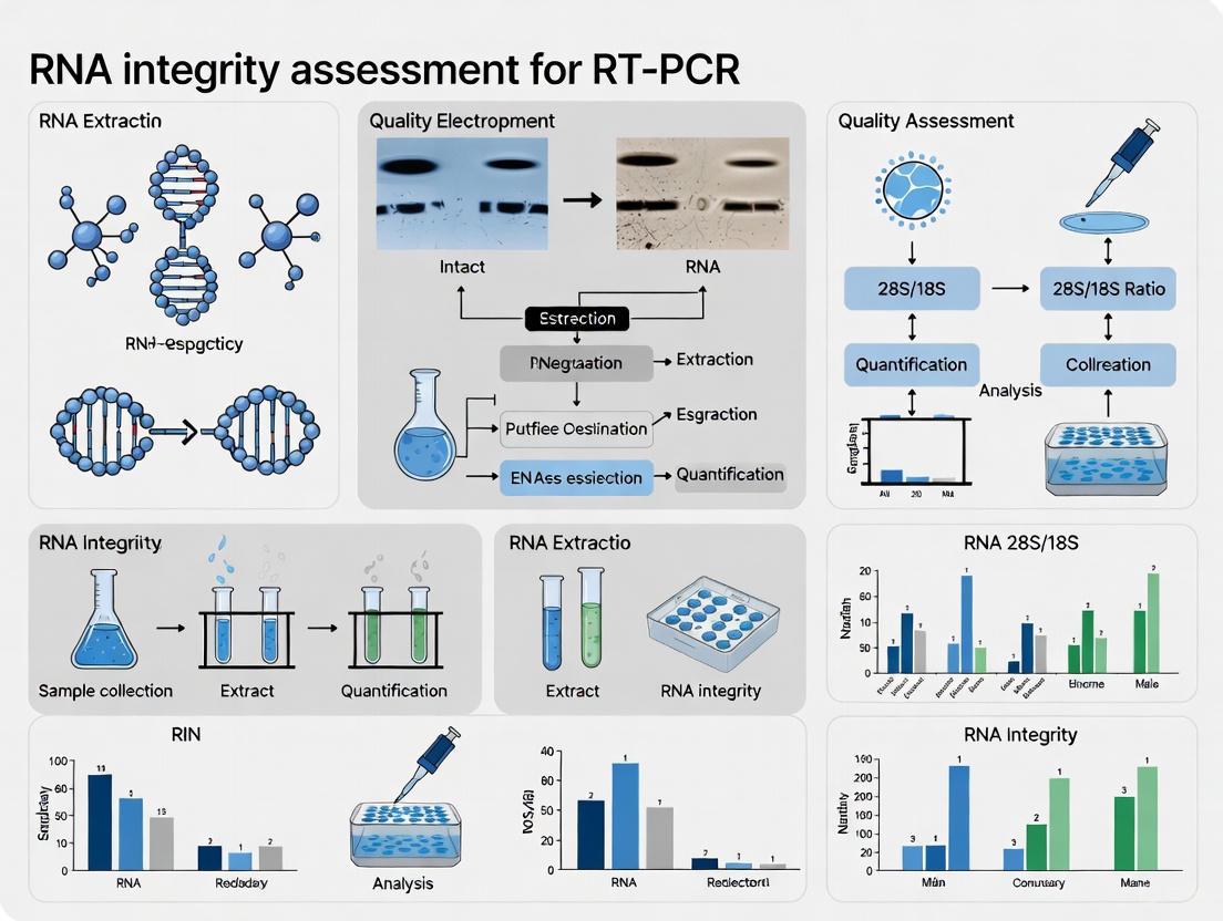
The integrity of Ribonucleic Acid (RNA) is a foundational element in molecular biology research, particularly for techniques that capture a snapshot of gene expression, such as quantitative reverse-transcription PCR (RT-PCR). RNA is a thermodynamically stable molecule but is highly susceptible to rapid degradation by nearly ubiquitous RNase enzymes. This degradation results in shorter RNA fragments that can critically compromise the results and reproducibility of downstream applications [25]. Historically, RNA integrity was assessed using agarose gel electrophoresis, visualizing the banding pattern of ribosomal RNA (rRNA) subunits. The ratio of the 28S to 18S rRNA bands, ideally around 2.0 for mammalian RNA, was the common measure. However, this method is subjective, prone to human interpretation error, and difficult to standardize across laboratories [25] [26]. The advent of microcapillary electrophoresis provided the basis for an automated, high-throughput, and objective approach to RNA quality control, leading to the development of standardized algorithms like the RNA Integrity Number (RIN) [25].
Understanding the Metrics: RIN and RQI
RNA Integrity Number (RIN)
The RIN is a software algorithm developed by Agilent Technologies to assign an integrity value to an RNA sample. It is generated using a Bayesian learning model trained on a large collection of eukaryotic RNA samples analyzed on the Agilent 2100 Bioanalyzer. The algorithm automatically selects features from the electrophoretic trace, or electropherogram, and constructs a regression model to predict integrity, eliminating the subjectivity of manual assessment [25] [27].
The Principle of RIN Calculation: The RIN algorithm moves beyond the simple 28S:18S rRNA ratio. It incorporates a holistic analysis of the entire electropherogram, taking into account the presence of degradation products and other anomalies. Key features used in the algorithm include [25] [26]:
- The total RNA ratio: The ratio of the area under the 18S and 28S rRNA peaks to the total area under the electropherogram.
- The height of the 28S peak: This large rRNA species is often degraded faster than the 18S rRNA.
- The "fast region": The area of the electropherogram representing shorter RNA fragments, which increases as degradation progresses.
- The marker region: The area where very small fragments accumulate, indicating extensive degradation.
RNA Quality Indicator (RQI) and Other Metrics
While RIN is a widely adopted metric, other instrumentation platforms have developed their own proprietary metrics for RNA integrity. For example, Bio-Rad's Experion system uses the RNA Quality Indicator (RQI). Although the exact algorithms differ, the underlying principle is similar: to provide a standardized, numerical assessment of RNA quality based on microfluidic electrophoretic separation. The consistent theme across platforms is the move away from subjective ratios to automated, software-generated scores that offer greater reproducibility and reliability for critical research applications.
Quantitative Interpretation of RIN Scores
The RIN system assigns a numerical value on a scale of 1 to 10, where 10 represents completely intact RNA and 1 represents fully degraded RNA. However, the interpretation of these scores for downstream applications is nuanced. The following table provides a general guideline for RIN score interpretation and its implications for common techniques, including RT-PCR.
Table 1: Interpretation of RIN Scores for Downstream Applications
| RIN Score Range | Integrity Level | Electropherogram Profile | Suitability for Downstream Applications |
|---|---|---|---|
| 9 - 10 | Excellent/Intact | Two sharp, distinct peaks for 28S and 18S rRNA; flat baseline. | Ideal for all sensitive applications, including RNA-Seq and microarrays. |
| 8 - 9 | Good | Clear 28S and 18S peaks; slight elevation in the fast region. | Highly suitable for most applications, including RT-PCR and qPCR. |
| 7 - 8 | Moderate | Visible 28S and 18S peaks, but with a reduced 28S:18S ratio; noticeable baseline shift. | Acceptable for RT-PCR and qPCR, but may affect sensitivity and accuracy of gene expression quantification. |
| 5 - 7 | Partially Degraded | 28S peak significantly diminished or absent; elevated fast region and baseline. | Marginal for RT-PCR; may be used for robust, short-amplicon qPCR targets with prior validation. |
| < 5 | Severely Degraded | No ribosomal peaks visible; high baseline signal with a smear of low molecular weight fragments. | Unsuitable for most gene expression studies, including RT-PCR. |
It is critical to note that while a RIN score of >7 is often considered acceptable for RT-PCR, the success of the experiment can also depend on other factors, such as the length of the target amplicon. Shorter amplicons are more tolerant of partially degraded RNA [27]. Therefore, RIN is a powerful guide but cannot, without prior validation, universally predict the success of a specific experimental setup [27].
Experimental Protocol: Assessing RNA Integrity
Workflow for RNA Quality Control Using Microcapillary Electrophoresis
The following diagram illustrates the standard workflow for preparing and analyzing an RNA sample to determine its RIN score.
Detailed Methodology
This protocol describes the process for using the Agilent 2100 Bioanalyzer, a mainstream instrument for this purpose [25].
4.2.1 Materials and Equipment
- Agilent 2100 Bioanalyzer system: The core instrument for microfluidic analysis.
- Appropriate LabChip kit: (e.g., RNA 6000 Nano or Pico Kit) containing the microfluidic chip, electrodes, syringe, and reagents.
- RNA samples: Diluted to the concentration range specified by the LabChip kit (typically 25-500 ng/µL). Concentrations below 25 ng/µL may yield inconsistent RIN scores [27].
- Thermal cycler or heat block: For sample denaturation.
- Vortex mixer and centrifuge.
4.2.2 Step-by-Step Procedure
- Chip Priming: Assemble the priming station. Place the microfluidic chip in the station. Pipette the gel matrix into the well marked "G". Use the syringe to dispense the gel, ensuring it fills the microchannels. This process is detailed in the kit-specific manual.
- Sample Denaturation: For each RNA sample, prepare a mixture of RNA and dye. A typical reaction uses 1 µL of RNA sample and 2 µL of dye. Mix thoroughly by pipetting. Denature the sample at 70°C for 2 minutes, then immediately cool on ice.
- Loading the Chip: Pipette 9 µL of the marker solution into the well marked with the ladder symbol. Load 5 µL of the marker into each of the 12 sample wells. Finally, pipette 1 µL of each denatured sample into separate sample wells. Avoid introducing air bubbles.
- Vortexing and Running the Chip: Place the chip in the vortex adapter and vortex for 60 seconds at the specified speed (e.g., 2400 rpm). This ensures proper mixing of the samples within the chip. Place the chip into the Agilent 2100 Bioanalyzer and start the run using the associated software.
- Data Analysis: Upon completion, the software will display the results as an electropherogram and a simulated gel image. The RIN number will be automatically calculated and displayed for each sample.
The Researcher's Toolkit for RNA Integrity
Table 2: Essential Research Reagent Solutions for RNA Integrity Analysis
| Item | Function in RNA QC | Key Considerations |
|---|---|---|
| Agilent 2100 Bioanalyzer | Microfluidic capillary electrophoresis system for automated separation, detection, and analysis of RNA samples. | The industry standard for RIN generation. Compatible with Nano and Pico kits for different concentration ranges. |
| RNA Integrity Number (RIN) | Software algorithm that assigns a numerical score (1-10) representing RNA integrity. | Provides an objective, reproducible metric. Superior to traditional 28S:18S ratio. Critical for reporting standards. |
| LabChip Kits (e.g., RNA 6000 Nano/Pico) | Disposable microchips containing gel matrix, dye, and wells for sample loading. | Enables high-throughput analysis of 12 samples per chip. The Pico kit is designed for very low-concentration samples. |
| Fluorescent RNA Dye | Intercalating dye that binds to RNA and is detected via laser-induced fluorescence (LIF) in the bioanalyzer. | Essential for visualizing the RNA fragments. The signal intensity is proportional to the amount of RNA. |
| RNase Inhibitors | Chemical additives or enzyme inhibitors used during RNA extraction and handling to prevent degradation. | Crucial for maintaining high RIN from the moment of cell lysis. Includes RNase-free water, plasticware, and dedicated workspace. |
Implications for RT-PCR Research
Within the context of RT-PCR, RNA integrity is non-negotiable for generating accurate and reliable gene expression data. Degraded RNA can lead to a significant underestimation of gene expression levels because the template for reverse transcription is fragmented [26]. The impact is more pronounced for longer transcript targets. The RIN metric provides a pre-experimental checkpoint, allowing researchers to qualify their input material objectively. By setting a RIN threshold (e.g., ≥7 or ≥8) for RT-PCR experiments, researchers can ensure the technical reproducibility of their data and draw more robust biological conclusions. Integrating RIN assessment as a mandatory step in the RT-PCR workflow is a best practice that strengthens the entire research process, from experimental design to data interpretation and publication.
Within the context of RT-PCR research, the accuracy of gene expression data is fundamentally dependent upon the quality of the starting RNA. Precise nucleic acid quantification and purity assessment are critical preliminary steps, as impurities or inaccurate concentration measurements can lead to failed reactions, non-reproducible results, and erroneous data interpretation [28] [29]. Two principal methodologies are employed for this purpose: spectrophotometry and fluorometry. Spectrophotometry provides a broad assessment of sample concentration and purity by measuring light absorption, while fluorometry offers exceptional sensitivity and specificity for quantifying a particular nucleic acid type through fluorescent dye binding [30] [31]. This application note details the principles, protocols, and comparative performance of these techniques, providing a structured framework for their application in RNA integrity assessment for RT-PCR.
Technical Principles and Comparative Performance
Core Measurement Principles
Spectrophotometry operates on the Beer-Lambert law, measuring the amount of ultraviolet (UV) light absorbed by a sample at specific wavelengths [31]. Nucleic acids display a characteristic absorption peak at 260 nm. The concentration is calculated based on this absorbance value, with an A260 of 1.0 corresponding to approximately 40 µg/mL for single-stranded RNA [29]. This method also calculates purity ratios, notably the A260/A280 ratio for protein contamination (with ~2.0 indicating pure RNA) and the A260/A230 ratio for contaminants like salts or organic compounds [28] [29].
Fluorometry relies on the use of fluorescent dyes that selectively bind to specific nucleic acid structures, such as double-stranded DNA (dsDNA) or RNA. Upon binding, these dyes emit light at a characteristic wavelength when excited by a specific light source. The intensity of the emitted fluorescence is directly proportional to the concentration of the target nucleic acid in the sample [30] [31]. This method does not directly assess sample purity but provides highly accurate quantification of the specific nucleic acid type bound by the dye.
Quantitative Performance Comparison
The following table summarizes the key operational characteristics and performance metrics of spectrophotometry and fluorometry, highlighting their complementary strengths.
Table 1: Comparative Analysis of Spectrophotometry and Fluorometry
| Feature | Spectrophotometry | Fluorometry |
|---|---|---|
| Measurement Principle | Absorbance of UV light [31] | Emission of fluorescent light from dye-bound nucleic acids [31] |
| Key Outputs | Nucleic acid concentration; Purity ratios (A260/A280, A260/230) [29] | Highly specific nucleic acid concentration (e.g., dsDNA, RNA) [30] |
| Sensitivity | Moderate (nanogram range) [30] | High (picogram range) [30] [31] |
| Sample Volume | Very low (1-2 µL) [29] | Small, but requires reagent mix (e.g., 1-20 µL sample) [32] |
| Speed | Very rapid (seconds per sample) [30] | Moderate, requires dye incubation (several minutes) [30] |
| Selectivity | Low; cannot distinguish between DNA, RNA, or free nucleotides [28] [30] | High; dye chemistry can be specific for dsDNA, ssDNA, or RNA [30] |
| Purity Assessment | Yes, via absorbance ratios [29] | No [28] |
| Cost & Complexity | Lower cost; simple operation [31] | Higher cost; requires specific dyes and calibrated standards [31] |
| Dynamic Range (Example) | NanoDrop One: 0.2 - 27,500 ng/µL (dsDNA) [29] | DeNovix Ultra High Sensitivity Assay: 0.5 - 300 pg/µL (dsDNA) [32] |
Experimental Workflow for RNA Integrity Assessment
The following diagram illustrates a recommended integrated workflow for comprehensive RNA sample assessment, combining the strengths of both spectrophotometry and fluorometry.
Detailed Experimental Protocols
Protocol A: Rapid Assessment via Spectrophotometry
This protocol uses a micro-volume spectrophotometer (e.g., NanoDrop, EzDrop) for quick concentration and purity checks [29].
Materials:
- Micro-volume spectrophotometer
- Lint-free wipes
- Nuclease-free water or TE buffer (blanking solution)
- RNA samples
Procedure:
- Initialize the spectrophotometer software and select the "RNA" application.
- Lift the sampling arm and carefully clean the upper and lower pedestals with a lint-free wipe.
- Pipette 1-2 µL of blanking solution onto the lower pedestal. Close the arm.
- Perform the blank measurement by selecting "Blank" in the software.
- Lift the arm, wipe the pedestals clean, and load 1-2 µL of the RNA sample.
- Lower the arm and initiate the sample measurement.
- Record the concentration (in ng/µL) and the A260/A280 and A260/230 ratios.
- Clean the pedestals thoroughly between samples.
Data Interpretation:
- Concentration: Calculated by the instrument based on A260.
- Purity: An A260/A280 ratio of ~2.0 is indicative of pure RNA. Significant deviation suggests protein or solvent contamination. An A260/230 ratio below 2.0 may indicate contamination with salts, EDTA, or carbohydrates [29].
Protocol B: Sensitive RNA Quantitation via Fluorometry
This protocol details RNA quantification using a fluorometer (e.g., Qubit, EzCube) and an RNA-specific assay kit [32].
Materials:
- Fluorometer
- RNA-specific fluorescence quantification assay kit (e.g., DeNovix RNA Assay, AccuGreen)
- RNase-free microcentrifuge tubes
- RNA samples and standards
Procedure:
- Prepare the working solution by diluting the concentrated Quantitation Dye (200X) in the provided Assay Buffer as per kit instructions [32].
- In RNase-free tubes, prepare standards and sample assays in duplicate:
- Standard Tubes: Add working solution and the provided RNA standard.
- Sample Tubes: Add working solution and a defined volume of your RNA sample (e.g., 1-10 µL, depending on expected concentration) [32].
- Mix each tube thoroughly by vortexing and incubate at room temperature for the specified time (typically 2-5 minutes), protected from light.
- While incubating, start the fluorometer and select the appropriate RNA assay protocol.
- After incubation, measure the standards first to generate a calibration curve, followed by the unknown samples.
- Record the concentration reported by the fluorometer software.
Protocol C: Assessing RNA Integrity via the 3'/5' Assay
This qPCR-based method is a highly sensitive functional test for RNA integrity, specifically for RT-PCR applications [33].
Materials:
- Quantitative PCR instrument
- LuminoCt ReadyMix or similar qPCR master mix
- cDNA synthesized from RNA sample using an anchored oligo(dT) primer
- Primers and probes for a target gene (e.g., GAPDH):
- A "3' assay" located near the 3' end of the transcript.
- A "5' assay" located approximately 1 kb upstream.
Procedure:
- cDNA Synthesis: Synthesize cDNA from a fixed amount of RNA (e.g., 100 ng) using an anchored oligo(dT) primer. This ensures reverse transcription starts from the poly-A tail.
- qPCR Setup: Prepare two separate qPCR reactions for each cDNA sample:
- One reaction with the 3' assay primers/probe.
- One reaction with the 5' assay primers/probe.
- qPCR Run: Perform qPCR amplification using optimized conditions.
- Data Analysis: Quantify the results and calculate the 3'/5' ratio for your target gene(s). Intact RNA will produce similar quantification cycle (Cq) values for both assays, resulting in a low 3'/5' ratio. Degraded RNA will show a higher Cq for the 5' assay due to fragmentation, leading to an elevated 3'/5' ratio [33].
Research Reagent Solutions
The following table lists essential materials and their functions for the protocols described.
Table 2: Essential Reagents and Materials for RNA Quality Control
| Item | Function/Description | Example Kits/Models |
|---|---|---|
| Micro-volume Spectrophotometer | Rapidly measures nucleic acid concentration and purity from 1-2 µL samples. | NanoDrop系列 [29], EzDrop系列 [30] |
| Fluorometer | Precisely quantifies specific nucleic acid types (dsDNA, RNA) using fluorescent dyes. | Qubit [28], EzCube系列 [30] |
| RNA Fluorometric Assay Kit | Contains dye, buffer, and standards for RNA-specific quantification on a fluorometer. | DeNovix RNA Assay [32], AccuGreen [28] |
| RNA Integrity Number (RIN) System | Provides an objective score (1-10) of RNA integrity via capillary electrophoresis. | Agilent 2100 Bioanalyzer [34] [2] |
| Anchored Oligo(dT) Primer | Ensures cDNA synthesis initiates from the 5' end of the mRNA poly-A tail for accurate 3'/5' assays. | Sigma O4387 [33] |
| qPCR Master Mix | Pre-mixed solution containing polymerase, dNTPs, and buffer for quantitative PCR. | LuminoCt ReadyMix [33] |
The data and protocols presented confirm that spectrophotometry and fluorometry are not mutually exclusive but are complementary techniques that should be used in tandem for critical RNA integrity assessment in RT-PCR research [28] [2]. Spectrophotometry is an indispensable first step for its rapid purity assessment, flagging samples contaminated with proteins or solvents that could inhibit enzymatic reactions [29]. However, its lack of specificity means it can overestimate functional RNA concentration in the presence of contaminants or other nucleic acids [28]. Fluorometry addresses this limitation by providing a highly accurate measurement of the actual RNA concentration, a crucial parameter for normalizing input across RT-PCR reactions [30] [31].
For the most demanding applications like RT-PCR, relying solely on spectrophotometry is insufficient. A comprehensive quality control workflow, as illustrated, should integrate both techniques. A sample with a good A260/A280 ratio (~2.0) and a high concentration as measured by fluorometry is a prime candidate for downstream use. For an added layer of confidence, especially with valuable or limited samples, the 3'/5' qPCR assay provides a functional integrity check that directly correlates with RT-PCR performance [33]. In conclusion, leveraging the combined strengths of spectrophotometry for purity and fluorometry for accurate quantification provides researchers with a robust strategy to ensure the reliability and reproducibility of their gene expression data.
The Limitations of rRNA Banding Patterns for mRNA Integrity Assessment
For decades, the assessment of RNA integrity has been a critical first step in gene expression analysis, with profound implications for the validity of downstream results in RT-PCR research, drug discovery, and clinical diagnostics. The scientific community has widely relied on ribosomal RNA (rRNA) banding patterns, visualized through denaturing agarose gel electrophoresis or microfluidic capillary electrophoresis, as a proxy for overall RNA quality [34] [35] [36]. This approach typically evaluates the sharpness and intensity ratio (approximately 2:1) of the 28S and 18S rRNA bands in eukaryotic samples, with the RNA Integrity Number (RIN) algorithm providing a standardized score from 1 (degraded) to 10 (intact) [35] [37].
However, within the context of modern molecular research—particularly studies focused on protein-coding genes—this traditional method presents significant limitations. This application note examines the technical and theoretical constraints of relying on rRNA banding patterns for mRNA integrity assessment and provides detailed protocols for implementing more direct and reliable alternatives suitable for pharmaceutical development and clinical research settings.
Key Limitations of rRNA-Based Assessment Methods
Fundamental Structural and Stability Differences
The core limitation of rRNA-based integrity assessment lies in the fundamental structural and functional differences between ribosomal RNA and messenger RNA:
- Structural composition: rRNAs form complex, compact secondary and tertiary structures with numerous post-transcriptional modifications that confer exceptional stability within functional ribosomes [37]. In contrast, mRNA exhibits a more linear structure without such extensive stabilization, making it inherently more susceptible to ribonuclease degradation and environmental stressors [37].
- Differential degradation rates: When exposed to RNases or environmental stressors such as elevated temperature, rRNA and mRNA degrade at different rates due to their structural differences [37]. Consequently, rRNA integrity may remain high while mRNA is significantly degraded, creating a false assurance of sample quality for gene expression studies.
Technical Limitations in Specialized Applications
- rRNA-deficient samples: Subcellular fractions, purified organelles, and synaptosomal preparations often lack sufficient rRNA for reliable assessment using traditional methods [37]. In such samples, rRNA-based quality metrics cannot be applied, necessitating alternative approaches.
- Low-input samples: While microfluidic capillary electrophoresis systems (e.g., Agilent 2100 Bioanalyzer) have improved sensitivity for precious clinical samples, the requirement for specialized equipment and chips may present practical limitations in resource-constrained settings [34] [35].
Table 1: Comparative Analysis of RNA Integrity Assessment Methods
| Method | Target Analyte | Sample Requirements | Key Limitations | Suitable Applications |
|---|---|---|---|---|
| Agarose Gel Electrophoresis | rRNA | 200 ng - 1 µg total RNA [34] [35] | Subjective interpretation; cannot detect mRNA degradation; requires significant RNA input [34] [37] | Basic RNA quality check when mRNA integrity is not critical |
| Microfluidic Capillary Electrophoresis (RIN) | rRNA | 5-500 ng/µL (RNA 6000 Nano assay) [35] | Poor correlation with mRNA integrity; expensive equipment; not suitable for rRNA-deficient samples [37] | Standard quality control for total RNA samples with sufficient rRNA content |
| 5':3' RT-qPCR Assay | mRNA | Varies by protocol; suitable for low-input samples [37] | Requires reference gene selection and primer optimization; not suitable for massively parallel applications [37] | Gene expression studies; subcellular fractions; clinical samples with limited material |
Superior Alternative Methods for mRNA Integrity Assessment
The 5':3' RT-qPCR Integrity Assay
The 5':3' RT-qPCR assay directly measures mRNA integrity by comparing the abundance of 5' and 3' fragments of a reference transcript, providing a targeted assessment specifically relevant to gene expression studies [38] [37].
Principle of Operation: The assay utilizes oligo-dT primers for reverse transcription, which bind to the polyadenylated tail of mature mRNA. Two sets of qPCR primers then quantify amplicons from the 5' and 3' regions of a long, constitutively expressed reference gene (e.g., PGK1) [37]. In intact mRNA, reverse transcription proceeds uninterrupted, generating full-length cDNA and resulting in approximately equal amplification of both regions. In degraded samples, fragmentation between the poly(A) tail and 5' region reduces the number of full-length transcripts, leading to disproportionately lower amplification of the 5' fragment compared to the 3' fragment [37].
Integrity Score Calculation: The 5':3' integrity value is calculated by dividing the efficiency-corrected quantity of the 5' amplicon by that of the 3' amplicon and multiplying by 10, producing a score from 10 (intact mRNA) to 0 (completely degraded mRNA) [37]. This scaling aligns with familiar RIN metrics while providing mRNA-specific integrity assessment.
Diagram 1: 5':3' mRNA Integrity Assay Workflow - This diagram illustrates the fundamental principle of the 5':3' assay, showing how intact and degraded mRNA molecules yield different patterns of cDNA synthesis and qPCR amplification, resulting in corresponding integrity scores.
RNA Disruption Assay (RDA) for Chemotherapy Response Monitoring
In clinical oncology research, particularly in cancer therapy response assessment, the RNA Disruption Assay (RDA) has emerged as a specialized tool that actually leverages rRNA degradation patterns as a biomarker for treatment efficacy [39]. Unlike traditional rRNA assessment that assumes intact rRNA indicates good quality, RDA quantitatively measures chemotherapy-induced rRNA fragmentation in tumor cells, which correlates with cell death and predicts treatment outcomes including complete tumor destruction and improved disease-free survival in breast cancer patients [39].
Table 2: Research Reagent Solutions for Advanced RNA Integrity Assessment
| Reagent/Kit | Primary Function | Application Context | Key Considerations |
|---|---|---|---|
| PGK1 Primer Sets (mouse/human) [37] | Amplification of 5' and 3' regions of PGK1 transcript for integrity assessment | 5':3' mRNA integrity assay; suitable for human and mouse brain tissue and subcellular fractions [37] | Requires efficiency correction; primers designed to span exon-exon junctions to avoid genomic DNA amplification [37] |
| Ribo-Zero Gold/Globin-Zero Kits [40] | Depletion of ribosomal RNA prior to RNA sequencing | RNA-seq library preparation from blood and tissue samples; enhances coverage of protein-coding transcripts [40] | More expensive than polyA+ selection; captures both polyA+ and polyA- transcripts including immature RNAs [40] |
| RNA 6000 Nano/Pico LabChip Kits (Agilent) [35] | Microfluidic capillary electrophoresis for RNA quality and quantity assessment | Standard quality control for total RNA samples; requires only nanogram to picogram amounts of RNA [35] | Primarily assesses rRNA integrity; limited correlation with mRNA quality [37] |
| SYBR Gold/SYBR Green II Stains [34] [36] | Fluorescent nucleic acid staining for gel-based RNA visualization | Sensitive detection of RNA in agarose gels; alternative to ethidium bromide with improved safety profile [34] [36] | 2.4X (SYBR Green II) to 7.9X (SYBR Gold) more sensitive than ethidium bromide; lower sample requirement [36] |
Detailed Experimental Protocols
Protocol 1: 5':3' mRNA Integrity Assay for Human Brain Tissue
This protocol adapts the methodology from recent studies demonstrating successful application in human and mouse brain tissues and synaptosomal preparations [37].
Step 1: RNA Sample Preparation and DNase Treatment
- Extract total RNA using your preferred method (e.g., guanidinium isothiocyanate-based isolation followed by organic extraction or column purification) [35].
- Treat 1-2 µg of total RNA with DNase I (RNase-free) to remove contaminating genomic DNA. Use 1 unit of DNase per µg of RNA in a 20 µL reaction with the supplied buffer. Incubate at 37°C for 30 minutes [37].
- Inactivate DNase by adding 2 µL of 25 mM EDTA and heating at 65°C for 10 minutes.
- Quantify RNA concentration using a fluorescence-based method (e.g., RiboGreen assay) for accurate measurement [36].
Step 2: Reverse Transcription with Oligo-dT Priming
- Use 100-500 ng of DNase-treated RNA in a 20 µL reverse transcription reaction.
- Add 1 µL of oligo-dT primer (50 µM) and 1 µL of dNTP mix (10 mM each).
- Incubate at 65°C for 5 minutes, then place on ice for 2 minutes.
- Add 4 µL of 5X reverse transcription buffer, 1 µL of RNase inhibitor, and 1 µL of reverse transcriptase.
- Incubate at 42°C for 50 minutes, followed by enzyme inactivation at 70°C for 15 minutes.
- Dilute cDNA with nuclease-free water to a final volume of 100 µL.
Step 3: Quantitative PCR with Efficiency-Corrected Primers
- Design primer pairs for 5' and 3' regions of a suitable reference gene (PGK1 recommended for human and mouse studies) [37]. Ensure one primer in each pair spans an exon-exon junction to prevent genomic DNA amplification.
- Determine primer efficiency using serial dilutions of a plasmid containing the cloned cDNA fragment or known intact RNA samples [37].
- Prepare qPCR reactions in triplicate for each sample and primer pair: 10 µL of 2X master mix, 0.5 µL of each primer (10 µM), 2 µL of diluted cDNA, and 7 µL of nuclease-free water.
- Use the following cycling conditions: 95°C for 10 minutes, followed by 40 cycles of 95°C for 15 seconds and 60°C for 1 minute.
Step 4: Integrity Value Calculation
- Calculate the efficiency-corrected quantity for each amplicon using the formula: Q = E^(ΔCq), where E is the primer efficiency and ΔCq is the difference between the sample Cq and a reference Cq [37].
- Compute the 5':3' integrity value using the formula: Integrity = (Q5' / Q3') × 10 [37].
- Interpret results: Values接近10 indicate intact mRNA; values below 5 suggest significant degradation that may compromise gene expression results.
Protocol 2: rRNA Depletion for RNA-seq of Clinical Blood Samples
This protocol is adapted from comparative studies of RNA-seq approaches for gene quantification in clinical samples [40].
Step 1: RNA Quality Assessment
- Evaluate total RNA quality using the Agilent 2100 Bioanalyzer with RNA 6000 Nano Kit to ensure initial RIN >7 for blood samples [40].
- Quantify RNA using fluorescence-based methods (e.g., RiboGreen assay) for accurate concentration measurement [36].
Step 2: rRNA Depletion with Globin-Zero Kit
- Use 100-500 ng of total RNA as input for the Ribo-Zero Globin depletion protocol following manufacturer's instructions.
- Incubate RNA with rRNA removal solution at 68°C for 10 minutes, then at room temperature for 5 minutes.
- Add magnetic beads, incubate at room temperature for 5 minutes, and place on magnetic stand to separate rRNA-bound beads.
- Transfer supernatant containing rRNA-depleted RNA to a new tube.
Step 3: RNA Clean-up and Library Preparation
- Purify the rRNA-depleted RNA using RNA clean-up beads or columns.
- Proceed with standard RNA-seq library preparation using fragmentation, reverse transcription, and adapter ligation.
- For blood-derived RNA, note that approximately 220% more sequencing reads are required with rRNA depletion compared to polyA+ selection to achieve equivalent exonic coverage [40].
Diagram 2: RNA Integrity Assessment Strategy Decision Tree - This workflow guides researchers in selecting the most appropriate RNA integrity assessment method based on their specific sample type and research objectives.
Application in Drug Development and Clinical Research
The implementation of mRNA-specific integrity assessment methods has significant implications for pharmaceutical research and clinical development:
Biomarker Discovery: In cancer research, the RNA Disruption Assay (RDA) has been used to measure chemotherapy-induced rRNA degradation in tumors, with higher disruption indices correlating with improved treatment response and disease-free survival in breast cancer patients [39]. This application actually leverages rRNA degradation as a positive biomarker rather than a quality concern.
Clinical Trial Quality Assurance: For mRNA-based therapeutic development, regulatory agencies have expressed concerns about batch-to-batch variability in intact mRNA content, with some commercial batches containing >55% intact mRNA [41]. Implementing direct mRNA integrity assessment ensures consistent product quality and therapeutic efficacy.
Adaptive Clinical Trials: RNA disruption measurements during neoadjuvant chemotherapy may inform treatment escalation or de-escalation decisions, potentially serving as an early response biomarker for adaptive trial designs [39].
Traditional rRNA banding patterns and derived metrics such as RIN provide inadequate assessment of mRNA integrity, potentially compromising gene expression studies and clinical research outcomes. The 5':3' RT-qPCR assay offers a targeted approach specifically evaluating mRNA integrity, while specialized methods like the RNA Disruption Assay leverage rRNA fragmentation as a therapeutic response biomarker in oncology. Researchers should select integrity assessment methods based on their specific sample types and research objectives, with mRNA-directed approaches providing superior relevance for gene expression studies in drug development and clinical research settings.
Practical Guide to RNA Integrity Assessment Methods
The integrity of RNA is a critical parameter in gene expression analysis, as it directly impacts the accuracy and reliability of downstream applications, including quantitative real-time RT-PCR and RNA sequencing [42] [5]. Degraded RNA can lead to skewed quantification, false results, and ultimately, erroneous scientific conclusions. Historically, RNA integrity was assessed using denaturing agarose gel electrophoresis, relying on the visual inspection of 28S and 18S ribosomal RNA bands and the calculation of their ratio, which is considered to be approximately 2:1 for intact eukaryotic RNA [34]. However, this method is subjective, requires a significant amount of sample, and lacks digital output for standardized comparison [34] [5].
The advent of microfluidic capillary electrophoresis has revolutionized RNA quality control by providing an automated, objective, and quantitative assessment. This technique, implemented on platforms like the Agilent 2100 Bioanalyzer, separates RNA molecules based on size within microfabricated channels and employs laser-induced fluorescence (LIF) for detection [34] [5]. The output is both a gel-like image and an electropherogram, which provides a detailed profile of the RNA species in the sample. To standardize the interpretation of these electropherograms, the RNA Integrity Number (RIN) was developed. The RIN is a software-generated algorithm that assigns an integrity value on a scale of 1 to 10, with 10 representing completely intact RNA [5]. This application note details the principles, protocols, and critical importance of using microfluidic capillary electrophoresis for RIN assignment within the context of RT-PCR research.
The Principle of RIN Assignment
The RIN algorithm represents a significant advancement over the traditional 28S/18S rRNA ratio. It is a sophisticated tool trained using a large collection of RNA electrophoretic measurements from various tissues and organisms. The algorithm employs machine learning methods, based on a Bayesian learning framework, to analyze the entire electrophoretic trace rather than just the two ribosomal bands [5].
Feature Analysis
The algorithm automatically selects and weighs multiple features from the electropherogram to compute the RIN. These features capture the complex changes in the trace that occur during degradation, which include:
- A decrease in the height of the 28S and 18S ribosomal peaks.
- An increase in the baseline signal between the 18S and 28S peaks, representing ribosomal fragments.
- An increase in the signal from low molecular weight fragments below the 18S band.
- The overall shift of the signal towards smaller fragment sizes [5].
By integrating information from these multiple regions, the RIN provides a robust and reliable prediction of RNA integrity that is far superior to the simple ribosomal ratio, which has been shown to be an inconsistent measure, especially for partially degraded samples [5].
The RIN Scale
The RIN scale is designed to categorize RNA samples based on their degree of degradation:
- RIN 10-9: Highly intact RNA. The electropherogram shows sharp, distinct ribosomal bands with a flat baseline and a high 28S/18S ratio.
- RIN 8-7: Good quality RNA. Suitable for most downstream applications, including RT-PCR.
- RIN 6-5: Partially degraded RNA. The ribosomal peaks are less distinct, and the baseline is elevated. Its use in gene expression studies requires caution and appropriate normalization strategies [42].
- RIN <5: Heavily degraded RNA. The ribosomal peaks are largely absent, and the electropherogram is dominated by a low molecular weight smear. Such samples are generally not suitable for quantitative analyses like RT-PCR.
Table 1: Comparison of RNA Integrity Assessment Methods
| Feature | Agarose Gel Electrophoresis | Microfluidic Capillary Electrophoresis (RIN) |
|---|---|---|
| Sample Consumption | High (≥ 200 ng) [34] | Very Low (as little as 5 ng total) [34] [5] |
| Output Format | Analog gel image | Digital electropherogram and gel-like image |
| Analysis Basis | 28S/18S rRNA ratio (subjective) | Multi-feature algorithm (objective) [5] |
| Throughput | Low | High (up to 12 samples per chip) [5] |
| Standardization | Low (user-dependent) | High (automated, software-generated score) |
| Sensitivity | Limited, requires ethidium bromide or similar | High, uses laser-induced fluorescence [34] |
Materials and Methods: Experimental Protocol for RIN Analysis
This protocol is adapted for systems like the Agilent 2100 Bioanalyzer using the RNA 6000 Nano LabChip kit.
Research Reagent Solutions
The following table lists the essential materials required for the analysis.
Table 2: Essential Research Reagents and Materials
| Item | Function / Description |
|---|---|
| Agilent 2100 Bioanalyzer | Instrumentation for microfluidic capillary electrophoresis and analysis. |
| RNA 6000 Nano LabChip | Disposable microfluidic chip containing interconnected channels and wells for sieving polymer and samples. |
| RNA Gel Matrix | A sieving polymer (e.g., polydimethylacrylamide) that separates RNA fragments by size during electrophoresis. |
| RNA Marker / Dye | An intercalating fluorescent dye (e.g., ethidium bromide or a proprietary dye) that stains RNA for LIF detection. |
| RNA Ladder | A standardized mixture of RNA fragments of known sizes used to calibrate the system and create a standard curve for sizing. |
| RNA 6000 Nano Marker | A solution used to prepare both the ladder and samples, ensuring consistent conditions. |
| Heating Block | Used to incubate samples and the gel-dye mix as per protocol specifications. |
| Vortexer and Centrifuge | For mixing and dispensing reagents into the chip wells without air bubbles. |
Step-by-Step Procedure
Chip Preparation:
- Place the LabChip on the priming station.
- Pipette 9 µL of the RNA gel matrix into the well marked with a "G" symbol. Ensure the pipette tip is seated against the bottom of the well.
- Close the priming station and press the plunger until it is held by the clip. Wait for exactly 30 seconds.
- Release the priming clip and wait for a further 5 seconds before opening the station.
- Pipette 9 µL of the gel matrix into the two other "G" wells on the chip.
Loading Ladder and Samples:
- Pipette 5 µL of the RNA 6000 Nano Marker into the ladder well and each of the 12 sample wells.
- Pipette 1 µL of the RNA ladder into the ladder well. Pipette 1 µL of each RNA sample into the respective sample wells.
- Avoid introducing air bubbles during pipetting, as they can disrupt the electrophoretic run.
Sample Preparation:
- The RNA samples and ladder are typically denatured by heating at 70°C for 2 minutes and then immediately cooled on ice prior to loading to remove secondary structures.
Vortexing and Centrifugation:
- Place the loaded chip into a vortex adapter and vortex for 1 minute at approximately 2400 rpm.
- Centrifuge the chip briefly to ensure all liquid is at the bottom of the wells and no air bubbles are trapped.
Electrophoretic Run:
- Place the chip into the Agilent 2100 Bioanalyzer and start the run using the associated software.
- The instrument will automatically apply a voltage, separating the RNA fragments through the gel matrix in the microfluidic channels. The fragments pass the LIF detector, generating the electropherogram data.
Data Analysis and RIN Assignment:
- After the run is complete (typically within 30 minutes), the software automatically analyzes the electropherograms, sizes the fragments based on the ladder, and assigns a RIN to each sample.
Diagram 1: RIN Analysis Workflow
Impact of RNA Integrity on RT-PCR Research
The integrity of the starting RNA template is a fundamental pre-analytical variable that significantly influences RT-PCR results. RNA degradation is not a uniform process; it often occurs in a 5' to 3' directional bias. This means that the 5' end of mRNA transcripts degrades faster than the 3' end [43].
Consequences for Quantification
This differential degradation directly impacts relative mRNA quantification in RT-PCR:
- Underestimation of Expression: If the PCR amplicon is located near the 5' end of a transcript, its amplification will be less efficient in a degraded sample compared to an amplicon located near the 3' end. This can lead to a systematic underestimation of the gene's expression level.
- Distorted Results: The use of reference genes (housekeeping genes) for normalization can be severely compromised if their degradation rates differ from those of the target genes. A reference gene with a stable 3' end might show consistent expression, while a target gene with a degraded 5' end would appear down-regulated, leading to incorrect conclusions [42].
The 3'/5' Assay for Integrity
A specialized qPCR-based method, the 3'/5' assay, can be used to detect this form of degradation. In this assay, two sets of primers and probes are designed for the same gene: one near the 3' end and one approximately 1 kb upstream near the 5' end. cDNA is synthesized using an anchored oligo-dT primer, which binds to the poly-A tail at the 3' end. In an intact RNA sample, the ratio of the quantification results (3'/5') should be close to 1. As the RNA degrades, the 5' target is less efficiently amplified, leading to an increase in the 3'/5' ratio [43]. This provides a sensitive, sequence-specific integrity check that complements the RIN.
Table 3: Relationship Between RIN, Electropherogram Profile, and Suitability for RT-PCR
| RIN Range | Electropherogram Profile | Suitability for RT-PCR |
|---|---|---|
| 10 - 9 | Sharp 28S and 18S peaks; 28S peak ~2x height of 18S; flat baseline. | Excellent. Ideal for all applications, including long amplicons and 5' targets. |
| 8 - 7 | Clear ribosomal peaks; slight baseline elevation between peaks. | Good. Suitable for most applications. Amplicons should be kept relatively short (<500 bp). |
| 6 - 5 | Ribosomal peaks less distinct; significant baseline elevation; lower 28S/18S ratio. | Moderate to Poor. Requires careful validation. Use short amplicons and stable reference genes validated for degradation. [42] |
| < 5 | Ribosomal peaks largely disappeared; dominant low molecular weight smear. | Not Recommended. Data from such samples is highly unreliable for quantitative gene expression. |
Diagram 2: Impact of RNA Integrity on qPCR
Microfluidic capillary electrophoresis, coupled with the RIN algorithm, has become the gold standard for RNA integrity assignment in modern molecular biology. It provides an automated, objective, and highly reproducible quality control metric that is essential for ensuring the validity of gene expression data, particularly in sensitive applications like RT-PCR. By replacing subjective gel-based methods with a quantitative digital score, the RIN empowers researchers to make informed decisions about their samples, select appropriate normalization strategies, and ultimately, generate more reliable and reproducible scientific data. The integration of RIN analysis as a mandatory step in the RNA workflow is a best practice for any rigorous RT-PCR research program.
The accuracy of reverse transcription quantitative polymerase chain reaction (RT-qPCR) data is highly dependent on the quality of the starting RNA material. Compromised RNA integrity is a significant source of bias, potentially leading to unreliable gene expression results and incorrect biological conclusions [44]. Unlike ribosomal RNA (rRNA), messenger RNA (mRNA) possesses a more linear structure and is more prone to degradation by environmental RNases. Because most gene expression studies focus on mRNA, directly assessing its integrity is paramount [37].
The 5'-3' mRNA integrity assay is a powerful QC tool that directly probes the integrity of a specific mRNA molecule, independently of rRNA. This method is particularly valuable when a large number of samples need analysis, or when degradation is subtle enough to escape detection by capillary electrophoresis systems but sufficient to affect qPCR results [37] [45]. This application note details the protocol and considerations for implementing this assay using HPRT1 and other reference genes, providing a critical component for robust RNA integrity assessment within RT-qPCR research.
Principle of the 5'-3' mRNA Integrity Assay
Core Concept and Workflow
The 5'-3' assay estimates mRNA integrity by comparing the abundance of 5' and 3' fragments from a long, constitutively expressed mRNA. The fundamental principle is that degradation occurs randomly along the mRNA transcript. In a partially degraded sample, the 5' end of the transcript is less represented in cDNA synthesized from an oligo(dT) primer because reverse transcription cannot proceed through breaks in the RNA strand [37] [45].
The following workflow illustrates the experimental process and underlying logic of the 5'-3' integrity assay:
Comparison of RNA Quality Assessment Methods
Researchers have several options for assessing RNA quality, each with strengths and limitations. The table below summarizes the key characteristics of major methodologies:
Table 1: Comparison of Common RNA Integrity Assessment Methods
| Method | Principle | What is Measured | Key Advantages | Key Limitations |
|---|---|---|---|---|
| 5'-3' mRNA Integrity Assay | qPCR of 5' and 3' ends of a specific mRNA | Integrity of a protein-coding mRNA transcript | Directly measures mRNA integrity; suitable for subcellular fractions lacking rRNA; highly sensitive [37]. | Requires prior validation; measures only the target mRNA. |
| RIN (Bioanalyzer) | Microfluidic capillary electrophoresis | Integrity of 18S and 28S ribosomal RNA | Gold standard; provides a snapshot of total RNA population; objective algorithm [46]. | Assumes rRNA integrity reflects mRNA integrity; may not be suitable for rRNA-lacking samples [37]. |
| RINe (TapeStation) | Microfluidic capillary electrophoresis | Integrity of ribosomal RNA | Higher throughput (96 samples/run) than Bioanalyzer [46]. | Algorithm different from RIN; values not directly interchangeable with RIN [46]. |
| DV200 | Microfluidic capillary electrophoresis | Percentage of RNA fragments >200 nucleotides | Useful for FFPE samples; simple metric [46]. | Does not provide information on the intactness of specific transcripts. |
| Gel Electrophoresis | Agarose gel separation | 28S:18S rRNA ratio | Low-cost; provides visual profile of RNA [46]. | Sample-demanding; subjective interpretation; low resolution [46]. |
Selecting a Reference Gene for the 5'-3' Assay
Candidate Genes and Selection Criteria
The choice of mRNA target is critical for a reliable 5'-3' assay. An ideal reference gene should be long, constitutively expressed, and stable across the experimental conditions of interest. Commonly used genes include HPRT1, PGK1, and GAPDH [44] [37] [45]. Using a long transcript ensures sufficient distance between the 5' and 3' primer pairs to detect degradation-induced breaks [37].
HPRT1 as a Prime Candidate
Evidence supports HPRT1 (Hypoxanthine Phosphoribosyltransferase 1) as an excellent candidate for this assay. A comprehensive study on RNA quality's impact on qPCR identified the HPRT1 5'-3' difference in quantification cycle (Cq) as a valuable parameter for assessing RNA integrity [44]. Furthermore, HPRT1 has been validated as a highly stable reference gene for mRNA expression normalization in challenging contexts, such as in canine dermal tissues post-radiation therapy, underscoring its robust expression stability [47].
The Scientist's Toolkit: Essential Reagents
The following table lists key reagents required to perform the 5'-3' mRNA integrity assay.
Table 2: Research Reagent Solutions for the 5'-3' mRNA Integrity Assay
| Reagent / Tool | Function / Description | Example Product / Note |
|---|---|---|
| High-Quality RNA Sample | The sample of interest; should have RIN > 6.5 as a starting point for reliable RT-qPCR [48]. | Verify purity and lack of genomic DNA contamination. |
| Anchored Oligo-dT Primers | For cDNA synthesis; primes from the poly-A tail of mRNA. | Ensures synthesis is initiated from the 3' end. |
| Reverse Transcriptase Enzyme | Synthesizes cDNA from the mRNA template. | Use a high-fidelity enzyme. |
| qPCR Master Mix | Contains DNA polymerase, dNTPs, buffers, and fluorescent dye for detection. | e.g., SYBR Green premix [49]. |
| Primer Pairs for 5' and 3' Ends | Gene-specific primers to amplify regions near the 5' and 3' ends of the target mRNA. | Designed for high and equal efficiency; one primer should span an exon-exon junction [37]. |
| DNase I, RNase-free | Removes contaminating genomic DNA from the RNA sample prior to cDNA synthesis. | Critical for accurate results. |
| Quantitative PCR Instrument | Platform to run and detect the qPCR reaction in real-time. | e.g., TaKaRa PCR Thermal Cycler Dice [49]. |
Detailed Experimental Protocol
Primer Design and Validation
- Target Selection: Choose a suitable reference gene (e.g., HPRT1, PGK1).
- Primer Design: Design two primer pairs:
- One pair amplifying a region close to the 5' end of the mature mRNA.
- One pair amplifying a region close to the 3' end.
- Amplicon sizes should be similar (recommended: 100-110 bp) [37].
- To avoid false positives from genomic DNA contamination, design at least one primer in each pair to span an exon-exon junction [37].
- Efficiency Validation: It is critical to determine the amplification efficiency (E) for both the 5' and 3' primer pairs using a serial dilution of a synthetic template or high-quality cDNA. The assay's accuracy heavily relies on both pairs having equal and near-perfect (90-110%) amplification efficiency. Correct for any efficiency differences in the final calculation [37].
cDNA Synthesis
- Treat total RNA with DNase I to remove genomic DNA.
- Synthesize cDNA using an anchored oligo-dT primer and a reverse transcriptase enzyme. Using an oligo-dT primer ensures that the cDNA synthesis starts from the 3' end of the poly(A) tail, which is essential for detecting 5' degradation.
- Dilute the synthesized cDNA 1:10 for use in the qPCR reaction [45].
Quantitative PCR (qPCR)
- Prepare two separate qPCR master mixes for each cDNA sample—one containing the 5'-end primer pair and the other containing the 3'-end primer pair.
- Run reactions in triplicate for each sample and primer pair. Include a no-template control (NTC) for each primer pair to check for contamination.
- Use a hot-start Taq polymerase and a three-step amplification protocol (e.g., denaturation at 95°C, annealing at 60°C, extension at 72°C) for 40 cycles, unless a two-step protocol is validated for your primers [45].
Table 3: Example qPCR Reaction Setup
| Component | Volume per Reaction (μL) | Final Concentration/Amount |
|---|---|---|
| 2x qPCR Master Mix (e.g., SYBR Green) | 10.0 μL | 1X |
| Forward Primer (e.g., 50 μM) | 0.2 μL | 0.5 μM |
| Reverse Primer (e.g., 50 μM) | 0.2 μL | 0.5 μM |
| PCR-grade Water | 4.6 μL | - |
| Template cDNA (diluted 1:10) | 5.0 μL | - |
| Total Reaction Volume | 20.0 μL | - |
Data Analysis and Interpretation
Calculating the Integrity Value
After the qPCR run, obtain the quantification cycle (Cq) values for both the 5' and 3' amplicons. The integrity value is calculated by correcting the raw Cq values for primer efficiency and then determining the ratio.
The formula for the integrity score (IS) is [37]: IS = [E{3'}^{-Cq(3')} / E{5'}^{-Cq(5')}] * F Where:
- E_{3'} and E_{5'} are the amplification efficiencies of the 3' and 5' primer pairs, respectively.
- Cq(3') and Cq(5') are the mean Cq values for the 3' and 5' amplicons.
- F is a scaling factor (e.g., 10) to bring the score to a familiar 1-10 scale, where 10 represents intact mRNA and lower values indicate degradation [37].
Interpreting Results
The relationship between the 5':3' ratio and RNA integrity is illustrated below. A low ratio indicates preferential loss of the 5' end, a hallmark of mRNA degradation.
For a reliable RT-qPCR experiment, it is essential to establish a minimum acceptable integrity score for your samples based on the requirements of your downstream application and the performance of your assay.
The 5'-3' mRNA integrity assay using reference genes like HPRT1 provides a targeted, sensitive, and direct method for evaluating mRNA quality, which is indispensable for generating accurate and reproducible RT-qPCR data. By implementing this protocol and establishing sample-specific quality thresholds, researchers can significantly reduce a major source of variability, thereby enhancing the reliability of their gene expression findings.
The molecular analysis of formalin-fixed paraffin-embedded (FFPE) tissues represents a cornerstone of modern biomedical research, particularly in oncology and biomarker discovery. These archival samples, accompanied by extensive clinical follow-up data, are an invaluable resource for investigating disease-associated alterations in gene expression [50]. However, RNA extracted from FFPE specimens presents significant analytical challenges due to extensive chemical modification and degradation that occurs during fixation and storage [50] [51]. Conventional RNA quality assessment methods, such as ribosomal RNA ratios or spectrophotometric measurements, fail to provide accurate information about the functional quality of RNA for reverse transcription polymerase chain reaction (RT-PCR) applications [50] [52].
Multiplex endpoint RT-PCR has emerged as a robust solution for quality assessment of FFPE-derived RNA. This technique enables researchers to evaluate RNA integrity, determine suitable amplicon sizes for downstream applications, and maximize the utility of often limited RNA samples [50] [53]. By simultaneously amplifying multiple target sequences of varying lengths, this approach provides a comprehensive assessment of RNA quality that directly correlates with performance in gene expression studies [50] [51]. This application note details standardized protocols and analytical frameworks for implementing multiplex endpoint RT-PCR in research settings, with particular emphasis on FFPE-derived RNA analysis.
Background and Principles
The Challenge of FFPE-Derived RNA
The process of formalin fixation introduces fundamental alterations to RNA molecules that profoundly impact downstream molecular applications. Formaldehyde causes chemical modifications through the addition of mono-methylol groups to RNA bases and the formation of methylene bridges between RNA bases and proteins [50]. These modifications, combined with fragmentation influenced by pre-fixation ischemia time, fixation duration, and storage conditions, result in RNA fragments typically less than 300 base pairs [50]. The fragmentation pattern is critical because it determines the maximum amplifiable fragment length possible in subsequent RT-PCR applications [50] [51].
Traditional RNA quality assessment methods exhibit significant limitations when applied to FFPE samples. The 28S/18S ribosomal RNA ratio, commonly used for intact RNA, is unsuitable for highly degraded FFPE RNA where ribosomal bands are often indistinguishable [50]. Similarly, spectrophotometric ratios (A260/A280 and A260/A230) provide information about purity but no insight into degradation levels or amplifiable fragment sizes [50]. The 3':5' ratio assay requires substantial fragment lengths (up to 1.2 kb) that exceed the typical length of FFPE-derived RNA fragments [50]. These limitations underscore the need for specialized quality assessment methods tailored to degraded RNA.
Multiplex Endpoint RT-PCR Fundamentals
Multiplex endpoint RT-PCR addresses these limitations by evaluating RNA through amplification efficiency across multiple target sizes. This approach uses a single reference gene amplified at different lengths (typically 92-300 bp) to create a degradation profile [50]. The TATA box binding protein (TBP) gene has proven particularly effective as a target due to its relatively stable expression across various tissues and tumor types [50]. The simultaneous amplification of multiple fragments in a single reaction conserves precious RNA samples—a critical advantage when working with limited clinical material [50] [53].
The fundamental principle underlying this technique is that RNA integrity directly correlates with the maximum amplifiable fragment size. Intact RNA will yield robust amplification across all target sizes, while degraded samples will show preferential amplification of shorter fragments [50]. This fragmentation profile enables researchers to match downstream RT-qPCR assays with appropriate amplicon sizes based on the quality of their RNA samples, thereby optimizing experimental success rates [50] [51].
Research Reagent Solutions
Table 1: Essential Research Reagents for Multiplex Endpoint RT-PCR
| Reagent Category | Specific Examples | Function and Application Notes |
|---|---|---|
| RNA Extraction Kits | RNeasy FFPE Kit (Qiagen), Maxwell 16 (Promega) | RNeasy FFPE provides superior yields; Maxwell 16 yields higher quality RNA suitable for RT-qPCR [52]. |
| Reverse Transcriptase | SuperScript III First Strand Synthesis System | Used with gene-specific primers for improved sensitivity in FFPE samples [53] [51]. |
| PCR Master Mix | Diamond Hotshot master mix | Provides robust amplification even with suboptimal templates containing PCR inhibitors [53] [54]. |
| Quality Assessment Kits | Agilent 2100 Bioanalyzer RNA 6000 Nano LabChip | Assesses RNA fragmentation extent via capillary electrophoresis; requires ≥5 ng/μl RNA concentration [52]. |
| DNA Removal Reagents | DNase I (included in RNeasy FFPE Kit) | Critical for eliminating genomic DNA contamination that could yield false positives in endpoint PCR [53] [52]. |
| Fluorometric Assays | Qubit HS RNA Assay (Life Technologies) | Provides accurate RNA quantification superior to spectrophotometric methods for FFPE-derived RNA [52]. |
Methodologies and Protocols
RNA Extraction and Quality Assessment
Optimal RNA extraction from FFPE tissues requires specialized protocols addressing cross-linked biomolecules. The following procedure has demonstrated efficacy for FFPE samples:
- Deparaffinization: Incubate FFPE sections in xylene (twice) followed by 100% ethanol (twice) using an automated protocol [52].
- Proteinase K Digestion: Digest tissues with proteinase K to reverse formaldehyde cross-links and release RNA fragments [51].
- RNA Isolation: Use silica-column or magnetic bead-based methods with optional DNase treatment [52]. The RNeasy FFPE kit has shown 3.25-fold higher yields compared to alternative methods [51].
- Quality Assessment: Evaluate RNA concentration using fluorometric methods (Qubit) rather than spectrophotometry for improved accuracy [52]. Assess fragmentation patterns via capillary electrophoresis (Bioanalyzer) when RNA concentration exceeds 5 ng/μl [52].
Multiplex Endpoint RT-PCR Assay
The core protocol for multiplex endpoint RT-PCR quality assessment:
Primer Design:
- Design primers for TBP gene (NM_003194) generating amplicons of 92, 161, 252, and 300 bp [50].
- Place primers in different exons spanning introns to prevent genomic DNA amplification [50].
- Avoid known polymorphic sites in primer binding regions [50].
- Maintain similar annealing temperatures (approximately 55°C) for all primers [54].
Reverse Transcription:
Multiplex PCR Amplification:
Product Analysis:
Figure 1: Experimental workflow for RNA quality assessment using multiplex endpoint RT-PCR
Data Analysis and Interpretation
Quantitative Assessment of RNA Quality
The multiplex endpoint RT-PCR assay generates distinct amplification patterns that correlate with RNA integrity and performance in downstream applications. Validation studies demonstrate that samples amplifying fragments >92 bp typically exhibit satisfactory performance in RT-qPCR assays with short amplicons [50].
Table 2: Correlation Between Multiplex PCR Results and RT-qPCR Performance
| Multiplex RT-PCR Result | Protocol Performance | Samples with Cq < 35.1 in RT-qPCR | Interpretation and Recommendation |
|---|---|---|---|
| No amplification | 3/90 (3%) for P1, 7/90 (8%) for P2 | 0/3 for P1, 0/7 for P2 | RNA severely degraded; not suitable for RT-qPCR without preamplification or alternative extraction |
| 92 bp only | 13/90 (15%) for P1, 18/90 (20%) for P2 | 5/13 for P1, 10/18 for P2 | RNA significantly degraded; suitable only for very short amplicons (<100 bp) |
| >92 bp amplification | 74/90 (82%) for P1, 65/90 (72%) for P2 | 73/74 for P1, 62/65 for P2 | RNA quality adequate for most RT-qPCR applications with amplicons up to 252 bp |
Impact of Methodological Variations
Several methodological factors significantly influence the success of gene expression analysis from FFPE samples. The choice of RNA extraction method affects both yield and quality, with the RNeasy FFPE kit producing 3.25-fold higher concentrations compared to alternative methods [51]. Perhaps more importantly, reverse transcription strategy dramatically impacts sensitivity, with gene-specific priming providing 4-fold improvement over whole-transcriptome approaches using oligo-dT and random primers [51]. For low-abundance targets, targeted preamplification can increase sensitivity by 172-fold in FFPE samples, enabling earlier detection by an average of 7.43 PCR cycles [51].
Figure 2: Methodological factors impacting RT-qPCR sensitivity with FFPE RNA
Applications and Validation
Research Applications
The multiplex endpoint RT-PCR quality assessment method supports diverse research applications:
Biomarker Discovery Studies: Enables reliable selection of FFPE samples with adequate RNA quality for gene expression profiling, maximizing resource utilization from valuable archival collections [50] [53].
RNA Extraction Protocol Evaluation: Provides quantitative comparison of different RNA isolation methods, as demonstrated in the assessment of two extraction protocols showing 82% versus 72% success rates for amplification of fragments >92 bp [50].
Clinical Assay Development: Facilitates development of robust molecular diagnostic tests for FFPE samples by establishing quality thresholds for reliable performance [53]. The method has been validated for lung cancer diagnostic tests measuring MYC, E2F1, and CDKN1A genes relative to ACTB [53].
Longitudinal Studies: Supports retrospective investigations utilizing archival tissues with varying storage durations by establishing objective RNA quality metrics [50].
Validation Framework
Comprehensive validation of the multiplex endpoint RT-PCR approach demonstrates its reliability for RNA quality assessment:
Analytical Validation: The assay successfully identified FFPE samples with adequate RNA quality, with validation through examination of Cq values in RT-qPCR assays with 87 bp amplicons [50]. Samples amplifying >92 bp fragments showed 73/74 (Protocol 1) and 62/65 (Protocol 2) with Cq values <35.1 [50].
Correlation with Alternative Methods: Results show strong correlation with independent quality metrics, including fluorometric quantification and capillary electrophoresis [52]. The method provides complementary information to RIN (RNA Integrity Number) scores, particularly for highly degraded samples where RIN may be uninformative [53] [52].
Inter-laboratory Reproducibility: The standardized protocol using defined amplicon sizes (92, 161, 252, and 300 bp) and controlled cycling parameters enables consistent implementation across different laboratory settings [50] [54].
Multiplex endpoint RT-PCR represents a robust, practical approach for quality assessment of fragmented RNA from FFPE tissues. This method addresses critical limitations of conventional RNA quality metrics by directly evaluating amplifiable fragment sizes relevant to RT-PCR applications. The standardized protocol, utilizing TBP gene amplification across four target sizes (92-300 bp), provides researchers with a reliable tool for selecting appropriate samples and optimizing downstream assays. Implementation of this quality assessment framework enhances the reliability of gene expression studies from precious FFPE samples, ultimately supporting advancements in biomarker discovery, molecular diagnostics, and personalized medicine.
The integrity of RNA is a critical determinant for the success of downstream molecular applications, particularly reverse transcription quantitative polymerase chain reaction (RT-qPCR) [36]. A profound understanding of RNA degradation kinetics and the development of robust quantification methods are essential for accurate gene expression analysis, especially when working with challenging sample types such as formalin-fixed paraffin-embedded (FFPE) tissues or clinical specimens [50] [55]. This application note delineates a mathematical framework that leverages the relationship between amplicon length and amplification efficiency to quantify RNA lesions, providing researchers with a precise tool for assessing RNA integrity. By integrating experimental protocols, mathematical models, and practical reagents, we establish a standardized approach for RNA quality assessment that is vital for ensuring reproducible and reliable research outcomes in drug development and diagnostic applications.
Theoretical Foundation: RNA Degradation and Amplification Efficiency
RNA degradation is an irreversible process that occurs through multiple pathways, including ribonuclease (RNase) activity, oxidative damage, and autocatalytic hydrolysis (self-cleavage) [56]. This process results in fragmented RNA molecules that directly impact the efficiency of reverse transcription and PCR amplification. The core principle underlying the mathematical model presented herein is that the probability of successful amplification of a target sequence decreases as the amplicon length increases, because a longer template has a higher probability of containing a lesion that interrupts polymerase processivity [50].
The degradation of RNA in biological samples follows measurable kinetics. A study investigating RNA degradation kinetics in blood revealed that the half-life of messenger RNA (mRNA) is approximately 16.4 hours, while those of circular RNA (circRNA), long non-coding RNA (lncRNA), and microRNA (miRNA) were 24.56 ± 5.2 h, 17.46 ± 3.0 h, and 16.42 ± 4.2 h, respectively [16]. This quantitative understanding of degradation rates is fundamental to designing experiments and interpreting data derived from the amplicon-length-based model.
Table 1: RNA Half-Lives in Whole Blood at Room Temperature
| RNA Type | Average Half-Life (Hours) | Standard Deviation (±) |
|---|---|---|
| mRNA | 16.4 | Not reported |
| circRNA | 24.56 | 5.2 |
| lncRNA | 17.46 | 3.0 |
| miRNA | 16.42 | 4.2 |
Established Methodologies for RNA Integrity Assessment
The Multiplex Endpoint RT-PCR Assay
A proven methodology for assessing mRNA integrity involves a single-tube multiplex endpoint RT-PCR assay that simultaneously amplifies multiple target regions of a reference gene [50]. This assay is designed to evaluate RNA extracted from FFPE tissues, which is often highly degraded and chemically modified. The protocol uses the TATA box binding protein (TBP) gene mRNA as a stable target to generate amplicons of 92 bp, 161 bp, 252 bp, and 300 bp [50]. The pattern of successful amplification across these different fragment sizes provides a direct visual assessment of the extent of RNA degradation and the maximum amplifiable fragment length for a given sample.
The 5':3' Integrity Assay
An alternative RT-qPCR-based assay estimates mRNA integrity by comparing the abundance of 3' and 5' mRNA fragments [38]. This method is particularly useful for gene expression studies focused on protein-coding mRNAs and can be applied to synaptosomal preparations that lack ribosomal RNAs. The assay calculates an integrity value by accounting for primer efficiency, providing a quantitative measure of RNA degradation.
Direct RT-ddPCR for RNA Genome Integrity
For lentiviral vectors (LVs) and other valuable samples, a direct reverse transcription droplet digital PCR (direct RT-ddPCR) approach can assess RNA genome integrity without the need for RNA extraction and purification [57]. This method uses RT-ddPCR assays targeted to four distant regions of the LV genome, allowing for simultaneous titer quantification and integrity evaluation. The direct approach reduces sample handling time and variability, making it suitable for quality control applications.
Experimental Protocol: Multiplex Endpoint RT-PCR for RNA Integrity
Sample Preparation and RNA Extraction
- Sample Types: This protocol is validated for total RNA extracted from FFPE tissues, fresh frozen tissues, and cell cultures [50].
- RNA Extraction: Use a high-efficiency RNA extraction kit suitable for your sample type. For FFPE samples, choose a kit designed to reverse cross-links and recover fragmented RNA.
- Quality Control: Quantify RNA using a fluorescent dye-based method for sensitivity. Assess purity via spectrophotometry (A260/A280 ratio of 1.8–2.2 and A260/A230 ratio of >1.7 are generally acceptable) [36]. Note that absorbance methods cannot determine integrity.
Primer Design and Validation
- Target Gene Selection: Select a stably expressed reference gene (e.g., TBP, GAPDH, β-actin). TBP has been shown to be stable across various tissues and tumor types [50].
- Amplicon Design: Design primer pairs to generate at least three amplicons of varying lengths (e.g., 100 bp, 200 bp, 300 bp). Primers should span exon-exon junctions to prevent genomic DNA amplification.
- Validation: Test each primer pair individually in a singleplex RT-PCR reaction to verify specificity, optimal annealing temperature, and the absence of primer-dimer formation.
cDNA Synthesis
- Reaction Setup: In a nuclease-free tube, combine 1 µg of total RNA (or a maximum volume of a limited sample), with primers (oligo(dT) and/or random hexamers) and reverse transcriptase according to the manufacturer's instructions.
- Thermal Cycling: Perform reverse transcription under the recommended conditions (e.g., 25°C for 10 min, 50°C for 30–60 min, 85°C for 5 min).
- Storage: Store synthesized cDNA at –20°C for short-term use or –80°C for long-term storage.
Multiplex Endpoint PCR
- Reaction Mixture: In a single tube, combine cDNA template, PCR master mix, and all pairs of primers for the different amplicon lengths. The primer concentrations may require optimization to ensure balanced amplification of all targets.
- Thermal Cycling:
- Initial Denaturation: 95°C for 5 min
- Amplification: 35–40 cycles of:
- Denaturation: 95°C for 30 sec
- Annealing: 60°C for 30 sec (optimize as needed)
- Extension: 72°C for 1 min (adjust based on amplicon length)
- Final Extension: 72°C for 7 min
Analysis by Gel Electrophoresis
- Gel Preparation: Prepare a 2–3% agarose gel containing a safe fluorescent nucleic acid stain (e.g., SYBR Gold or SYBR Green).
- Electrophoresis: Load the multiplex PCR products alongside a DNA ladder suitable for the expected fragment sizes.
- Visualization and Interpretation: Visualize under appropriate light. Intact RNA will yield visible bands for all amplicon sizes. Degraded RNA will show a loss of longer amplicons (e.g., only the 92 bp and 161 bp bands are visible) [50].
Diagram 1: Multiplex RT-PCR Workflow for RNA Integrity.
Mathematical Modeling of Degradation from Amplification Data
The core model quantifies the number of lesions per base by analyzing the relative amplification efficiency of different amplicon lengths. The probability of amplifying a fragment of length L without encountering a lesion is proportional to e^(–λL), where λ is the lesion frequency (lesions per base) [50].
Model Formulation
The quantitative data from the endpoint PCR (e.g., band intensity from gel densitometry) or from qPCR (Cq values) is used. The logarithmic ratio of the amplification products for a long amplicon versus a short, stable amplicon is linearly related to the lesion frequency.
For two amplicons of lengths L (long) and S (short), the lesion frequency λ can be estimated as: λ ≈ –ln(IL / IS) / (L – S) Where:
- IL and IS are the intensities (or quantities) of the long and short amplicons, respectively.
- The model assumes the short amplicon is sufficiently short that its amplification is largely unaffected by lesions.
Data Integration and Calculation
Data from the multiplex endpoint RT-PCR assay can be tabulated to facilitate the calculation of the lesion frequency.
Table 2: Sample Data from Multiplex Endpoint RT-PCR for Lesion Calculation
| Target Gene | Amplicon Length (bp) | Band Intensity (Arbitrary Units) | Normalized Intensity (Relative to 92 bp) | Lesion Frequency (λ) Calculated |
|---|---|---|---|---|
| TBP | 92 | 9500 | 1.00 | - |
| TBP | 161 | 7200 | 0.76 | 1.77 x 10⁻³ /bp |
| TBP | 252 | 3800 | 0.40 | 1.73 x 10⁻³ /bp |
| TBP | 300 | 2100 | 0.22 | 1.75 x 10⁻³ /bp |
Example Calculation (using 92 bp and 300 bp amplicons):
- IS (92 bp) = 9500
- IL (300 bp) = 2100
- IL / IS = 2100 / 9500 ≈ 0.221
- λ ≈ –ln(0.221) / (300 – 92) ≈ –(–1.51) / 208 ≈ 7.26 x 10⁻³ lesions per base
This calculated value provides a quantitative metric of RNA degradation, with a higher λ indicating a more degraded sample.
Diagram 2: Mathematical Model Logic Flow.
The Scientist's Toolkit: Research Reagent Solutions
The successful implementation of this RNA integrity assessment protocol relies on several key reagents and instruments.
Table 3: Essential Research Reagents and Tools for RNA Integrity Analysis
| Item | Function/Description | Example Use Case |
|---|---|---|
| High-Efficiency RNA Extraction Kit | Isolves intact total RNA while removing contaminants and genomic DNA. | RNA extraction from blood, tissues, or FFPE samples [16]. |
| DNase I, RNase-free | Digests residual genomic DNA post-extraction to prevent false positives in PCR. | Treatment of RNA samples before cDNA synthesis [36]. |
| Reverse Transcriptase Enzyme | Synthesizes complementary DNA (cDNA) from an RNA template. | First-strand cDNA synthesis for RT-PCR [50]. |
| Hot-Start DNA Polymerase | Reduces non-specific amplification and primer-dimer formation by requiring heat activation. | Multiplex endpoint PCR for specific amplification of multiple targets [50]. |
| SYBR Gold Nucleic Acid Gel Stain | Highly sensitive fluorescent dye for visualizing RNA or DNA in gels; safer alternative to EtBr. | Staining agarose gels to visualize multiplex PCR products [34]. |
| DNA Ladder (Low Molecular Weight) | Provides size references for gel electrophoresis to confirm amplicon sizes. | Verification of expected PCR product sizes (e.g., 92, 161, 252, 300 bp) [50]. |
| Agilent 2100 Bioanalyzer | Microfluidics-based system for automated RNA integrity assessment (RIN) and quantification. | Alternative to gel electrophoresis, providing an electropherogram and RIN number [34] [36]. |
The integration of multiplex RT-PCR with the mathematical model of RNA degradation presented here provides a robust and quantitative framework for assessing RNA integrity. This approach directly addresses the critical need for accurate quality control in RNA-based research, particularly for suboptimal samples like those from FFPE tissues. By quantifying lesions per base, researchers can make informed decisions about the suitability of their RNA samples for specific downstream applications, such as long-amplicon RT-PCR or high-resolution gene expression studies, thereby enhancing the reliability and reproducibility of their scientific findings. This protocol serves as a vital component within a broader thesis on RNA integrity, underpinning rigorous and credible RT-PCR research.
Assessing Inhibitors with the SPUD Assay to Ensure PCR Purity
In the context of a broader thesis on RNA integrity assessment for reverse transcription polymerase chain reaction (RT-PCR) research, ensuring the absence of enzymatic inhibitors is a critical prerequisite. Inhibitors that co-purify with nucleic acids during isolation from various biological sources can severely compromise the efficiency and accuracy of RT-qPCR, leading to false negative results or inaccurate quantification [18] [58]. The SPUD assay (SPUD: A Quantitative PCR Assay for the Detection of Inhibitors in Nucleic Acid Preparations) serves as an essential quality control tool designed to detect these inhibitors, thereby lending greater confidence to data reporting, especially when targets are present at low copy numbers or when analyzing a large number of samples [59] [60]. This application note provides a detailed protocol and framework for implementing the SPUD assay within a rigorous RNA integrity assessment strategy.
The Critical Role of the SPUD Assay in RNA Integrity Workflows
The Problem of PCR Inhibition
PCR inhibitors can originate from multiple sources, including sample collection (e.g., heparin, bile salts), reagents used during nucleic acid isolation (e.g., phenol, chloroform, detergents), or the biological sample itself (e.g., hemoglobin, immunoglobulin G, polysaccharides, polyphenolics) [61] [58]. These substances can alter the activity of reverse transcriptase and DNA polymerase enzymes, reducing amplification efficiency. Even small amounts of inhibitor, which may not be detected by spectrophotometric purity ratios (A260/A280), can significantly impact the sensitive detection of low-abundance targets [61]. This is particularly critical in clinical diagnostics and drug development, where prognostic, therapeutic, or diagnostic conclusions depend on reliable results [18].
Principle of the SPUD Assay
The SPUD assay is based on a simple principle: an artificial template—a 101 bp sequence derived from a potato photoreceptor gene (Solanum tuberosum phyB gene) with no homology to known human sequences—is spiked into the qPCR reaction [18] [61] [62]. This template is amplified using sequence-specific primers in the presence of a detection dye (e.g., SYBR Green I) or a specific hydrolysis probe.
- Uninhibited Reaction: When the SPUD template is amplified in a clean sample (or nuclease-free water), it produces a characteristic quantification cycle (Cq).
- Inhibited Reaction: When the same SPUD template is spiked into a sample containing inhibitors, the amplification is compromised, resulting in a delayed Cq value [59] [61].
The core of the assay involves comparing the Cq of the SPUD amplicon in the presence of the test sample to the Cq of a control reaction without the sample. A difference greater than one cycle (delta-Cq > 1) is typically considered indicative of the presence of inhibitors [18].
Detailed SPUD Assay Protocol
Equipment and Reagents
Table 1: Research Reagent Solutions for the SPUD Assay
| Item | Function / Description | Example Products / Components |
|---|---|---|
| qPCR Instrument | Platform for performing real-time PCR amplification and fluorescence detection. | Various calibrated instruments. |
| Master Mix | Provides optimized buffer, enzymes, dNTPs, and dye for qPCR. | KiCqStart SYBR Green I ReadyMix; LuminoCt qPCR ReadyMix (for probe-based detection) [59]. |
| SPUD Primer Set | Forward and reverse primers specific for the artificial potato template. | 10 µM stock solutions [59]. |
| SPUD Template | Artificial inhibitor-detection template (potato-derived sequence). | Oligo diluted to ~20,000 copies/µL [59]. |
| Nuclease-Free Water | Solvent and negative control; must be free of contaminants. | PCR grade water (e.g., W1754) [59]. |
| Optical Plates/Tubes | Reaction vessels compatible with the qPCR instrument. | 96-well or 384-well plates, or individual PCR tubes [59]. |
Experimental Workflow
The following diagram illustrates the core logical workflow of the SPUD assay for interpreting results.
Step-by-Step Procedure
Preparation:
- Thaw all reagents (Master Mix, primers, template, water) and keep them on ice.
- Briefly mix and centrifuge all tubes to collect contents at the bottom.
- Prepare the test samples (e.g., cDNA or gDNA) diluted as for standard qPCR (often at a 1:10 dilution) [59].
Master Mix Preparation:
- Calculate the number of reactions needed, including duplicates for each test sample and two "No Test Sample" controls (containing water instead of sample).
- Prepare a master mix for all reactions according to the table below, adding a 10% excess to account for pipetting error. Mix the master mix thoroughly by pipetting or gentle vortexing.
Table 2: SPUD qPCR Master Mix Formulation (per 25 µL reaction)
Component Volume per Reaction (µL) Final Concentration/Amount SYBR Green I ReadyMix (2X) 12.5 1X Forward Primer (10 µM) 0.75 0.3 µM Reverse Primer (10 µM) 0.75 0.3 µM SPUD Template (~20,000 copies/µL) 2.0 ~40,000 copies Nuclease-Free Water 4.0 - Total Master Mix Volume 20 - cDNA Sample or Water (Control) 5.0 - Total Reaction Volume 25 - Reaction Setup:
- Aliquot 20 µL of the SPUD qPCR master mix into each qPCR tube or well.
- Add 5 µL of the test cDNA sample to the respective tubes/wells. For the "No Test Sample" controls, add 5 µL of nuclease-free water.
- Cap the tubes or seal the plate, ensuring labels do not obstruct the instrument's optical path.
qPCR Amplification:
- Place the plate/tubes in the qPCR instrument.
- Use the following standard two-step cycling protocol [59] [18]:
- Polymerase Activation: 95°C for 10 minutes.
- Amplification (40 cycles):
- Denaturation: 95°C for 15 seconds.
- Annealing/Extension: 60°C for 60 seconds.
- Melting Curve Analysis (if using SYBR Green): Data collection from 60°C to 95°C.
Data Analysis and Interpretation
Calculation and Quality Threshold
After the run, collect the Cq values for the SPUD amplicon in both the control and test sample reactions.
- Calculate the Delta-Cq (dCq): dCq = (Mean Cq of SPUD in test sample) - (Mean Cq of SPUD in control) [18].
- Interpretation:
- dCq ≤ 1: The sample is considered clean, with no significant inhibition detected. The nucleic acid preparation is suitable for downstream qPCR applications [18].
- dCq > 1: The sample contains inhibitors that interfere with the qPCR reaction. The data obtained from this sample may be unreliable, and further purification or dilution of the nucleic acid sample is recommended before proceeding with gene expression analysis [18] [61].
Expected Results and Quantitative Data
In optimally functioning assays, the SPUD control (without sample) should yield a Cq of approximately 25 when using the recommended template copy number [59]. The following table summarizes expected outcomes:
Table 3: SPUD Assay Results Interpretation
| Sample Type | Expected SPUD Cq | Delta-Cq (vs. Control) | Interpretation |
|---|---|---|---|
| No Template Control (NTC) | No amplification | N/A | Assay is specific. |
| Negative Control (Water) | ~25 [59] | 0 | Baseline for comparison. |
| Clean Test Sample | ~25 | ≤ 1 [18] | No significant inhibition; sample is PCR-ready. |
| Inhibited Test Sample | >26 | > 1 [18] | Inhibition detected; data is unreliable; sample requires cleanup. |
One study utilizing the SPUD assay reported that reactions spiked with approximately 15,500 copies of the SPUD amplicon yielded a Cq of 27.39 ± 0.28, while reactions with about 7,750 copies of a SPUD plasmid yielded a Cq of 23.82 ± 0.15, demonstrating the assay's consistency [58].
Integration in a Comprehensive RNA Quality Assessment Strategy
While the SPUD assay is highly effective for detecting enzymatic inhibitors, a holistic assessment of RNA integrity for RT-qPCR should include multiple parameters. RNA quality is multifaceted, encompassing not only purity from inhibitors but also integrity, which can be assessed via methods like microfluidic capillary electrophoresis (yielding 18S/28S ratios and RNA Quality Index - RQI) and 5'/3' mRNA integrity assays (e.g., measuring the Cq difference for the 5' and 3' ends of a reference gene like HPRT1) [18]. It has been demonstrated that RNA quality significantly impacts the variation of reference gene expression and the performance of multigene risk classification signatures, underscoring the necessity of rigorous quality control [18]. The SPUD assay, therefore, is a vital component within a larger toolkit that ensures the generation of reliable and reproducible RT-qPCR data in both research and clinical diagnostics.
The success of any Reverse Transcription-Polymerase Chain Reaction (RT-PCR) experiment is fundamentally dependent on the quality of the starting RNA material. RNA integrity is a critical parameter that directly impacts the accuracy, reproducibility, and reliability of gene expression data [34]. Isolated RNA must be intact and free from degradation for effective reverse transcription and subsequent amplification [34]. The choice of protocol—whether for RNA assessment, extraction, or amplification—must be carefully matched to both the sample type and available laboratory resources to ensure valid experimental outcomes.
This guide provides a structured framework for selecting appropriate methodologies throughout the RT-PCR workflow, with a particular emphasis on evaluating RNA integrity as a prerequisite for successful research.
Assessing RNA Integrity: A Prerequisite for Reliable RT-PCR
Before proceeding with cDNA synthesis, it is essential to verify that the RNA is not degraded. The assessment method chosen can depend on the quantity of RNA available and the required level of resolution.
Denaturing Agarose Gel Electrophoresis
This is the most traditional method for assessing the integrity of total RNA [34].
- Principle: RNA is run on a denaturing agarose gel stained with ethidium bromide (EtBr) to eliminate secondary structure and allow separation by size [34].
- Interpretation: Intact eukaryotic total RNA displays sharp, clear 28S and 18S ribosomal RNA bands. A key quality indicator is a 2:1 intensity ratio (28S:18S) [34]. Partially degraded RNA appears smeared, with faint or absent rRNA bands, while completely degraded RNA manifests as a low molecular weight smear [34].
- Sample Requirement: This method typically requires at least 200 ng of RNA for visualization with EtBr, though more sensitive stains like SYBR Gold can reduce the required amount to as little as 1 ng [34].
Microfluidics-Based Analysis (e.g., Agilent 2100 Bioanalyzer)
This automated, chip-based system offers a more sensitive and comprehensive alternative.
- Principle: The system performs electrophoretic separation of RNA samples in microchannels filled with a sieving polymer and fluorescent dye [34] [63].
- Output: Results are presented as an electropherogram and a gel-like image. Intact RNA produces distinct peaks corresponding to the 18S and 28S rRNAs [34] [63].
- Advantages: It requires only a very small sample volume (e.g., 1 µl at 10 ng/µl) and provides simultaneous information on RNA concentration, integrity, and purity in a single analysis [34].
Table 1: Comparison of RNA Integrity Assessment Methods
| Method | Principle | Sample Requirement | Key Quality Indicator | Best For |
|---|---|---|---|---|
| Denaturing Agarose Gel | Size separation on a gel matrix | ~200 ng (with EtBr) | Sharp 28S & 18S rRNA bands; 2:1 ratio | Labs with standard equipment; qualitative assessment |
| Microfluidics (Bioanalyzer) | Electrophoresis on a microchip | ~1 µl of 10 ng/µl sample | Distinct 18S and 28S peaks on an electropherogram | Precious samples; high-throughput; quantitative assessment |
The following workflow outlines the logical decision process for selecting an RNA assessment method based on sample quantity and resource availability:
Sample-Specific RNA Extraction and Stabilization Protocols
The vast diversity of biological samples demands tailored approaches for RNA extraction to ensure high yield and integrity.
General Guidelines for Successful RNA Purification
Regardless of sample type, several universal practices are critical:
- RNase-Free Environment: Wipe work surfaces with RNase decontamination reagents (e.g., RNaseZap), use dedicated RNase-free pipettes and plasticware, and always wear gloves [64].
- Rapid Processing: Process fresh samples immediately after harvest, flash-freeze them in liquid nitrogen for later use, or store them in a specialized DNA/RNA stabilization reagent to prevent degradation [64].
- Complete Homogenization: Ensure samples are thoroughly disrupted to maximize RNA release. For tough tissues or cells, mechanical homogenization with a bead-beater is often necessary [64].
- DNase Treatment: If genomic DNA (gDNA) contamination is a concern for downstream applications, perform an on-column or in-solution DNase I treatment during the purification process [64].
Protocols for Challenging Sample Types
Table 2: Recommended RNA Extraction Protocols for Different Sample Types
| Sample Type | Recommended Lysis/Homogenization Method | Critical Steps & Considerations | Stabilization Method |
|---|---|---|---|
| Mammalian Cells | Chemical lysis with guanidinium-thiocyanate-based buffers | For cell culture pellets, brief thawing before adding lysis buffer improves yield [64]. | Pellet and freeze; or use stabilization reagent [64]. |
| Tissue | Mechanical homogenization (bead mill or rotor-stator) followed by Proteinase K digestion | Pre-heat block to 55°C for Proteinase K step. Homogenize on ice to prevent overheating [64]. | Flash-freeze; or immerse in >5 vol. of stabilization reagent [64]. |
| Blood | Osmotic lysis, centrifugation, or column-based kits | Use specialized kits for whole blood, plasma, or serum. Dilution can help reduce PCR inhibitors [65] [66]. | Collect in EDTA tubes; use commercial RNA stabilization tubes. |
| Lentiviral Vectors | Commercial viral RNA mini kits (e.g., QIAamp) or direct lysis | Direct RT-ddPCR methods bypass extraction, assessing titer and RNA integrity simultaneously [57]. | Store particles at -80°C in appropriate buffer (e.g., HBSS) [57]. |
Matching RT-PCR Methodology to Experimental Goals
Once high-quality RNA is obtained, the choice of RT-PCR protocol depends on the experimental question, sample throughput, and the number of targets.
One-Step vs. Two-Step RT-PCR
The core methodological divide in RT-PCR is between one-step and two-step protocols [67].
One-Step RT-PCR: Combines the reverse transcription and PCR amplification in a single tube and reaction mix. This method uses gene-specific primers for both steps [67].
- Advantages: Faster, with less hands-on time; reduced risk of cross-contamination due to minimal pipetting; easier to automate, making it ideal for high-throughput applications [67].
- Disadvantages: Not suitable for analyzing multiple targets from the same sample; the cDNA product cannot be stored for future use; offers less flexibility for individual reaction optimization [67].
Two-Step RT-PCR: Involves two separate, discrete reactions. First, RNA is reverse-transcribed into cDNA using oligo-dT, random hexamers, or gene-specific primers. Second, an aliquot of the cDNA is used as a template for PCR amplification with gene-specific primers [67].
- Advantages: The synthesized cDNA library can be used to analyze numerous targets from the same RNA sample; both reverse transcription and PCR steps can be optimized independently; cDNA can be stored long-term for future validation or analysis of new targets [67].
- Disadvantages: More time-consuming and requires more pipetting, which increases the risk of contamination and variability [67].
Qualitative vs. Quantitative Detection
- Qualitative RT-PCR: Traditional RT-PCR followed by end-point detection via agarose gel electrophoresis. It is useful for confirming the presence or absence of a specific RNA sequence and can provide semi-quantitative data based on band intensity [67].
- Quantitative RT-qPCR: Real-time monitoring of cDNA amplification using fluorescent reporters (e.g., SYBR Green, TaqMan probes). This method allows for precise quantification of initial RNA levels, making it essential for gene expression studies [67]. Common detection chemistries include:
The decision workflow below helps select the appropriate RT-PCR strategy based on key experimental parameters:
The Scientist's Toolkit: Essential Reagents and Kits
Selecting the right reagents is crucial for the success of each stage in the RT-PCR workflow. The market offers a wide array of optimized kits for different applications and sample types.
Table 3: Essential Reagents and Kits for the RT-PCR Workflow
| Workflow Stage | Reagent/Kits | Primary Function | Example Application/Note |
|---|---|---|---|
| Sample Stabilization | Monarch DNA/RNA Protection Reagent; RNA stabilization tubes | Preserves nucleic acid integrity at room temperature post-collection | Essential for clinical/field samples; prevents degradation [64]. |
| RNA Extraction | Monarch Total RNA Miniprep Kit; QIAamp Viral RNA Mini Kit; TRIzol | Isolate pure, intact RNA from various sample matrices | Select kits based on sample type (cells, blood, virus) [64] [57] [66]. |
| DNase Treatment | DNase I, Amplification Grade | Degrades contaminating genomic DNA to prevent false-positive PCR results | Can be performed on-column during purification or in-tube afterwards [64] [66]. |
| Reverse Transcription | Cells-to-cDNA II Kit; ReadyScript cDNA Synthesis Mix | Converts RNA template into stable cDNA using reverse transcriptase | Kits include buffers, enzymes, dNTPs. Choose based on one-step/two-step need [68] [66]. |
| PCR Amplification | SensiFAST SYBR/Probe Kits; FastStart Taq DNA Polymerase | Amplifies target cDNA sequence with high specificity and efficiency | SYBR Green for cost-efficiency; Probe-based for multiplexing/higher specificity [69] [66]. |
| Inhibition Resistance | Inhibitor Resistant PCR ReadyMixes; Betaine, DMSO | Overcomes PCR inhibition from compounds in complex biological samples | Critical for direct PCR from blood, soil, or plant extracts [65] [66]. |
Selecting the optimal protocol for RT-PCR is a multi-factorial decision that begins with a rigorous assessment of RNA integrity and extends through every step of the experimental workflow. There is no universal solution; the best choice is always dictated by the specific sample type, the biological question (qualitative vs. quantitative, single vs. multiple targets), and the available laboratory resources (equipment, budget, time). By using the guidelines and decision frameworks provided in this document, researchers can make informed choices that enhance the efficiency, reliability, and reproducibility of their RT-PCR experiments, thereby strengthening the foundation of their gene expression research.
Solving Common RNA Integrity Challenges in RT-PCR Workflows
The success of transcriptomic studies, particularly within the context of reverse transcription quantitative polymerase chain reaction (RT-qPCR) research, is fundamentally dependent on the quality of the input RNA and the effectiveness of mRNA enrichment [18]. Since ribosomal RNA (rRNA) constitutes 80-90% of total RNA in a typical cell, its overwhelming presence can obscure the detection of messenger RNA (mRNA), effectively drowning out subtler biological signals beneath a sea of noise [70]. Consequently, the selection of an appropriate enrichment strategy is not merely a preliminary step but a critical determinant of data reliability and biological insight. This application note details the two principal methodologies for mRNA enrichment—poly(A) selection and rRNA depletion—framed within the essential practice of RNA integrity assessment. We provide detailed protocols, a comparative analysis of quantitative performance, and strategic guidance to enable researchers and drug development professionals to optimize their experimental outcomes for RT-qPCR and broader gene expression analysis.
RNA Integrity Assessment: A Prerequisite for Reliable Enrichment
Prior to any enrichment procedure, a rigorous assessment of RNA integrity is imperative. The suitability of an RNA sample for downstream applications is heavily influenced by its quality, and requirements can vary significantly depending on the chosen technique [36]. For instance, while qPCR-based assays with short amplicons may tolerate partially degraded RNA, microarray experiments often demand RNA with specific integrity values.
Several established methods are available for RNA quality control:
- UV Spectrophotometry: Measurements of absorbance at 260nm, 280nm, and 230nm provide estimates of nucleic acid concentration and purity. Standard acceptable ratios are A260/A280 ≈ 1.8–2.2 and A260/A230 > 1.7 [36]. A key limitation is that absorbance cannot distinguish between RNA and DNA, nor can it detect RNA degradation, as individual nucleotides will still contribute to the 260nm reading [36].
- Agarose Gel Electrophoresis: This method provides a visual assessment of RNA integrity. For eukaryotic RNA, intact total RNA run on a denaturing gel will display sharp 28S and 18S rRNA bands, with the 28S band approximately twice as intense as the 18S band, indicating a 2:1 ratio characteristic of high-quality RNA [34]. Degraded RNA will appear as a smear lacking these distinct bands.
- Microfluidic Capillary Electrophoresis: Systems like the Agilent 2100 Bioanalyzer provide an automated, quantitative assessment of RNA integrity using minimal sample volume (as little as 1µl of 10 ng/µl) [34]. This analysis yields an RNA Integrity Number (RIN) or similar index, which is a more objective measure of quality than gel-based methods. This approach is highly recommended for rigorous studies [18].
- qPCR-Based Integrity Assays: These methods directly probe the integrity of the mRNA fraction, which is the actual target for RT-qPCR. One common assay involves measuring the difference in quantification cycle (Cq) between qPCR assays targeting the 5' and 3' ends of a reference gene (e.g., HPRT1). A larger 5'–3' Cq difference indicates a higher degree of RNA degradation [18].
Table 1: Summary of RNA Quality Assessment Methods
| Method | Information Provided | Sample Requirement | Key Advantage | Key Limitation |
|---|---|---|---|---|
| UV Spectrophotometry | Concentration; purity (A260/A280, A260/A230 ratios) | 0.5-2 µl | Fast; inexpensive | Cannot detect degradation; lacks specificity [36] |
| Agarose Gel Electrophoresis | Visual integrity (28S:18S ratio); degradation smear | ≥ 200 ng | Low cost; intuitive | Semi-quantitative; requires more RNA [34] |
| Microfluidic Capillary Electrophoresis | RNA Integrity Number (RIN); electrophoregram | ~5-50 ng | High sensitivity; quantitative; small sample volume [34] | Higher instrument cost |
| qPCR-based Assay | mRNA integrity directly | Minimal (used in RT-qPCR) | Most relevant for RT-qPCR workflow | Requires assay optimization [18] |
mRNA Enrichment Methodologies
Poly(A) Selection
3.1.1 Principle and Workflow Poly(A) selection, or mRNA enrichment, is a targeted method that leverages the polyadenylated tail present on most mature eukaryotic mRNAs. The process involves hybridizing these poly(A) tails to oligo(dT) sequences immobilized on magnetic beads or other solid supports [70]. Following a series of wash steps to remove non-poly(A)-tailed RNA (including rRNA and tRNA), the purified mRNA is eluted from the beads, becoming the predominant species for downstream library construction or cDNA synthesis [70].
The following diagram illustrates the core workflow for Poly(A) selection:
3.1.2 Detailed Protocol: NEBNext Poly(A) mRNA Magnetic Isolation Module This protocol is designed for 0.1–5 µg of high-quality total RNA.
- RNA Preparation: Dilute the total RNA sample to 50 µl using nuclease-free water.
- Binding: Add 50 µl of Oligo d(T) Bead Suspension to the RNA. Mix thoroughly by pipetting.
- Incubation: Heat the mixture at 65°C for 5 minutes to denature secondary structures, then place on a thermal mixer at 25°C for 5 minutes with mixing to allow binding.
- Capture and Washes:
- Place the tube on a magnetic separator to capture the beads. Discard the supernatant.
- With the tube on the magnet, wash the beads twice with 200 µl of Wash Buffer 1.
- Perform a third wash with 200 µl of Wash Buffer 2.
- Elution:
- Remove the tube from the magnet and resuspend the beads in 20 µl of 10 mM Tris-HCl (pH 7.5).
- Heat at 80°C for 2 minutes, then immediately place on the magnet.
- Transfer the supernatant, which contains the enriched poly(A)+ RNA, to a new tube.
- Quality Control: Assess the concentration and integrity of the eluted RNA using a method suitable for low-concentration samples, such as a microfluidic capillary electrophoresis system [70] [36].
3.1.3 Advantages and Limitations Poly(A) selection efficiently enriches for mature, protein-coding transcripts, resulting in a high fraction of usable exonic reads. Studies show that poly(A) selection yields ~70-71% usable exonic reads from blood and colon tissue, making it highly cost-effective for gene expression studies as it requires fewer total sequencing reads to achieve the same exonic coverage [71]. However, this method is strongly biased towards RNAs with intact poly(A) tails. It will miss important non-polyadenylated RNA species, such as replication-dependent histone mRNAs and many long non-coding RNAs (lncRNAs) [72]. Furthermore, its performance is highly dependent on RNA integrity; with degraded or fragmented RNA (e.g., from FFPE samples), the workflow produces a strong 3' bias and under-represents long transcripts [72].
rRNA Depletion
3.2.1 Principle and Workflow rRNA depletion broadly describes the removal of unwanted, abundant ribosomal RNA species from a total RNA sample. This is most frequently achieved using sequence-specific DNA probes that hybridize to cytosolic and mitochondrial rRNAs. Following hybridization, the RNA-DNA hybrids are removed, often through RNase H digestion which specifically degrades the RNA strand, or via affinity capture [70] [72]. The remaining RNA pool, which includes both poly(A)+ and non-polyadenylated RNAs, is then recovered.
The following diagram illustrates the core workflow for rRNA depletion:
3.2.2 Detailed Protocol: NEBNext rRNA Depletion Workflow This protocol utilizes targeted DNA probes and RNase H.
- Probe Hybridization: Combine 0.1–5 µg of total RNA with the predesigned DNA probe mix (e.g., from the NEBNext rRNA Depletion Kit). Heat the mixture to denature the RNA and then incubate at a defined temperature to allow the probes to bind specifically to their rRNA targets.
- RNase H Digestion: Add RNase H to the hybridization mixture and incubate. The enzyme will cleave the RNA strand of the RNA-DNA hybrids, specifically degrading the rRNA targets.
- DNase Digestion: Add DNase to digest the DNA probes, preventing them from interfering with downstream steps.
- RNA Clean-up: Purify the remaining RNA using a magnetic bead-based clean-up system (e.g., RNAClean XP Beads) or column-based purification.
- Quality Control: Quantify the depleted RNA and check for residual rRNA using a Bioanalyzer or similar system. A successful depletion will show a significant reduction or elimination of the 18S and 28S rRNA peaks [70].
3.2.3 Advantages and Limitations The primary advantage of rRNA depletion is its breadth of coverage. It retains a diverse population of RNA biotypes, including pre-mRNA, lncRNAs, and other non-coding RNAs that lack poly(A) tails, providing a more comprehensive view of the transcriptome [72] [71]. Furthermore, because it does not rely on an intact 3' poly(A) tail, it is more resilient and performs better with degraded RNA samples, such as those derived from FFPE tissues [72]. The main trade-off is a lower yield of exonic reads. To achieve exonic coverage comparable to poly(A) selection, rRNA depletion can require 50% (colon) to 220% (blood) more sequencing reads, increasing the cost and data handling burden [71]. The presence of intronic and other non-coding reads also increases bioinformatic complexity [71].
Strategic Selection and Comparative Data
Choosing between poly(A) selection and rRNA depletion depends on the organism, RNA quality, and research objectives. The following decision matrix and comparative table summarize the key factors to guide this choice.
Table 2: Method Selection Guide
| Situation / Research Goal | Recommended Method | Rationale | What to Watch Out For |
|---|---|---|---|
| Eukaryotic RNA, good integrity (RIN >7), coding-mRNA focus | Poly(A) Selection | Concentrates reads on exons; cost-effective for gene-level differential expression [72] [71] | Coverage skews to 3' end as RNA integrity falls [72] |
| Degraded or FFPE RNA (RIN <7) | rRNA Depletion | More tolerant of fragmentation; does not rely on intact poly(A) tails [72] | Intronic and intergenic fractions rise; confirm probe match to organism |
| Need for non-polyadenylated RNAs (e.g., lncRNAs, histone mRNAs) | rRNA Depletion | Retains both poly(A)+ and non-poly(A) species in a single assay [73] [72] | Residual rRNA can be high if probes are off-target |
| Prokaryotic transcriptomics | rRNA Depletion | Poly(A) capture is not appropriate as bacterial mRNA lacks stable poly(A) tails [72] | Must use species-matched or broad-coverage rRNA probes |
| High-throughput, cost-sensitive gene expression screening | Poly(A) Selection | High exonic read yield reduces required sequencing depth and cost [71] | Limited to polyadenylated transcripts; not suitable for discovery beyond mRNA |
Table 3: Quantitative Performance Comparison
| Feature | Poly(A) Enrichment | rRNA Depletion | Implications for Research |
|---|---|---|---|
| Usable Exonic Reads (Blood) | 71% [71] | 22% [71] | Poly(A) is vastly more efficient for blood mRNA profiling. |
| Usable Exonic Reads (Colon) | 70% [71] | 46% [71] | Poly(A) is more efficient, but the gap is smaller than in blood. |
| Extra Reads for Same Exonic Coverage | — | +220% (blood), +50% (colon) [71] | rRNA depletion requires significantly deeper, more expensive sequencing. |
| Transcript Types Captured | Mature, coding mRNAs | Coding + non-coding RNAs (lncRNAs, pre-mRNA, etc.) [72] [71] | rRNA depletion enables discovery of non-polyadenylated transcripts. |
| 3′–5′ Coverage Bias | Pronounced 3′ bias | More uniform coverage [71] | rRNA depletion is better for isoform and splicing analysis. |
| Performance with Low-Quality/FFPE RNA | Reduced efficiency; strong 3' bias | Robust [72] | rRNA depletion is the clear choice for compromised samples. |
The Scientist's Toolkit: Research Reagent Solutions
Table 4: Essential Reagents and Kits for mRNA Enrichment
| Product / Reagent | Function | Example Kits / Suppliers |
|---|---|---|
| Oligo(dT) Magnetic Beads | Captures poly(A)+ RNA from total RNA lysates through complementarity to the poly(A) tail. | NEBNext Poly(A) mRNA Magnetic Isolation Module (NEB) [70] |
| rRNA Depletion Probes & Enzyme Mix | Sequence-specific DNA probes hybridize to rRNA, followed by enzymatic (RNase H) degradation of the rRNA. | NEBNext rRNA Depletion Kits (Human/Mouse/Rat, Bacteria) [70] |
| RNA Clean-up Beads | Purifies and concentrates RNA after enzymatic reactions (e.g., depletion), removing salts, enzymes, and short fragments. | RNAClean XP Beads (Beckman Coulter) |
| Microfluidic Capillary Electrophoresis Kits | Provides quantitative assessment of RNA integrity (RIN) and concentration before and after enrichment. | RNA 6000 Nano Kit (Agilent) [34] |
| Fluorometric Quantification Kits | Enables highly sensitive quantification of low-yield RNA samples post-enrichment using RNA-binding dyes. | QuantiFluor RNA System (Promega) [36] |
The choice between poly(A) selection and rRNA depletion is a foundational decision that defines the transcriptome an experiment will measure. For RT-qPCR and gene expression studies relying on high-quality, eukaryotic RNA where the target is mature mRNA, poly(A) selection remains the gold standard, offering exceptional efficiency and cost-effectiveness. In contrast, when working with degraded samples, FFPE tissues, or when the research question demands a comprehensive profile that includes non-coding and nascent transcripts, rRNA depletion is the unequivocally superior and more robust strategy. By integrating a rigorous assessment of RNA integrity with a strategically selected enrichment method, researchers can ensure that their downstream results are both reliable and biologically meaningful.
Formalin-fixed paraffin-embedded (FFPE) tissues represent an invaluable resource for biomedical research, particularly in oncology and retrospective studies. However, RNA derived from FFPE specimens is typically degraded, fragmented, and chemically modified, presenting significant challenges for reliable gene expression analysis [74] [75]. The inherent fragmentation and cross-linking of RNA in FFPE tissues compromises transcript integrity, potentially skewing downstream results and reducing assay sensitivity.
Within this context, targeted preamplification strategies coupled with gene-specific reverse transcription (RT) have emerged as critical tools for rescuing meaningful transcriptional data from these suboptimal samples. These methods enable researchers to effectively analyze gene expression even when working with severely compromised RNA, thereby unlocking the potential of vast FFPE tissue archives stored in clinical biobanks worldwide. This application note details standardized protocols for implementing these rescue strategies within a comprehensive RNA integrity assessment framework.
Assessing RNA Integrity in FFPE Samples
Beyond RIN: Alternative Metrics for FFPE-Derived RNA
While the RNA Integrity Number (RIN) has traditionally been used to assess RNA quality, it has significant limitations when applied to FFPE-derived RNA. The RIN algorithm primarily evaluates the ratio of ribosomal RNA bands, which may not accurately reflect messenger RNA integrity, especially in degraded samples [76]. Studies have demonstrated substantial inconsistencies between RIN values and actual mRNA quality in postmortem human brains, suggesting that RIN is not a reliable standalone measure for FFPE sample usability [76].
For FFPE samples, the DV200 value (percentage of RNA fragments >200 nucleotides) provides a more meaningful quality metric. Research indicates that samples with DV200 values exceeding 30% are generally suitable for RNA-seq protocols, even with noticeable degradation [74]. Other relevant metrics include the RNA Quality Indicator (RQI) and direct assessment of 3'/5' amplification efficiency in housekeeping genes.
Practical Assessment Workflow
Materials Required:
- Agilent 2100 Bioanalyzer with RNA 6000 Nano/Pico Kit or TapeStation system
- Fluorometric quantitation system (Qubit with RNA HS Assay)
- Thermal cycler
- qPCR instrumentation
Procedure:
- Quantification and Qualification: Determine RNA concentration using fluorometric methods (avoiding UV spectrophotometry due to contamination sensitivity). Assess fragmentation profile using microfluidic electrophoresis systems.
- DV200 Calculation: Calculate the percentage of RNA fragments >200 nucleotides from the electrophoregram.
- Amplification Efficiency Test: Perform 3'/5' integrity assays using housekeeping genes (e.g., GAPDH, β-actin) to determine the extent of 5' degradation.
- Quality Threshold Determination: Establish sample-specific acceptability criteria based on intended applications (e.g., DV200 >30% for RNA-seq, >20% for targeted assays).
Table 1: RNA Quality Metrics and Interpretation for FFPE Samples
| Quality Metric | Optimal Range | Marginal Range | Unacceptable | Application Suitability |
|---|---|---|---|---|
| DV200 | >50% | 30-50% | <30% | RNA-seq requires >30% |
| 3'/5' Ratio | 1-3 | 3-5 | >5 | Target amplicons should be positioned accordingly |
| Concentration | >10 ng/μL | 1-10 ng/μL | <1 ng/μL | Adjust input volume for low concentrations |
| rRNA Ratio (28S/18S) | >1.5 | 1.0-1.5 | <1.0 | Less relevant for severely degraded FFPE RNA |
Gene-Specific Reverse Transcription for Suboptimal RNA
Strategic Primer Design for Fragmented RNA
Gene-specific reverse transcription maximizes sensitivity by directing cDNA synthesis to targets of interest, particularly crucial for fragmented FFPE-derived RNA. This approach circumvents the inefficiency of random hexamers when template integrity is compromised.
Key Considerations for Primer Design:
- Amplicon Positioning: Design primers to amplify 50-100 bp products positioned near the 3' end of transcripts to accommodate 5' degradation.
- Melting Temperature (Tm): Maintain consistent Tm of 60-65°C across all primers.
- Specificity Validation: BLAST all primer sequences against reference databases to ensure target specificity.
- Multiplex Compatibility: When designing for multiple targets, ensure primers have similar thermodynamic properties to maintain balanced amplification.
Optimized Reverse Transcription Protocol
Research Reagent Solutions:
- SuperScript VILO cDNA Synthesis Kit (Thermo Fisher): Provides robust reverse transcriptase with enhanced processivity for challenging templates.
- RNaseOUT Ribonuclease Inhibitor: Protects RNA templates from degradation during cDNA synthesis.
- NucleoSpin TotalRNA FFPE Kit (Machery-Nagel): Optimized for RNA extraction from FFPE tissues.
Procedure:
- RNA Input Preparation: Use 10-100 ng of total FFPE RNA in 8 μL nuclease-free water. For very limited samples, inputs as low as 5 ng can be successful with the LINCATRA method, which provides ~1000-fold amplification [77].
- Primer Mix Preparation: Prepare a 2X primer mix containing 1 μM gene-specific primers for each target in nuclease-free water.
- Denaturation: Combine RNA and primer mix, incubate at 65°C for 5 minutes, then immediately place on ice.
- Master Mix Preparation: Prepare the following reaction mix per sample:
- 4 μL 5X RT buffer
- 1 μL dNTP mix (10 mM each)
- 1 μL RNaseOUT (40 U/μL)
- 1 μL SuperScript IV RT (200 U/μL)
- 5 μL nuclease-free water
- cDNA Synthesis: Add master mix to denatured RNA-primer complex. Incubate at 50°C for 30 minutes, followed by 80°C for 10 minutes to inactivate the enzyme.
- Post-RT Processing: Dilute cDNA 1:5 with nuclease-free water before preamplification or qPCR.
Targeted Preamplification Strategy
Principle and Applications
Targeted preamplification employs limited-cycle PCR to selectively enrich genes of interest before quantitative analysis, dramatically improving detection sensitivity for low-quality RNA samples. This approach is particularly valuable when working with limited FFPE material where RNA quantity and quality are constrained.
Studies comparing library preparation methods have demonstrated that kits employing targeted amplification strategies (such as the TaKaRa SMARTer Stranded Total RNA-Seq Kit) can achieve comparable gene expression quantification while requiring 20-fold less input RNA than conventional methods [74] [75].
Preamplification Protocol
Materials:
- Amplification Primers: Pool of gene-specific primers (100 nM each final concentration)
- AmpliTaq Gold 360 Master Mix (2X concentration)
- Low-EDTA TE buffer (10 mM Tris-HCl, 0.1 mM EDTA, pH 8.0)
Procedure:
- Reaction Setup: Prepare preamplification mix on ice:
- 12.5 μL AmpliTaq Gold 360 Master Mix (2X)
- 2.5 μL Primer Pool (100 nM each primer)
- 5 μL cDNA (from RT reaction)
- 5 μL nuclease-free water
- Thermal Cycling:
- Initial activation: 95°C for 10 minutes
- Amplification: 14 cycles of:
- Denaturation: 95°C for 15 seconds
- Annealing/Extension: 60°C for 4 minutes
- Final hold: 4°C
- Reaction Cleanup: Dilute preamplified products 1:5 with low-EDTA TE buffer.
- Quality Assessment: Analyze preamplification efficiency by running 2 μL of product on Agilent Bioanalyzer using DNA High Sensitivity Chip (expect smear of products rather than discrete bands).
Table 2: Troubleshooting Common Issues in FFPE RNA Analysis
| Problem | Potential Causes | Solutions |
|---|---|---|
| High Ct values in qPCR | Excessive RNA degradation, insufficient input, inhibitor carryover | Increase input RNA, reposition amplicons to 3' end, implement additional cleanup steps |
| Inconsistent replicate results | Low RNA quality, pipetting errors with viscous solutions | Use wide-bore pipette tips, ensure complete RNA dissolution, include additional replicates |
| Amplification failure | Severe RNA degradation, RT enzyme inhibition, primer design issues | Implement target preamplification, test alternative reverse transcriptases, validate primers on control RNA |
| Reduced 5' target detection | Expected with FFPE RNA due to fragmentation | Design assays toward 3' end, use random primer:gene-specific primer mixtures in RT |
Integrated Workflow for FFPE RNA Analysis
The following workflow diagram illustrates the complete integrated process for rescuing data from FFPE samples:
Research Reagent Solutions
Table 3: Essential Research Reagents for FFPE RNA Analysis
| Reagent/Category | Specific Examples | Function & Application Notes |
|---|---|---|
| RNA Extraction Kits | NucleoSpin TotalRNA FFPE (Macherey-Nagel), RecoverAll Total Nucleic Acid (Thermo Fisher) | Optimized for fragmented RNA recovery; include DNase treatment steps to remove genomic DNA contamination [78] [79] |
| Reverse Transcription Kits | SuperScript VILO (Thermo Fisher), SMARTer Universal Low Input (Takara Bio) | Provide high-efficiency cDNA synthesis from degraded templates; SMARTer technology incorporates random priming for fragmented RNA [79] |
| Preamplification Systems | TaqMan PreAmp Master Mix (Thermo Fisher), SMARTer Universal Low Input RNA Kit (Takara Bio) | Enable limited-cycle amplification of multiple targets before quantification; critical for low-input samples [77] [79] |
| RNA Quality Assessment | Agilent 2100 Bioanalyzer RNA 6000 Pico Kit, Qubit RNA HS Assay | Provide accurate qualification (DV200) and quantification of degraded RNA samples; fluorometric methods preferred over spectrophotometry [74] |
| Targeted NGS Solutions | Oncomine Focus Assay (Thermo Fisher), RiboGone - Mammalian (Takara Bio) | Enable comprehensive mutation profiling from FFPE RNA; RiboGone effectively removes rRNA (to 0.6%) without polyA selection [78] [79] |
The integration of gene-specific reverse transcription with targeted preamplification provides a robust framework for extracting reliable gene expression data from compromised FFPE-derived RNA. By implementing appropriate RNA integrity assessment metrics like DV200, optimizing primer design for fragmented templates, and employing selective amplification strategies, researchers can effectively rescue valuable transcriptional information from these challenging yet invaluable clinical specimens. These approaches enable the utilization of extensive FFPE tissue archives for both retrospective research and contemporary biomarker studies, bridging pathological archives with modern molecular analysis techniques.
The integrity of RNA is a foundational consideration in any gene expression study, as it directly impacts the accuracy and reproducibility of experimental results. Techniques such as reverse transcription quantitative polymerase chain reaction (RT-qPCR), microarrays, and next-generation sequencing (RNA-Seq) aim to capture a snapshot of gene expression at the moment of RNA extraction [5] [25]. However, RNA is a labile molecule that is susceptible to rapid degradation by nearly ubiquitous RNase enzymes, leading to shorter RNA fragments that can compromise downstream applications [5] [25] [34]. The RNA Integrity Number (RIN) was developed as a standardized, automated algorithm to objectively assess RNA quality, replacing subjective methods like the 28S:18S ribosomal RNA ratio which has been shown to be inconsistent and unreliable [5] [25] [26]. This algorithm, developed by Agilent Technologies, utilizes microcapillary electrophoresis data from the Agilent 2100 Bioanalyzer to generate a integrity score on a scale of 1 (completely degraded) to 10 (perfectly intact) [5] [25] [26].
The RIN algorithm employs a combination of features extracted from the electrophoretic trace to provide a robust measure of integrity. Key features considered in the calculation include the total RNA ratio (area of 18S and 28S peaks relative to total area), the height of the 28S region, the fast area ratio (region between 18S and 5S peaks), and the marker region (indicating small degradation products) [26]. This multi-feature approach allows for a more comprehensive assessment of RNA degradation compared to traditional methods. The algorithm was developed using machine learning techniques trained on a large dataset of 1,208 RNA samples from various tissues and organisms, with integrity values assigned by experts [5] [25]. This ensures that the RIN score provides a consistent, user-independent standard for RNA quality assessment across different laboratories and experimental conditions [5] [25] [26].
The RIN 7.0 Benchmark: Rationale and Experimental Validation
Significance of the RIN 7.0 Threshold
The RIN value of 7.0 has emerged as a widely accepted quality threshold for many downstream applications in molecular biology, particularly for sensitive techniques like RNA-Seq. This benchmark signifies that the RNA sample, while not perfectly intact, retains sufficient quality to generate reliable gene expression data [80]. RNA samples with RIN values below 7.0 are considered to be of poor quality, as they show clear signs of degradation including reduced ribosomal peaks, elevated baselines between ribosomal bands, and increased signal in lower molecular weight regions corresponding to degradation products [80] [34]. Such degradation can introduce substantial biases in transcriptome profiling, including uneven gene coverage, 3'-5' transcript bias, and potentially erroneous biological conclusions [80].
The practical implications of using degraded RNA are particularly pronounced in RNA-Seq experiments. High-quality RNA with minimal degradation (RIN ≥ 7) is essential for successful library construction, as degraded RNA can lead to preferential sequencing of shorter fragments and underrepresentation of longer transcripts [80]. For mammalian RNA, the Agilent Bioanalyzer system can determine RNA quality by producing an RIN between 1 and 10, with the highest quality samples designated with a RIN of 10 [80]. The recommendation that RNA samples should score a RIN of ≥ 7 before proceeding with subsequent treatments in transcriptomic studies is based on empirical observations that samples below this threshold show significant degradation that compromises data quality [80].
Experimental Workflow for RNA Integrity Assessment
The following diagram illustrates the complete experimental workflow for RNA sample preparation, integrity assessment, and downstream application, highlighting the critical RIN evaluation step:
Figure 1: Experimental workflow for RNA integrity assessment and downstream application decision-making.
Detailed Protocol: RNA Quality Assessment Using the Agilent 2100 Bioanalyzer
Principle: The Agilent 2100 Bioanalyzer system uses microfluidics technology to perform electrophoretic separation of RNA samples in gel-filled channels followed by laser-induced fluorescence detection. The resulting electropherogram provides a detailed profile of the RNA population in the sample, which is used to calculate the RIN value [5] [25] [34].
Materials and Equipment:
- Agilent 2100 Bioanalyzer instrument
- RNA 6000 Nano LabChip kit (includes chips, reagents, and electrodes)
- RNA standards and markers provided with the kit
- Heating block (set to 70°C)
- Vortex mixer
- Centrifuge with plate adapters
- Pipettes and specific RNase-free tips (0.5-10 µL, 10-100 µL, 100-1000 µL)
Procedure:
- Chip Preparation:
- Remove the LabChip from its foil pouch and place it on the chip priming station.
- Pipette 9 µL of gel matrix into the well marked with a "G" (position 3). Close the syringe to dispense the gel.
- Wait for exactly 30 seconds, then slowly pull back the syringe plunger to the 1 mL mark.
- Pipette 9 µL of gel matrix into the remaining wells marked "G" (positions 4, 5, 6, and 7).
Sample Preparation:
- Thaw the RNA 6000 Nano dye and RNA markers on ice.
- Prepare the sample mix by adding 1 µL of RNA 6000 Nano dye to 65 µL of RNA 6000 Nano marker for each chip to be run. Mix thoroughly by vortexing and centrifuge briefly.
- Pipette 9 µL of the sample mix into the well marked with the ladder symbol and all sample wells (positions 1, 2, 4, 5, 6, 7, 8, 9, 10, 11, 12).
- Pipette 5 µL of RNA 6000 Nano ladder (provided with the kit) into the well marked with the ladder symbol (position 1).
- Pipette 1 µL of each RNA sample into the respective sample wells (positions 2-12).
Chip Run:
- Place the prepared chip into the adapter and vortex for 1 minute at 2400 rpm.
- Insert the chip into the Agilent 2100 Bioanalyzer and start the run within 5 minutes.
- The run will be completed in approximately 30 minutes.
Data Analysis:
- The Bioanalyzer software will automatically generate electropherograms, gel-like images, and tabular data including concentration estimates.
- The RIN value is automatically calculated by the proprietary algorithm implemented in the Bioanalyzer software [5] [25].
- Interpret results: High-quality RNA (RIN ≥ 8) shows sharp 28S and 18S ribosomal peaks with a 28S:18S ratio of approximately 2:1. As degradation progresses, these peaks diminish while the baseline between peaks rises due to accumulation of degradation products [5] [34].
Troubleshooting Tips:
- If RIN values are consistently low, check for RNase contamination during RNA extraction and handling.
- Ensure all reagents are fresh and properly stored.
- If the baseline is elevated, the RNA sample may be degraded or contaminated.
When to Deviate from the RIN 7.0 Benchmark
Sample-Specific Considerations
While the RIN 7.0 benchmark provides a valuable general guideline, there are several well-justified scenarios where deviation from this standard is necessary or acceptable. The table below summarizes key situations where alternative RNA quality thresholds may be appropriate:
Table 1: Guidelines for deviating from the RIN 7.0 benchmark
| Scenario | Recommended Threshold | Rationale | Downstream Applications |
|---|---|---|---|
| Challenging Sample Types (FFPE tissues, autopsy samples) [80] [81] | RIN 2.0-6.0 may be acceptable | Formalin fixation causes RNA cross-linking and fragmentation; these samples rarely achieve high RIN values [80] [81] | Targeted RT-qPCR with short amplicons; specialized RNA-Seq protocols for degraded RNA |
| Sample Limitation (laser capture microdissection, single-cell analysis) [80] | Lower thresholds often necessary | Limited starting material results in poor yield; amplification may compensate for quality issues [80] | Single-cell RNA-Seq; pre-amplification protocols |
| Experimental Goal (analysis of small RNAs) [80] | RIN less critical | Small RNA molecules (miRNA, siRNA, piRNA) remain intact despite degradation of ribosomal RNAs [80] | Small RNA sequencing; miRNA profiling |
| Technique Selection (short-amplicon RT-qPCR) [34] [36] | RIN 5.0-7.0 may be sufficient | Short target amplicons (<100 bp) can be successfully amplified from partially degraded RNA [34] [36] | RT-qPCR with amplicons <100 bp |
Alternative Quality Metrics for Challenging Samples
For samples that cannot meet the RIN 7.0 threshold, particularly FFPE tissues, alternative quality metrics have been developed. The DV200 value (percentage of RNA fragments larger than 200 nucleotides) has emerged as a particularly useful metric for FFPE-derived RNA, as it better accommodates the expected fragmentation pattern in these samples [81]. While RIN values for FFPE samples are typically low (often between 2.0 and 5.0), the DV200 metric has shown better correlation with successful downstream applications [81]. Studies have demonstrated that DV200 values >50-70% can predict successful RNA-Seq library preparation even when RIN values are suboptimal [81].
Additionally, the RNA Quality Score (RQS) is another parameter used to assess the integrity of RNA, particularly in FFPE samples where traditional RIN assessment may be less informative [81]. Like RIN, RQS is derived from the size distribution of the RNA and represents the degree of integrity/degradation, with a score of 10 corresponding to intact RNA and a score of 1 corresponding to highly degraded RNA [81].
For prokaryotic samples or studies involving eukaryotic-prokaryotic interactions, it's important to note that the standard RIN algorithm has limitations. The algorithm was primarily developed and validated for mammalian RNA and may be unable to properly differentiate between eukaryotic, prokaryotic, and chloroplastic ribosomal RNAs, potentially leading to serious quality index underestimation in these situations [26].
Essential Research Reagent Solutions
Successful RNA integrity assessment requires specific reagents and equipment. The following table details key solutions for implementing proper RNA quality control in research settings:
Table 2: Essential research reagents and equipment for RNA quality assessment
| Reagent/Equipment | Function | Application Notes |
|---|---|---|
| Agilent 2100 Bioanalyzer [80] [5] [34] | Microfluidics-based platform for RNA integrity analysis | Provides RIN calculation; requires specific RNA LabChip kits; minimal sample consumption (1 µL at 10 ng/µL) [80] [34] |
| RNA 6000 Nano/Pico LabChip Kits [5] [25] [34] | Microfluidic chips with separation matrix and fluorescent dye | Nano kit for 5-500 ng/µL range; Pico kit for lower concentrations [5] [34] |
| RiboMinus/RiboZero Kits [80] | Selective depletion of ribosomal RNA | Removes abundant rRNA (95% of cellular pool) before library construction for successful transcriptome profiling [80] |
| DNase I (TURBO DNase) [80] | Removal of contaminating genomic DNA | Essential step before RNA quality assessment to prevent DNA contamination; enzymes and salts must be removed after treatment [80] |
| Proteinase K [81] | Digests proteins and assists in breaking formalin crosslinks | Crucial for FFPE RNA extraction; digests crosslinks formed by formalin fixation [81] |
| RNA Stabilization Reagents | Preserve RNA integrity during sample collection | Critical for maintaining high RIN values, especially with difficult tissues or during extended collection procedures |
| Fluorescent Nucleic Acid Stains (SYBR Gold, SYBR Green II) [34] [36] | High-sensitivity detection of RNA in gels | Increased sensitivity compared to ethidium bromide; as little as 1-2 ng RNA can be detected [34] [36] |
Establishing appropriate RNA integrity thresholds is essential for generating reliable, reproducible data in gene expression studies. The RIN 7.0 benchmark serves as a valuable general standard for most applications, particularly for RNA-Seq experiments using high-quality RNA from fresh or frozen tissues. However, rigid adherence to this threshold without consideration of experimental context can unnecessarily exclude valuable samples, particularly those from biobanked FFPE tissues or limited clinical materials. Researchers should view the RIN 7.0 benchmark as a guideline rather than an absolute rule, applying scientific judgment to determine appropriate quality thresholds based on their specific sample types, experimental goals, and technical approaches. By understanding both the utility and limitations of RIN values, and when appropriate employing complementary metrics like DV200, scientists can make informed decisions that maximize both data quality and sample utilization in RNA-based research.
For researchers conducting RT-PCR and other gene expression analyses, the choice of sample stabilization method is a critical first step that fundamentally impacts RNA quality, experimental feasibility, and data reliability. The integrity of RNA at the time of sample preservation directly influences the accuracy of transcript quantification in downstream applications. Within the context of a broader thesis on RNA integrity assessment for RT-PCR research, this application note provides a detailed comparison of three predominant stabilization methods: RNAlater stabilization solution, snap-freezing in liquid nitrogen, and formalin-fixed paraffin-embedding (FFPE). We present quantitative data on RNA stability and quality across these methods, detailed experimental protocols for implementation, and guidance for selecting the optimal approach based on specific research requirements.
Comparative Analysis of Stabilization Methods
Mechanism of Action and Practical Considerations
Each stabilization method employs a distinct mechanism to preserve RNA, with significant implications for experimental workflow:
RNAlater: This aqueous, nontoxic solution rapidly permeates tissues to stabilize and protect cellular RNA by inactivating RNases immediately upon immersion. It eliminates the immediate need for processing or freezing in liquid nitrogen, providing significant flexibility for field collection and simplifying sample logistics [82]. Tissues can be stored at 4°C for approximately one month, at 25°C for one week, or at -20°C indefinitely, offering various options for temporary and long-term storage [82].
Snap-Freezing: This method involves rapidly freezing fresh tissues in liquid nitrogen or on dry ice to achieve vitrification temperatures below -150°C within seconds, effectively halting all biochemical activity including RNase degradation. While highly effective, it necessitates immediate access to cryogenic materials and continuous cold-chain management, making it less suitable for remote collection sites [83].
FFPE (Formalin-Fixed Paraffin-Embedded): This traditional histopathology method preserves tissue architecture through cross-linking fixatives. Formalin fixation creates methylene bridges between proteins, effectively trapping nucleic acids within the cross-linked matrix. While excellent for morphological preservation, this process fragments RNA and introduces modifications that challenge downstream molecular applications, though advances in extraction and analysis have improved utility [84].
Quantitative Comparison of RNA Quality and Stability
The following table summarizes key performance metrics for each stabilization method, essential for planning RT-PCR experiments where RNA integrity directly impacts data quality:
Table 1: Quantitative Comparison of RNA Stabilization Methods for RT-PCR Research
| Parameter | RNAlater | Snap-Freezing | FFPE |
|---|---|---|---|
| Optimal Storage Conditions | 4°C: 1 month; 25°C: 1 week; -20°C: Indefinitely [82] | -80°C or liquid nitrogen vapor phase [83] | Room temperature (darkness recommended) [84] |
| RNA Integrity Number (RIN) | Maintains high RIN (≥8) when protocols followed [83] | High when properly executed and stored | Low to moderate (inherent fragmentation) [84] |
| Stability Duration | At least one year at ≤-20°C [85] | Long-term with proper cold chain | Decades, though with progressive degradation [84] |
| Impact on Downstream Applications | Compatible with most RNA isolation procedures; excellent for transcriptome analysis [85] [82] | Gold standard for most molecular applications | Requires specialized protocols; 3' bias in sequencing [84] [86] |
| Tissue Morphology Preservation | Preserves histology suitable for pathological examination [82] | Poor unless specifically embedded after freezing | Excellent structural preservation |
| Recommended Tissue Size | <0.5 cm in one dimension; 10-30 mg optimal for RNA extraction [82] [83] | 0.5-1 g standard; <30 mg optimal for extraction [83] | Standard clinical sections (5-10 µm thickness) |
| Compatibility with Proteomics | Compatible with protein recovery for western blotting [82] [87] | Excellent for proteomic analysis [87] | Challenging due to cross-linking |
Impact of Handling Variables on RNA Quality
For cryopreserved tissues, several handling factors significantly impact RNA integrity. Recent research demonstrates that preservatives, tissue aliquot sizes, and thawing methods critically influence RNA quality in frozen tissues originally stored without preservatives:
Table 2: Impact of Handling Variables on RNA Quality in Cryopreserved Tissues
| Variable | Condition | Impact on RNA Integrity (RIN) | Recommendation |
|---|---|---|---|
| Thawing Method | Ice thawing | Significantly greater RNA integrity vs. room temperature thawing [83] | Recommended for small aliquots (≤100 mg) |
| -20°C thawing | Better for larger tissue aliquots (250-300 mg) [83] | Preferred for larger samples | |
| Preservative Application | RNAlater during thawing | Best performance in maintaining high-quality RNA (RIN ≥8) [83] | Add during thawing for frozen tissues |
| TRIzol during thawing | Effective but less than RNAlater [83] | Suitable alternative | |
| Tissue Aliquot Size | 10-30 mg | Maintains RIN ≥8 even with processing delays [83] | Optimal for RNA extraction |
| 250-300 mg | Significantly lower RIN with ice thawing [83] | Requires careful thawing protocol | |
| Freeze-Thaw Cycles | 3-5 cycles | Notable RIN variability, especially in larger aliquots [83] | Minimize cycles; aliquot appropriately |
Detailed Experimental Protocols
RNAlater Stabilization Protocol
Principle: RNAlater solution rapidly permeates tissue to stabilize RNA by inactivating RNases, allowing flexible storage conditions without immediate freezing [82].
Materials:
- RNAlater Stabilization Solution
- Forceps and surgical scissors
- RNase-free microcentrifuge tubes or cryovials
- Container for ice (if working with heat-sensitive tissues)
Procedure:
- Tissue Harvesting: Excise target tissue promptly after sacrifice or collection.
- Size Reduction: Trim tissue to dimensions not exceeding 0.5 cm in any direction to ensure complete penetration of solution [82]. For very dense tissues, create smaller fragments.
- Immersion: Immediately submerge tissue in 5 volumes of RNAlater solution (e.g., add approximately 2.5 mL RNAlater to 0.5 g tissue) [82].
- Initial Incubation: Incubate samples at 4°C overnight to allow complete penetration of the solution.
- Storage: Transfer samples to long-term storage conditions:
- ≤ 7 days: Room temperature (25°C)
- ≤ 1 month: 4°C
- Long-term: -20°C to -80°C [82]
- RNA Isolation: Remove tissue from RNAlater and proceed with standard RNA isolation protocols. Most tissues can be homogenized directly in lysis buffers.
Technical Notes:
- For cell pellets, resuspend in a small amount of PBS before adding 5-10 volumes of RNAlater [82].
- RNAlater-preserved tissues can also be used for histology and protein analysis with appropriate processing [82] [87].
- If precipitate forms during storage, warm solution to 37°C with agitation to redissolve before use.
Snap-Freezing Protocol for Optimal RNA Preservation
Principle: Ultra-rapid freezing halts cellular metabolism and RNase activity, preserving RNA in its native state at collection.
Materials:
- Liquid nitrogen in appropriate dewar
- Cryovials pre-chilled on dry ice
- Forceps, scissors
- Isopentane pre-chilled with liquid nitrogen (optional, for improved morphology)
- Marking pen resistant to cryogenic temperatures
Procedure:
- Preparation: Pre-chill cryovials in liquid nitrogen or on dry ice.
- Tice Dissection: Rapidly dissect tissue to appropriate size (≤0.5 cm thickness optimal).
- Rapid Freezing:
- Method A (Direct Immersion): Using forceps, quickly submerge tissue piece in liquid nitrogen for 10-15 seconds until freezing is complete.
- Method B (Isopentane Mediation): Freeze tissue in isopentane pre-chilled by liquid nitrogen to prevent cracking and better preserve morphology.
- Transfer: Immediately transfer frozen tissue to pre-chilled cryovials.
- Storage: Maintain samples at -80°C or in liquid nitrogen vapor phase (-150°C or below) continuously.
Technical Notes:
- Avoid repeated freeze-thaw cycles which degrade RNA quality [83].
- For RNA extraction from frozen tissues, pre-chill mortar and pestle with liquid nitrogen and grind tissue to powder while frozen.
- Transfer powdered tissue directly to lysis buffer for RNA extraction.
RNA Quality Assessment Protocol
Principle: Assessing RNA integrity before RT-PCR is essential for obtaining reliable gene expression data. Multiple complementary methods provide comprehensive quality evaluation [34] [36].
Materials:
- Spectrophotometer (NanoDrop or equivalent)
- Agilent 2100 Bioanalyzer with RNA Nano chips
- Denaturing agarose gel equipment
- Fluorescent nucleic acid stains (SYBR Gold, EtBr)
Procedure:
- Spectrophotometric Analysis:
- Use 1-2 µL RNA sample for analysis.
- Record concentration (ng/µL) and purity ratios (A260/A280 and A260/A230).
- Acceptable quality ranges: A260/A280 = 1.8-2.2; A260/A230 > 1.7 [36].
Microfluidics Analysis (Bioanalyzer):
- Load 1 µL RNA onto RNA Nano chip.
- Run according to manufacturer's instructions.
- Record RNA Integrity Number (RIN) or DV200 values.
- For FFPE samples, rely on DV200 (% fragments >200 nucleotides) rather than RIN [84].
Agarose Gel Electrophoresis:
- Prepare denaturing agarose gel (1.5%).
- Load 200 ng RNA per lane alongside RNA markers.
- Run gel at 5-6 V/cm until adequate separation.
- Stain with SYBR Gold or EtBr and visualize.
- Intact RNA shows sharp 28S and 18S rRNA bands with 2:1 intensity ratio [34].
Interpretation Guidelines:
- High Quality RNA: RIN ≥8.0, clear 28S:18S band ratio, minimal degradation smear.
- Moderate Quality: RIN 5.0-7.9, altered 28S:18S ratio, some degradation evident.
- Poor Quality: RIN <5.0, no distinct ribosomal bands, extensive smearing.
Decision Framework for Method Selection
The following workflow diagram illustrates the decision process for selecting the appropriate stabilization method based on research objectives, sample type, and logistical constraints:
The Scientist's Toolkit: Essential Research Reagent Solutions
Table 3: Essential Reagents for RNA Stabilization and Quality Assessment
| Reagent/Solution | Primary Function | Application Notes |
|---|---|---|
| RNAlater Stabilization Solution | Rapidly penetrates tissue to stabilize RNA by inactivating RNases | Aqueous, nontoxic; enables flexible storage without immediate freezing; compatible with most RNA isolation methods [82] |
| RNAlater-ICE | Prevents RNA degradation during thawing of frozen tissues | Used for frozen tissue transition; simply submerge frozen tissue at -20°C overnight [82] [83] |
| TRIzol Reagent | Monophasic solution of phenol and guanidine isothiocyanate for simultaneous RNA/DNA/protein extraction | Effective for fresh tissues; can be applied during thawing of frozen tissues as a preservative alternative [83] |
| RL Lysis Buffer | Component of many RNA extraction kits for tissue homogenization | Can function as a preservative when applied to thawing frozen tissues [83] |
| SYBR Gold Nucleic Acid Gel Stain | Highly sensitive fluorescent dye for RNA detection in gels | 7.9X more sensitive than ethidium bromide; enables visualization of as little as 1-2 ng RNA [34] |
| Agilent RNA 6000 Nano LabChip | Microfluidics chip for RNA integrity assessment | Requires only 1 µL of sample; provides RIN and DV200 values crucial for quality assessment [34] [36] |
| AllPrep DNA/RNA FFPE Kit | Simultaneous extraction of DNA and RNA from FFPE samples | Optimized for cross-linked tissues; enables multi-omics from limited archival samples [84] |
The selection of an appropriate sample stabilization method represents a fundamental decision point in RT-PCR research that profoundly influences data quality and experimental possibilities. RNAlater offers an optimal balance of RNA preservation quality and practical flexibility, particularly valuable when immediate freezing is impractical. Snap-freezing remains the gold standard for maximum RNA integrity when logistics permit, while FFPE preservation provides unique advantages for morphological studies and utilization of archival collections despite RNA quality challenges. By implementing the protocols and guidelines presented herein, researchers can make informed decisions that ensure RNA integrity throughout their experimental workflows, forming a solid foundation for reliable gene expression analysis in RT-PCR applications.
Overcoming Challenges in Bacterial RNA Isolation and Quantitation
The isolation of high-quality bacterial RNA from complex biological environments is a cornerstone of reliable gene expression analysis in infectious disease research. This process is particularly critical for understanding host-pathogen interactions during active infection, yet it remains technically challenging due to the inherent instability of RNA and the persistent risk of host nucleic acid contamination. Successful downstream applications, especially quantitative reverse transcription PCR (RT-qPCR), depend entirely on obtaining RNA of sufficient purity and integrity [88] [36]. This application note details optimized protocols and quality control strategies to overcome these challenges, providing researchers with a robust framework for bacterial RNA isolation and assessment, specifically framed within the context of RT-PCR research.
Critical Challenges in Bacterial RNA Isolation
Isolating bacterial RNA for in vivo gene expression studies presents several distinct hurdles that can compromise data integrity if not properly addressed.
- Host RNA Contamination: During infection, host tissues contain abundant eukaryotic RNA that can overwhelm bacterial signals. Without effective separation, this contamination leads to inaccurate quantification and false results in downstream assays [88].
- RNA Degradation: Ribonucleases (RNases) are ubiquitous in the environment and can rapidly degrade labile bacterial transcripts. The half-life of bacterial mRNA is typically short, making rapid processing and proper stabilization crucial for preserving RNA integrity [36].
- Low Bacterial Biomass: In early or controlled infections, bacterial load in tissue samples can be exceptionally low, necessitating highly sensitive isolation methods that can recover detectable RNA from minimal starting material [88].
- Co-purification of Inhibitors: Substances like guanidine thiocyanate from purification kits or contaminants from complex sample matrices can co-purify with RNA and inhibit downstream enzymatic reactions in RT-PCR [36].
Optimized Protocol for Selective Bacterial RNA Isolation
The following protocol has been adapted from a recently published workflow for selective isolation of bacterial RNA from Streptococcus agalactiae during in vivo infection and is designed to maximize yield while minimizing host contamination [88].
Materials and Equipment
Table 1: Key Research Reagent Solutions for Bacterial RNA Isolation
| Item | Function | Example Products/Composition |
|---|---|---|
| High-frequency bead-beater | Mechanical disruption of bacterial cell walls | TissueLyser III (QIAGEN) [89] |
| 106 μm glass beads | Enhances mechanical lysis efficiency | Acid-washed glass beads [88] |
| Differential centrifugation | Separates bacterial cells from host cell debris | Refrigerated centrifuge [88] |
| Selective osmotic lysis buffer | Lyses eukaryotic cells while preserving bacteria | Hypotonic buffer solutions [88] |
| RNA purification kit | Isolves total RNA from bacterial lysate | Monarch Total RNA Miniprep Kit (NEB) [90] |
| DNase I treatment | Removes contaminating genomic DNA | RNase-free DNase [36] |
Step-by-Step Workflow
Sample Collection and Stabilization
- Collect infected tissue (e.g., mouse peritoneal lavage fluid) directly into RNA stabilization reagent.
- Process samples immediately or flash-freeze in liquid nitrogen and store at -80°C.
Selective Bacterial Enrichment
- Subject samples to differential centrifugation at low speed (e.g., 500 × g for 10 min) to pellet large host cells and debris.
- Transfer supernatant containing predominantly bacterial cells to a new tube.
- Optionally, use selective osmotic lysis with a hypotonic buffer to lyse remaining eukaryotic cells while bacterial cells remain intact [88].
Mechanical Bacterial Lysis
- Pellet bacterial cells by centrifugation at higher speed (e.g., 4,000 × g for 15 min).
- Resuspend pellet in lysis buffer containing β-mercaptoethanol.
- Transfer suspension to a tube containing 106 μm glass beads.
- Homogenize using a high-frequency bead-beating system for 45-60 seconds [88].
- Centrifuge to pellet debris and transfer the clear lysate to a new tube.
RNA Purification and DNase Treatment
Comprehensive RNA Quality Assessment for RT-PCR
RNA quality assessment is a critical prerequisite for reliable RT-PCR results. The following methods provide complementary information about RNA concentration, purity, and integrity.
Spectrophotometric Analysis (Absorbance)
UV absorbance provides a rapid assessment of RNA concentration and purity, though it lacks specificity for RNA integrity assessment [36].
- Procedure: Use 0.5-2 μL of RNA sample in a microvolume spectrophotometer (e.g., NanoDrop).
- Interpretation:
- Concentration: Calculated from A260 reading (A260 of 1.0 = 40 μg/mL for RNA).
- Purity: Acceptable A260/A280 ratios are 1.8–2.2; A260/A230 ratios are generally >1.7 [36].
- Limitations: Cannot distinguish between RNA forms (rRNA, mRNA, tRNA) or detect degradation, as single nucleotides also contribute to A260 reading.
Fluorometric Quantitation
For samples with low concentration, fluorescent dye-based methods offer superior sensitivity compared to absorbance.
- Procedure: Incubate RNA samples with RNA-binding fluorescent dye (e.g., QuantiFluor RNA System) and measure fluorescence using a fluorometer or plate reader [36].
- Advantages: Detects as little as 1 pg/μL RNA, ideal for low-yield samples.
- Caveat: Most dyes are not RNA-specific and will bind to DNA; DNase treatment is recommended before quantification [36].
Integrity Analysis
Table 2: Methods for Assessing RNA Integrity
| Method | Principle | Sample Requirement | Information Provided | Suitability for RT-PCR |
|---|---|---|---|---|
| Denaturing Agarose Gel Electrophoresis | Size separation of RNA fragments | 200 ng | Visual assessment of rRNA bands; 28S:18S ratio (~2:1 indicates intact RNA) [34] | Moderate (assesses overall integrity) |
| Microfluidics (Bioanalyzer) | Microcapillary electrophoresis | 5-25 ng | RNA Integrity Number (RINe); electrophoretogram [34] [36] | High (precise integrity score) |
| 3'/5' Assay (qPCR-based) | Amplification from 3' vs 5' ends of transcript | cDNA | Ratio of 3' to 5' amplification; increased ratio indicates degradation [91] | Excellent (directly tests template quality) |
The 3'/5' Integrity Assay Protocol
This qPCR-based method is particularly valuable for detecting degradation that might affect RT-PCR efficiency but is insufficient to be detected by other methods [91].
cDNA Synthesis:
- Generate cDNA from 100-500 ng total RNA using reverse transcription with an anchored oligo-dT primer.
- Dilute cDNA 1:10 with nuclease-free water.
qPCR Setup:
- Select a reference gene (e.g., GAPDH) and design two assays: one near the 3' end (<300 bp from poly-A tail) and another approximately 1 kb upstream.
- Prepare qPCR reactions using LuminoCt ReadyMix according to Table 3.
- Run each sample in duplicate for both 3' and 5' assays, including no-template controls.
Table 3: qPCR Reaction Setup for 3'/5' Assay
| Component | Volume per Reaction (μL) | Final Concentration |
|---|---|---|
| 2× LuminoCt ReadyMix | 10.0 | 1× |
| Forward Primer (50 μM) | 0.4 | 1 μM |
| Reverse Primer (50 μM) | 0.4 | 1 μM |
| Probe (10 μM) | 0.2 | 0.1 μM |
| PCR-grade Water | 8.0 | - |
| Template cDNA | 5.0 | - |
| Total Volume | 25.0 | - |
qPCR Cycling Conditions:
- Initial denaturation: 95°C for 2 minutes
- 40 cycles of:
- Denaturation: 95°C for 15 seconds
- Annealing/Extension: 60°C for 60 seconds
Data Analysis:
- Calculate the quantity of product for both 3' and 5' assays using standard curves.
- Determine the 3'/5' ratio for each sample.
- Compare the ratio to that derived from high-quality control RNA. An increased ratio indicates RNA degradation, as the 5' end amplifies less efficiently.
Troubleshooting and Technical Considerations
Successful bacterial RNA isolation requires attention to several technical aspects that can significantly impact outcomes.
- Bead-beating Optimization: Parameters including bead size, homogenization time, and bacterial input are critical. For Streptococcus agalactiae, 106 μm glass beads with 45-60 seconds homogenization enabled recovery of detectable RNA from as few as 4 × 10^5 CFU/mL [88].
- Handling Low-Input Samples: For precious samples with limited material, prioritize fluorometric quantification (for sensitivity) and the 3'/5' assay (for integrity assessment), as both require minimal RNA input [36] [91].
- Minimizing Contamination: The NEBNext rRNA Depletion Kit can introduce DNA oligonucleotides during bacterial rRNA removal. If detected, additional DNase treatment is necessary [90].
- Timing of Infection: For phage-infected bacterial studies, harvest cells later in the infection cycle to maximize phage mRNA yield relative to host transcripts [90].
Concluding Remarks
Isolating high-quality bacterial RNA from complex samples remains challenging but achievable through methodical optimization and comprehensive quality control. The integrated approach presented here—combining selective bacterial enrichment, mechanical lysis, and multi-parameter quality assessment—provides a robust framework for obtaining reliable RNA suitable for sensitive downstream applications like RT-qPCR. As evidenced by recent research, this workflow enabled the detection of biologically significant findings, such as an 11-fold upregulation of the adhesin gene pbsP in vivo compared to in vitro conditions [88]. By implementing these protocols and quality control measures, researchers can significantly enhance the reliability of their bacterial gene expression data in host-pathogen interaction studies.
Adapting Workflows for Low-Abudance Transcripts and Long Amplicons
Within the broader context of a thesis on RNA integrity assessment for RT-PCR research, adapting methodologies for low-abundance transcripts and long amplicons presents distinct challenges. High-quality, intact RNA is the foundational requirement for successful reverse transcription polymerase chain reaction (RT-PCR), particularly when targeting rare transcripts or generating lengthy amplicons [34] [36]. RNA integrity directly impacts the efficiency of cDNA synthesis and the accuracy of subsequent quantification. Degraded RNA samples can lead to false negatives, biased quantification, and complete amplification failure, especially for long amplicons [92] [34]. This application note details specialized protocols and workflows designed to overcome these challenges, ensuring reliable and reproducible results in gene expression studies for research and drug development.
RNA Quality Assessment and Quantification
Methods for Evaluating RNA Integrity
The sensitivity of RT-PCR for low-abundance targets necessitates rigorous RNA quality control. While UV absorbance provides a quick concentration check, it fails to report on integrity and is susceptible to interference from common contaminants [36]. A RNA Integrity Number (RIN) greater than 7 is generally recommended for high-quality sequencing and complex RT-PCR workflows [92]. The table below summarizes the primary methods for RNA quality assessment.
Table 1: Methods for RNA Quality and Quantity Assessment
| Method | Principle | Information Provided | Sample Required | Advantages | Disadvantages |
|---|---|---|---|---|---|
| UV Absorbance (e.g., NanoDrop) | Nucleic acid absorbance at 260 nm [36] | Concentration; Purity (A260/A280 and A260/A230 ratios) [36] | 0.5-2 µL [36] | Fast, minimal sample volume [36] | Does not assess integrity; overestimates concentration if contaminants present [36] |
| Fluorometric Assay (e.g., Qubit) | RNA-specific fluorescent dyes [93] | Accurate RNA concentration, specific for RNA [93] | 1-20 µL [36] | Highly specific and sensitive; accurate for low-concentration samples [36] [93] | No integrity information; requires standards [36] |
| Denaturing Agarose Gel Electrophoresis | Size separation of RNA fragments [34] | Integrity (sharp 28S and 18S rRNA bands in a 2:1 ratio) [34] | ≥ 200 ng [34] | Low cost; visual integrity check [34] | Low sensitivity; semi-quantitative; hazardous stains [34] |
| Microfluidics Capillary Electrophoresis (e.g., Agilent Bioanalyzer) | Electrochemical separation in microchannels [36] | Concentration, Integrity Number (RIN), electropherogram [92] [34] | As little as 5 ng [34] | High sensitivity; small sample volume; quantitative integrity score [34] [36] | Higher cost per sample; requires specialized equipment [36] |
For low-abundance transcripts, where sample is often precious, fluorometric quantification and microfluidics analysis are the most reliable methods. They provide the accuracy needed to normalize inputs correctly and the integrity data to preemptively identify samples likely to fail in downstream long-amplicon PCR [36] [93].
The Scientist's Toolkit: Essential Reagents for RNA Workflow
Table 2: Research Reagent Solutions for Sensitive RT-PCR
| Item | Function | Example Product(s) | Considerations for Low-Abundance/Long Amplicons |
|---|---|---|---|
| RNA Isolation Kit | Purifies RNA from biological samples | PureLink RNA Mini Kit, mirVana miRNA Isolation Kit [93] | Select based on sample type and size. Kits preserving small/large RNA are essential for full transcriptome access [93]. |
| DNase I, RNase-free | Digests contaminating genomic DNA | Included in many isolation kits [93] | Critical to prevent false positives in qPCR. A dedicated DNase step is recommended [36]. |
| RNA-Specific Quantification Assay | Accurately measures RNA concentration | Qubit RNA assays, Quant-iT RiboGreen [36] [93] | Provides specific RNA concentration, unlike UV absorbance, which is skewed by contaminants or nucleotides [93]. |
| Reverse Transcriptase | Synthesizes cDNA from RNA template | SuperScript IV [93] | Use a high-processivity, high-fidelity enzyme to ensure full-length cDNA synthesis from long or structured transcripts. |
| RT Primers | Initiates cDNA synthesis | Random Primers, Oligo(dT), Gene-Specific [94] | Random primers are preferred for degraded RNA or non-polyadenylated targets. Oligo(dT) enriches for mRNA but requires an intact poly-A tail [94]. |
| Robust DNA Polymerase | Amplifies cDNA target | Qiagen Quantitect SYBR Green kit [7] | A polymerase mix optimized for long amplicons and high processivity is essential for successful amplification. |
| Sequence-Specific Primers | Defines the amplicon for qPCR | Designed with Primer3, BLAST-checked [7] | Design amplicons 150-300 bp for degraded RNA. For intact RNA, design primers to span an exon-exon junction to avoid genomic DNA amplification [94] [7]. |
Specialized Protocols for Challenging Targets
Two-Step RT-PCR Protocol for Low-Abundance Transcripts
This optimized two-step protocol provides greater flexibility and sensitivity for detecting rare transcripts, as the cDNA synthesized can be used for multiple qPCR reactions [7] [1].
A. cDNA Synthesis
- Denature: Combine 5 µg of total RNA and 100 ng/µL of random primers (e.g., random hexamers) in a nuclease-free tube. Adjust volume to 12 µL with PCR-grade water. Denature at 85°C for 3 minutes, then immediately place on ice [7].
- Prepare Master Mix: For each reaction, add 4 µL of 5x First Strand Buffer, 2 µL of 0.1 M DTT, and 1 µL of 15 mM dNTP Mix. Mix thoroughly [7].
- Reverse Transcribe: Add the master mix to the denatured RNA-primer mixture. Incubate at 42°C for 2 minutes. Add 1 µL (200 U/µL) of Superscript II reverse transcriptase. Incubate at 42°C for 60 minutes [7].
- Inactivate Enzyme: Heat the reaction at 85°C for 10 minutes to terminate the reaction. The resulting cDNA can be diluted (e.g., 1:16) for use in qPCR [7].
B. Quantitative PCR (qPCR)
- Reaction Setup: Prepare a master mix for each target on ice. Per reaction, combine:
- 1 µL upstream primer (6.25 µM)
- 1 µL downstream primer (6.25 µM)
- 10 µL of 2x SYBR Green Master Mix (e.g., Qiagen Quantitect)
- 8 µL of diluted cDNA
- Total volume: 20 µL [7]
- Thermal Cycling: Run the reaction with the following parameters:
- Initial Denaturation: 95°C for 15 min (for hot-start polymerase activation)
- Amplification (40 cycles):
- Denature: 94°C for 30 sec
- Anneal: 55°C for 30 sec (optimize temperature based on primer Tm)
- Extend: 72°C for 1 min (extend time depends on amplicon length; 1 min/kb is a general rule)
- Plate Read: Acquire fluorescence at a temperature just below the amplicon melting temperature (e.g., 80°C) to reduce signal from primer-dimers [7].
- Melting Curve Analysis: Perform from 65°C to 95°C, reading every 0.2°C, to verify amplicon specificity [7].
Workflow Diagram: RNA Integrity Assessment to Specialized RT-PCR
The following diagram illustrates the decision-making workflow for adapting your RT-PCR strategy based on initial RNA quality assessment.
Data Analysis and Troubleshooting
Quantification Strategies and Validation
Accurate quantification is paramount. The cycle threshold (Ct) value is the fundamental metric in qPCR, representing the cycle number at which fluorescence crosses a defined threshold [94]. Lower Ct values indicate higher starting quantities of the target transcript.
Two primary methods are used for quantification:
- Absolute Quantification: Determines the exact copy number of the target RNA by comparing Ct values to a standard curve of known concentrations. This is ideal for applications requiring precise copy number data [1].
- Relative Quantification (Comparative Ct Method): Determines the relative change in gene expression between samples (e.g., treated vs. untreated). The 2–ΔΔCt method is commonly used, which normalizes the target gene's Ct value to an endogenous reference gene (housekeeping gene) and then to a calibrator sample [1]. For this method to be valid, the amplification efficiencies of the target and reference genes must be approximately equal [1].
Validation of Low-Abundance Targets:
- Melting Curve Analysis: Essential for verifying amplicon specificity and absence of primer-dimers, especially when using SYBR Green chemistry [7] [1].
- Sequencing RT-PCR Products: For definitive confirmation, dilute the qPCR product (e.g., 1:100) and sequence it using a higher concentration of primer (e.g., 25 µM) [7].
- Use of Internal Controls: Always include validated primers for housekeeping genes (e.g., Cyclophilin A, GAPDH) to control for variations in RNA input and cDNA synthesis efficiency [7].
Successful detection and quantification of low-abundance transcripts and the generation of long amplicons are critically dependent on a meticulously controlled workflow that begins with high-quality RNA. By integrating rigorous quality assessment methods, such as RIN analysis, with specialized protocols involving robust reverse transcriptases, random primers, and optimized qPCR designs, researchers can overcome the significant challenges associated with these difficult targets. The protocols and guidelines provided here form a solid foundation for obtaining reliable, reproducible, and meaningful gene expression data, thereby advancing research and drug development projects where precision is key.
Validating RNA Quality and Comparative Analysis of Assessment Techniques
Correlating RNA Integrity Metrics with RT-PCR Performance Outcomes
Ribonucleic acid (RNA) integrity is a foundational parameter determining the success and reliability of reverse transcription polymerase chain reaction (RT-PCR) analyses. The quality of the starting RNA template directly influences key RT-PCR performance outcomes, including sensitivity, accuracy, and reproducibility [95]. Degraded RNA can lead to false-negative results, reduced detection sensitivity, and inaccurate quantification, ultimately compromising experimental conclusions and diagnostic decisions [95] [96]. This application note provides a detailed framework for assessing RNA integrity and correlates specific integrity metrics with expected RT-PCR performance. By establishing clear guidelines and protocols, we aim to empower researchers and drug development professionals to standardize RNA quality control, thereby ensuring the generation of robust and meaningful RT-PCR data.
The Critical Link Between RNA Integrity and RT-PCR
RNA integrity refers to the structural preservation and completeness of RNA molecules. During RT-PCR, the reverse transcription (RT) step is particularly vulnerable to RNA degradation, as it involves enzymes synthesizing complementary DNA (cDNA) from the RNA template. Fragmented RNA molecules can result in truncated cDNA transcripts, which subsequently fail to amplify in the qPCR stage or lead to inaccurate quantification of gene expression levels [95]. The impact of degradation is not uniform across all target genes; transcripts of longer length or those with lower abundance are disproportionately affected, potentially skewing experimental results [15].
Studies have conclusively demonstrated that RT-PCR performance is directly affected by the integrity of the input RNA. Research involving various bovine tissues and cell cultures established that while PCR efficiency itself may not be significantly impacted, the overall qRT-PCR performance is compromised with low-quality RNA [95]. The same research recommends a RNA Integrity Number (RIN) higher than 5 as indicative of good total RNA quality and a RIN greater than 8 as perfect for downstream RT-PCR applications [95]. This correlation is critical in all contexts, from basic research to clinical diagnostics, as exemplified during the COVID-19 pandemic where the integrity of SARS-CoV-2 viral RNA in patient samples was a prerequisite for accurate RT-PCR diagnosis and subsequent public health actions [97].
Quantitative Correlation of RNA Integrity and PCR Performance
The relationship between RNA integrity and RT-PCR outcomes can be quantified to establish operational thresholds. The following table summarizes the key metrics and their impact on experimental data.
Table 1: Correlation of RNA Integrity Metrics with RT-PCR Performance Outcomes
| RNA Integrity Metric | Threshold Value | Expected RT-PCR Performance Outcome | Recommended Application |
|---|---|---|---|
| RNA Integrity Number (RIN) | RIN ≥ 8 [95] | Optimal performance; high sensitivity and accuracy. | Publication-quality gene expression studies; absolute quantification. |
| 5 < RIN < 8 [95] | Acceptable performance; potential for mild reduction in sensitivity for long amplicons. | Routine qualitative and quantitative diagnostics; screening assays. | |
| RIN ≤ 5 [95] | Compromised performance; high risk of false negatives and quantification bias. | Not recommended for reliable RT-PCR; requires RNA re-extraction. | |
| rRNA Ratio (28S/18S) | ~2.0 (eukaryotic) | Indicator of high integrity. | Historical quality assessment; supportive data for RIN. |
| < 2.0 | Indicator of degradation; severity of performance loss is sample-dependent. | Historical quality assessment; supportive data for RIN. | |
| Fragment Analysis (DV200) | DV200 > 70% | Good performance for short amplicons. | Suitable for FFPE and other challenging samples [96]. |
The RIN value, which is algorithmically derived from an electrophoretic trace, has become the industry standard due to its objectivity and reproducibility [95]. The data clearly indicates that a RIN below 5 poses a significant risk to data fidelity. For samples where degradation is unavoidable, such as Formalin-Fixed Paraffin-Embedded (FFPE) tissues, optimizing the assay by designing short amplicons (< 150 bp) can partially mitigate the effects of fragmentation [96].
Protocols for RNA Integrity Assessment
Protocol: RNA Quality Assessment Using Capillary Electrophoresis
This protocol describes the standardized procedure for evaluating RNA integrity using automated capillary electrophoresis systems, such as the Agilent Bioanalyzer or Bio-Rad Experion.
I. Principle RNA samples are separated based on size via microcapillary electrophoresis. Fluorescent dye intercalation allows for the visualization of ribosomal RNA peaks, and software algorithms calculate quantitative integrity scores like the RIN.
II. Materials and Equipment
- Agilent 2100 Bioanalyzer system or equivalent.
- RNA Nano or Pico LabChip kit (contains gel matrix, dye, spin filters, and ladder).
- RNase-free pipette tips and microcentrifuge tubes.
- Thermal cycler or block heater.
- Vortex mixer and magnetic stirrer.
III. Procedure
- Chip Preparation: Place the LabChip on the provided chip priming station. Pipette the gel matrix into the appropriate well and press the plunger. Allow to settle for 30 seconds.
- Sample Preparation: Dilute the RNA samples to within the dynamic range of the kit (e.g., 25-500 ng/µL for Nano). Heat-denature the RNA ladder and samples at 70°C for 2 minutes to remove secondary structures, then immediately place on ice.
- Loading: Pipette 1 µL of the RNA ladder into the designated ladder well. Load 1 µL of each prepared sample into separate sample wells.
- Run: Place the loaded chip into the Bioanalyzer and start the run procedure. The separation is typically complete in ~30 minutes.
- Analysis: The software automatically generates an electropherogram, gel-like image, and calculates the RIN and rRNA ratio.
IV. Data Interpretation
- High Integrity RNA: Electropherogram shows two sharp, dominant peaks for 18S and 28S rRNA, with a 28S:18S ratio close to 2:1 and a high baseline between the peaks. RIN value is typically ≥ 8.
- Degraded RNA: Electropherogram shows a reduction in the ribosomal peaks, an increase in the low molecular weight fragments, and a elevated baseline. RIN value is low (< 5).
Protocol: Assessing RNA Integrity via Long-Range RT-dPCR
For specialized applications, such as evaluating viral RNA integrity in complex matrices like wastewater, a Long-Range Reverse Transcription digital PCR (LR-RT-dPCR) method can provide a more nuanced view of genome fragmentation [15].
I. Principle A long-range reverse transcription using a single specific primer generates contiguous cDNA. This cDNA is then partitioned, and a multiplex amplification is performed on targets located at the 3′ end, middle, and 5′ end of the genome. The differential detection frequency across these regions provides a map of RNA integrity [15].
II. Key Steps
- Long-Range RT: Perform reverse transcription on the extracted RNA using a primer specific to the 3' end of the target genome to generate a full-length or near-full-length cDNA transcript.
- Multiplex dPCR Setup: Partition the cDNA sample into thousands of nanoliter-sized droplets or wells.
- Endpoint PCR: Run a multiplex PCR within each partition using primer/probe sets targeting distinct regions (e.g., 3', mid, 5') of the genome.
- Reading and Analysis: Count the positive and negative partitions for each target. A significant drop in detection frequency for the 5' target compared to the 3' target indicates 5' degradation.
This method was successfully used to show that the S3-ORF3a region of the SARS-CoV-2 genome appears particularly stable compared to other regions, highlighting that fragmentation is not always linear and can be influenced by intrinsic genomic properties [15].
Experimental Workflow Visualization
The following diagram illustrates the logical and procedural pathway for ensuring RNA integrity and its subsequent verification in the RT-PCR workflow.
The Scientist's Toolkit: Essential Research Reagent Solutions
The following table catalogs key reagents and kits critical for conducting robust RNA integrity analysis and RT-PCR.
Table 2: Essential Reagents and Kits for RNA Integrity and RT-PCR Analysis
| Item Name | Function / Purpose | Application Notes |
|---|---|---|
| Agilent RNA 6000 Nano/Pico Kit | Reagents for capillary electrophoresis-based RNA quality assessment. | The industry standard for generating RIN values. Choose Pico kit for limited samples (< 50 ng/µL). |
| TRIzol Reagent | Monophasic solution of phenol and guanidine isothiocyanate for RNA isolation. | Effective for a wide variety of sample types; maintains RNA integrity during lysis by inactivating RNases. |
| RNase-Free DNase Set | Digests DNA contamination during RNA purification. | Critical for preventing false positives in RT-PCR, especially in gene expression studies. |
| High-Capacity cDNA Reverse Transcription Kit | Converts intact RNA into cDNA for subsequent PCR. | Contains random hexamers and oligo(dT) primers for comprehensive cDNA synthesis [98]. |
| TaqMan Gene Expression Assays | Predesigned primer-probe sets for specific, sensitive qPCR detection. | Ideal for quantitative applications. Design assays with short amplicons (60-150 bp) for degraded RNA. |
| RNase Inhibitor | Protects RNA templates from degradation by RNases. | An essential additive during reverse transcription and RNA handling to preserve integrity. |
| SPRIselect Beads | Solid-phase reversible immobilization beads for nucleic acid purification. | Used in automated, high-throughput workflows to clean up RNA and cDNA, removing enzymes and inhibitors. |
RNA integrity is a critical prerequisite for obtaining reliable and reproducible data in gene expression analysis. The susceptibility of RNA molecules to degradation can significantly compromise downstream applications, including reverse transcription quantitative real-time PCR (RT-qPCR) [4]. Within the context of a broader thesis on RNA integrity assessment for RT-PCR research, this application note provides a comparative evaluation of three fundamental RNA quality assessment methods: the RNA Integrity Number (RIN), the 5':3' assay, and the use of multiplex PCR. Each method offers distinct advantages and limitations, making them differentially suitable for various sample types and research scenarios. This document outlines detailed protocols, provides a comparative analysis supported by quantitative data, and offers guidance for researchers and drug development professionals in selecting the appropriate integrity assessment strategy for their experimental workflows.
Methodologies and Experimental Protocols
RNA Integrity Number (RIN) Assessment
The RIN algorithm, generated by the Agilent 2100 Bioanalyzer system, provides a quantitative score from 1 (completely degraded) to 10 (fully intact) based on the entire electrophoretic trace of an RNA sample, including the presence of 18S and 28S ribosomal RNA bands [99] [34].
Detailed Protocol:
- Equipment and Reagents: Agilent 2100 Bioanalyzer, RNA 6000 Nano LabChip kit, RNA ladder, and all necessary reagents.
- Sample Preparation: Dilute 1 µL of total RNA to a concentration within the range of 25-500 ng/µL.
- Chip Preparation: Load the gel-dye mix into the designated well of the LabChip. Pipette 5 µL of the RNA marker into the ladder and sample wells.
- Loading: Add 1 µL of the RNA ladder to the designated ladder well. Add 1 µL of each sample to the remaining sample wells.
- Run and Analysis: Place the chip in the Bioanalyzer and run the assay. The software automatically calculates the RIN value based on the electrophoregram.
Quality Interpretation: Intact eukaryotic total RNA exhibits sharp 28S and 18S rRNA bands, where the 28S band should be approximately twice as intense as the 18S band (Figure 1) [34]. RIN values above 8.0 indicate high-quality RNA, values between 5.0 and 8.0 indicate moderate degradation, and values below 5.0 suggest severely degraded samples that may not be suitable for reliable RT-qPCR [100].
5':3' Integrity Assay
This RT-qPCR-based method quantitatively assesses messenger RNA (mRNA) integrity by measuring the relative expression of two amplicons located at the 3' and 5' ends of a reference gene transcript [100].
Detailed Protocol:
- Primer Design: Design two PCR primer sets spanning exon junctions and targeting the 3' and 5' regions of a stable housekeeping gene, such as Phosphoglycerate kinase 1 (Pgk1) in rats. The lengthy sequence between amplicons increases sensitivity to degradation.
- cDNA Synthesis: Synthesize cDNA from the RNA sample using anchored oligo-dT primers to initiate reverse transcription from the poly-A tail.
- qPCR Amplification: Perform separate qPCR reactions for the 3' and 5' amplicons. A typical reaction volume of 20 µL may contain 10 µL of 2x qPCR master mix, forward and reverse primers (e.g., 5 pmol each), and 3 µL of cDNA template.
- Thermal Cycling: Conditions are as follows: 95°C for 3 min (DNA polymerase activation), followed by 40 cycles of 95°C for 15 s (denaturation) and 60°C for 30 s (annealing/extension).
- Data Analysis: Calculate the 3':5' ratio using the relative quantification method (e.g., 2^(-ΔΔCt)). A ratio approaching 1.0 indicates intact mRNA, while higher ratios indicate degradation, as the 5' amplicon fails to amplify efficiently [100].
Integrity Assessment via Multiplex PCR
Multiplex PCR assays can serve as a functional check for RNA integrity by simultaneously amplifying multiple targets of varying lengths. Successful amplification across all targets indicates sufficient RNA quality.
Detailed Protocol (Representative 12-Plex Pathogen Detection Assay):
- Nucleic Acid Extraction: Extract total DNA/RNA from 100 µL of sample using a commercial automated system. Analyze content and purity via spectrophotometry (A260/A280 ratio >1.8).
- Assay Design: Design primer and probe sets for 12 distinct targets. For RNA viruses, include a one-step RT-PCR protocol. Probes are labeled with different fluorophores (e.g., FAM, HEX, CalRed610) for multiplex detection.
- Reaction Setup: Prepare a 20 µL reaction mixture containing:
- 10 µL of 2x probe qPCR mix
- 2.5 µL of a primer-probe mixture (e.g., 5 pmol each primer and probe)
- 3 µL of template DNA/RNA
- Thermal Cycling (for RNA targets):
- Reverse Transcription: 50°C for 2 min
- Enzyme Activation: 95°C for 2 min
- Amplification (40 cycles): 95°C for 3 s (denaturation) and 55°C for 30 s (annealing/extension)
- Data Interpretation: Analyze amplification curves. A positive result is typically assigned when the cycle threshold (Ct) value is below 35. The successful detection of a predetermined panel of targets indicates that the sample integrity is sufficient for the assay [101].
Comparative Data Analysis
Table 1: Comparative Analysis of RNA Integrity Assessment Methods
| Feature | RIN (Bioanalyzer) | 5':3' Assay | Multiplex PCR |
|---|---|---|---|
| Principle | Microfluidic electrophoresis of rRNA [99] [34] | qPCR of 3' vs. 5' mRNA ends [100] | Simultaneous amplification of multiple targets [101] |
| Sample Throughput | High (Sequential chip analysis) | Medium (96-well plate scale) | High (96-well plate scale) |
| Sample Consumption | Very Low (~1 µL, 5-500 ng) [34] | Low (~50 ng per reaction) | Low to Moderate (varies) |
| Quantitative Output | RIN score (1-10) [99] | 3':5' ratio (unitless) | Cycle threshold (Ct) for each target |
| Cost per Sample | High (specialized chips/reagents) | Low (uses standard qPCR reagents) | Medium (commercial kits) |
| Suitability for FFPE/Degraded Samples | Low (RIN is often <5) [99] | High (Informs on mRNA integrity) | High (Can be optimized for short amplicons) |
| Information on mRNA Integrity | Indirect (via rRNA) | Direct | Direct (functional assessment) |
| Best For | Standardized quality control of high-quality RNA (e.g., cell lines, fresh tissue) [100] | Assessing suitability of degraded samples for mRNA analysis (e.g., FFPE, archived samples) [100] | Validating RNA quality in the context of a specific downstream diagnostic or detection assay [101] |
Table 2: Performance Characteristics of Multiplex PCR Assays from Cited Literature
| Assay Name / Target | Sample Type | Sensitivity | Specificity | Overall Agreement/Accuracy | Citation |
|---|---|---|---|---|---|
| SARS-CoV-2 Variants II (Allplex) | Nasopharyngeal Swabs | 96-100% | 100% | 96.9% to 100% | [102] [103] |
| One-step RV real-time PCR | Respiratory Samples (Sputum, BAL) | 94.1% | 96.6% | Concordance with Sequencing: 95.5% | [104] |
| Seeplex RV 12 Detection | Respiratory Samples (Sputum, BAL) | 83.3% | 95.2% | Concordance with Sequencing: 89.8% | [104] |
| mRT-PCR (12 pathogens in mice) | Fecal/Cecal Samples | Detection Limit: 1-100 copies/µL | 100% (No cross-reactivity) | Perfect match with sequencing (κ = 1) | [101] |
The Scientist's Toolkit: Research Reagent Solutions
Table 3: Essential Materials for RNA Integrity Assessment
| Item | Function / Application | Example Product / Specification |
|---|---|---|
| Agilent 2100 Bioanalyzer | Instrument for microfluidic analysis to generate RIN scores. | Agilent Technologies |
| RNA 6000 Nano LabChip Kit | Consumable kit containing gels, dyes, and chips for RNA analysis on the Bioanalyzer. | Agilent Technologies (Catalog #: 5067-1511) |
| Anchored Oligo-dT Primers | To ensure cDNA synthesis initiates from the poly-A tail for accurate 5':3' assay results. | Various Suppliers (e.g., Thermo Fisher Scientific) |
| Probe-based qPCR Master Mix | For sensitive and specific detection in 5':3' and multiplex PCR assays. | Thunderbird Probe qPCR Mix [101] |
| Multiplex PCR Assay Kits | Pre-optimized kits for simultaneous detection of multiple targets from a single sample. | Allplex SARS-CoV-2 Variants Assay [102], One-step RV real-time PCR [104] |
| Automated Nucleic Acid Extractor | For high-throughput, consistent purification of nucleic acids, minimizing manual errors. | Miracle-AutoXT System [101], M2000sp [105] |
| Real-time PCR Thermal Cycler | Instrument for running qPCR and RT-qPCR reactions with multiplex fluorescence detection. | CFX96 System (Bio-Rad) [102] [101] |
The choice of an RNA integrity assessment method is not one-size-fits-all and must be aligned with the sample type and research objectives. The RIN value provides a standardized, quick overview of total RNA quality and is ideal for initial QC of pristine samples. For gene expression studies, particularly those involving challenging sample types like FFPE tissues, the 5':3' assay offers a direct, functional, and cost-effective assessment of mRNA integrity. Finally, multiplex PCR serves a dual purpose: validating that RNA quality is sufficient for a specific multi-target diagnostic assay and providing a robust tool for pathogen detection itself. Integrating these methods according to the specific research context ensures the reliability of downstream gene expression data, a cornerstone of valid RT-PCR research and drug development.
Validation Frameworks for Clinical and Diagnostic RNA-Based Testing
The translation of RNA-based tests from research tools to clinically applicable diagnostics has been significantly hampered by a lack of technical standardization and reproducibility [106]. For quantitative reverse transcription PCR (qRT-PCR) assays, this noticeable lack of standardization remains a huge obstacle in clinical translation, affecting the clinical management of patients regarding diagnosis, prognosis, prediction, and monitoring of therapeutic response [106]. These guidelines bridge the critical gap between Research Use Only (RUO) assays and fully certified In Vitro Diagnostics (IVD) by defining a "Clinical Research" (CR) assay level, providing researchers with a validated framework for biomarker development without requiring full IVD certification initially [106]. The recommendations encompass all pre-analytical and analytical phases, from sample acquisition through experimental design, with a focus on maintaining RNA integrity throughout the process.
Analytical Validation Parameters for RNA-Based Assays
Comprehensive validation of RNA-based testing frameworks requires establishing key performance parameters to ensure analytical reliability and clinical utility. These parameters must be validated according to a "fit-for-purpose" concept, where the level of validation rigor is sufficient to support the specific context of use [106].
Table 1: Essential Validation Parameters for RNA-Based Clinical Research Assays
| Validation Parameter | Definition | Acceptance Criteria | Experimental Approach |
|---|---|---|---|
| Analytical Sensitivity (Limit of Detection) | The minimum detectable concentration of the target analyte [106] | Consistent detection at the required dilution level [107] | Serial dilution of positive control or standard in relevant matrix [107] |
| Analytical Specificity | Ability to distinguish target from non-target analytes [106] | 100% specificity for target; no cross-reaction with non-targets [107] | Testing against a panel of genetically similar non-target organisms [107] [108] |
| Inclusivity | Ability to detect all target strains/isolates [108] | Detection of all intended genetic variants | Testing against diverse collection of target isolates [107] |
| Precision | Closeness of agreement between repeated measurements [106] | CV% within acceptable range for the assay | Repeated testing of identical samples across multiple runs, days, and operators |
| Linearity & Dynamic Range | Range over which signal is proportional to analyte concentration [108] | R² ≥ 0.980; efficiency 90-110% [108] | Seven 10-fold dilution series of standard in triplicate [108] |
| Accuracy/Trueness | Closeness of measured value to true value [106] | Recovery within specified percentage of known value | Comparison to reference method or certified reference material |
The relationship between these validation parameters forms a comprehensive framework for establishing assay reliability, as visualized in the following workflow:
RNA Integrity and Quality Assessment
RNA integrity is the most critical parameter determining the success of transcriptomic analysis, as degraded RNA leads to erroneous results [109]. Proper assessment of RNA quality and quantity should be performed after extraction and prior to any downstream application.
RNA Quality Control Methods
Table 2: Methods for RNA Quantity and Quality Assessment
| Method | Information Provided | Advantages | Disadvantages | Suitable RQN/RIN |
|---|---|---|---|---|
| UV Spectrophotometry (NanoDrop) | Concentration, A260/A280, A260/A230 ratios [35] [36] | Fast, small sample volume, no reagents required [36] | Does not assess integrity; contaminant interference [36] | Not applicable |
| Fluorescent Dye-Based (RiboGreen) | Highly sensitive concentration [35] [36] | Detects as little as 1 ng/ml; wide linear range [35] | Not specific for RNA; no integrity/purity data [36] | Not applicable |
| Agarose Gel Electrophoresis | Ribosomal band integrity, degradation assessment [35] [36] | Low cost; visual quality assessment | Low sensitivity; subjective; requires significant RNA [35] | Qualitative (2:1 28S:18S ratio) [35] |
| Bioanalyzer/Fragment Analyzer | RNA Integrity Number (RIN)/RNA Quality Number (RQN) [109] [36] | Objective numerical quality score; minimal sample requirement | Higher cost; specialized equipment required | ≥7 for transcriptomics [109] |
For clinical applications, the RNA Integrity Number (RIN) or RNA Quality Number (RQN) provides an objective measure of RNA quality on a scale of 1 (degraded) to 10 (intact) [35] [109]. Values of RQN above 7 are representative of well-preserved, high-quality RNA suitable for analyses such as qPCR and RNA-seq [109].
Sample Collection, Preservation and RNA Extraction
The pre-analytical phase is critical for successful RNA-based testing, as RNA is labile and susceptible to degradation by ubiquitous RNases [109]. Sample collection and preservation methods significantly impact downstream RNA quality and assay results.
Sample Preservation Method Comparison
A 2025 study comparing preservation methods for ovine placenta demonstrated significant differences in RNA quality parameters between snap-freezing and RNAlater preservation [109]:
- RNA Concentration: Higher in RNAlater (70.39 ± 6.3 ng/μL) versus snap-frozen (49.77 ± 10.5 ng/μL)
- Purity (A260/A280): Higher in snap-frozen (2.06 ± 0.01) versus RNAlater (2.03 ± 0.01)
- RNA Quality (RQN): Significantly higher in snap-frozen (6.81 ± 0.24) versus RNAlater (2.84 ± 0.24)
This demonstrates that the snap-freezing method provided superior RNA quality despite slightly lower concentration measurements, highlighting the importance of method selection based on the specific tissue type [109].
Experimental Protocol: Validation of a qRT-PCR Assay for Clinical Research
This protocol provides a detailed framework for validating a qRT-PCR assay according to Clinical Research (CR) standards, filling the gap between RUO and IVD applications [106].
Pre-Analytical Phase: RNA Extraction and Quality Control
Materials: RNase-free tubes and tips, RNase decontamination solution, personal protective equipment, appropriate tissue homogenizer, recommended RNA extraction kit, RNAlater or liquid nitrogen according to sample type, DNase treatment kit, spectrophotometer/fluorometer, and Bioanalyzer/Fragment Analyzer.
Procedure:
- Sample Preservation: Preserve samples immediately after collection either by snap-freezing in liquid nitrogen or immersion in RNAlater, depending on tissue type and validation data [109].
- RNA Extraction: Perform RNA extraction using a validated method. Include a DNase digestion step to remove contaminating genomic DNA [35].
- Quantity Assessment: Measure RNA concentration using UV spectrophotometry (e.g., NanoDrop). Acceptable purity ratios: A260/A280 = 1.8-2.2; A260/A230 > 1.7 [36]. For low-concentration samples, use fluorescent dye-based methods (e.g., RiboGreen) for improved accuracy [35] [36].
- Quality Assessment: Analyze RNA integrity using Agilent 2100 Bioanalyzer or similar system to determine RIN/RQN. For transcriptomic applications, require RIN/RQN ≥ 7 [109]. Visually inspect electropherograms for distinct 18S and 28S ribosomal peaks.
- Storage: Store RNA at -80°C in RNase-free conditions if not used immediately.
Analytical Validation Phase: qRT-PCR Assay Performance
Materials: Validated primer/probe sets, reverse transcription kit, qPCR master mix, qPCR instrument, nuclease-free water, positive control templates, negative template controls.
Procedure:
Inclusivity Testing:
- In silico analysis: Check oligonucleotide sequences against genetic databases for homology with all intended targets [108].
- Experimental validation: Test assay against a panel of well-defined target strains (up to 50 recommended) representing genetic diversity [107] [108]. The assay should demonstrate 100% detection of all target variants [107].
Exclusivity Testing:
- In silico analysis: Check for sequence similarities with non-targets that may cause cross-reactivity [108].
- Experimental validation: Test against a panel of genetically similar non-target organisms. For Tobamovirus detection, this included 11 non-target viruses and 7 viroids with no cross-reactivity [107]. The assay should generate no amplification with non-targets.
Dynamic Range and Linearity:
Limit of Detection (LoD) Determination:
- Perform serial dilution of target RNA in relevant matrix until consistent detection is no longer achieved.
- For the Tobamovirus assay, the detection limit was determined to be at 10⁻⁵ dilution (0.25pg/μl) in plant samples [107].
- Test the proposed LoD concentration in at least 20 replicates, with ≥95% detection rate.
Precision Assessment:
- Test intra-assay precision by running multiple replicates of the same sample in the same run.
- Test inter-assay precision by running the same sample across different runs, days, and operators.
- Calculate coefficient of variation (CV%) for Ct values, with lower CV indicating higher precision.
The following diagram illustrates the complete validation workflow from sample to validated assay:
The Scientist's Toolkit: Essential Research Reagent Solutions
Table 3: Essential Materials and Reagents for RNA-Based Assay Validation
| Reagent/Equipment | Function/Purpose | Examples/Notes |
|---|---|---|
| RNA Stabilization Solution | Preserves RNA integrity during sample storage/transport [109] | RNAlater; validation needed for specific tissues [109] |
| Automated Nucleic Acid Purification System | Standardized RNA extraction with minimal contamination | EZ1 Advanced XL with appropriate RNA extraction kits [110] |
| Fragment Analyzer/Bioanalyzer | Objective RNA quality assessment via RIN/RQN [109] [36] | Agilent 2100 Bioanalyzer with RNA kits; essential for integrity number [35] |
| Fluorometric Quantitation Systems | Highly sensitive nucleic acid concentration measurement | Quantus Fluorometer; RiboGreen assay for low-concentration samples [36] |
| One-Step RT-PCR Kits | Combined reverse transcription and PCR in single tube | Reduces handling steps and potential contamination [107] |
| DNase Treatment Kits | Removal of contaminating genomic DNA from RNA preparations | Critical for accurate RNA quantification and specific amplification [35] [36] |
Implementing these validation frameworks for RNA-based testing ensures the development of robust, reliable Clinical Research assays that generate reproducible results. By addressing all phases from sample collection through analytical validation, researchers can bridge the critical gap between basic research and clinical application, ultimately supporting the translation of RNA biomarkers into clinically useful tools for patient management. The consistent application of these standards across laboratories will enhance the reproducibility of research findings and accelerate the implementation of RNA-based testing in clinical practice.
Impact of RNA Quality on High-Throughput Sequencing and Multigene Signatures
The integrity of RNA is a foundational prerequisite for obtaining reliable and reproducible data in gene expression analysis, particularly in high-throughput sequencing and the application of multigene signatures. Ribonucleic acid (RNA) is an inherently labile molecule, susceptible to degradation by ubiquitous RNases, which can introduce significant bias and noise into downstream analytical results [111]. The quality of RNA is typically defined by two key parameters: purity (the absence of contaminants such as genomic DNA, proteins, or reagents) and integrity (the structural preservation of the RNA molecules themselves) [111] [36]. For techniques like Next-Generation Sequencing (NGS) and RT-qPCR, which are central to modern oncology research, molecular diagnostics, and drug development, compromised RNA quality can lead to inaccurate gene expression measurements, failed experiments, and erroneous clinical interpretations [112] [111]. This application note details the critical impact of RNA integrity on these advanced genomic applications and provides standardized protocols for its rigorous assessment, ensuring data fidelity in research and clinical settings.
The Critical Role of RNA Integrity in Downstream Applications
The requirement for high-quality RNA varies significantly across different molecular techniques, influencing the choice of quality control method.
Table 1: RNA Quality Requirements for Common Transcriptomic Applications
| Application | Recommended Quality Metric | Minimum Recommended RIN/ DV200 | Tolerance to Degradation | Rationale |
|---|---|---|---|---|
| Whole Transcriptome RNA-seq (wtRNAseq) | RIN, DV200 | RIN > 7 [112] | Low | Requires full-length transcript representation for accurate alignment and quantification across the entire gene body [112]. |
| Targeted RNA-seq & RT-qPCR (short amplicons) | DV200, 3'/5' Assay | DV200 > 30% [112] | Moderate | Targets specific, often shorter regions (< 1 kb). More tolerant of partial degradation if the target region is intact [34] [111]. |
| Microarray Analysis | RIN, 28S/18S Ratio | RIN > 8 | Very Low | Relies on hybridization of long, labeled cDNA; degradation severely compromises signal intensity and specificity. |
| cDNA Library Construction | 28S/18S Ratio | 2:1 Ratio [34] | Very Low | Requires long, intact poly-A tails for reverse transcription; degraded RNA yields truncated libraries [111]. |
The consequences of using degraded RNA are profound. In NGS, RNA degradation introduces substantial 3' bias, where sequencing coverage is skewed towards the 3' end of transcripts. This misrepresentation invalidates the assumption of uniform coverage, leading to inaccurate quantification of gene expression levels and potential failure to detect important 5' isoforms or mutations [112]. Similarly, for multigene signatures—such as those used for breast cancer subtyping (e.g., OncotypeDX, EndoPredict)—degradation can alter the measured expression of individual signature genes disproportionately, potentially leading to incorrect risk stratification and therapeutic recommendations [112]. One study demonstrated that while some multigene signatures are highly robust across variable RNA quality, others can be discordant, with the reliability depending on the specific genes and algorithms used [112].
Methods for Assessing RNA Quality and Integrity
A combination of methods is recommended for a comprehensive assessment of RNA quality. The following workflow outlines a standardized approach for RNA quality control in a research or clinical setting.
Assessment of RNA Purity and Concentration
1. UV Spectrophotometry
- Principle: Measures the absorbance of ultraviolet light by nucleic acids and common contaminants at specific wavelengths [36].
- Protocol:
- Use 0.5-2 µL of RNA sample on a microvolume spectrophotometer (e.g., NanoDrop).
- Record absorbance values at 230nm, 260nm, and 280nm.
- Calculate concentrations and ratios:
- Concentration (ng/µL) = A260 × 40 ng/µL × Dilution Factor
- Purity Ratios: A260/A280 ≈ 1.8-2.0; A260/A230 > 1.7 [36].
- Interpretation: An A260/A280 ratio below 1.8 indicates protein contamination, while a low A260/A230 ratio suggests residual guanidine salts or other organic compounds from the extraction process [36].
2. Fluorometric Methods
- Principle: Fluorescent dyes (e.g., RiboGreen) bind to RNA, resulting in a signal proportional to concentration. This is more sensitive and specific than spectrophotometry, especially for low-concentration samples [36].
- Protocol:
- Prepare a dilution series of an RNA standard.
- Mix standards and unknown samples with the fluorescent dye.
- Incubate according to the manufacturer's instructions (typically 5-10 minutes).
- Measure fluorescence with a fluorometer or plate reader.
- Generate a standard curve to determine the concentration of unknowns.
- Interpretation: This method provides highly accurate concentration data but offers no information on integrity. Treating samples with DNase is recommended to avoid overestimation from gDNA contamination [36].
Assessment of RNA Integrity
1. Denaturing Agarose Gel Electrophoresis
- Principle: Separates RNA molecules by size to visualize the integrity of ribosomal RNA bands, which constitute the majority of total RNA [34].
- Protocol:
- Prepare a 1.5% denaturing agarose gel with a formaldehyde or glyoxal/DMSO buffer system.
- Load 200-500 ng of total RNA alongside an RNA molecular weight marker.
- Run the gel at 5-6 V/cm until the dye front has migrated sufficiently.
- Stain with SYBR Gold or EtBr and visualize under UV light [34] [111].
- Interpretation: Intact eukaryotic RNA displays two sharp bands: the 28S rRNA approximately twice the intensity of the 18S rRNA. Degraded RNA appears as a smear with faint or absent ribosomal bands [34].
2. Microfluidics-Based Analysis (e.g., Agilent Bioanalyzer)
- Principle: This automated system uses microfluidics and capillary electrophoresis to provide an electrophoregram and gel-like image of the RNA sample, along with quantitative integrity scores [34] [36].
- Protocol:
- Use the RNA Nano or Pico Kit according to the manufacturer's instructions.
- Load 1 µL of RNA sample (concentration as low as 50 pg/µL for Pico) onto the primed chip.
- Run the chip in the 2100 Bioanalyzer instrument.
- Interpretation: The software generates an RNA Integrity Number (RIN) on a scale of 1 (degraded) to 10 (intact) or a DV200 value (the percentage of RNA fragments > 200 nucleotides) [112]. A RIN > 7 or a DV200 > 30% is often considered acceptable for many NGS applications [112].
3. The 3'/5' Assay (qPCR-Based Integrity Check)
- Principle: This functional assay compares the quantification of a reference gene target near the 3' end (less affected by degradation) with a target near the 5' end (more affected) [113].
- Protocol:
- cDNA Synthesis: Convert RNA to cDNA using an anchored oligo(dT) primer to ensure priming from the poly-A tail start [113].
- qPCR Amplification: Perform two parallel qPCR reactions for the same gene (e.g., GAPDH):
- One assay with amplicon close to the 3' UTR.
- One assay with amplicon ~1 kb upstream.
- Calculation: Determine the Cq values for both assays and calculate the ratio of 3' quantity to 5' quantity (3'/5' ratio). A ratio of 1 indicates intact RNA, while increasing ratios indicate greater degradation [113].
Table 2: Comparison of RNA Integrity Assessment Methods
| Method | Principle | Sample Required | Throughput | Key Output(s) | Advantages | Limitations |
|---|---|---|---|---|---|---|
| Agarose Gel | Size separation | 200-500 ng [34] | Low | 28S/18S ratio, visual smear | Low cost, simple | Qualitative, low-throughput, requires more RNA [111] |
| Bioanalyzer | Microfluidics electrophoresis | 1 µL (as low as 50 pg) [34] | Medium | RIN, DV200, electropherogram | Quantitative, high sensitivity, digital output | Higher cost, specialized equipment [36] |
| 3'/5' qPCR Assay | Differential amplification | Low (depends on qPCR) | High | 3'/5' ratio | Functional assessment, high sensitivity, uses downstream workflow | Requires primer design/optimization, gene-specific [113] |
The Scientist's Toolkit: Research Reagent Solutions
Table 3: Essential Reagents and Kits for RNA Quality Control
| Item | Function | Example Use Case |
|---|---|---|
| DNase I Enzyme | Degrades contaminating genomic DNA to prevent false positives in qPCR and inaccurate RNA quantification [111]. | Treatment of RNA samples prior to cDNA synthesis or fluorometric quantification. |
| RNA-Specific Fluorescent Dyes (e.g., RiboGreen, Quant-iT RNA Assay) | Enable highly sensitive quantification of RNA concentration, especially for low-yield samples [36]. | Quantifying RNA extracted from liquid biopsies or laser-capture microdissected samples. |
| Microfluidics Kits (e.g., Agilent RNA 6000 Nano/Pico Kit) | Provide all reagents and chips for automated RNA integrity and concentration analysis on the Bioanalyzer or TapeStation systems [34] [112]. | Standardized QC of RNA samples prior to costly whole transcriptome sequencing. |
| Anchored Oligo(dT) Primers | Ensure cDNA synthesis initiates from the very start of the poly-A tail, which is critical for the accuracy of the 3'/5' integrity assay [113]. | Reverse transcription step in the 3'/5' qPCR assay to correctly assess 5' degradation. |
| SYBR Gold Nucleic Acid Gel Stain | A sensitive, safer alternative to ethidium bromide for visualizing RNA in gels, detecting as little as 1 ng of RNA [34]. | Staining denaturing agarose gels to visualize ribosomal RNA bands with high sensitivity. |
Experimental Protocol: A Standardized Workflow for RNA QC Prior to Multigene Signature Analysis
This protocol is designed for validating RNA samples before use in RT-qPCR-based multigene assays, such as prognostic signatures in cancer.
Objective: To comprehensively assess the quality and functionality of an RNA sample to ensure its suitability for accurate gene expression profiling.
Materials:
- RNA sample(s)
- NanoDrop or similar spectrophotometer
- Agilent 2100 Bioanalyzer with RNA Nano Kit
- DNase I (e.g., RNase-Free DNase Set, Qiagen)
- Reverse Transcription Kit with anchored oligo(dT) primers
- qPCR Master Mix (e.g., LuminoCt ReadyMix)
- Primers and probes for 3' and 5' assays of a reference gene (e.g., GAPDH)
Procedure:
Step 1: Concentration and Purity Check
- Dilute 1-2 µL of RNA in nuclease-free water as needed.
- Using the NanoDrop, measure the absorbance and record the concentration, A260/A280, and A260/A230 ratios.
- Acceptance Criterion: Proceed if A260/A280 is between 1.8-2.2 and concentration is sufficient for subsequent steps.
Step 2: DNase Treatment (Optional but Recommended)
- Treat the RNA sample with DNase I according to the manufacturer's protocol to remove genomic DNA contamination [111].
- Purify the DNase-treated RNA if required by the kit.
Step 3: Integrity Analysis via Bioanalyzer
- Follow the Agilent RNA 6000 Nano Kit protocol to prepare and run the RNA sample.
- Record the RIN and DV200 values from the software report.
- Acceptance Criterion: For multigene signatures relying on robust amplification, a RIN ≥ 7.0 or a DV200 ≥ 30% is typically recommended [112].
Step 4: Functional Integrity via 3'/5' Assay
- Reverse Transcription: Synthesize cDNA from 100-500 ng of DNase-treated RNA using an anchored oligo(dT) primer [113].
- qPCR Setup:
- Prepare two separate qPCR master mixes for the target gene: one for the 3' assay and one for the 5' assay.
- Use the following table as a guide for a 20 µL reaction (volumes may vary by master mix):
- qPCR Run:
- Use the following cycling conditions (optimized for human GAPDH) [113]:
- Initial Denaturation: 95°C for 10 minutes
- 40 Cycles of:
- Denature: 95°C for 15 seconds
- Anneal/Extend: 60°C for 60 seconds
- Use the following cycling conditions (optimized for human GAPDH) [113]:
- Data Analysis:
- Determine the Cq values for both the 3' and 5' assays.
- Calculate the relative quantity using the formula: Quantity = 2^(Cq).
- Calculate the 3'/5' ratio = (3' Quantity) / (5' Quantity).
- Acceptance Criterion: A ratio close to 1 (e.g., 0.8 - 3.0) indicates intact RNA. A high ratio suggests significant 5' degradation, and the sample should be used with caution or excluded [113].
Rigorous assessment of RNA quality is not a mere formality but a critical step that underpins the validity of data generated from high-throughput sequencing and multigene diagnostic signatures. By integrating the quantitative and qualitative methods described—spectrophotometry, fluorometry, microfluidics, and functional qPCR assays—researchers and clinical scientists can establish a robust quality control pipeline. This proactive approach mitigates the risk of analytical failure, ensures the reproducibility of research findings, and safeguards the accuracy of clinical diagnostics, thereby reinforcing the foundation of precision medicine.
The accuracy of molecular diagnostics in oncology is paramount, directly influencing patient risk stratification and subsequent treatment decisions. Reverse Transcription-Polymerase Chain Reaction (RT-PCR) has emerged as a cornerstone technology for profiling gene expression in cancer tissues. However, the reliability of its results is critically dependent on the quality of the input RNA. This application note examines the direct impact of RNA Integrity Number (RIN) on the quantitative results of RT-PCR assays and demonstrates how RNA degradation can skew risk classification in cancer prognostics. We provide detailed protocols for standardized RNA quality assessment and data interpretation to ensure reproducible and reliable molecular diagnostics.
The Critical Link Between RNA Integrity and RT-PCR Quantification
RNA Integrity Number (RIN) as a Quality Metric
The RNA Integrity Number (RIN) is an algorithm-based assessment of RNA quality, assigned on a scale of 1 (completely degraded) to 10 (perfectly intact) [114]. This metric is superior to the traditional 28S:18S ribosomal RNA ratio, as it evaluates the entire electrophoretic trace from microcapillary electrophoresis, providing a more robust and user-independent quality control measure [25]. The RIN algorithm automatically selects features from signal measurements and constructs regression models based on a Bayesian learning technique, taking characteristics of several regions of the recorded electropherogram into account to achieve a robust and reliable prediction of RNA integrity [25].
For cancer research utilizing RT-PCR, a RIN value of ≥7.0 is generally recommended, while a RIN value of ≥8.0 is considered optimal for more sensitive applications like next-generation sequencing [114]. In brain tissue research, for instance, a RIN cutoff value of greater than or equal to 6 is considered optimal [114].
Mechanism of Impact on RT-PCR Results
Real-time PCR quantification relies on the principle that the initial quantity of the target gene determines when the amplification signal emerges from the baseline during the PCR cycles [115]. This is measured as the threshold cycle (Ct) value—the cycle number at which the fluorescence signal crosses a defined threshold [115].
RNA degradation introduces bias in RT-PCR results through several mechanisms. Degradation occurs in a non-uniform manner across different mRNA transcripts; longer transcripts are more susceptible to fragmentation than shorter ones. This differential degradation leads to skewed representation of gene transcripts during the reverse transcription and amplification processes. Consequently, the measured Ct values for longer transcripts are artificially elevated (indicating lower apparent abundance) compared to shorter transcripts, compromising the accuracy of gene expression quantification that is essential for reliable cancer risk classification [25] [116].
Experimental Data: RIN Correlation with PCR Performance
Case Study: RNA Integrity in Breast Cancer Tissues
A systematic study monitoring RNA stability in devitalized tumor tissue demonstrated significant inter- and intra-sample variation in RNA decay rates [114]. The research revealed that while all analyzed specimens had initial RIN values >6.0, the rates of decay were distinct for each specimen, indicating tumor heterogeneity. Most samples maintained RIN values ≥6.0 for up to 45 minutes post-devitalization, though degradation patterns varied significantly between samples [114]. This highlights the importance of standardized tissue collection and processing protocols to preserve RNA integrity for accurate downstream analysis.
Quantitative Impact on Gene Expression Measurements
Table 1 summarizes experimental data demonstrating how varying RIN values affect RT-PCR outcomes and subsequent interpretation in cancer research contexts.
Table 1: Impact of RNA Integrity on Experimental Outcomes in Various Tissues
| Tissue Type | RIN Value | 28S/18S Ratio | Effect on RT-PCR Results | Impact on Risk Classification |
|---|---|---|---|---|
| Mouse Cornea (Method 2) | 8.9 ± 0.13 | 2.2 ± 0.10 | Minimal bias in transcript representation | Reliable gene expression signature |
| Mouse Cornea (Method 1) | 5.7 ± 1.00 | 0.9 ± 0.17 | Significant Ct value shift for longer transcripts | Erroneous risk categorization |
| Mouse Cornea (Method 3) | 5.8 ± 1.91 | 1.0 ± 0.37 | High variability in technical replicates | Unreliable prognostic prediction |
| Brain Tissue | ≥6.0 | N/A | Recommended minimum for reliable data | Acceptable for robust classification |
| RPE Cells | >8.0 | N/A | Optimal for microarray and qPCR studies | High-confidence prognostic scoring |
Research on mouse cornea digestion methods showed that samples with RIN values of 8.9 ± 0.13 maintained nearly normal 28S/18S ratios (2.2 ± 0.10), while samples with lower RIN values (5.7 ± 1.00) showed significantly compromised ribosomal ratios (0.9 ± 0.17) [114]. This degradation directly impacts the accuracy of gene expression measurements in RT-PCR assays, potentially leading to misclassification of risk in cancer prognostics.
Methodologies and Protocols
Protocol 1: RNA Quality Assessment Using Microcapillary Electrophoresis
Principle
Microcapillary electrophoresis separates RNA molecules based on their size using microfluidics technology, voltage-induced size separation in gel-filled channels, and laser-induced fluorescence (LIF) detection [25] [116]. The resulting electropherogram provides a visual representation of the RNA size distribution, which software algorithms use to calculate the RIN value [25].
Equipment and Reagents
- Agilent 2100 Bioanalyzer or similar microcapillary electrophoresis system
- RNA-specific LabChip kit (e.g., RNA 6000 Nano or Pico LabChip kits)
- RNA 6000 Ladder (size standard)
- Fluorescent dye that intercalates with nucleic acids
- Nuclease-free water and pipette tips
Step-by-Step Procedure
- Chip Preparation: Prime the microfluidics chip according to manufacturer's instructions.
- Standard Preparation: Prepare the RNA 6000 Ladder by thawing and mixing thoroughly. Avoid multiple freeze-thaw cycles by aliquoting.
- Sample Preparation: Dilute RNA samples in nuclease-free water to a concentration between 25-500 ng/μL (optimal range: 50-250 ng) [116].
- Loading: Load 1 μL of ladder and samples into designated wells on the RNA Lab Chip.
- Run: Place the chip in the bioanalyzer and run the separation protocol.
- Analysis: Review the generated electropherograms and gel-like images for quality assessment.
Data Interpretation
- High-quality RNA: Electropherogram shows two distinct peaks (28S and 18S ribosomal RNA) with the 28S peak approximately twice the height of the 18S peak, and a flat baseline between the regions [25] [116].
- Degraded RNA: Decreased signal intensities for ribosomal bands, elevated baseline between ribosomal bands indicating RNA fragmentation, and increased signal in lower molecular weight regions [25].
- RIN Calculation: The software algorithm automatically calculates RIN based on multiple features of the electropherogram, including total RNA ratio, 28S region characteristics, and baseline signal [25].
Protocol 2: RT-PCR Quality Control and Validation
Principle
Real-time PCR detects amplification of a specific genetic sequence after each PCR cycle using fluorescently labeled probes or dyes [115]. The Ct value represents the cycle number at which amplification signal exceeds a defined threshold, providing quantitative information about the initial amount of the target sequence [115].
Equipment and Reagents
- Real-time PCR instrument
- RT-PCR kit (reverse transcriptase, PCR mix, fluorescent probes)
- Sequence-specific primers and probes
- Nuclease-free water and reaction plates/tubes
Step-by-Step Procedure
- RNA Qualification: Verify RNA quality using RIN values before proceeding. Use only samples with RIN ≥7.0 for prognostic assays.
- Reverse Transcription: Convert RNA to cDNA using reverse transcriptase according to manufacturer's protocol.
- PCR Setup: Prepare reactions containing cDNA, primers, probe, and PCR master mix.
- PCR Program: Run the real-time PCR program typically for 40 cycles.
- Threshold Setting: Set the threshold within the exponential phase of amplification for consistent Ct value calculations [115].
- Data Analysis: Calculate gene expression using the comparative Ct (ΔΔCt) method for relative quantification [115].
Quality Control Measures
- Include a control gene for sample mass normalization [115].
- For qualitative assays, a control gene Ct value that is too high may indicate problems with sample collection, extraction, or presence of inhibitors [115].
- Set thresholds within the exponential phase of PCR, identifiable on a log scale y-axis where they appear as parallel lines with a positive slope [115].
- Visually assess thresholds in the amplification plot to ensure they are in an acceptable range [115].
Experimental Workflow Visualization
The following diagram illustrates the complete workflow from sample collection to risk classification, highlighting critical checkpoints for RNA quality assessment:
Figure 1: RNA Quality Workflow for Cancer Prognostics
The Scientist's Toolkit: Essential Research Reagents
Table 2: Essential Research Reagents for RNA Integrity Analysis in Cancer Prognostics
| Reagent/Kit | Primary Function | Quality Control Application |
|---|---|---|
| Agilent RNA 6000 Nano Kit | Microcapillary electrophoresis of RNA samples | Provides RIN values and electropherogram profiles for RNA quality assessment [116] [114] |
| RNA 6000 Ladder | Size standard for electrophoresis | Enables accurate sizing and quantitation of RNA fragments during bioanalyzer runs [116] |
| RNase-free DNase | Removal of contaminating DNA | Eliminates DNA contamination that could interfere with accurate RNA quantification and downstream RT-PCR results [116] |
| RNA Stabilization Reagents (e.g., RNAlater) | RNA preservation at collection | Prevents RNA degradation during sample collection and transport [114] |
| Fluorescent Nucleic Acid Dye | RNA detection in electrophoresis | Enables visualization and quantification of RNA fragments during microcapillary separation [116] |
| TaqMan Probes & Primers | Target-specific amplification & detection | Enable accurate quantification of specific gene targets in RT-PCR assays [115] |
RNA integrity, as quantified by the RIN metric, serves as a critical determinant in the reliability of cancer risk classification based on RT-PCR gene expression profiling. Implementation of standardized protocols for RNA quality assessment throughout the experimental workflow—from sample acquisition to data analysis—is essential for generating clinically actionable results. The methodologies and quality thresholds outlined in this application note provide a framework for maintaining RNA quality standards in cancer prognostic research, ultimately supporting more accurate patient risk stratification and treatment decisions.
Implementing a Rigorous Quality Control Pipeline for Reproducible Research
The integrity of RNA serves as a fundamental prerequisite for obtaining reliable and reproducible results in molecular biology research, particularly in quantitative reverse transcription PCR (RT-qPCR) experiments. High-quality RNA is required for many downstream applications, with specific quality requirements varying depending on the intended application [36]. RNA unsuitable for one application may provide perfectly acceptable results in another; for instance, microarray experiments may require RNA samples with specific concentration, purity, and integrity values, while qPCR-based assays may accept samples with lower quality scores because the amplicons are small [36]. The assessment of RNA integrity represents a critical first step in obtaining meaningful gene expression data, as working with low-quality RNA may strongly compromise experimental results of downstream applications which are often labor-intensive, time-consuming, and highly expensive [117].
The establishment of a rigorous quality control (QC) pipeline ensures not only the reliability of individual experiments but also the reproducibility of research findings across laboratories—a cornerstone of the scientific method. Transparent, clear, and comprehensive description and reporting of all experimental details are necessary to ensure the repeatability and reproducibility of qPCR results [118]. This application note provides detailed protocols and frameworks for implementing a comprehensive RNA quality assessment pipeline within the context of RT-qPCR research, aligned with established quality guidelines such as the MIQE (Minimum Information for Publication of Quantitative Real-Time PCR Experiments) guidelines [119] [118].
RNA Quality Assessment Methods
Fundamental QC Parameters and Measurement Techniques
A robust RNA QC pipeline must evaluate multiple parameters to fully characterize RNA quality. The most common methods for RNA quantification and analysis each provide distinct information about the RNA sample, with correct interpretation of this data being essential for determining suitability for downstream applications [36].
Table 1: RNA Quality Assessment Methods and Their Characteristics
| Method | Parameters Measured | Sensitivity | Sample Volume | Advantages | Limitations |
|---|---|---|---|---|---|
| UV Absorbance | Concentration via A260; Purity via A260/A280 and A260/A230 ratios | 2 ng/μL (NanoDrop) | 0.5-2 μL | Fast, no reagents required | Cannot distinguish between RNA and DNA; cannot detect degradation |
| Fluorescence-Based | RNA concentration | 100 pg (QuantiFluor RNA System) | 1-100 μL | High sensitivity, suitable for low-concentration samples | Requires standard curves; may not be RNA-specific |
| Agarose Gel Electrophoresis | RNA integrity via ribosomal band pattern; DNA contamination | Few ng | Varies | Low cost, visual integrity assessment | Time-consuming; subjective interpretation; hazardous stains |
| Automated Capillary Electrophoresis | RNA Integrity Number (RIN); concentration; ribosomal ratios | Varies by system | 1 μL | Quantitative integrity score; high reproducibility | Higher equipment cost; specialized chips |
Interpretation of QC Metrics
UV Absorbance Measurements: Absorbance at 260nm is used to measure the amount of nucleic acid present in the sample, with concentrations calculated using the extinction coefficient for RNA (A260 of 1.0 = 40μg/ml) [36]. For purity assessment, the ratio of absorbance at different wavelengths is calculated. Acceptable ratios for purity vary with downstream application, though typical requirements are:
- A260/A280 ratio: 1.8–2.2, indicating minimal protein contamination
- A260/A230 ratio: Generally >1.7, indicating minimal contamination from compounds such as guanidine thiocyanate [36]
It is important to note that the A230 is often constant for nucleic acid purified using a specific kit, while the amount of nucleic acid can vary depending on the sample source. Thus, the A260/A230 ratio will often decrease when small amounts of nucleic acids are isolated [36].
RNA Integrity Number (RIN): The RIN algorithm provides a numerical assessment of RNA integrity on a scale of 1-10, with higher numbers indicating better integrity. Based on research findings, a RIN higher than five is considered good total RNA quality, while a RIN higher than eight is considered perfect total RNA for downstream applications [117]. This metric is particularly valuable as it provides an objective, standardized measure that facilitates comparison across samples and laboratories.
Experimental Protocols for RNA Quality Assessment
Sample Collection and Preservation
Proper sample collection and preservation represent the most critical steps in maintaining RNA integrity, as RNA is extremely susceptible to degradation due to the ubiquitous presence of RNases in the environment [36]. For tissues, immersive cryopreservation in liquid nitrogen (LN) has been widely adopted as an effective method for tissue stabilization, achieving vitrification temperatures below -150°C within seconds to effectively inhibit RNase activity and preserve RNA integrity [83].
Protocol: Optimization of RNA Quality in Cryopreserved Tissues
Tissue Aliquot Sizes: Standard operating procedures typically recommend tissues divided into aliquots measuring 0.5 × 0.5 × 0.4 cm³ or weighing 0.5–1 g. However, most commercial RNA extraction kits are optimized for ≤30 mg of tissue inputs. Reducing aliquot sizes to meet kit specifications helps maintain RNA quality, though this increases processing time and storage costs [83].
Thawing Methods: For frozen tissues originally stored without preservatives:
- For small tissue aliquots (≤100 mg), thaw on ice
- For larger tissue aliquots (250-300 mg), thaw at -20°C [83]
Preservative Application: Adding RNALater during thawing significantly improves RNA integrity. In validation experiments, RNALater-treated tissues consistently maintained high-quality RNA integrity (RIN ≥8) [83].
Freeze-Thaw Cycles: Minimize freeze-thaw cycles, as they significantly impact RNA integrity. After 3-5 freeze-thaw cycles, tissues show notably greater variability in RIN, particularly in larger tissue aliquots [83].
RNA Integrity Assessment Using Capillary Electrophoresis
Automated capillary electrophoresis systems have become the standard in RNA quality assessment, providing information on RNA concentration, allowing visual inspection of RNA integrity, and generating approximated ratios between the mass of ribosomal subunits [117].
Protocol: RNA Quality Assessment Using Bioanalyzer 2100
Sample Preparation: Dilute RNA samples to an appropriate concentration (typically 25-500 ng/μL) in nuclease-free water.
Chip Preparation:
- Prime the RNA chip with the provided gel matrix
- Load 1 μL of RNA marker into appropriate wells
- Load 1 μL of RNA ladder and sample into designated wells
Run Conditions:
- Place the chip in the Bioanalyzer 2100 instrument
- Run the RNA assay according to manufacturer specifications
- Analysis time is approximately 30 minutes
Data Interpretation:
- Examine electrophoregrams for distinct ribosomal peaks (18S and 28S for mammalian RNA)
- Record the RNA Integrity Number (RIN) provided by the software
- A RIN value ≥8 indicates high-quality RNA suitable for sensitive downstream applications like RT-qPCR [117]
mRNA Integrity Assessment Using RT-qPCR
While traditional RNA integrity evaluation is based on ribosomal RNAs (rRNAs), gene expression studies typically focus on protein-coding messenger RNAs (mRNAs). An RT-qPCR-based assay can estimate mRNA integrity by comparing the abundance of 3′ and 5′ mRNA fragments [38].
Protocol: 5':3' mRNA Integrity Assay
Primer Design:
- Design two primer sets for each target gene: one amplifying a region near the 5' end and another near the 3' end
- Ensure amplicon lengths are similar (typically 80-120 bp)
cDNA Synthesis:
- Use consistent reverse transcription conditions across all samples
- Use oligo(dT) or random hexamer primers as appropriate for your application
qPCR Amplification:
- Run qPCR reactions for both 5' and 3' amplicons for each sample
- Include standards for calculating primer efficiency
- Perform reactions in technical triplicates
Data Analysis:
- Calculate the ratio of 3' to 5' amplification for each sample
- Include primer efficiency in calculations for accurate integrity values
- Higher ratios indicate greater degradation of the 5' end relative to the 3' end [38]
Implementation of Quality Control Framework
End-to-End QC Framework for Transcriptomic Studies
A comprehensive QC framework must be implemented across all stages of experimental workflow to ensure reliable results. Next-generation RNA sequencing (RNA-seq) enables comprehensive transcriptomic profiling for disease characterization, biomarker discovery, and precision medicine, but variability introduced during processing and analysis remains a key barrier to its clinical adoption [120].
Multilayered Quality Metrics: Establish quality metrics across preanalytical, analytical, and postanalytical processes. Among all QCs, preanalytical metrics (specimen collection, RNA integrity, and genomic DNA contamination) typically exhibit the highest failure rates [120].
Genomic DNA Contamination Control: Implement additional DNase treatment where necessary, as this has been shown to significantly lower intergenic read alignment and provide sufficient RNA for downstream sequencing and analysis [120].
Reference Gene Validation: For RT-qPCR studies, carefully select and validate reference genes based on their expression stability in different experimental conditions. The best reference genes are those that exhibit the highest expression stability—minimal variation across different tissues and/or experimental conditions [21]. Using inappropriate reference genes affects the precision and reliability of results [21].
Table 2: Stability of Candidate Reference Genes in Sweet Potato Tissues (as a model system)
| Gene | Fibrous Roots | Tuberous Roots | Stems | Leaves | Overall Ranking |
|---|---|---|---|---|---|
| IbACT | Most stable | Third most stable | - | Second most expressed | High stability |
| IbARF | Most stable | Second most stable | Second most stable | - | High stability |
| IbCYC | - | Least stable | Most stable | Third most expressed | Variable stability |
| IbGAP | Most stable | Most stable | - | Most expressed | High stability |
| IbRPL | Least stable | Least stable | - | Most expressed | Low stability |
| IbCOX | Least stable | Least stable | Least stable | Least expressed | Low stability |
Adherence to MIQE Guidelines for RT-qPCR
The MIQE guidelines provide a standardized framework for the execution and reporting of qPCR assays, aimed at achieving reproducibility and credibility of experimental results [119]. The updated MIQE 2.0 guidelines reflect recent advances in qPCR technology, offering clear recommendations for sample handling, assay design, and validation, along with guidance on qPCR data analysis [118].
Key MIQE Compliance Requirements:
Sample Quality Documentation: Report RNA quality metrics including RIN values, absorbance ratios, and evidence of absence of significant genomic DNA contamination [119] [117].
Assay Validation: Provide detailed information on assay design, including primer and probe sequences or assay identifiers. For predesigned TaqMan assays, publication of a unique identifier such as the Assay ID is typically sufficient, though to fully comply with MIQE guidelines on assay sequence disclosure, the probe or amplicon context sequence in addition to the Assay ID will need to be provided [119].
Data Analysis and Reporting: Quantification cycle (Cq) values should be converted into efficiency-corrected target quantities and reported with prediction intervals, along with detection limits and dynamic ranges for each target, based on the chosen quantification method [118].
The Researcher's Toolkit: Essential Materials and Reagents
Implementing a rigorous QC pipeline requires specific reagents, instruments, and consumables. The following table details key research reagent solutions essential for RNA quality assessment:
Table 3: Essential Research Reagent Solutions for RNA Quality Assessment
| Item | Function/Application | Examples/Specifications |
|---|---|---|
| RNALater Stabilization Solution | Preserves RNA integrity in tissues during collection, storage, and thawing | Effective for frozen tissues originally stored without preservatives [83] |
| TRIzol Reagent | Simultaneous RNA, DNA, and protein purification; effective for challenging samples | Suitable for fresh tissues; maintains RNA integrity during processing [83] |
| DNase Treatment Kits | Removal of genomic DNA contamination from RNA preparations | Critical for reducing intergenic read alignment in sequencing studies [120] |
| Fluorescent RNA Binding Dyes | Sensitive RNA quantification | QuantiFluor RNA System (detection as low as 100 pg) [36] |
| Automated Capillary Electrophoresis Systems | Comprehensive RNA quality assessment providing RIN values | Bioanalyzer 2100, Experion; provides quantitative integrity scores [117] |
| RNA Extraction Kits | High-quality RNA purification optimized for specific sample types | Qiagen RNeasy, Promega ReliaPrep; optimized for ≤30 mg tissue inputs [83] |
| Reference Gene Assays | Normalization of RT-qPCR data | Validated reference genes with stable expression across experimental conditions [21] |
Workflow Diagram: Comprehensive RNA QC Pipeline
Diagram 1: Comprehensive RNA quality control workflow spanning preanalytical, analytical, and postanalytical phases.
Implementing a rigorous quality control pipeline for RNA integrity assessment is fundamental to ensuring reproducible research outcomes, particularly in RT-qPCR studies. This comprehensive approach encompasses proper sample handling and preservation, utilization of appropriate assessment technologies, validation of reference genes, and adherence to established reporting guidelines such as MIQE. By integrating these protocols into routine laboratory practice, researchers can significantly enhance the reliability and credibility of their gene expression data, thereby advancing scientific knowledge with robust, reproducible findings.
Conclusion
RNA integrity is not merely a preliminary check but a fundamental determinant of success in any RT-PCR-based gene expression study. A holistic approach that combines quantitative assessment methods like RIN with functional PCR-based assays provides the most robust evaluation of sample quality. Proactive optimization of RNA extraction, stabilization, and enrichment protocols, coupled with tailored reverse transcription and preamplification strategies for compromised samples, can significantly salvage data and enhance sensitivity. As molecular diagnostics increasingly rely on RNA from diverse and challenging sources like FFPE archives, integrating these rigorous integrity assessment and correction methodologies becomes paramount. Future directions should focus on developing standardized, universally applicable degradation correction factors and embedding RNA quality metrics as mandatory reporting criteria to ensure the reproducibility and clinical translatability of biomedical research.
References
- 1. The Basics: RT-PCR [https://www.thermofisher.com/us/en/home/references/ambion-tech-support/rtpcr-analysis/general-articles/rt--pcr-the-basics.html]
- 2. RNA preservation in human dental pulp for transcriptomic ... [https://pmc.ncbi.nlm.nih.gov/articles/PMC12592039/]
- 3. RT-PCR | Reverse transcription PCR [https://www.qiagen.com/us/knowledge-and-support/knowledge-hub/bench-guide/pcr/real-time-rt-pcr/guidelines-for-rt-pcr]
- 4. RNA integrity and the effect on the real-time qRT-PCR ... [https://www.sciencedirect.com/science/article/abs/pii/S0098299705000944]
- 5. The RIN: an RNA integrity number for assigning integrity ... [https://bmcmolbiol.biomedcentral.com/articles/10.1186/1471-2199-7-3]
- 6. Optimization of genomic DNA and total RNA extraction ... [https://bmcmethods.biomedcentral.com/articles/10.1186/s44330-025-00038-3]
- 7. RT-PCR Protocols | Office of Scientific Affairs [https://osa.stonybrookmedicine.edu/research-core-facilities/dna-sequencing/rtpcr/protocols]
- 8. RT-PCR Protocol [https://www.creative-biogene.com/support/rt-pcr-protocol.html]
- 9. RNA Stability: A Review of the Role of Structural Features and ... [https://pmc.ncbi.nlm.nih.gov/articles/PMC11676819/]
- 10. Improving mRNA Stability [https://rna.bocsci.com/support/mrna-stability-storage-degradation-factors-and-stabilization-strategies.html]
- 11. Factors Affecting Stability of RNA – Temperature, Length, ... [https://www.sciencedirect.com/science/article/pii/S0022354923004987]
- 12. RNA modifications in cellular metabolism: implications for ... [https://www.nature.com/articles/s41392-024-01777-5]
- 13. The genetic and biochemical determinants of mRNA ... [https://genomebiology.biomedcentral.com/articles/10.1186/s13059-022-02811-x]
- 14. Calculating RNA degradation rates using large-scale ... [https://pmc.ncbi.nlm.nih.gov/articles/PMC10495639/]
- 15. Assessing the integrity of SARS-CoV-2 and F-specific RNA ... [https://www.sciencedirect.com/science/article/pii/S0043135425019050]
- 16. Factors influencing degradation kinetics of mRNAs and half ... [https://www.nature.com/articles/s41598-022-11339-w]
- 17. Quantitative expression profiling of highly degraded RNA ... [https://www.sciencedirect.com/science/article/pii/S0023683722019146]
- 18. Measurable impact of RNA quality on gene expression results ... [https://pmc.ncbi.nlm.nih.gov/articles/PMC3089491/]
- 19. A PCR-based quantitative assay for the evaluation of ... [https://www.sciencedirect.com/science/article/pii/S2214753517302176]
- 20. Assessing the RNA integrity in dry seeds collected from ... [https://www.frontiersin.org/journals/plant-science/articles/10.3389/fpls.2025.1585631/full]
- 21. Evaluation and validation of reference genes for RT-qPCR ... [https://www.nature.com/articles/s41598-025-22650-7]
- 22. Validation of reference genes for the normalization of RT ... [https://pmc.ncbi.nlm.nih.gov/articles/PMC5431254/]
- 23. Validation of Housekeeping Genes for Normalizing RNA ... [https://www.mdpi.com/1422-0067/26/22/11219]
- 24. Validation of Reference Genes for Quantitative Real-Time ... [https://www.frontiersin.org/journals/genetics/articles/10.3389/fgene.2021.716137/full]
- 25. The RIN: an RNA integrity number for assigning integrity ... [https://pmc.ncbi.nlm.nih.gov/articles/PMC1413964/]
- 26. RNA integrity number [https://en.wikipedia.org/wiki/RNA_integrity_number]
- 27. What is the RNA Integrity Number (RIN)? [https://www.cd-genomics.com/longseq/resource-rna-integrity-number.html]
- 28. Performance of Spectrophotometric and Fluorometric DNA ... [https://www.mdpi.com/2673-4532/3/3/25]
- 29. The Nanodrop Spectrophotometer: Quantification Made Easy [https://bitesizebio.com/25329/nanodrop-spectrophotometer-pros-and-cons-2/]
- 30. Choosing the Right Tool for DNA/RNA Quantification [https://www.blue-raybio.com/en/blog/Spectrophotometers-vs-Fluorometers-Choosing-the-Right-Tool-for-DNARNA-Quantification]
- 31. Spectrophotometers vs. Fluorometers [https://aelabgroup.com/spectrophotometer-vs-fluorometer/]
- 32. RNA Assay Short Protocol | Technical Note 199 [https://www.denovix.com/tn-199-denovix-rna-assay-short-protocol/]
- 33. The 3'/5' Assay for Analysis of RNA Integrity Protocol [https://www.sigmaaldrich.com/US/en/technical-documents/protocol/genomics/qpcr/3-5-assay-for-analysis-of-rna-integrity?srsltid=AfmBOor8bn2-p_bSKxZx7KBGXajzeJOa7kj32AcR0j4H3B2iAy5U1izX]
- 34. Is Your RNA Intact? Methods to Check RNA Integrity [https://www.thermofisher.com/us/en/home/references/ambion-tech-support/rna-isolation/tech-notes/is-your-rna-intact.html]
- 35. Analysis of RNA Yield, Integrity, and Purity [https://www.biopharminternational.com/view/validating-rna-quantity-and-quality-analysis-rna-yield-integrity-and-purity]
- 36. Methods of RNA Quality Assessment [https://www.promega.com/resources/pubhub/methods-of-rna-quality-assessment/]
- 37. Integrity assay for messenger RNA in mouse and human brain ... [https://pmc.ncbi.nlm.nih.gov/articles/PMC11301070/]
- 38. Integrity assay for messenger RNA in mouse and human ... [https://www.sciencedirect.com/science/article/pii/S2589004224016444]
- 39. Ribosomal RNA Degradation (RNA Disruption) in Tumour Cells [https://pmc.ncbi.nlm.nih.gov/articles/PMC12427290/]
- 40. Evaluation of two main RNA-seq approaches for gene ... [https://www.nature.com/articles/s41598-018-23226-4]
- 41. Current Analytical Strategies for mRNA-Based Therapeutics [https://pmc.ncbi.nlm.nih.gov/articles/PMC11990077/]
- 42. Comparison of relative mRNA quantification models and ... [https://pubmed.ncbi.nlm.nih.gov/16900335/]
- 43. The 3'/5' Assay for Analysis of RNA Integrity Protocol [https://www.sigmaaldrich.com/US/en/technical-documents/protocol/genomics/qpcr/3-5-assay-for-analysis-of-rna-integrity?srsltid=AfmBOoqT6_FjO3p3wt-30opfPim-c6doBgs1Q9eqNq8rPCT0VonUWb8O]
- 44. Measurable impact of RNA quality on gene expression ... [https://pubmed.ncbi.nlm.nih.gov/21317187/]
- 45. The 3'/5' Assay for Analysis of RNA Integrity Protocol [https://www.sigmaaldrich.com/US/en/technical-documents/protocol/genomics/qpcr/3-5-assay-for-analysis-of-rna-integrity?srsltid=AfmBOopTbJw0u8SSqm3NXYO7Xt1a82IMnzpKnmo85uQ0Ir2bv4fv9Lwr]
- 46. A Comparison of TapeStation 4200 and Bioanalyzer 2100 ... [https://pmc.ncbi.nlm.nih.gov/articles/PMC10531353/]
- 47. HPRT1 Most Suitable Reference Gene for Accurate ... [https://pubmed.ncbi.nlm.nih.gov/36360165/]
- 48. An RNA-seq Protocol to Identify mRNA Expression ... [https://pmc.ncbi.nlm.nih.gov/articles/PMC3743099/]
- 49. Analysis of mRNA by RT-PCR [https://bio-protocol.org/exchange/minidetail?id=6811242&type=30]
- 50. A multiplex endpoint RT-PCR assay for quality assessment ... [https://pmc.ncbi.nlm.nih.gov/articles/PMC3016381/]
- 51. Straightforward and sensitive RT-qPCR based gene ... [https://www.nature.com/articles/srep21418]
- 52. Nucleic acid extraction from formalin-fixed paraffin-embedded ... [https://bmcclinpathol.biomedcentral.com/articles/10.1186/s12907-016-0039-3]
- 53. A Multiplex Two-Color Real-Time PCR Method for Quality ... [https://journals.plos.org/plosone/article?id=10.1371/journal.pone.0089395]
- 54. A Simple Two-Stage PCR Method for Quality Amplification ... [https://clinmedjournals.org/articles/ijccr/ijccr-2-018.php]
- 55. Generative and integrative modeling for transcriptomics with ... [https://translational-medicine.biomedcentral.com/articles/10.1186/s12967-025-07031-y]
- 56. Quantifying RNA Degradation with Single-Molecule ... [https://pmc.ncbi.nlm.nih.gov/articles/PMC12590466/]
- 57. Quantitation and integrity evaluation of RNA genome in ... [https://www.nature.com/articles/s41598-023-41644-x]
- 58. SPUD qPCR Assay Confirms PREXCEL-Q Software's ... [https://pmc.ncbi.nlm.nih.gov/articles/PMC2891329/]
- 59. SPUD Assay for Detection of Assay Inhibitors Protocol [https://www.sigmaaldrich.com/US/en/technical-documents/protocol/genomics/qpcr/spud-assay-for-detection-of-assay-inhibitors?srsltid=AfmBOopJWnrOTlmgf9jDOqq5OQ6I0i9jXH-kDIPpiFJpCrEGTk14qVKf]
- 60. a quantitative PCR assay for the detection of inhibitors in ... [https://pubmed.ncbi.nlm.nih.gov/16524557/]
- 61. SPUD's Your Bud When it Comes to Sensitive qPCR [https://bitesizebio.com/28936/spuds-your-bud-when-it-comes-to-sensitive-qpcr/]
- 62. (PDF) SPUD: a quantitative PCR assay for the detection of ... [https://www.academia.edu/2088488/SPUD_a_quantitative_PCR_assay_for_the_detection_of_inhibitors_in_nucleic_acid_preparations?uc-g-sw=54005590]
- 63. RNA integrity and concentration analysis [https://bio-protocol.org/exchange/minidetail?id=2858001&type=30]
- 64. General Guidelines for Successful RNA Purification Using ... [https://www.neb.com/en-us/tools-and-resources/usage-guidelines/general-guidelines-for-successful-rna-purification-using-the-monarch-total-rna-miniprep-kit?srsltid=AfmBOoqQs3lHpstTAlxHBPbjNDXN4NklvN4xXvBRxtdInuXi8TnaH745]
- 65. A simple and cost-effective real-time PCR method using ... [https://www.nature.com/articles/s41598-024-78802-8]
- 66. Complete Solutions for PCR Assay Development [https://www.sigmaaldrich.com/US/en/technical-documents/technical-article/genomics/dna-and-rna-purification/pcr-assay-development?srsltid=AfmBOorePGeGDhtIt0TN9WgT3vLOkYDaRUVbYjtkEMwpwqhfEVHPfzhX]
- 67. From RNA to results: the essential guide to RT-PCR [https://www.integra-biosciences.com/united-states/en/blog/article/rna-results-essential-guide-rt-pcr]
- 68. Sample Preparation for PCR, Real time PCR & RT-PCR [https://www.thermofisher.com/us/en/home/references/ambion-tech-support/rtpcr-analysis.html]
- 69. sensifast-real-time-pcr-reagents-selection-guide [https://www.bioline.com/sensifast-real-time-pcr-reagents-selection-guide]
- 70. RNA Depletion & mRNA Enrichment [https://www.neb.com/en-us/products/next-generation-sequencing-library-preparation/rna-depletion-and-mrna-enrichment?srsltid=AfmBOoq6bIUrCHkxcliwXF7b9XU_3MaXj3fJdBnOpQf3cpiPNOzbx3i4]
- 71. Poly(A) Enrichment vs. rRNA Depletion Strategy [https://rna.cd-genomics.com/resource/choose-polya-vs-rrna-depletion.html]
- 72. Choosing Poly(A) Selection or rRNA Depletion for RNA-seq [https://www.revvity.com/blog/polya-selection-vs-rrna-depletion-guide-picking-right-upstream-method]
- 73. Whole Transcriptome vs 3' mRNA-Seq: How to Choose ... [https://www.lexogen.com/blog/choosing-between-whole-transcriptome-total-rna-seq-and-3-mrna-seq/]
- 74. Comparative analysis of library preparation approaches for ... [https://www.nature.com/articles/s41598-025-12992-7]
- 75. Comparative analysis of library preparation approaches for ... [https://pmc.ncbi.nlm.nih.gov/articles/PMC12343946/]
- 76. RNA Integrity Number - an overview [https://www.sciencedirect.com/topics/biochemistry-genetics-and-molecular-biology/rna-integrity-number]
- 77. Two-cycle method to amplify RNA for transcriptome ... [https://www.sciencedirect.com/science/article/pii/S2405844024089278]
- 78. Real-world application of targeted next-generation ... [https://pmc.ncbi.nlm.nih.gov/articles/PMC12004552/]
- 79. Prepare RNA-seq libraries from FFPE samples [https://www.takarabio.com/learning-centers/next-generation-sequencing/rna-seq/technotes/nonstranded-libraries-from-ffpe-inputs?srsltid=AfmBOoqInKOvJ07m-x3GzMhiXPXAL-wHttpUTDDhAd-fp325xaSviz6C]
- 80. RNA Integrity Number - an overview [https://www.sciencedirect.com/topics/agricultural-and-biological-sciences/rna-integrity-number]
- 81. Systematic comparison of quantity and quality of RNA ... [https://translational-medicine.biomedcentral.com/articles/10.1186/s12967-024-05890-5]
- 82. RNAlater Solutions for RNA Stabilization and Storage [https://www.thermofisher.com/us/en/home/brands/product-brand/rnalater.html]
- 83. Optimizing RNA quality in cryopreserved tissues without ... [https://bmcbiotechnol.biomedcentral.com/articles/10.1186/s12896-025-01059-0]
- 84. Reliable RNA-seq analysis from FFPE specimens as a means ... [https://pmc.ncbi.nlm.nih.gov/articles/PMC12011225/]
- 85. RNAlater® Tissue Collection [https://www.thermofisher.com/us/en/home/references/ambion-tech-support/rna-isolation/tech-notes/rnalater-solution-around-the-world-preserving-rna-quality-for-transcriptome-analysis.html]
- 86. snPATHO-seq, a versatile FFPE single-nucleus RNA ... [https://www.nature.com/articles/s42003-024-07043-2]
- 87. Comparing the proteome of snap frozen, RNAlater ... [https://www.sciencedirect.com/science/article/pii/S2212968515300222]
- 88. A workflow for selective isolation of bacterial RNA from ... [https://pubmed.ncbi.nlm.nih.gov/41176290/]
- 89. Emerging Drivers to Drive DNA/RNA Sample Extraction [https://www.openpr.com/news/4272888/emerging-drivers-to-drive-dna-rna-sample-extraction]
- 90. Challenges of isolating bacteriophage mRNA for chemical ... [https://research.arcadiascience.com/pub/negative-data-phage-rna-isolation]
- 91. The 3'/5' Assay for Analysis of RNA Integrity Protocol [https://www.sigmaaldrich.com/US/en/technical-documents/protocol/genomics/qpcr/3-5-assay-for-analysis-of-rna-integrity?srsltid=AfmBOorn9QB1HkLyqU_sTkNjtG6L5GKdMUZlEYUzJRCSQelaEqe3F577]
- 92. Commentary: a review of technical considerations for ... [https://pmc.ncbi.nlm.nih.gov/articles/PMC12522984/]
- 93. Real-Time PCR RNA Quantification Workflow [https://www.thermofisher.com/us/en/home/life-science/pcr/real-time-pcr/real-time-pcr-applications/gene-expression-using-real-time-pcr/real-time-rna-quantification.html]
- 94. Brief guide to RT-qPCR - PMC [https://pmc.ncbi.nlm.nih.gov/articles/PMC11612376/]
- 95. RNA integrity and the effect on the real-time qRT-PCR ... [https://portal.fis.tum.de/en/publications/rna-integrity-and-the-effect-on-the-real-time-qrt-pcr-performance/]
- 96. Assessing RNA Integrity from FFPE samples for NGS, ... [https://www.protocols.io/view/assessing-rna-integrity-from-ffpe-samples-for-ngs-81wgbr27ylpk/v1]
- 97. RT-qPCR Testing and Performance Metrics in the COVID- ... [https://www.mdpi.com/1422-0067/25/17/9326]
- 98. Guidelines for RNA Analysis by Reverse Transcription ... [https://pubmed.ncbi.nlm.nih.gov/40699418/]
- 99. A preliminary study of RNA integrity number (RIN) and ... [https://www.sciencedirect.com/science/article/abs/pii/S0379073824000574]
- 100. A PCR-based quantitative assay for the evaluation of ... [https://pmc.ncbi.nlm.nih.gov/articles/PMC6006387/]
- 101. Performance of three multiplex real-time PCR assays for ... [https://www.frontiersin.org/journals/veterinary-science/articles/10.3389/fvets.2024.1421427/full]
- 102. Comparative Analysis of Five Multiplex RT-PCR Assays in ... [https://pmc.ncbi.nlm.nih.gov/articles/PMC8876857/]
- 103. Comparative Analysis of Five Multiplex RT-PCR Assays in ... [https://pubmed.ncbi.nlm.nih.gov/35208761/]
- 104. Comparison of two multiplex PCR assays for the detection of ... [https://pmc.ncbi.nlm.nih.gov/articles/PMC7162222/]
- 105. Comparison of four multiplex PCR assays for the detection of ... [https://pmc.ncbi.nlm.nih.gov/articles/PMC7119551/]
- 106. Consensus guidelines for the validation of qRT-PCR ... [https://pmc.ncbi.nlm.nih.gov/articles/PMC8792405/]
- 107. Validation of a one-step reverse transcription PCR ... [https://www.frontiersin.org/journals/plant-science/articles/10.3389/fpls.2025.1535175/full]
- 108. Quantitative PCR validation for research scientists: Part 1 of 2 [https://virologyresearchservices.com/2023/01/13/quantitative-pcr-validation-part-1-of-2/]
- 109. Comparison Between Two Methodologies of Sample ... [https://pmc.ncbi.nlm.nih.gov/articles/PMC11939649/]
- 110. An updated comparison of microarray and RNA-seq for ... [https://bmcgenomics.biomedcentral.com/articles/10.1186/s12864-025-11548-3]
- 111. qPCR: RNA Quality and Why It Matters [https://bitesizebio.com/20778/the-importance-of-rna-quality/]
- 112. The impact of RNA extraction method on ... - BMC Cancer [https://bmccancer.biomedcentral.com/articles/10.1186/s12885-019-6363-0]
- 113. The 3'/5' Assay for Analysis of RNA Integrity Protocol [https://www.sigmaaldrich.com/US/en/technical-documents/protocol/genomics/qpcr/3-5-assay-for-analysis-of-rna-integrity?srsltid=AfmBOopd4MFKFwG2zTHIB8mO6f9UEDncorEOjBfcIgT2sCBIQCkXcCa1]
- 114. RNA Integrity Number - an overview [https://www.sciencedirect.com/topics/neuroscience/rna-integrity-number]
- 115. Understanding Ct Values in Real-Time PCR [https://www.thermofisher.com/blog/behindthebench/understanding-ct-values/]
- 116. Quantitating RNA | Thermo Fisher Scientific - US [https://www.thermofisher.com/us/en/home/references/ambion-tech-support/rna-isolation/tech-notes/quantitating-rna.html]
- 117. RNA integrity and the effect on the real-time qRT-PCR ... [https://pubmed.ncbi.nlm.nih.gov/16469371/]
- 118. MIQE 2.0: Revision of the Minimum Information for ... [https://pubmed.ncbi.nlm.nih.gov/40272429/]
- 119. MIQE Guidelines | Thermo Fisher Scientific - US [https://www.thermofisher.com/us/en/home/life-science/pcr/real-time-pcr/real-time-pcr-assays/why-choose-taqman-assays/publish-real-time-pcr-results.html]
- 120. Development of an End-to-End Total RNA Sequencing Quality ... [https://pubmed.ncbi.nlm.nih.gov/40578551/]