PCR Fidelity Evaluation: A Comprehensive Guide to Polymerase Selection and Optimization for Research and Drug Development
This article provides a systematic evaluation of PCR fidelity across different DNA polymerases, a critical consideration for researchers, scientists, and drug development professionals.
PCR Fidelity Evaluation: A Comprehensive Guide to Polymerase Selection and Optimization for Research and Drug Development
Abstract
This article provides a systematic evaluation of PCR fidelity across different DNA polymerases, a critical consideration for researchers, scientists, and drug development professionals. It covers foundational concepts of polymerase error rates and proofreading mechanisms, explores methodological approaches for fidelity measurement and reaction optimization, offers troubleshooting strategies for common issues, and delivers a comparative analysis of high-fidelity enzymes. By integrating current data and best practices, this guide empowers scientists to select the optimal polymerase and reaction conditions to ensure sequence accuracy in sensitive downstream applications like cloning, next-generation sequencing, and diagnostic assay development.
Understanding PCR Fidelity: Mechanisms, Metrics, and Critical Polymerase Properties
DNA polymerase fidelity is a critical concept in molecular biology, defining the accuracy with which these enzymes replicate genetic material. It is commonly quantified as the error rate, representing the number of mistakes (misincorporated nucleotides) made per base synthesized per duplication event [1]. High-fidelity DNA replication is essential for maintaining genetic integrity, preventing mutations that could drive carcinogenesis, and ensuring reliability in biotechnological applications like PCR, cloning, and next-generation sequencing [2] [3]. This guide provides an objective comparison of performance characteristics among commercially available DNA polymerases, presenting experimental data to inform selection for research and diagnostic applications.
Comparative Analysis of DNA Polymerase Fidelity
The fidelity of DNA polymerases varies significantly across different enzyme families and commercial formulations. This variation stems from intrinsic properties, such as the presence of 3'→5' proofreading exonuclease activity, and extrinsic factors like reaction buffer composition [4] [5].
The table below summarizes quantitative error rate data for several common PCR enzymes, enabling direct comparison of their replication accuracy.
Table 1: Error Rates and Properties of Common DNA Polymerases
| DNA Polymerase | Reported Error Rate (Errors per bp per duplication) | Proofreading Activity | Relative Fidelity (vs. Taq) |
|---|---|---|---|
| Taq | 1.0–20.0 × 10⁻⁵ [3] | No | 1x [3] |
| AccuPrime-Taq HF | ~1.0 × 10⁻⁵ [3] | No | ~9x better [3] |
| KOD Hot Start | Not explicitly stated (High Fidelity) | Yes | >4x better [3] |
| Pfu | 1.3 × 10⁻⁶ [5] | Yes | 6–10x better [3] |
| Pwo | Not explicitly stated (High Fidelity) | Yes | >10x better than Taq [3] |
| Phusion Hot Start | 4.0 × 10⁻⁷ (HF buffer) [3] | Yes | >50x better [3] |
The data reveals a clear fidelity hierarchy. Taq polymerase, lacking proofreading activity, exhibits the highest error rate. Enzymes with 3'→5' exonuclease (proofreading) activity, such as Pfu, Pwo, and Phusion, demonstrate significantly higher accuracy, with error rates up to 50-fold lower than Taq [3] [5]. The family B enzyme Pfu, isolated from Pyrococcus furiosus, is a benchmark for high-fidelity PCR, typically providing error rates around 1.3 × 10⁻⁶ [5].
Experimental Methodologies for Fidelity Assessment
Accurately determining polymerase error rates requires controlled experiments and specific analytical workflows. The following sections detail key methodologies cited in the comparison data.
Direct Sequencing of Cloned PCR Products
This method involves amplifying a target gene, cloning the products into a plasmid vector, and then sequencing individual clones to identify mutations introduced during PCR [3].
- Template: A diverse set of 94 unique plasmid templates (360 bp to 3.1 kb inserts) to sample a broad DNA sequence space [3].
- PCR Amplification: Reactions use small amounts of plasmid template (e.g., 25 pg) and a high number of cycles (e.g., 30) to maximize the number of doublings and amplify any errors [3].
- Cloning and Sequencing: PCR products are cloned using a system like Gateway recombination. Numerous individual clones are then Sanger sequenced [3].
- Error Rate Calculation: The error rate is calculated using the formula: Error Rate = (Total Mutations Observed) / (Total Base Pairs Sequenced × Number of Doublings). The number of doublings is derived from the measured fold-amplification [3].
PCR-based Forward Mutation Assay
This established screening method uses the loss of function in a reporter gene to rapidly identify mutants.
- Template: A gene whose function is easily assayed, such as lacZ, is used. Mutations in a specific region of this gene lead to a colorimetric change in colonies [5] [1].
- PCR and Cloning: The target gene is amplified and cloned into a vector. The resulting plasmids are used to transform bacteria, which are then plated on indicator media [1].
- Mutation Screening: Colonies are screened for the loss of reporter gene function. The mutant frequency is the ratio of mutant plaques to total plaques [5].
- Sequence Analysis: The DNA from mutant clones is sequenced to determine the specific sequence alterations and the mutational spectrum [5].
High-Throughput Single-Molecule Sequencing
Modern approaches like Pacific Biosciences (PacBio) SMRT sequencing allow for highly accurate, large-scale fidelity profiling without cloning [2] [1].
- Workflow: PCR products are sequenced directly using long-read, single-molecule sequencing technology [2].
- Advantage: This method generates millions of high-quality consensus bases, providing deep sampling that is particularly useful for characterizing high-fidelity polymerases. It can also capture other types of PCR errors, such as template-switching and recombination events [1].
- Analysis: A highly accurate consensus sequence for each read is derived and compared to the known template sequence to identify replication errors [1].
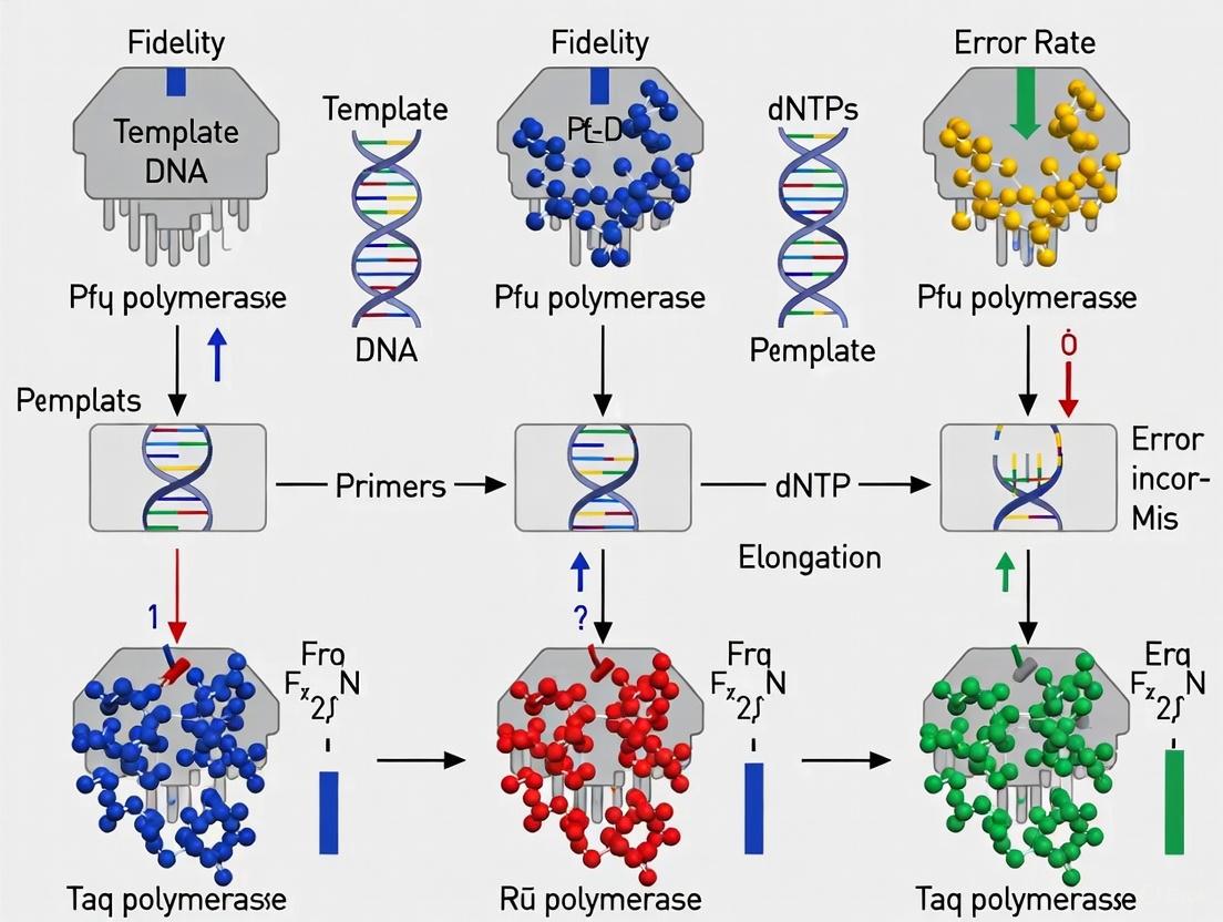
The following diagram illustrates the logical relationship between polymerase characteristics, experimental factors, and the resulting types of errors, as revealed by these methodologies.
The Scientist's Toolkit: Key Reagents for Fidelity Analysis
Successful experimentation in polymerase fidelity relies on specific reagents and instruments. The table below lists essential materials derived from the cited experimental protocols.
Table 2: Essential Research Reagents and Tools for Fidelity Studies
| Reagent / Instrument | Function / Application | Example Use in Fidelity Research |
|---|---|---|
| High-Fidelity Polymerases | Accurate amplification for cloning and sequencing. | Pfu polymerase used for high-fidelity amplification of target genes [4]. |
| dNTP Set | Nucleotide substrates for DNA synthesis. | Concentration optimization (100-300 µM each) to maximize fidelity [5]. |
| MgSO₄ / MgCl₂ | Essential cofactor for polymerase activity. | Concentration titration (e.g., 2-3 mM MgSO₄ for Pfu) to optimize accuracy [5]. |
| Cloning Kit | Insertion of PCR products into vectors for sequencing. | Gateway cloning system used for high-throughput clone preparation [3]. |
| Sanger Sequencing | Gold standard for validating sequences and identifying mutations. | Direct sequencing of cloned PCR products to count mutations [3]. |
| Pacific Biosciences Sequencer | Single-molecule, long-read sequencing for deep error profiling. | Used in a highly accurate workflow to measure DNAP error rates and profiles [2]. |
Discussion and Research Implications
The selection of a DNA polymerase is a critical methodological decision that directly impacts data integrity. While proofreading enzymes are the default choice for high-fidelity applications, researchers must consider that error profiles are family-specific [2]. Furthermore, for the most accurate polymerases like Q5, factors beyond intrinsic polymerase fidelity, such as DNA damage introduced during thermocycling, can become the dominant source of errors in the final amplification product [1].
Understanding the nuances of polymerase performance, as quantified in this guide, supports robust experimental design in fields ranging from functional genomics and synthetic biology to the development of novel molecular diagnostics, ultimately ensuring the reliability and reproducibility of scientific findings.
In the realm of molecular biology, the accuracy of DNA replication—whether in vivo or in vitro through the Polymerase Chain Reaction (PCR)—is paramount. PCR fidelity refers to the ability of a DNA polymerase to correctly incorporate nucleotides complementary to the template strand without introducing errors. The 3'→5' exonuclease activity, often termed proofreading, is a critical mechanism that significantly enhances this fidelity by providing a built-in error-correction system [6] [7]. This activity allows certain DNA polymerases to recognize and excise misincorporated nucleotides from the 3' end of the growing DNA chain before further extension occurs. For researchers, scientists, and drug development professionals, selecting a polymerase with high fidelity is crucial for applications where sequence accuracy is non-negotiable, such as cloning, sequencing, and functional gene analysis. The presence or absence of proofreading activity represents a fundamental distinction between commercially available DNA polymerases and directly impacts the reliability of experimental outcomes.
The Molecular Mechanism of Proofreading
The proofreading process is an exquisite example of intramolecular quality control. DNA polymerases with 3'→5' exonuclease activity possess a distinct exonuclease domain that is spatially separate from the polymerase active site. Following nucleotide incorporation, the newly formed primer terminus can partition between these two sites. A correctly paired terminus is preferentially channeled to the polymerase site for further extension. In contrast, a mismatched base pair, due to its distorted geometry, is more likely to be shuttled to the exonuclease domain [6].
The exonuclease domain itself is a highly coordinated catalytic center. Structural studies on various polymerase families, including the unique Family D archaeal polymerases, reveal that these domains typically bind two metal ions (often Mg²⁺ or Mn²⁺) that are essential for catalyzing the hydrolysis of the phosphodiester bond, thereby releasing the incorrect nucleotide as a deoxynucleoside monophosphate [8] [6]. After excision, the now-correct primer terminus is returned to the polymerase active site to continue with faithful DNA synthesis. This proofreading cycle dramatically reduces error rates, sometimes by a hundredfold or more, as it provides a second opportunity for correct nucleotide selection after the initial incorporation step.
The following diagram illustrates the sequential mechanism of the proofreading process:
Experimental Evidence: Quantifying the Proofreading Advantage
Direct Comparison of Error Rates
The most direct evidence for the proofreading advantage comes from studies comparing the fidelity of wild-type polymerases to engineered exonuclease-deficient mutants. Research on human DNA polymerase δ (Pol δ), a replicative enzyme, demonstrated this elegantly. Scientists constructed exonuclease-domain mutants (e.g., D515V and D402A/D515A) where conserved aspartate residues crucial for metal ion coordination were altered. These mutants lost over 95% of their exonuclease activity, leading to a severe decrease in proofreading capability compared to the wild-type enzyme [9]. While these studies focused on how exonuclease activity affects bypass of damaged DNA templates, the underlying principle—that the loss of proofreading compromises fidelity—is firmly established.
The consequence of diminished proofreading extends beyond in vitro experiments. An age-dependent study in rats revealed that the 3'→5' exonuclease activity in liver cells declined by approximately 30% in 24-month-old rats compared to 4-month-old rats. This decline was correlated with an observed increase in non-complementary nucleotide misincorporations during DNA synthesis, linking reduced proofreading to a higher frequency of replication errors in a biological system [7].
Impact on Translesion Synthesis and Complex Templates
Proofreading activity also plays a nuanced role when polymerases encounter damaged DNA. The exonuclease activity can act as a kinetic barrier to translesion synthesis (TLS). For some polymerases, the exonuclease activity efficiently removes a nucleotide incorporated opposite a DNA lesion, a process known as enzymatic idling, which can prevent the stable bypass of the lesion. The efficiency of this idling is dependent on both the DNA polymerase and the type of DNA lesion [6]. This indicates that the role of proofreading is not merely binary but is modulated by the context of the DNA template, adding a layer of complexity to its error-correction function.
Comparative Performance of Polymerases With and Without Proofreading
The table below summarizes key characteristics of selected DNA polymerases, highlighting the impact of intrinsic proofreading activity and protein engineering on performance.
Table 1: Comparison of DNA Polymerase Features and Fidelity
| Polymerase | Proofreading (3'→5' Exo) | Relative Error Rate | Primary Applications | Key Features & Notes |
|---|---|---|---|---|
| Taq Polymerase | No | ~1 x 10⁻⁵ | Routine PCR, qPCR | Low fidelity; suitable for simple amplification where high accuracy is not critical [10]. |
| KlenTaq | No | ~1 x 10⁻⁵ | PCR, qPCR | N-terminal truncation of Taq; lacks 5'→3' exonuclease activity [11]. |
| Pfu Polymerase | Yes | ~1.5 x 10⁻⁶ | High-fidelity PCR, cloning | High fidelity due to proofreading; slower extension rate than Taq [12]. |
| Sso7d-Fused Polymerases | Varies (depends on base polymerase) | Lower than non-fused counterpart | Fast PCR, amplification of difficult templates | Fusion protein technology drastically increases processivity, improving efficiency on long amplicons and in the presence of inhibitors [10] [12]. |
| Engineered RT-active Taq variants | No | Data not explicitly provided | Single-enzyme RT-PCR, multiplex qPCR | Novel variants (e.g., RT-KTq, Mut_RT) combine reverse transcriptase and DNA polymerase activity; enable multiplex RNA detection without viral RTs [11]. |
Advanced Engineering: Enhancing Polymerase Performance
Protein engineering has further expanded the toolkit of available polymerases, creating enzymes with tailored properties. A prominent example is the fusion of Sso7d, a non-specific double-stranded DNA-binding protein from Sulfolobus solfataricus, to DNA polymerases like Taq and Pfu. This fusion does not directly provide proofreading activity but confers a remarkable increase in processivity—the number of nucleotides incorporated per binding event. For instance, the median processivity of a mutant Taq polymerase increased from 2.9 to 51.0 nucleotides when fused to Sso7d [12]. This enhanced processivity translates to better performance in challenging PCR applications, such as amplifying long targets or templates with high GC content, and can provide greater tolerance to common PCR inhibitors [10].
Another frontier of engineering is the development of novel polymerase variants capable of catalyzing both reverse transcription (RT) and PCR amplification. A 2025 study recombined mutation pools from two engineered Taq variants to create a library of novel polymerases. These enzymes enable single-enzyme, single-tube quantitative multiplex RT-PCR without requiring separate viral reverse transcriptases, simplifying workflows for molecular diagnostics and multiplexed RNA detection [11].
Essential Research Reagents and Experimental Protocols
The Scientist's Toolkit: Key Reagents for Fidelity Studies
Table 2: Essential Research Reagents for Studying Polymerase Fidelity
| Reagent / Tool | Function in Fidelity Assessment |
|---|---|
| Exonuclease-Deficient Mutants | Engineered polymerases (e.g., Pol δ D515V) serve as critical controls to isolate the contribution of proofreading by comparison with their wild-type counterparts [9]. |
| Defined Mismatch-Containing Primers/Templates | Synthetic oligonucleotides with a single, site-specific mismatch are used to directly measure exonuclease activity and partitioning between polymerase and exonuclease sites [8]. |
| Damaged DNA Templates | Templates containing specific lesions (e.g., abasic sites, 8-oxoguanine) help evaluate how proofreading activity influences translesion synthesis and error-prone bypass [9] [6]. |
| Capillary Electrophoresis (CE) | A high-resolution method used to separate and quantify primer extension and excision products in real-time, providing kinetic data on exonuclease activity [8]. |
| Pyrosequencing / NGS | Next-generation sequencing platforms allow for comprehensive analysis of PCR products to quantify mutation frequencies and spectra introduced by different polymerases. |
A Standard Experimental Workflow for Assessing Proofreading
A typical biochemical assay to characterize proofreading activity involves incubating the DNA polymerase with a double-stranded DNA substrate where the primer is labeled with a fluorophore at its 5' end. This substrate can be either perfectly matched or contain a defined mismatch at the 3'-primer terminus.
Detailed Protocol:
- Substrate Preparation: A synthetic oligonucleotide primer, labeled with a fluorophore (e.g., FAM) at its 5' end, is annealed to a complementary template strand in a suitable buffer. For proofreading assays, a substrate with a single mismatch at the 3'-end of the primer is often used alongside a matched control [8].
- Reaction Setup: The polymerase of interest is added to the substrate mixture in the presence of Mg²⁺ or Mn²⁺ (essential cofactors for both polymerase and exonuclease activities) and dNTPs. The reaction is typically carried out at the enzyme's optimal temperature.
- Time-Course Sampling: Aliquots are taken from the reaction at various time points (e.g., 0, 1, 2, 5, 10, 20 minutes) and immediately quenched by adding a stop solution like EDTA (which chelates Mg²⁺ ions and inactivates the enzyme) or formamide.
- Product Analysis: The quenched samples are denatured and analyzed by high-resolution capillary electrophoresis. This technique separates DNA molecules by size, allowing for the resolution of the full-length primer from shorter excision products.
- Data Quantification: The fluorescence intensity of the full-length product and the shorter bands (representing excision intermediates) is quantified. For a proofreading-active polymerase, a mismatch-containing substrate will show a higher proportion of excision intermediates and a slower accumulation of full-length product compared to a matched substrate. The number of visible excision intermediates can also indicate how far back from the primer terminus the exonuclease can remove nucleotides, as demonstrated in studies with PolD where mismatches up to 5 nucleotides back were excised [8].
The 3'→5' exonuclease proofreading activity is an indispensable feature of high-fidelity DNA polymerases, providing a critical frontline defense against replication errors. Direct experimental comparisons show that the absence of this activity can lead to an order-of-magnitude increase in error rates. For the scientific and drug development community, the choice of polymerase is a fundamental determinant of data integrity. While non-proofreading polymerases may be sufficient for routine amplification, applications such as cloning, sequencing, and functional genetics necessitate the use of high-fidelity, proofreading enzymes to ensure sequence accuracy. Emerging technologies, including polymerase fusion proteins for enhanced processivity and novel engineered variants with expanded functions like single-enzyme RT-PCR, continue to push the boundaries of PCR applications. However, the core principle remains: the proofreading advantage is a cornerstone of reliable and reproducible molecular biology.
The fidelity of a DNA polymerase is defined as the accuracy with which it copies a DNA template sequence, and it is a critical parameter for experiments whose outcomes depend upon the correct DNA sequence, such as cloning, single-nucleotide polymorphism (SNP) analysis, and next-generation sequencing (NGS) applications [13]. Maintaining sequence integrity during the Polymerase Chain Reaction (PCR) is paramount for the accurate transfer of genetic information and for preventing artifacts that can confound downstream analysis. The concept of fidelity is most commonly expressed as the mean error rate per base per duplication, which represents the probability that the polymerase will incorporate an incorrect nucleotide during a single replication event [14] [13]. This metric allows for the direct comparison of different enzymes, ranging from traditional polymerases like Taq to modern, ultra-high-fidelity alternatives. Understanding and interpreting these error rates is not merely an academic exercise; it is a fundamental practice for researchers, scientists, and drug development professionals who rely on the integrity of amplified DNA sequences to draw meaningful conclusions from their experiments.
Core Fidelity Metrics and Error Rate Comparisons
At its core, polymerase fidelity is governed by the enzyme's ability to select the correct nucleoside triphosphate and incorporate it into the growing DNA strand, maintaining Watson-Crick base pairing. This accuracy is quantified as the error rate, typically reported in scientific literature as errors per base per duplication [14]. For example, an error rate of 1 × 10⁻⁵ signifies that, on average, one error is expected for every 100,000 nucleotides incorporated. The inverse of this error rate is often referred to as accuracy—the number of bases over which one substitution error is expected [13]. Therefore, an enzyme with an error rate of 1 × 10⁻⁶ possesses an accuracy of 1,000,000, meaning one error per million bases.
A key differentiator among DNA polymerases is the presence of 3′→5′ proofreading exonuclease activity. Polymerases lacking this domain, such as Taq, are unable to correct misincorporated nucleotides, leading to higher error rates. In contrast, high-fidelity enzymes like Q5, Pfu, and Phusion contain this proofreading domain, which actively checks and removes mismatched nucleotides during polymerization, resulting in a dramatic reduction in error frequency [14] [13].
Table 1: Comparison of DNA Polymerase Fidelity Metrics
| DNA Polymerase | Proofreading Activity | Reported Error Rate (errors/base/duplication) | Accuracy (1/error rate) | Fidelity Relative to Taq |
|---|---|---|---|---|
| Taq | No | 1.5 × 10⁻⁴ to 2.28 × 10⁻⁵ [14] [13] [1] | ~4,500 - 6,500 [13] | 1x |
| Q5 | Yes | 5.3 × 10⁻⁷ [14] [13] | ~1,870,000 [13] | 280x |
| Phusion | Yes | 3.9 × 10⁻⁶ [13] | ~255,000 [13] | 39x |
| Pfu | Yes | 5.1 × 10⁻⁶ [13] | ~195,000 [13] | 30x |
| Deep Vent | Yes | 4.0 × 10⁻⁶ [13] | ~251,000 [13] | 44x |
The data in Table 1, synthesized from multiple studies, illustrates the profound impact of proofreading activity. Taq polymerase exhibits the highest error rate, while Q5 High-Fidelity DNA Polymerase demonstrates an error rate nearly three orders of magnitude lower, making it one of the most accurate enzymes available [13]. It is crucial to note that error rates can vary based on reaction conditions, including buffer composition, dNTP and magnesium concentration, and the specific DNA template being amplified [1].
Experimental Protocols for Measuring Fidelity
Several established experimental methods exist for determining polymerase fidelity, each with its own throughput, cost, and detection limit considerations. These protocols enable the empirical comparison of enzymes and are foundational to the data presented in manufacturer specifications and scientific publications.
Blue/White Colony Screening (LacZ Assay)
This classical method involves amplifying a reporter gene, typically lacZ, and cloning the PCR products into a vector. The ligated DNA is then used to transform competent E. coli cells, which are plated on media containing a chromogenic substrate like X-gal.
- Workflow: PCR Amplification of lacZ → Cloning → Transformation → Plating & Colony Color Screening → Sequencing (optional) [13].
- Principle: Functional β-galactosidase enzyme produced from the error-free lacZ gene metabolizes X-gal, resulting in blue colonies. Mutations that disrupt the gene's function lead to white colonies [13] [3].
- Data Analysis: The error rate is calculated based on the ratio of white to total colonies, after accounting for the number of detectable sites within the gene and the number of PCR doublings [13].
- Advantages/Limitations: This is a high-throughput, cost-effective screening method. However, it is an indirect measure of fidelity, as only mutations within a specific region (e.g., 349 bases of the 1.9 kb lacZ gene) that inactivate the protein result in a color change, potentially obscuring the true error spectrum [13] [3].
Direct Sequencing of Cloned PCR Products
This method provides a direct and comprehensive view of all mutations within an amplified sequence.
- Workflow: PCR Amplification → Cloning → Colony Picking → Sanger Sequencing of individual clones → Sequence Alignment and Variant Calling [14] [3].
- Principle: Multiple clones derived from the PCR product are sequenced and compared to the known original template sequence. Any discrepancies (substitutions, insertions, deletions) are recorded as polymerase errors [3].
- Data Analysis: The raw error frequency is normalized to the number of template doublings during PCR to calculate the error rate per base per duplication [14] [1].
- Advantages/Limitations: This method detects all types of errors across the entire sequenced fragment, providing a direct and unambiguous measurement. Its main limitation is lower throughput and higher cost compared to screening assays, especially for quantifying the fidelity of very accurate polymerases, which requires sequencing a large number of clones [3].
Next-Generation Sequencing (NGS) Approaches
NGS technologies overcome the throughput limitations of Sanger sequencing by enabling the analysis of millions of DNA molecules in parallel.
- Workflow: PCR Amplification → NGS Library Preparation → High-Throughput Sequencing → Bioinformatic Analysis [13] [1].
- Principle: The entire population of PCR products is sequenced, and sophisticated algorithms are used to distinguish true polymerase errors from sequencing errors. Some protocols use unique molecular identifiers (UMIs) to tag individual template molecules before amplification [1].
- Data Analysis: After aligning sequences to a reference, the frequency and spectrum of errors are computed. Methods like Pacific Biosciences SMRT sequencing can generate highly accurate consensus sequences from multiple passes of a single molecule, providing a low-background error rate suitable for measuring ultra-high-fidelity polymerases [13] [1].
- Advantages/Limitations: NGS provides a massive dataset for a statistically robust analysis of error rates and mutational spectra. The primary challenges are the cost and complexity of data analysis, though this is becoming more accessible [13].
Diagram 1: Experimental workflow for determining DNA polymerase fidelity, comparing classical (blue) and next-generation sequencing (green) methods.
The Scientist's Toolkit: Research Reagent Solutions
A successful fidelity experiment relies on a suite of specialized reagents and materials. The following table details key components and their functions in a standard fidelity assessment workflow.
Table 2: Essential Research Reagents for PCR Fidelity Experiments
| Reagent/Material | Function in Fidelity Assay | Examples & Notes |
|---|---|---|
| DNA Polymerases | The enzyme under evaluation; catalyzes DNA synthesis. | Test polymerases with different fidelities (e.g., Taq, Q5, Pfu) and a high-fidelity control [14] [13]. |
| Control Template | A well-characterized DNA sequence used as the amplification template. | Plasmid containing lacZ or another reporter gene; must have a known reference sequence for error detection [13] [3]. |
| Cloning Vector & Host | System for isolating and propagating individual PCR molecules for sequencing. | pGEM plasmid vectors and competent E. coli DH5-α cells are commonly used [14]. |
| NGS Platform | For high-throughput sequencing of the entire PCR product population. | Pacific Biosciences SMRT sequencing or Illumina platforms (the latter often requiring UMIs) [13] [1]. |
| Chromogenic Substrate | Allows visual identification of mutant clones in phenotypic assays. | X-gal; used in combination with IPTG for blue/white screening [13]. |
Implications of Fidelity in Research Applications
The choice of DNA polymerase has direct and significant consequences across various research applications. In cloning and sequencing, low-fidelity polymerases can introduce mutations that alter the encoded amino acid sequence or regulatory elements of a gene, leading to incorrect functional data [3]. This is particularly critical for large-scale projects, such as the creation of ORFeomes, where even high-fidelity enzymes will generate some mutant clones given a sufficiently large pool of targets [3].
In the context of detecting intraindividual genetic variation, such as mitochondrial DNA heteroplasmy, the use of a low-fidelity polymerase like Taq can lead to a substantial overestimation of genetic diversity. One study demonstrated that Taq polymerase generated a significant increase in singleton haplotypes per individual compared to Q5, most of which were A→G/T→C transitions characteristic of Taq errors rather than true biological variation [14].
For next-generation sequencing, PCR is integral to library preparation and target enrichment. Mistakes made during this amplification appear in the sequencing data as false mutations, which can confound the detection of rare genetic or somatic variants [1]. Furthermore, errors are not limited to single-base substitutions. PCR-mediated recombination—where a partially extended primer anneals to a different template—can occur at a frequency comparable to base substitution errors in Taq polymerase, creating chimeric sequences that are particularly problematic in 16S ribosomal RNA sequencing or HLA genotyping [1].
It is also important to recognize that not all errors are enzymatic. Studies using single-molecule sequencing have revealed that DNA damage introduced during thermocycling can be a major contributor to observed mutations in amplification products, sometimes exceeding the base substitution rate of ultra-high-fidelity polymerases like Q5 [1]. This highlights that achieving the highest possible data fidelity requires attention to the entire experimental process, not just the choice of polymerase.
Interpreting the key fidelity metric of error rate per base per duplication is fundamental to designing robust and reliable molecular biology experiments. As the comparative data shows, the selection of a DNA polymerase—from standard Taq to proofreading-enabled, ultra-high-fidelity enzymes like Q5—can influence error rates by several hundred-fold. Researchers must align the fidelity requirements of their specific application, be it diagnostic assay development, rare variant detection, or cloning for protein expression, with the demonstrated performance of available polymerases. By understanding the methodologies behind fidelity measurements, the capabilities of different research reagents, and the potential sources of error beyond polymerase misincorporation, scientists can make informed decisions that ensure the integrity of their amplified DNA and the validity of their scientific conclusions.
The fidelity of DNA polymerase enzymes is a cornerstone of modern molecular biology, directly influencing the reliability of applications ranging from basic cloning to next-generation sequencing and molecular diagnostics. This guide provides a comparative analysis of polymerase error spectra, focusing on the rates and types of errors—substitutions, insertions, and deletions—across different enzyme families and reaction conditions. Understanding these error profiles is essential for selecting appropriate polymerases for specific applications where accuracy is paramount, such as in gene synthesis, long-amplicon PCR, and library preparation for sequencing. The evaluation of PCR fidelity extends beyond simple error rate measurements to encompass the sequence context dependencies that influence mutagenesis, providing a framework for assessing polymerase performance in research and diagnostic contexts.
Error-corrected sequencing (ECS) technologies have emerged as transformative methods for in vivo mutagenicity assessment, enabling direct, highly sensitive measurement of mutation frequency and spectrum with error rates as low as 5 errors per billion base pairs [15]. These advanced sequencing methods have revealed that polymerase fidelity is not merely a function of intrinsic enzymatic properties but is modulated by sequence context, buffer composition, and template characteristics. This analysis synthesizes current understanding of polymerase error spectra to guide researchers in making evidence-based decisions for experimental design and interpretation.
Comparative Error Profiles of DNA Polymerases
Quantitative Comparison of Polymerase Fidelity
The fidelity of DNA polymerases varies significantly across enzyme families and commercial formulations. A comparative analysis of 14 different PCR kits using a mock eukaryotic community DNA sample revealed statistically significant differences (p < 0.05) across seven parameters: quality, chimera formation, BLAST top hit accuracy, deletions, insertions, base substitutions, and amplification bias among species [16]. These findings highlight that the choice of polymerase system substantially impacts experimental outcomes in applications requiring high accuracy.
Table 1: Comparative Error Profiles of DNA Polymerases
| Polymerase Type | Error Rate (mutations/bp) | Primary Substitution Errors | Indel Frequency | Sequence Context Dependence | Recommended Applications |
|---|---|---|---|---|---|
| Wild-type Taq | 10⁻⁴–10⁻⁵ | A•T→G•C transitions | High | Strong sequence context effects | Routine PCR, gel detection |
| High-Fidelity Taq (>50X) | 10⁻⁶–10⁻⁷ | Reduced A•T→G•C | Moderate | Moderate context dependence | Cloning, site-directed mutagenesis |
| KOD plus Neo | ~10⁻⁶ | Balanced spectrum | Low | Minimal context bias | NGS library prep, long amplicons |
| HotStart Taq | ~10⁻⁵ | A•T→G•C predominant | Moderate-high | Strong sequence context | Diagnostic PCR, multiplex assays |
| Engineered Taq variants | 10⁻⁷–10⁻⁸ | Variable by design | Low | Context-dependent | Reverse transcription PCR, specialized applications |
The market for high-fidelity DNA polymerases reflects this diversity, with an estimated global market size exceeding 150 million units annually and concentrations in commercial preparations typically ranging from 250,000 units/mL to 5,000,000 units/mL [17]. Different polymerases exhibit characteristic error profiles, with some demonstrating superior performance in specific metrics. For instance, kits containing KOD plus Neo (TOYOBO) and HotStart Taq DNA polymerase (BiONEER) at an annealing temperature of 65°C displayed better results in parameters associated with chimeras, top hit similarity, and deletions [16].
Sequence Context Dependence of Errors
Sequence context significantly influences polymerase error rates and types. Analysis of error-corrected sequencing data from normal adult cells, which typically carry a mutation burden of approximately 10⁻⁷ mutations per base pair, has revealed distinct mutational signatures across genomic regions [15]. These signatures reflect the interplay between polymerase fidelity, DNA sequence context, and exogenous mutagens.
Trinucleotide contexts particularly influence error susceptibility, with certain triplets demonstrating significantly higher mutation rates. For example, cytosine residues in CpG dinucleotides are notably prone to C→T transitions due to spontaneous deamination, while repetitive sequences foster slippage-induced indels. Polymerases vary in their susceptibility to these context effects, with high-fidelity enzymes generally exhibiting more uniform error distribution across sequence contexts.
The development of novel Thermus aquaticus DNA polymerase I (Taq pol) variants with reverse transcription capability illustrates ongoing efforts to engineer enzymes with altered fidelity profiles. These engineered variants combine mutations such as L459M, S515R, I638F, and M747K to enhance thumb domain flexibility and stabilize substrate binding, resulting in altered error characteristics [11].
Experimental Protocols for Fidelity Assessment
Error-Corrected Sequencing Methodologies
Error-corrected sequencing (ECS) represents the gold standard for assessing polymerase fidelity, enabling discrimination between true mutations and technical artifacts. ECS methods enhance accuracy by matching sequences of the forward and reverse strands of original DNA fragments to build a consensus sequence, as true mutations will be present on both strands [18]. Most approaches employ unique molecular identifiers (UMIs) attached to both strands or rely on shear point alignments to the reference genome to uniquely identify mutations, with adapter asymmetry informing strand orientation.
Table 2: Error-Corrected Sequencing Methods for Fidelity Assessment
| Method | Error Rate | Coverage | Key Features | Applications |
|---|---|---|---|---|
| Duplex Sequencing | <5×10⁻⁹ | Targeted or genome-wide | Uses representative panels of endogenous loci | Mutation frequency and spectrum analysis |
| NanoSeq | <5×10⁻⁹ | Whole-exome and targeted capture | Avoids error transfer via restriction enzyme fragmentation | Single-molecule mutation detection in any tissue |
| SMM-seq | ~10⁻⁷ | Genome-scale | Provides comprehensive distribution of mutational events | Genome-wide mutation profiling |
| HiDEF-seq | ~10⁻⁸ | Targeted | Duplex sequencing with optimized library prep | Ultra-sensitive variant detection |
The experimental workflow for ECS typically involves: (1) DNA extraction and quality assessment; (2) library preparation with UMI ligation; (3) target enrichment (for targeted approaches); (4) high-throughput sequencing; (5) bioinformatic processing including consensus sequence generation; and (6) variant calling and annotation [18] [15]. For polymerase fidelity assessment, a standard template is amplified with the test polymerase, and the resulting amplicons are subjected to ECS to identify errors introduced during amplification.
Recent advancements include the development of nanorate sequencing (NanoSeq) with error rates below five errors per billion base pairs, compatible with whole-exome and targeted capture [15]. This method avoids error transfer by using restriction enzyme fragmentation without end repair and dideoxynucleotides during A-tailing, achieving unprecedented accuracy for mutation detection in single DNA molecules.
Mock Community and Synthetic Template Approaches
The use of mock community samples provides a robust approach for comparative assessment of polymerase fidelity. In one study, researchers prepared a mock eukaryotic community from the marine environment by mixing equal amounts of plasmid DNA from 40 microalgal species, then performed amplicon sequencing using different polymerase systems [16]. This approach enables quantitative comparison of error profiles across multiple enzymes under standardized conditions.
Synthetic DNA templates with known sequences provide an alternative fidelity assessment method. These templates typically include defined regions of varying GC content, homopolymer stretches, and secondary structure potential to evaluate sequence context-dependent errors. After amplification with test polymerases, the products are sequenced, and variants are called against the reference sequence to determine error rates and spectra.
The experimental protocol for mock community analysis includes: (1) preparation of reference DNA templates; (2) PCR amplification with test polymerases using standardized cycling conditions; (3) library preparation and sequencing; (4) bioinformatic processing including read alignment and variant calling; and (5) statistical analysis of error profiles across seven parameters: quality, chimera formation, BLAST top hit accuracy, deletions, insertions, base substitutions, and amplification bias [16].
Experimental Workflow Visualization
Polymerase Fidelity Assessment Workflow
This workflow illustrates the standardized process for comparing polymerase error spectra, from initial template preparation through final comparative analysis. The critical step of error-corrected library preparation ensures that identified variants represent true polymerase errors rather than sequencing artifacts [18] [15].
The Scientist's Toolkit: Essential Research Reagents
Table 3: Research Reagent Solutions for Polymerase Fidelity Studies
| Reagent/Category | Specific Examples | Function in Fidelity Assessment | Key Characteristics |
|---|---|---|---|
| High-Fidelity DNA Polymerases | Q5 (NEB), KOD FX (TOYOBO), Phusion (Thermo Scientific) | Provide benchmarks for comparison studies | Error rates 50-100× lower than Taq, enhanced processivity |
| Error-Corrected Sequencing Kits | NanoSeq, Duplex Sequencing, SMM-seq | Enable discrimination of true mutations from artifacts | Ultra-low error rates (<10⁻⁸), UMI incorporation |
| Reference DNA Templates | Mock community plasmids, synthetic control templates | Standardized substrates for fidelity assessment | Known sequence composition, defined variant positions |
| Library Preparation Kits | Illumina Nextera, Swift Biosciences Accel-NGS | Prepare amplification products for sequencing | Compatible with error correction methods, minimal bias |
| Bioinformatic Tools | dNdScv, GATK, custom consensus callers | Identify and classify polymerase errors | Error correction algorithms, mutation signature analysis |
The high-fidelity DNA polymerase market continues to evolve, with leading players like Thermo Scientific, New England Biolabs, and QIAGEN driving innovation through novel enzyme formulations [17]. These companies and others offer specialized polymerases optimized for specific applications, with engineered properties including improved fidelity, enhanced thermostability, and expanded capabilities for challenging templates.
Recent innovations include the development of ultra-high-fidelity enzymes with even lower error rates for demanding applications like gene synthesis and genome editing. Additionally, the integration of AI and machine learning technologies accelerates enzyme design and optimization processes, leading to polymerases with customized fidelity profiles for specialized applications [17].
Comparative analysis of polymerase error spectra reveals substantial differences in fidelity across enzyme families, with error rates spanning several orders of magnitude from wild-type Taq to engineered ultra-high-fidelity variants. The sequence context dependence of these errors further complicates polymerase selection, as enzymes perform differently across genomic regions with varying sequence characteristics.
Error-corrected sequencing methodologies have revolutionized fidelity assessment, enabling discrimination of true mutations from technical artifacts at unprecedented resolution. These technologies have revealed that polymerase errors are non-randomly distributed, with characteristic spectra influenced by both enzymatic properties and template sequence.
For researchers requiring high accuracy in applications such as cloning, sequencing, or diagnostic assay development, careful consideration of polymerase error spectra is essential. The data presented in this guide provide a framework for evidence-based polymerase selection, though specific experimental requirements may necessitate empirical testing to identify optimal enzymes for particular applications. As enzyme engineering continues to advance, the availability of polymerases with customized fidelity profiles will further enhance experimental capabilities across molecular biology applications.
Strategies for High-Fidelity Amplification: From Reaction Setup to Complex Templates
The pursuit of high-fidelity polymerase chain reaction (PCR) is a cornerstone of reliable genetic analysis, cloning, and sequencing. While the selection of a polymerase with proofreading capability is often the initial focus in fidelity-centric protocols, the ultimate success and accuracy of amplification are profoundly governed by the precise optimization of fundamental reaction components. The concentration of magnesium ions (Mg2+), the balance of deoxynucleoside triphosphates (dNTPs), and the strategic use of enhancing additives form a critical triumvirate that directly influences DNA polymerase kinetics, specificity, and error rate. This guide objectively compares the performance of these components under varied conditions, providing supporting experimental data to equip researchers with a systematic framework for optimizing PCR fidelity within a broader thesis on polymerase evaluation.
Magnesium Ion (Mg2+) Concentration: The Essential Cofactor
Magnesium ion (Mg2+) is an indispensable cofactor for all DNA polymerases, serving a dual role by stabilizing the enzyme's active site for dNTP incorporation and facilitating the primer-template binding through charge neutralization [19] [20]. Its concentration is arguably the single most critical variable to optimize for any given PCR assay.
Optimal Concentration Range and Meta-Analysis Insights
A comprehensive meta-analysis of 61 peer-reviewed studies established a clear optimal range for MgCl2 between 1.5 mM and 3.0 mM for efficient PCR performance [21]. Within this range, a precise logarithmic relationship between MgCl2 concentration and DNA melting temperature was quantified. The analysis revealed that for every 0.5 mM increase in MgCl2, the DNA melting temperature increases by approximately 1.2°C [21]. This thermodynamic effect directly impacts the stringency of primer annealing and must be accounted for when calibrating thermal cycling conditions.
Consequences of Improper Mg2+ Concentration
The effects of deviating from the optimal Mg2+ range are profound and well-documented:
- Low Mg2+ Concentration: Reduces DNA polymerase activity to suboptimal levels, leading to significantly poor reaction yield or complete amplification failure [19] [20]. The enzyme lacks sufficient cofactor for efficient catalysis.
- High Mg2+ Concentration: Promotes a significant loss of reaction specificity and fidelity. This occurs because elevated Mg2+ reduces the enzyme's stringency for correct base pairing, leading to increased misincorporation rates and nonspecific amplification, often visible as smearing or multiple bands on an agarose gel [19] [21].
Template-Dependent Optimization
The complexity of the DNA template directly influences the optimal Mg2+ requirement. The meta-analysis found that genomic DNA templates consistently require higher Mg2+ concentrations compared to more straightforward templates like plasmid DNA or synthetic oligonucleotides [21]. This is likely due to the greater sequence complexity and potential for secondary structures in genomic DNA.
Table 1: Effects of MgCl2 Concentration on PCR Performance
| MgCl2 Concentration | Efficiency | Specificity | Fidelity | Typical Application |
|---|---|---|---|---|
| Low (<1.5 mM) | Greatly Reduced | High (but yield is low) | Potentially High | Rarely optimal; may require optimization |
| Optimal (1.5 - 3.0 mM) | High | High | High | Standard PCR; most templates [21] |
| High (>3.0 - 4.5 mM) | High | Low | Reduced | May be required for complex genomic DNA [21] |
| Very High (>4.5 mM) | Unpredictable | Very Low | Very Low | Not recommended |
dNTP Balance and Concentration: The Building Blocks of Fidelity
Deoxynucleoside triphosphates (dNTPs) are the foundational substrates for DNA synthesis. Their concentration and purity are paramount for achieving high yield and, crucially, low error rates.
Standard Concentration and Molar Balance
The four dNTPs (dATP, dTTP, dCTP, dGTP) should be provided in equimolar ratios to prevent misincorporation due to nucleotide imbalance [22] [20]. A final concentration of 0.2 mM for each dNTP is widely considered a standard and effective starting point for most PCR applications [20] [23]. This concentration ensures that dNTP levels remain above the Km (Michaelis constant) of most DNA polymerases throughout the amplification process, preventing premature reaction termination.
dNTPs and Fidelity Trade-offs
The relationship between dNTP concentration and fidelity is complex. While sufficient dNTPs are necessary for efficient amplification, elevated concentrations can increase error rates. This is because high dNTP levels can reduce the efficiency of the polymerase's proofreading activity by promoting the extension of mismatched primer termini [24] [23]. Consequently, for applications requiring ultra-high fidelity, such as cloning or sequencing, lowering dNTP concentrations to 0.01–0.05 mM (with a proportional reduction in Mg2+) can improve accuracy when using non-proofreading polymerases [20]. However, this must be balanced against a potential reduction in product yield.
Interaction with Mg2+
A critical, often overlooked, interaction is that between dNTPs and Mg2+. Mg2+ ions bind to dNTPs in the reaction mix to form the actual substrate for the polymerase. Therefore, the Mg2+ concentration must always be in molar excess of the total dNTP concentration. A common recommendation is to set the Mg2+ concentration at least 0.5-1.0 mM higher than the total dNTP concentration to ensure a sufficient pool of free Mg2+ for the polymerase [20].
Table 2: Optimizing dNTPs for Different PCR Applications
| Application Goal | Recommended [each dNTP] | Rationale | Considerations |
|---|---|---|---|
| Standard PCR | 0.2 mM | Balances high yield with good fidelity [20] | Standard starting point for most assays |
| High-Fidelity PCR | 0.01 - 0.05 mM | Reduces misincorporation and promotes proofreading [20] | May lower yield; requires Mg2+ titration |
| Long-Range PCR | 0.4 mM | Ensures sufficient substrates for synthesis of long products | Higher dNTPs may require increased Mg2+ [23] |
| GC-Rich PCR | 0.2 - 0.4 mM | Helps polymerase traverse complex secondary structures | Often used in combination with additives |
Chemical Additives: Enhancing Efficiency and Specificity
PCR enhancers are a class of chemical additives that improve amplification efficiency, particularly for problematic templates such as those with high GC content or complex secondary structures.
Common Additives and Their Mechanisms
- DMSO (Dimethyl Sulfoxide): Used at 2-10% (v/v), DMSO interferes with the DNA hydrogen bonding network, effectively lowering the melting temperature of the template. This helps denature GC-rich regions that are prone to forming stable secondary structures [19] [25].
- Betaine: Used at a concentration of 1-2 M, betaine acts as a stabilizing osmolyte. It homogenizes the base stacking energies between GC-rich and AT-rich regions, effectively equalizing the melting temperature across the amplicon and preventing the "breathing" of AT-rich zones and the stable occlusion of GC-rich zones [19] [22].
- Formamide and Related Amides: Like DMSO, formamide destabilizes DNA duplexes. A structure-activity study found that other low molecular weight amides, such as 2-pyrrolidone and N-methylpyrrolidone (NMP), can also serve as potent novel PCR enhancers, improving both potency (yield) and specificity [25].
Additive Selection Guide
The choice of additive is often template-dependent. Betaine is frequently the preferred agent for GC-rich templates, while DMSO is a common general-purpose destabilizer. It is crucial to note that these additives can inhibit PCR at high concentrations, so titration is essential [22] [25].
Experimental Protocols for Systematic Optimization
Protocol 1: Mg2+ Titration
Objective: To empirically determine the optimal MgCl2 concentration for a specific primer-template system. Methodology:
- Prepare a master mix containing all standard PCR components except MgCl2.
- Aliquot the master mix into a series of tubes (or a multi-well plate).
- Add MgCl2 from a stock solution to create a concentration gradient across the reactions. A recommended range is 1.0 mM to 4.0 mM in 0.5 mM increments [19] [21].
- Run the PCR using a standardized thermal cycling protocol.
- Analyze the products using agarose gel electrophoresis. The condition that produces the highest yield of the specific product with the least background smearing indicates the optimal MgCl2 concentration.
Protocol 2: dNTP and Additive Titration
Objective: To optimize dNTP concentration and evaluate the effect of enhancers. Methodology:
- Set up a series of reactions with a fixed, optimal Mg2+ concentration.
- For dNTP titration: Vary the concentration of each dNTP (e.g., 0.05, 0.1, 0.2, 0.4 mM) while keeping them equimolar [23].
- For additive screening: Test different additives (e.g., 5% DMSO, 1 M Betaine, 2-pyrrolidone) at their common working concentrations alongside a no-additive control [25].
- Analyze by gel electrophoresis. For dNTPs, seek the lowest concentration that gives robust yield. For additives, identify the agent that eliminates nonspecific products or enhances the target band intensity.
The Scientist's Toolkit: Research Reagent Solutions
Table 3: Essential Reagents for PCR Optimization
| Reagent | Critical Function | Optimization Consideration |
|---|---|---|
| MgCl2 Solution | DNA polymerase cofactor; stabilizes nucleic acids. | The most critical variable; requires titration for every new primer pair. |
| dNTP Mix (equimolar) | Building blocks for new DNA strand synthesis. | Purity is vital; concentrations >0.2 mM each can reduce fidelity. |
| Betaine (1-2 M stock) | Equalizes Tm of DNA; essential for GC-rich targets. | Final concentration typically 1.0-1.7 M; do not exceed 2 M. |
| DMSO (100% stock) | Destabilizes DNA secondary structure. | Use at 2-10% (v/v); can inhibit PCR and polymerase at high levels. |
| High-Fidelity PCR Buffer | Provides pH, salts, and often a baseline Mg2+ level. | Use the buffer recommended for your specific polymerase. |
The meticulous optimization of Mg2+ concentration, dNTP balance, and chemical additives is not a mere preliminary step but a fundamental requirement for achieving robust and reliable PCR results, especially in a research context focused on evaluating polymerase fidelity. Data synthesized from multiple studies confirms that Mg2+ should be titrated between 1.5-3.0 mM, with genomic DNA often requiring higher levels. dNTPs should be used at the lowest concentration that provides sufficient yield, typically starting at 0.2 mM each, to balance efficiency with fidelity. Finally, challenging templates often necessitate rescue with additives like betaine or DMSO. By systematically applying the comparative data and experimental protocols outlined in this guide, researchers can deconvolute the interplay of these core components and establish PCR conditions that ensure the highest standards of data integrity for their work.
In the rigorous context of evaluating PCR fidelity across different polymerases, the calibration of thermal cycling parameters transcends routine optimization and becomes a fundamental determinant of data integrity. The precision with which annealing temperature stringency and PCR cycle number are controlled directly influences the yield, specificity, and accuracy of the amplified product, which are critical for downstream applications in cloning, sequencing, and functional genomics research [26] [19]. Flawed amplification can introduce errors that compromise the validity of entire experimental lineages, making thermal cycling not just a technical step, but a cornerstone of reproducible science.
This guide provides a systematic, data-driven comparison of how these parameters interact with various polymerase classes. By presenting consolidated experimental data and detailed protocols, we aim to equip researchers and drug development professionals with the evidence needed to make informed decisions that enhance the reliability and fidelity of their PCR-based research.
The Critical Interplay of Annealing Temperature and Polymerase Fidelity
The Fundamental Role of Annealing Stringency
The annealing temperature (T~a~) is arguably the most pivotal variable governing PCR specificity. It dictates the stringency of primer-template binding, acting as a molecular gatekeeper that permits only perfect or near-perfect complementarity to proceed to extension [27]. The relationship between the primer's melting temperature (T~m~) and the applied T~a~ is foundational: using a T~a~ too far below the T~m~ allows primers to bind to non-complementary sequences, leading to spurious amplification and reduced yield of the desired product. Conversely, a T~a~ that is too high prohibits efficient primer binding altogether, resulting in PCR failure [28] [19].
Systematic Optimization via Gradient PCR
Determining the optimal T~a~ empirically is a non-negotiable step for any new primer set. Modern thermal cyclers with gradient functionality are indispensable for this, enabling the simultaneous testing of a temperature range across a single block [26]. A standard optimization protocol involves setting a gradient from approximately 5°C below to 5°C above the calculated average T~m~ of the primer pair. The results are then analyzed by gel electrophoresis to identify the temperature that produces a single, robust band of the expected size [29] [28].
For advanced optimization, particularly with challenging templates, a 2D-Gradient PCR can be employed. This method simultaneously tests a range of denaturation temperatures (T~d~) along one axis of the block and a range of annealing temperatures (T~a~) along the other. This allows for the rapid identification of the ideal T~a~/T~d~ combination from 96 different conditions in a single run, dramatically improving specificity and yield for difficult assays such as those involving long or GC-rich amplicons [29].
Polymerase-Specific Buffer Chemistry
The optimal T~a~ is not solely a function of the primer sequence; it is also profoundly influenced by the buffer chemistry of the polymerase system. Standard T~m~ calculations assume a certain ionic environment, but specialized buffers containing isostabilizing agents or co-solvents can alter duplex stability [27] [28]. For instance, the presence of 10% DMSO can lower the effective T~a~ by 5.5–6.0°C [28]. Some proprietary buffer systems are designed to enable a "universal" annealing temperature (e.g., 60°C) for a wide variety of primers, thereby streamlining workflows and reducing optimization time [28]. This interplay between enzyme, buffer, and thermal profile underscores the necessity of a holistic approach to parameter calibration.
Diagram 1: A logical workflow for the systematic optimization of the PCR annealing temperature (Ta) using a thermal gradient, leading to specific amplification.
Cycle Number Optimization: Balancing Yield and Error Accumulation
The Plateau Effect and the Risk of Spurious Products
The number of amplification cycles is a direct driver of PCR yield, but it is subject to the law of diminishing returns. As cycles progress, reaction components (dNTPs, primers, enzyme) are depleted, and the accumulation of pyrophosphate molecules and non-specific products inhibits the reaction. This leads to the plateau phase, where product yield ceases to increase exponentially [28]. Critically, continuing amplification beyond this point (typically >35-40 cycles for standard samples) is counterproductive, as it selectively amplifies nonspecific artifacts and primer-dimers, which can be mistaken for genuine products [28] [19].
A Data-Driven Approach for Low Biomass Samples
While high cycle numbers are detrimental for high-template reactions, they are often essential for samples with low microbial or target biomass, such as blood, milk, or single-cell samples. A 2020 study systematically evaluated this trade-off by performing 16S rRNA gene amplicon sequencing on matched low-biomass samples (bovine milk, murine pelage, and blood) using different PCR cycle numbers (25, 30, 35, and 40) [30] [31].
The key findings, summarized in the table below, demonstrate that increased cycle number successfully increases sequence coverage without significantly altering the perceived microbial community structure, supporting the use of higher cycles for such challenging applications.
Table 1: Influence of PCR Cycle Number on Sequencing Results from Low Biomass Samples [30] [31]
| Sample Type | PCR Cycles Tested | Effect on Coverage/Read Count | Effect on Community Richness (Alpha Diversity) | Effect on Community Structure (Beta-Diversity) |
|---|---|---|---|---|
| Bovine Milk | 25, 30, 35, 40 | Significantly increased with higher cycles | No significant differences detected | No significant differences detected |
| Murine Pelage | 25, 40 | Significantly increased at 40 cycles | No significant differences detected | No significant differences detected |
| Murine Blood | 25, 40 | Significantly increased at 40 cycles | No significant differences detected | No significant differences detected |
Template Copy Number Dictates Optimal Cycling
The primary determinant for cycle number is the initial copy number of the target sequence. The following table provides general guidelines based on template abundance, balancing the need for sufficient yield against the risks of error accumulation and spurious amplification.
Table 2: Recommended PCR Cycle Number Based on Template Abundance [28] [19]
| Initial Target Copy Number | Recommended Cycle Number | Rationale and Considerations |
|---|---|---|
| High Abundance (e.g., plasmid DNA, colony PCR) | 25 - 30 | Minimizes polymerase-induced errors and prevents plateau phase entry for the purest product, ideal for cloning. |
| Medium Abundance (e.g., genomic DNA for genotyping) | 30 - 35 | Standard range offering a robust yield for most analytical applications without excessive nonspecific background. |
| Low Abundance / Low Biomass (e.g., single-copy genes, pathogen detection, microbiome samples) | 35 - 40 | Necessary to generate sufficient product for detection; the benefit of increased coverage can outweigh concerns about errors, which can be filtered bioinformatically [30]. |
| Very Low Copy (<10 copies) | Up to 40-45* | *Use with caution. Nonspecific bands often appear beyond 45 cycles. Requires meticulous negative controls to confirm specificity. |
Experimental Data: Comparing Polymerase Performance Under Stringent Conditions
The fidelity of a PCR reaction is ultimately a measure of the polymerase's ability to faithfully copy the template DNA. Different polymerases possess varying intrinsic error rates due to the presence or absence of 3'→5' proofreading exonuclease activity.
Table 3: Polymerase Fidelity and Its Interaction with Cycling Parameters [26] [19]
| Polymerase Type | Proofreading Activity | Estimated Error Rate (per bp per cycle) | Impact of High Cycle Number | Recommended Application Context |
|---|---|---|---|---|
| Standard Taq | No | ~1 x 10⁻⁴ | High: Significant cumulative errors, not suitable for long cycles or high-fidelity needs. | Routine screening, genotyping, diagnostic assays where speed is prioritized over sequence perfection. |
| High-Fidelity (e.g., Pfu, KOD) | Yes | ~1 x 10⁻⁶ to 1 x 10⁻⁷ | Lower: Greatly reduced error accumulation, making them robust for high-cycle applications. | Cloning, sequencing, gene expression analysis, and any application where sequence accuracy is paramount. |
| Hot-Start Taq | No | ~1 x 10⁻⁴ | Medium: Errors similar to standard Taq, but improved specificity from hot-start reduces nonspecific products at all cycle numbers. | All applications requiring high specificity; reduces primer-dimer formation in low-template reactions. |
This data clearly demonstrates that for research focused on evaluating PCR fidelity, the choice of a high-fidelity, proofreading polymerase is non-negotiable, especially when cycle numbers must be pushed to their practical limits to amplify scarce targets.
Essential Protocols for Parameter Calibration
Protocol 1: One-Dimensional Annealing Temperature Gradient
Objective: To determine the optimal annealing temperature for a specific primer-template pair. Materials: Thermal cycler with gradient functionality, PCR reagents, primers, template DNA. Method:
- Calculate the T~m~ of both forward and reverse primers using the nearest-neighbor method.
- Program the thermal cycler with a denaturation step (e.g., 98°C for 10-30s) and an extension step (e.g., 72°C for 1 min/kb).
- Set the annealing step to a gradient spanning from 5°C below the lowest T~m~ to 5°C above the highest T~m~.
- Run the PCR for 30 cycles.
- Analyze the results by agarose gel electrophoresis. The optimal T~a~ is the highest temperature that produces a strong, specific band of the correct size [29] [28].
Protocol 2: Cycle Number Titration for Low-Target Applications
Objective: To establish the minimum number of cycles required for adequate yield from a low-copy-number sample without excessive background. Materials: PCR reagents, low-copy-number template DNA, validated primer set. Method:
- Set up a master mix with all PCR components and aliquot into multiple tubes.
- Program the thermal cycler with optimized T~a~ and T~d~.
- Run identical reactions but set the cycle number to different values (e.g., 30, 35, 38, 40, 45).
- Analyze the products by gel electrophoresis and/or quantitative methods (e.g., qPCR melt curve analysis, Fragment Analyzer).
- Identify the cycle number that provides a clear, specific product before nonspecific amplification becomes dominant [30] [28].
The Scientist's Toolkit: Key Reagents for PCR Fidelity Research
Table 4: Essential Materials and Reagents for Optimizing Thermal Cycling Parameters
| Item | Function/Application | Key Considerations |
|---|---|---|
| Gradient Thermal Cycler | Simultaneous optimization of annealing/denaturation temperatures across a block. | Look for models with high temperature uniformity (<±0.5°C) and precise gradient control [26]. |
| High-Fidelity Polymerase Mix | Amplification for applications requiring maximal sequence accuracy. | Select enzymes with documented proofreading activity (e.g., Pfu, KOD) and low error rates [19]. |
| Hot-Start Polymerase | Prevents non-specific amplification and primer-dimer formation during reaction setup. | Essential for sensitive applications; improves specificity across all cycle numbers and annealing temperatures [19]. |
| PCR Additives (DMSO, Betaine) | Aids in denaturation of GC-rich templates and reduces secondary structures. | Titrate concentrations (e.g., DMSO 2-10%, Betaine 0.5-2 M) as they can lower the effective T~a~ [28] [19]. |
| MgCl₂ Solution | Essential cofactor for DNA polymerase activity. | Requires optimization (0.5-5.0 mM); concentration affects enzyme processivity, fidelity, and primer annealing [27] [19]. |
| dNTP Mix | Building blocks for DNA synthesis. | Use balanced, high-quality dNTPs; excessive concentrations can reduce fidelity by promoting misincorporation [19]. |
Diagram 2: A decision pathway for selecting the appropriate polymerase and cycling strategy based on experimental application and template characteristics.
Calibrating annealing temperature stringency and cycle number is a critical, non-negotiable process in the broader thesis of PCR fidelity research. As demonstrated, these parameters are deeply intertwined with the choice of polymerase and the nature of the template. Proofreading polymerases provide a fundamental safeguard against error accumulation, especially when high cycle numbers are unavoidable. The experimental data and protocols provided here offer a roadmap for researchers to systematically optimize these variables, ensuring that the results driving scientific discovery and drug development are built upon a foundation of precision and reliability.
In polymerase chain reaction (PCR) applications, from basic research to molecular diagnostics, scientists frequently encounter templates that resist efficient amplification. Two particularly problematic categories are GC-rich sequences and long amplicons, each presenting unique biochemical challenges that can lead to amplification failure, reduced yield, or inaccurate representation of template abundance. GC-rich regions, characterized by guanine-cytosine content exceeding 65%, form stable secondary structures that can block polymerase progression [32] [28]. These stable structures arise due to the three hydrogen bonds between G-C base pairs compared to only two between A-T pairs, creating particularly stable hairpins and loops that hinder denaturation and primer annealing. Meanwhile, long amplicons challenge the processivity and fidelity of DNA polymerases, with error rates increasing proportionally to product length due to the cumulative effect of minor incorporation inaccuracies [3].
The amplification of difficult templates is not merely an academic concern but has significant practical implications across molecular biology applications. In clinical diagnostics, failure to efficiently amplify GC-rich regions of clinical relevance, such as the epidermal growth factor receptor (EGFR) promoter (with GC content up to 88%), can compromise mutation detection and subsequent treatment decisions [32]. In research settings, inaccurate amplification of long amplicons can introduce errors in cloning projects and synthetic biology applications. Quantitative molecular techniques, including multi-template PCR used in high-throughput sequencing library preparation, are especially vulnerable to amplification biases, where sequence-specific efficiency variations can dramatically skew abundance data and compromise experimental results [33]. Understanding and addressing these challenges is therefore essential for obtaining reliable, reproducible results across diverse PCR applications.
Polymerase Fidelity: A Critical Determinant for Success
The choice of DNA polymerase fundamentally influences success rates when amplifying challenging templates. Polymerase fidelity—the accuracy of DNA synthesis—varies significantly among enzymes and becomes increasingly critical with longer amplicons where error accumulation is multiplicative. A comprehensive comparison of six commonly used DNA polymerases revealed substantial differences in error rates, measured by direct sequencing of cloned PCR products across 94 unique DNA targets [3].
Table 1: Error Rate Comparison of DNA Polymerases
| Enzyme | Published Error Rate (errors/bp/duplication) | Fidelity Relative to Taq | Key Characteristics |
|---|---|---|---|
| Taq | 1–20 × 10⁻⁵ | 1x | Standard for routine PCR; lowest fidelity |
| AccuPrime-Taq HF | N/A | 9x better | Engineered variant with improved accuracy |
| KOD Hot Start | N/A | 4-50x better | High processivity; good for long amplicons |
| Pfu | 1-2 × 10⁻⁶ | 6-10x better | Proofreading activity; high fidelity |
| Pwo | Comparable to Pfu | >10x better | Proofreading activity; high fidelity |
| Phusion Hot Start | 4.0 × 10⁻⁷ (HF buffer) | >50x better | Engineered enzyme; highest fidelity |
The fidelity differences observed in these enzymes stem from both structural variations and the presence of proofreading mechanisms. Family A polymerases like Taq lack 3'→5' exonuclease activity, while Family B polymerases like Pfu and Pwo possess proofreading capabilities that remove misincorporated nucleotides [34]. The exceptional fidelity of engineered enzymes like Phusion demonstrates how rational design can enhance replication accuracy. For challenging applications, high-fidelity polymerases with proofreading activity are strongly recommended, particularly for long amplicons where cumulative error rates become problematic [3].
Strategic Optimization for GC-Rich Templates
Biochemical Additives and Reaction Composition
GC-rich templates demand specialized reaction conditions to overcome their structural challenges. Several additives can significantly improve amplification efficiency by disrupting stable secondary structures:
- DMSO (Dimethyl Sulfoxide): Adding 5% DMSO has proven necessary for successful amplification of extremely GC-rich targets like the EGFR promoter [32]. DMSO interferes with base pairing by disrupting hydrogen bonds and reduces the melting temperature of DNA, facilitating denaturation of stable structures.
- Betaine: Also known as trimethylglycine, betaine can be used at concentrations of 1-1.3 M to equalize the contribution of GC and AT base pairs to DNA stability. It reduces the strand separation temperature and helps prevent secondary structure formation [28].
- GC-Rich Solutions: Commercial solutions often contain a proprietary mix of enhancing agents that destabilize secondary structures and improve polymerase processivity through GC-rich regions.
Beyond additives, template DNA concentration critically impacts success with difficult targets. Research has demonstrated that DNA concentrations of at least 2 μg/ml are necessary for reliable amplification of GC-rich sequences, with samples below 1.86 μg/ml often failing to amplify altogether [32].
Thermal Cycling Parameters for GC-Rich Targets
Temperature optimization is crucial for GC-rich amplification. Standard cycling parameters frequently fail, requiring the following adjustments:
- Higher Denaturation Temperatures: While standard denaturation occurs at 94-95°C, GC-rich templates often benefit from temperatures of 98°C or higher, particularly when using buffers with high salt concentrations [28].
- Longer Denaturation Times: Increasing initial denaturation time from 0-5 minutes progressively improves yield of GC-rich fragments. For complex genomic DNA targets, extended denaturation of 3-5 minutes is recommended [28].
- Optimized Annealing Temperature: Although calculated annealing temperature for the EGFR promoter was 56°C, experimental optimization revealed 63°C as optimal—7°C higher than calculated [32]. This highlights the importance of empirical verification over theoretical calculation.
Table 2: Optimization Parameters for Challenging Templates
| Parameter | Standard Conditions | GC-Rich Optimization | Long Amplicon Optimization |
|---|---|---|---|
| Initial Denaturation | 94-95°C, 1-3 min | 98°C, 3-5 min | 94-98°C, 2-3 min |
| Denaturation Cycles | 94-95°C, 0.5-1 min | 98°C, 0.5-2 min | 94-98°C, 0.5-1 min |
| Annealing Temperature | Calculated Tm - (3-5°C) | May require increase up to 7°C above calculated | Calculated Tm - (3-5°C) |
| Extension Time | 1 min/kb (Taq) | Standard or slightly increased | 2-3 min/kb for >10 kb targets |
| MgCl₂ Concentration | 1.5-2.0 mM | 1.5-2.0 mM (requires optimization) | 1.5-2.5 mM (requires optimization) |
| Additives | None | 5% DMSO, betaine | May benefit from enhancers |
Experimental Design and Validation Approaches
Dilution-Replicate Design for Reliable Quantification
Traditional qPCR experimental design employs identical replicates to assess technical variation, but this approach may be inefficient for challenging targets. A novel dilution-replicate design strategy has been developed that requires fewer reactions while providing robust quantification [35]. This approach uses dilution series instead of identical replicates for each test sample, creating standard curves for every sample that simultaneously estimate both PCR efficiency and initial DNA quantity.
The mathematical foundation for this method derives from the PCR amplification equation:
Cq = -log(d)/log(E) + log(T/Q(0)) / log(E)
Where Cq is the quantification cycle, d is the dilution factor, E is PCR efficiency, T is the threshold, and Q(0) is initial quantity [35]. This relationship enables a semi-log plot of Cq versus log(dilution) to yield both efficiency (from slope) and relative quantity (from y-intercept). The dilution-replicate design offers particular advantages for challenging templates by allowing identification and exclusion of outlier points that may occur at extreme dilutions, rather than requiring complete repetition of problematic reactions.
GC-Rich PCR Optimization Workflow
Fidelity Assessment Methodologies
Accurately determining polymerase error rates is essential for selecting appropriate enzymes for challenging applications. Several methodological approaches have been developed:
- Direct Sequencing: The most straightforward method involving cloning and sequencing PCR products to identify mutations. This approach benefits from interrogating diverse sequence contexts but becomes resource-intensive for high-fidelity enzymes [3].
- Pacific Biosciences SMRT Sequencing: This long-read, non-PCR-amplification-based platform uses circular consensus sequencing (CCS) to repeatedly read the same DNA molecule, achieving extremely high accuracy in fidelity measurements [34].
- LacZα Complementation Assays: A screening-based method that uses functional loss of β-galactosidase activity to detect mutations, though limited to specific target sequences [3] [34].
Recent advances in deep learning have further enhanced our ability to predict amplification efficiency based on sequence features alone. Convolutional neural networks (1D-CNNs) trained on synthetic DNA pools can now predict sequence-specific amplification efficiencies with high accuracy (AUROC: 0.88), enabling the design of inherently homogeneous amplicon libraries and identification of problematic sequences before experimental validation [33].
Advanced Applications and Future Directions
Novel Polymerase Engineering for Challenging Applications
Protein engineering has produced remarkable advances in polymerase capabilities, with novel variants overcoming traditional limitations. Recent development of Thermus aquaticus DNA polymerase I (Taq pol) variants demonstrates how single enzymes can now catalyze both reverse transcription and DNA amplification simultaneously without viral reverse transcriptases [11]. These engineered variants, created by combining mutation pools from independently evolved RT-active KTq polymerases, maintain excellent thermostability (up to 95°C) and enable multiplex detection of various RNA targets in a single tube with a single enzyme.
The engineering strategy employed combinatorial investigation of two mutation pools—L459M, S515R, I638F, M747K from RT-KTq and N483K, E507K, K540Y, V586G, I614K from Mut_RT—generating 256 Taq pol variants that were screened for enhanced reverse transcriptase and PCR activity [11]. The successful variants demonstrate that polymerase capabilities can be substantially expanded through rational design, opening new possibilities for challenging amplification applications.
The Scientist's Toolkit: Essential Reagents and Materials
Table 3: Research Reagent Solutions for Challenging Amplification
| Reagent/Category | Specific Examples | Function/Application |
|---|---|---|
| High-Fidelity Polymerases | Pfu, Phusion, KOD | Reduced error rates for long amplicons; proofreading activity |
| GC-Rich Enhancers | DMSO, betaine, commercial GC-rich solutions | Disrupt secondary structures; improve denaturation efficiency |
| MgCl₂ Solutions | Various concentrations (0.5-2.5 mM) | Cofactor optimization; critical for polymerase activity |
| Optimized Buffer Systems | HF buffers, GC buffers, isostabilizing formulations | Enhance specificity; reduce optimization requirements |
| dNTP Mixtures | Balanced dNTPs, 7-deaza-dGTP | Substrate provision; 7-deaza-dGTP reduces secondary structure |
| Quality Template Preparation | FFPE-specific kits, high-purity isolation | Ensure template integrity; minimize inhibitors |
Amplifying challenging templates requires a systematic approach addressing both biochemical and physical parameters of the PCR reaction. The strategic combination of polymerase selection, reaction optimization, and appropriate experimental design dramatically improves success rates with GC-rich regions and long amplicons. Key recommendations include:
Enzyme Selection: Prioritize high-fidelity polymerases with proofreading activity for long amplicons, while considering specialized variants for specific applications like multiplex reverse transcription-PCR [11] [3].
Comprehensive Optimization: Implement orthogonal optimization strategies addressing additives, magnesium concentration, and thermal cycling parameters rather than single-factor adjustments [32] [28].
Experimental Design: Consider efficient design strategies like dilution-replicate approaches that provide robust quantification while minimizing reagent use and processing time [35].
Validation Rigor: Employ appropriate fidelity assessment methods and efficiency prediction tools to verify amplification accuracy and avoid skewed results in quantitative applications [34] [33].
As polymerase engineering continues to advance and computational prediction of amplification efficiency improves, researchers are increasingly equipped to tackle even the most challenging templates. The strategies outlined herein provide a foundation for reliable amplification, ensuring accurate results across diverse molecular biology applications.
qPCR Experimental Design Comparison
Primer Design Principles for Maximizing Specificity and Yield
In the context of evaluating PCR fidelity across different polymerases, primer design emerges as a fundamental determinant of success. Polymerase Chain Reaction (PCR) serves as a cornerstone of molecular biology, enabling the amplification of specific DNA sequences for applications ranging from basic research to clinical diagnostics and drug development [36]. While the choice of DNA polymerase significantly influences amplification efficiency and error rate, the primers themselves dictate the specificity and yield of the entire reaction. Properly designed primers ensure accurate binding to the intended target sequence, while poorly designed primers can lead to nonspecific amplification, primer-dimer formation, and reduced product yield, thereby compromising experimental results and downstream applications [37] [38].
This guide objectively compares the performance of different primer design strategies and their interaction with various DNA polymerases, providing supporting experimental data to establish evidence-based best practices. By integrating principles of oligonucleotide design with polymerase characteristics, researchers can optimize PCR assays for maximum reliability and reproducibility in demanding scientific contexts.
Fundamental Principles of Primer Design
Effective primer design balances multiple parameters to ensure specific annealing and efficient amplification. The following principles represent the consensus from established molecular biology protocols and commercial reagent providers.
Core Design Parameters
- Length: Optimal primers are typically 18-30 nucleotides long. Shorter primers may lack specificity, while longer primers can exhibit slower hybridization rates and increased production costs [37] [38] [39].
- Melting Temperature (Tm): Primers should exhibit a Tm between 60-75°C, with forward and reverse primers having Tms within 2-5°C of each other to ensure simultaneous annealing [38] [39]. The annealing temperature (Ta) should be set approximately 3-5°C below the primer Tm for optimal binding efficiency [39].
- GC Content: Ideal GC content ranges from 40-60%, with a GC clamp (one or more G or C bases) at the 3' end to strengthen binding due to stronger hydrogen bonding of GC base pairs compared to AT pairs [37] [38].
- Secondary Structures: Primers must be free of significant secondary structures, including hairpins, self-dimers, and cross-dimers, which can interfere with template binding. The free energy (ΔG) of any secondary structure should be weaker than -9.0 kcal/mol [39].
Specificity Considerations
To maximize specificity, primer sequences should be unique to the target region, which can be verified using tools like NCBI BLAST [39]. Avoid templates with repetitive sequences or long stretches of single bases (more than 3-4 consecutive identical nucleotides), as these promote mispriming [38]. Additionally, primers should not contain regions of high complementarity to other sequences in the reaction mixture.
Table 1: Optimal Primer Design Parameters Based on Current Guidelines
| Parameter | Optimal Range | Rationale | Citation |
|---|---|---|---|
| Length | 18-30 bases | Balances specificity with efficient hybridization | [37] [39] |
| GC Content | 40-60% | Provides sequence complexity without extreme stability | [37] [38] |
| Melting Temp (Tm) | 60-75°C | Compatible with standard PCR cycling conditions | [38] [39] |
| Tm Difference | ≤2-5°C between primers | Ensures simultaneous annealing of both primers | [37] [39] |
| 3'-End Stability | G or C base (GC clamp) | Enhances binding initiation due to stronger hydrogen bonding | [37] [38] |
Advanced Considerations for Challenging Applications
Primer Design for Cloning Applications
When designing primers for cloning applications, additional 5' extensions containing restriction enzyme sites are often necessary. To ensure efficient enzyme recognition and cleavage, include a 3-6 base pair "clamp" upstream of the restriction site [37] [38]. For cartridge-purified primers are recommended as a minimum purification standard to ensure product quality and cloning efficiency [38].
qPCR Probe Design
For quantitative PCR (qPCR) applications using hydrolysis probes, additional design considerations apply. Probes should have a Tm 5-10°C higher than the corresponding primers to ensure they remain bound to the template during primer extension [39]. Double-quenched probes incorporating internal quencher molecules (e.g., ZEN or TAO) provide lower background fluorescence compared to single-quenched probes, especially for longer probe sequences [39]. When using intercalating dyes like SYBR Green, rigorous validation of primer specificity is essential to prevent false positive signals from primer-dimers or nonspecific products [40].
Experimental Determination of PCR Efficiency
Assessing Amplification Efficiency in qPCR
In quantitative PCR, amplification efficiency represents the percentage of template that is duplicated in each cycle, with 100% efficiency indicating perfect doubling [41] [40]. Efficiency can be calculated using a standard curve method with serial dilutions of template DNA. The efficiency (E) is derived from the slope of the curve plotting Ct values against log template concentration: E = 10(-1/slope) [40]. Ideal reactions demonstrate efficiencies between 90-110%, with deviations indicating potential issues with primer design, reaction conditions, or enzyme performance [41].
Efficiencies exceeding 100% typically indicate the presence of PCR inhibitors in concentrated samples, which become diluted along with the template and artificially flatten the standard curve slope [41]. Conversely, efficiencies below 90% often suggest suboptimal primer design, including inappropriate Tm, secondary structures, or mispriming [41] [40].
Visual Assessment Method
As an alternative to standard curves, visual assessment of amplification plots provides a qualitative efficiency measure. Reactions with 100% efficiency display parallel linear phases when plotted with a log y-axis scale [40]. This method avoids potential errors associated with dilution series preparation and can quickly identify problematic assays without additional experimentation.
DNA Polymerase Selection and Fidelity Considerations
Polymerase Fidelity Comparison
DNA polymerase fidelity, defined as the accuracy of DNA sequence replication, varies significantly between enzymes and directly impacts PCR reliability. Fidelity is commonly expressed as error rate (errors per base pair per duplication) or as relative fidelity compared to Taq polymerase.
Table 2: Error Rates of Common DNA Polymerases Based on Experimental Data
| DNA Polymerase | Error Rate (errors/bp/duplication) | Fidelity Relative to Taq | Primary Application | Citation |
|---|---|---|---|---|
| Taq | 1-20 × 10⁻⁵ | 1x | Routine PCR | [3] |
| AccuPrime-Taq HF | ~1.0 × 10⁻⁵ | ~9x better | High-fidelity standard PCR | [3] |
| KOD Hot Start | Similar to Pfu | ~4-50x better | High-temperature PCR | [3] |
| Pfu | 1-2 × 10⁻⁶ | 6-10x better | Cloning, mutagenesis | [42] [3] |
| Phusion Hot Start | 4.0 × 10⁻⁷ (HF buffer) | >50x better | Ultimate fidelity applications | [3] |
Error rate measurement methodologies vary between studies, with more recent approaches utilizing direct sequencing of cloned PCR products across multiple templates to sample a broader DNA sequence space [3]. This method provides comprehensive mutational spectra in addition to error rates, revealing that high-fidelity polymerases primarily produce transition mutations with minimal bias toward specific mutation types [3].
Polymerase Characteristics Impacting Primer Performance
Different DNA polymerases exhibit distinct biochemical properties that interact with primer design:
- Proofreading Activity: Polymerases with 3'→5' exonuclease activity (e.g., Pfu, Phusion) correct misincorporated nucleotides during amplification, significantly enhancing fidelity but potentially extending reaction times [42].
- Processivity: Highly processive enzymes incorporate more nucleotides per binding event, improving amplification of long templates and GC-rich sequences that may challenge primers with moderate binding strength [42].
- Hot-Start Capability: Antibody-mediated or chemical inhibition of polymerase activity at room temperature prevents mispriming before thermal cycling, significantly enhancing specificity particularly for challenging primer templates [42].
Reaction Component Optimization
Magnesium Chloride Optimization
Magnesium chloride (MgCl₂) concentration critically influences PCR efficiency and specificity by serving as a DNA polymerase cofactor and affecting DNA strand thermodynamics. A comprehensive meta-analysis of 61 studies established an optimal MgCl₂ range of 1.5-3.0 mM for most applications [21]. Within this range, each 0.5 mM increase in MgCl₂ raises DNA melting temperature by approximately 1.2°C, directly impacting primer annealing efficiency [21].
Template characteristics significantly influence optimal MgCl₂ requirements, with complex genomic DNA templates typically requiring higher concentrations than simple synthetic templates [21]. This relationship demonstrates the importance of matching reaction conditions to both primer design and template properties.
Research Reagent Solutions
Table 3: Essential Reagents for Optimized PCR
| Reagent | Function | Optimization Guidelines |
|---|---|---|
| DNA Polymerase | Catalyzes DNA synthesis | Select based on fidelity requirements, template properties, and application needs |
| Primers | Provide sequence-specific initiation sites | Design according to length, Tm, GC content, and specificity parameters |
| MgCl₂ | Polymerase cofactor, stabilizes nucleic acids | Titrate between 1.5-3.0 mM; increase for complex templates |
| dNTPs | Building blocks for DNA synthesis | Maintain balanced concentrations (typically 0.2-0.8 mM total) |
| Buffer Components | Maintain optimal pH and ionic strength | Include potassium salts (typically 50 mM) and stabilizers |
Experimental Protocols for Validation
Protocol: Primer Efficiency Validation
Objective: Determine amplification efficiency of primer pairs for qPCR applications.
Methodology:
- Prepare a 5-10 point serial dilution series of template DNA (e.g., 10-fold dilutions)
- Perform qPCR with all dilution points using the candidate primers
- Plot Ct values against log template concentration
- Calculate slope and determine efficiency using E = 10(-1/slope)
- Validate primer specificity by melt curve analysis (for intercalating dyes) or electrophoresis
Interpretation: Ideal primers yield efficiency values of 90-110% with a single distinct peak in melt curve analysis and a single band of expected size on gels [41] [40].
Protocol: Fidelity Assessment for Polymerase Comparisons
Objective: Compare error rates between different DNA polymerases using the same primer-template system.
Methodology:
- Amplify target sequence (≥1 kb) with each test polymerase using identical primer pairs
- Clone PCR products into sequencing vector
- Sequence adequate number of clones (typically 20-50 per polymerase)
- Align sequences to reference template to identify mutations
- Calculate error rates using formula: Error rate = (total mutations)/(total bp sequenced × number of doublings)
Interpretation: High-fidelity enzymes should demonstrate error rates in the range of 10⁻⁶ mutations/bp/duplication, significantly lower than standard Taq polymerase (10⁻⁵ range) [3].
Integrated Workflow for Optimal Primer and Polymerase Selection
The following diagram illustrates the systematic approach to selecting and validating primer-polymerase combinations for high-fidelity PCR:
Optimal PCR outcomes require integrated consideration of primer design principles and polymerase characteristics. Primer parameters including length (18-30 bases), Tm (60-75°C), GC content (40-60%), and specificity constraints provide the foundation for successful amplification. These factors interact significantly with DNA polymerase properties, particularly fidelity, proofreading capability, and processivity. Magnesium concentration (1.5-3.0 mM) serves as a critical bridge between primer binding and polymerase activity, requiring template-specific optimization.
Experimental validation of primer efficiency and polymerase fidelity remains essential for applications demanding high accuracy, particularly in drug development and clinical research contexts. By systematically applying these principles and validation protocols, researchers can achieve maximal specificity and yield across diverse PCR applications.
Troubleshooting PCR Errors: A Practical Guide to Overcoming Low Yield and Sequence Artifacts
Diagnosing Non-Specific Amplification and Primer-Dimer Formation
In polymerase chain reaction (PCR), the accurate amplification of a specific target sequence is paramount for reliable data across research, diagnostic, and drug development applications. However, non-specific amplification and primer-dimer (PD) formation are two prevalent artifacts that compete for reaction reagents, reduce amplification efficiency, and can severely compromise quantification accuracy, particularly in quantitative PCR (qPCR) and reverse transcription qPCR (RT-qPCR) [43] [44]. Non-specific amplification refers to the synthesis of off-target DNA products, which can be longer than the intended amplicon due to partial primer homology to non-target sequences [44]. Primer-dimers are short, unintended by-products typically between 30-50 base pairs (bp) in length that form when two primers hybridize to each other via complementary bases and are extended by the DNA polymerase [43] [45]. The formation of these artifacts is not merely a nuisance; it is a key variable in the broader evaluation of PCR fidelity, directly impacting the sensitivity, specificity, and reproducibility of genetic assays [42]. For professionals engaged in assay development and validation, understanding the mechanisms, detection methods, and prevention strategies for these artifacts is fundamental to ensuring data integrity.
Mechanisms and Causes of Artifact Formation
The Formation Pathway of Primer-Dimers
A primer-dimer is formed and amplified in a multi-step process. Initially, two primers anneal at their 3' ends due to strings of complementary bases (Step I). If this hybridized construct is sufficiently stable, the DNA polymerase binds and extends both primers, synthesizing a short, double-stranded DNA fragment (Step II). A critical factor contributing to the initial stability is a high GC-content and the length of the overlapping complementary region at the 3' ends. In the subsequent PCR cycle, the newly synthesized PD strand serves as a template for fresh primers, leading to exponential amplification of the PD product itself [43]. This process is facilitated by low-temperature conditions (e.g., during reaction setup) where DNA polymerase, even if not fully active, can still possess some polymerizing activity [43].
Drivers of Non-Specific Amplification
Non-specific amplification occurs when primers anneal to regions with low homology under suboptimal annealing conditions. The frequency of such artifacts is determined by a complex interplay of reaction components rather than a single factor [44]. Key drivers include:
- Low Annealing Temperature: Reduces the stringency of primer binding, allowing primers to anneal to partially complementary sites [45].
- Excessive Primer Concentration: A high primer-to-template ratio increases the probability of primers interacting with each other (forming dimers) or with off-target sequences [44] [46].
- Prolonged Bench Time During Setup: The time taken to complete pipetting of a qPCR plate can be a critical, yet often neglected, factor. Longer bench times at room temperature allow more time for primer interactions and low-temperature extension by the polymerase before the first denaturation step, significantly increasing artifact formation [44].
- Suboptimal Primer Design: Primers with self-complementary regions or complementary 3' ends are particularly prone to forming dimers and other artifacts [43] [45].
Experimental Protocols for Detection and Diagnosis
A robust diagnostic workflow is essential for identifying and confirming the presence of PCR artifacts. The following protocols are standard in the field.
Gel Electrophoresis for Endpoint PCR Analysis
After conventional PCR, products are separated by size using agarose gel electrophoresis and visualized with intercalating dyes like ethidium bromide [43] [36].
- Procedure: A portion of the PCR product is loaded onto an agarose gel. A DNA ladder is run alongside for size reference. Following electrophoresis, the gel is imaged under UV light.
- Diagnosis: Primer-dimers typically appear as a fuzzy smear or a diffuse band around 30-50 bp, distinct from the sharper, higher molecular weight band of the specific target (usually >50 bp) [43] [45]. Running the gel for a longer duration can help separate and distinguish small primer-dimers from the desired product [45]. Non-specific amplification may appear as multiple bands or a band at an unexpected size.
Melting Curve Analysis for Real-Time PCR
In qPCR using intercalating dyes like SYBR Green I, melting curve analysis is a powerful, in-tube method for assessing reaction specificity [43] [44].
- Procedure: After the final amplification cycle, the reaction temperature is gradually increased from low (e.g., 60°C) to high (e.g., 95°C) while continuously monitoring fluorescence. As the double-stranded DNA (dsDNA) denatures, the fluorescence decreases. The negative derivative of fluorescence over temperature (-dF/dT) is plotted against temperature, producing a melting curve.
- Diagnosis: A specific PCR product yields a single, sharp peak at a characteristic melting temperature (Tm). Primer-dimers, being shorter and less stable, denature at a lower Tm, producing a distinct, earlier peak [43] [46]. The presence of multiple peaks indicates either non-specific products or primer-dimer contamination.
The Critical Role of the No-Template Control (NTC)
The inclusion of a No-Template Control (NTC) is a non-negotiable control for diagnosing artifact formation [46] [45].
- Procedure: An NTC reaction contains all PCR components—master mix, primers, water—except for the template DNA or cDNA.
- Diagnosis: Any amplification signal in the NTC must be due to artifact formation (like primer-dimers) or reagent contamination. In SYBR Green assays, amplification in the NTC with a low Tm peak in the melt curve is a classic signature of primer-dimer [46]. If amplification in the NTC is random and at varying quantification cycle (Cq) values, it may indicate sporadic template contamination during plate setup. Consistent amplification across NTC replicates suggests systematic contamination of one or more reagents [46].
The following diagram illustrates the core diagnostic workflow for identifying these PCR artifacts.
Comparative Performance of Polymerases and Reaction Conditions
The choice of DNA polymerase and optimization of reaction conditions are among the most critical factors in suppressing artifacts. The following table summarizes key experimental data and observations from troubleshooting studies.
Table 1: Impact of Reaction Components and Conditions on PCR Artifacts
| Factor Evaluated | Experimental Protocol/Context | Key Finding/Effect on Artifacts |
|---|---|---|
| Primer Concentration | Checkerboard titration of forward and reverse primers (50-400 nM each) in qPCR [46]. | High primer concentrations (>200 nM) strongly promote primer-dimer formation. Optimal concentration is primer-specific and must be determined empirically [44] [46]. |
| cDNA/Non-Template Input | Two-way design varying template (plasmid DNA) and non-template (mouse cDNA) concentrations [44]. | High non-template DNA concentration increases frequency of artifacts. Valid quantification depends on non-template concentration, questioning dilution series where both are reduced [44]. |
| Hot-Start vs. Standard Polymerase | Comparison of antibody-based hot-start and non-hot-start enzymes in PCR with human gDNA [42]. | Hot-start polymerase showed no activity without heat activation, eliminating pre-PCR mispriming and primer-dimer, resulting in high specific yield. Non-hot-start showed nonspecific amplification and lower yield [42]. |
| Annealing Temperature | Gradient PCR with annealing temperatures ranging below and above the primer Tm [45]. | Higher annealing temperatures (e.g., 3-5°C below Tm) reduce nonspecific binding and primer-dimer. Touchdown PCR (starting 5-10°C above Tm) further enhances specificity [47]. |
| DNA Extraction Method | Comparison of three DNA extraction methods for detecting Theileria equi via PCR [48]. | Methods with higher sensitivity and lower detection limits (FTA cards, Qiagen kits) produced more reliable amplification with fewer artifacts compared to simple filter paper methods [48]. |
The Critical Role of Hot-Start Polymerases
Hot-start DNA polymerases are engineered to be inactive at room temperature during reaction setup. This is achieved through antibody-based inhibition, chemical modification, or aptamer binding [43] [42]. Activation occurs only after a prolonged high-temperature incubation (e.g., 95°C for 2-5 minutes) at the start of cycling. This simple modification is highly effective because it prevents the polymerase from extending primers that have annealed to each other or to non-target sequences during the reaction setup on the bench, a period identified as a key source of artifacts [44] [42]. The data consistently shows that true hot-start polymerases provide a significant reduction in primer-dimer and non-specific products compared to standard polymerases [42].
High-Fidelity Polymerases and Primer Design
While all polymerases can produce artifacts, high-fidelity DNA polymerases are engineered for superior performance. These enzymes often possess 3'→5' exonuclease (proofreading) activity, which corrects misincorporated nucleotides and increases the overall accuracy of DNA replication [42]. The fidelity of a polymerase is expressed relative to Taq polymerase, with modern high-fidelity enzymes offering error rates that are 50-300 times lower [42]. This inherent accuracy, combined with optimized buffer systems, makes them less prone to extending mis-annealed primers. Furthermore, primer-design software is indispensable, using algorithms to check for self-complementarity, cross-complementarity, and secondary structures, thereby minimizing the root cause of dimer formation [43].
Research Reagent Solutions for Artifact Prevention
Successful PCR requires a combination of well-designed reagents and proper technique. The following table catalogs essential solutions for mitigating non-specific amplification and primer-dimer formation.
Table 2: Key Research Reagents and Methods for Troubleshooting PCR Artifacts
| Reagent/Method | Primary Function | Role in Preventing Artifacts |
|---|---|---|
| Hot-Start DNA Polymerase | Enzyme remains inactive until a high-temperature activation step. | Prevents low-temperature extension of mis-annealed primers and primer-dimers during reaction setup, dramatically improving specificity [43] [42]. |
| SYBR Green Melting Curve Analysis | Post-amplification analysis of product dissociation. | Diagnoses reaction specificity; distinguishes specific product (high Tm peak) from primer-dimer (low Tm peak) [43] [46]. |
| UNG/Uracil-DNA Glycosylase | Enzyme incorporated into master mixes. | Prevents carryover contamination from previous PCRs by degrading uracil-containing DNA, helping to rule out contamination as a cause of false positives [46]. |
| Primer Design Software | In silico analysis of primer sequences. | Identifies and helps avoid primers with self-dimers, cross-dimers, and stable secondary structures before synthesis [43]. |
| Optimized PCR Buffers/Additives | Commercial master mixes with proprietary components. | May include DMSO, betaine, or other enhancers that improve specificity, especially for difficult templates like GC-rich sequences [47]. |
| Sequence-Specific Probes | Uses fluorescently labeled probes (e.g., TaqMan, Molecular Beacons). | Provides signal only upon binding to the specific target sequence, preventing false-positive detection from primer-dimer or non-specific amplicons [43]. |
Integrated Workflow for Troubleshooting and Prevention
A systematic approach combining optimized reagents, rigorous protocols, and appropriate controls is the most effective strategy for managing PCR artifacts. The following diagram synthesizes the key strategies into a cohesive troubleshooting and prevention workflow.
- Primer Design: The first line of defense. Design primers 20-25 bp long with similar Tm (within 1°C) and minimal 3' end complementarity. Utilize software tools to predict and avoid self-dimers and hairpins [43] [44].
- Reaction Setup: Use a verified hot-start DNA polymerase to prevent pre-PCR amplification. Optimize primer concentrations through checkerboard titration (e.g., 100-400 nM) to find the lowest concentration that yields efficient amplification without dimers [46] [42]. Minimize the time reactions spend on the bench before thermal cycling [44].
- Thermal Cycling Parameters: Increase the annealing temperature to the highest possible that still allows efficient target amplification. Consider touchdown PCR, which starts with a high annealing temperature to favor only the most specific primer-template interactions, then gradually decreases to the optimal temperature [47]. For SYBR Green assays, a "four-step PCR" that includes a brief data acquisition step at a temperature above the Tm of the primer-dimer (but below the Tm of the specific product) can avoid measuring fluorescence from artifacts [43].
- Alternative Strategies: For persistent problems, consider advanced primer modifications like the Homo-Tag Assisted Non-Dimer System (HANDS) or the use of self-avoiding molecular recognition systems (SAMRS). These introduce nucleotide analogs that prevent primer-primer interactions while allowing binding to natural DNA targets [43]. Switching to a sequence-specific probe-based detection system (e.g., TaqMan) entirely bypasses signal generation from primer-dimers and non-specific products [43].
Polymersse chain reaction (PCR) failure remains a significant challenge in molecular biology, particularly in research and drug development where results directly impact scientific conclusions and therapeutic discoveries. Successful amplification hinges on the intricate balance of multiple reaction components and conditions. This guide objectively compares the performance of different PCR polymerases and provides supporting experimental data, framing the discussion within a broader thesis on evaluating PCR fidelity. By systematically addressing common failure points related to template quality, inhibitors, and reaction component integrity, researchers can significantly improve experimental reproducibility and reliability.
PCR Fidelity Across DNA Polymerases: A Quantitative Comparison
The choice of DNA polymerase fundamentally influences PCR success, particularly through its intrinsic fidelity and error rate. Different polymerases exhibit varying capacities for accurate nucleotide incorporation, which becomes critically important in applications like cloning, sequencing, and functional genomics where sequence accuracy is paramount.
Table 1: Error Rate Comparison of Common DNA Polymerases
| DNA Polymerase | Published Error Rate (errors/bp/duplication) | Fidelity Relative to Taq | Primary Application Context |
|---|---|---|---|
| Taq | 1–20 × 10⁻⁵ | 1x (Reference) | Standard PCR, genotyping |
| AccuPrime-Taq High Fidelity | N/A | ~9x better [3] | High-fidelity standard PCR |
| KOD Hot Start | N/A | ~4-50x better [3] | High-throughput cloning |
| Pfu | 1-2 × 10⁻⁶ | 6–10x better [3] | Cloning, mutagenesis |
| Pwo | N/A | >10x better [3] | High-fidelity applications |
| Phusion Hot Start | 4 × 10⁻⁷ (HF buffer) | >50x better [3] | Highest fidelity requirements |
Experimental Protocol for Fidelity Determination
The quantitative data presented in Table 1 was generated through direct sequencing of cloned PCR products, a method that allows interrogation across extensive DNA sequence space. The experimental methodology can be summarized as follows [3]:
Template Preparation: 94 unique plasmid templates with inserts ranging from 360 bp to 3.1 kb (median 1.4 kb) and GC content from 35% to 52% were used.
PCR Amplification: Each polymerase was used to amplify all templates under vendor-recommended buffer conditions with minimal template input (25 pg/reaction) to maximize the number of doublings.
Cycling Parameters: 30 amplification cycles were performed with extension times of 2 minutes/cycle for targets ≤2 kb and 4 minutes/cycle for targets >2 kb.
Cloning and Sequencing: Purified PCR products were cloned using the Gateway recombination system, and resulting clones were directly sequenced.
Error Rate Calculation: Error rates were calculated based on the number of mutations observed across the total base pairs sequenced, normalized for the number of template doublings that occurred during PCR.
This extensive analysis revealed that Pfu, Phusion, and Pwo polymerases all demonstrated error rates more than 10-fold lower than Taq polymerase, with mutation spectra dominated by transition mutations [3].
Diagram 1: PCR failure causes and their relationships. This diagram illustrates the primary categories of PCR failure and their specific manifestations, providing a systematic framework for troubleshooting.
Template Quality and Integrity
Template DNA quality represents one of the most fundamental variables in PCR success. The composition, purity, and structural integrity of the template directly influence amplification efficiency and specificity.
Template Quality Assessment and Optimization
Table 2: Template-Related PCR Issues and Solutions
| Issue | Impact on PCR | Recommended Solution | Experimental Evidence |
|---|---|---|---|
| Poor Integrity | Smearing, high background, or no amplification [49] | Minimize shearing during isolation; evaluate by gel electrophoresis; store in molecular-grade water or TE buffer (pH 8.0) [49] | Degraded templates show smearing on agarose gels versus discrete bands for intact DNA [49] |
| Low Purity | Inhibition of DNA polymerase; reduced or failed amplification [36] [49] | Repurify template; use ethanol precipitation; select polymerases with high inhibitor tolerance [49] | Direct comparison shows >50% yield reduction with contaminated templates versus purified DNA [49] |
| Insufficient Quantity | Weak or no amplification [49] | Increase input DNA; choose high-sensitivity polymerases; increase cycle number to 40 for <10 copy targets [49] | Titration experiments demonstrate threshold effects with very low template amounts [20] |
| Complex Targets (GC-rich, secondary structures) | Reduced efficiency or amplification failure [49] | Use high-processivity enzymes; additives (DMSO, GC enhancers); increase denaturation time/temperature [49] | Specialized polymerases with GC enhancers successfully amplify >80% GC targets that fail with standard enzymes [49] |
| Excess DNA Input | Nonspecific amplification; primer-dimers [49] | Titrate input DNA; use 0.1–1 ng plasmid DNA or 5–50 ng gDNA in 50 μL reactions [20] | Optimization curves show distinct optimal ranges for different template types [20] |
Experimental Protocol: Template Quality Assessment
A robust protocol for evaluating template quality before proceeding with large-scale PCR experiments includes [49] [20]:
Gel Electrophoresis: Analyze 100-200 ng of template DNA on 0.8-1.0% agarose gel with appropriate DNA size markers. Intact genomic DNA should appear as a high-molecular-weight band with minimal smearing downward. Plasmid DNA should show appropriate supercoiled, linear, and nicked circle forms.
Spectrophotometric Analysis: Measure A260/A280 and A260/A230 ratios. Optimal purity values are 1.8-2.0 for A260/A280 and >2.0 for A260/A230. Significant deviations indicate protein or chemical contamination.
Functional Testing: Perform test PCR with a well-characterized control gene and optimized conditions. Compare amplification efficiency with a known high-quality template.
Template Dilution Series: Include a dilution series (e.g., 1:10, 1:100, 1:1000) to identify inhibition and determine optimal input amount.
PCR inhibitors represent a diverse category of substances that interfere with amplification through various mechanisms, primarily through interaction with DNA or interference with DNA polymerase activity [50]. These inhibitors may originate from the original sample (blood, tissues, soil) or be introduced during sample processing and DNA extraction [50].
Common Inhibitors and Removal Strategies
Carryover Substances: Phenol, EDTA, proteinase K, ionic detergents (SDS, sarkosyl), heparin, and hemoglobin can inhibit PCR by degrading essential components or chelating cofactors [36] [50]. Removal strategies include ethanol precipitation, chloroform extraction, chromatography, or selecting DNA polymerases with high processivity that display tolerance to common inhibitors [36] [49].
Pipetting Errors: Loose-fitting pipette tips allow air entry causing volume inaccuracies, while improper technique introduces air bubbles that disrupt reactions [51]. Prevention includes using manufacturer-recommended tips, proper insertion technique, and allowing reagents to acclimate to room temperature to prevent volume shifts [51].
Environmental Factors: High humidity causes moisture inside tips, affecting measurement accuracy [51]. Storage of tips in dry, sealed containers with desiccants is recommended to maintain optimal performance [51].
Experimental Protocol: Inhibition Testing and Resolution
To systematically address potential inhibition issues, implement the following protocol [50] [49]:
Inhibition Detection: Add a known amount of control template to the investigated reaction mixture and compare its amplification to the same template in a clean reaction. Reduced amplification in the sample mixture indicates presence of inhibitors.
DNase Treatment for RNA Samples: When preparing RNA for RT-PCR, treat with DNase I to remove contaminating DNA using 2 units per ~10 μg RNA in 25-100 μL reaction with Mg²⁺ and Ca²⁺ containing buffer [52]. For heavily contaminated preparations, use 4-6 units with 1-hour incubation at 37°C.
Polymerase Selection: Choose DNA polymerases with demonstrated tolerance to inhibitors from specific sample types (blood, soil, plants). Increase polymerase amount 1.5-2× when inhibitors are suspected.
Additive Incorporation: For difficult samples, include bovine serum albumin (BSA) at 0.1-0.5 μg/μL to counteract inhibitors in blood-based samples [50].
Reaction Component Integrity and Optimization
The integrity and precise formulation of PCR components significantly impact amplification success. Even with high-quality templates, suboptimal reaction components can lead to complete amplification failure or unreliable results.
Critical Component Analysis
Table 3: Reaction Component Optimization Guide
| Component | Common Issues | Optimal Concentration | Performance Impact |
|---|---|---|---|
| Primers | Problematic design; old primers; insufficient quantity [49] | 0.1–1 μM (0.3–1 μM for degenerate primers or long PCR) [20] | Higher concentrations cause mispriming; lower concentrations reduce yield [20] |
| dNTPs | Unbalanced concentrations; degradation after freeze-thaw [49] [53] | 0.2 mM each dNTP; free dNTPs ≥0.010–0.015 mM [20] | Unbalanced dNTPs increase error rate; insufficient dNTPs cause early termination [49] |
| Mg²⁺ | Incorrect concentration; wrong salt type [49] | 1.5–4.0 mM (optimize for each primer-template system) [20] | Excess Mg²⁺ reduces specificity; insufficient Mg²⁺ limits polymerase activity [49] |
| DNA Polymerase | Incorrect type; insufficient quantity; not hot-start [49] | 1–2 units/50 μL reaction (adjust for inhibitors) [20] | Excessive enzyme increases nonspecific products; insufficient enzyme reduces yield [20] |
| Additives | Wrong type; incorrect concentration [49] | DMSO: 1–10%; BSA: 0.1–0.5 μg/μL [49] | Excessive additives can inhibit PCR; optimal concentrations enhance specificity [49] |
Experimental Protocol: Reaction Component Titration
Systematic optimization of reaction components follows this methodology [49] [20]:
Mg²⁺ Titration: Perform a gradient from 1.0 mM to 5.0 mM in 0.5 mM increments while keeping all other components constant. Identify the concentration yielding the strongest specific amplification with minimal background.
Primer Concentration Optimization: Test primer concentrations from 0.05 μM to 1.0 μM in 0.05-0.1 μM increments. Balance sufficient amplification yield against nonspecific products and primer-dimer formation.
Annealing Temperature Optimization: Using a thermal cycler with gradient capability, test annealing temperatures from 45°C to 72°C in 2°C increments. The optimal temperature is typically 3–5°C below the lowest primer Tm [49].
Additive Screening: Test different additives including DMSO (1-10%), formamide (1-5%), glycerol (1-10%), and betaine (0.5-2.0 M) for difficult templates. Use the lowest effective concentration.
Diagram 2: PCR optimization workflow and common issues. This diagram outlines a systematic approach to troubleshooting PCR failures, linking optimization steps to their corresponding common problems.
Research Reagent Solutions for PCR Optimization
A carefully selected toolkit of research reagents is essential for addressing PCR failures and optimizing amplification conditions. The following reagents represent critical components for successful PCR experiments.
Table 4: Essential Research Reagent Solutions for PCR
| Reagent Category | Specific Examples | Function | Application Notes |
|---|---|---|---|
| High-Fidelity DNA Polymerases | Pfu, Phusion, Pwo [3] | Provide accurate DNA amplification with low error rates | Essential for cloning, sequencing; error rates >10x lower than Taq [3] |
| Inhibitor-Tolerant Polymerases | Specialized formulations for soil, blood, plants [49] | Resist common PCR inhibitors in complex samples | Enable amplification without extensive purification; require optimization [49] |
| DNase I | RNase-free DNase I [52] | Degrades contaminating DNA in RNA preparations | Critical for RT-PCR; requires Mg²⁺ and Ca²⁺ for activity; 2 units/10 μg RNA [52] |
| PCR Additives | DMSO, BSA, betaine, GC enhancers [49] | Improve amplification of difficult templates | Use lowest effective concentration; DMSO 1-10%; BSA 0.1-0.5 μg/μL [49] |
| Nuclease-Free Water | Molecular grade water | Solvent for reagent preparation | Ensures no nuclease contamination; proper pH maintenance [53] |
| dNTP Solutions | Balanced dNTP mixes (dATP, dCTP, dGTP, dTTP) [20] | Provides nucleotide substrates for DNA synthesis | Use 0.2 mM each dNTP; unbalanced concentrations increase error rate [49] [20] |
PCR failure represents a multidimensional challenge requiring systematic investigation of template quality, inhibitors, and reaction component integrity. The experimental data presented demonstrates that polymerase selection alone can influence error rates by more than an order of magnitude, with high-fidelity enzymes like Pfu and Phusion providing significantly improved accuracy over standard Taq polymerase. Successful amplification requires careful attention to template integrity, with optimization of input amounts ranging from 0.1-1 ng for plasmid DNA to 5-50 ng for genomic DNA in standard 50 μL reactions. Furthermore, proactive management of PCR inhibitors through appropriate purification techniques, additive incorporation, and polymerase selection can rescue otherwise failed reactions. By implementing the systematic troubleshooting approaches and experimental protocols outlined in this guide, researchers can significantly enhance PCR reliability, ultimately supporting robust scientific conclusions in polymerase fidelity research and drug development applications.
In the realm of molecular biology, the fidelity of a DNA polymerase is paramount, referring to its accuracy in incorporating nucleotides during DNA replication. Achieving high fidelity in Polymerase Chain Reaction (PCR) is not the result of a single factor but a delicate balance of multiple reaction conditions. For researchers and drug development professionals, understanding and optimizing these parameters is critical for applications where sequence accuracy is non-negotiable, such as cloning, next-generation sequencing, and functional gene analysis. Low-fidelity amplification can introduce unintended mutations, compromising experimental results and leading to erroneous conclusions. This guide provides a systematic comparison of polymerase performance and the experimental optimization strategies necessary to correct for and minimize misincorporation, providing a practical framework for ensuring amplification accuracy in sensitive molecular applications.
The fundamental sources of error in PCR stem from the intrinsic properties of the DNA polymerase and the reaction environment. Standard polymerases like Taq lack proofreading ability, leading to an error rate of approximately 1 in 10,000 bases [54]. Misincorporated nucleotides that are not corrected can become fixed in the amplified product, particularly when errors occur early in the amplification process. Fortunately, through strategic polymerase selection and meticulous optimization of chemical and thermal cycling parameters, it is possible to significantly enhance fidelity, reducing error rates to a range of 10⁻⁶ to 10⁻⁷ mutations per base pair duplicated [19] [3]. The subsequent sections will dissect the experimental data, protocols, and reagent solutions that empower researchers to achieve this high standard of amplification.
Comparative Fidelity of DNA Polymerases
Quantitative Error Rate Analysis
The choice of DNA polymerase is the most significant factor determining PCR fidelity. "High-fidelity" polymerases incorporate a 3'→5' exonuclease (proofreading) activity, which allows them to identify and excise misincorporated nucleotides, thereby drastically lowering the error rate [55]. Direct sequencing studies comparing cloned PCR products have provided robust, quantitative data on the performance of various enzymes.
Table 1: Error Rate Comparison of Common DNA Polymerases
| DNA Polymerase | Proofreading Activity | Reported Error Rate (errors/bp/duplication) | Fidelity Relative to Taq | Primary Applications |
|---|---|---|---|---|
| Taq | No | 1–20 × 10⁻⁵ [3] | 1x | Routine PCR, genotyping [19] |
| AccuPrime-Taq HF | Yes | ~1.0 × 10⁻⁵ [3] | ~9x better than Taq [3] | Standard high-fidelity PCR |
| KOD Hot Start | Yes | ~3 × 10⁻⁶ [3] | >50x better than Taq [55] | High-fidelity & long-range PCR [19] |
| Pfu | Yes | 1–2 × 10⁻⁶ [3] | 6–10x better than Taq [3] [55] | Cloning, sequencing [19] |
| Pwo | Yes | ~3 × 10⁻⁶ [3] | >10x better than Taq [3] | Cloning, sequencing |
| Phusion Hot Start | Yes | 4.0 × 10⁻⁷ (HF buffer) [3] | >50x better than Taq [3] | Ultimate accuracy applications |
| Pfu-X (engineered) | Yes (enhanced) | 2.5 × 10⁻⁷ [56] | ~50x better than Taq, 2x better than wild-type Pfu [56] | Applications requiring the highest accuracy |
As illustrated in Table 1, proofreading enzymes like Pfu, Pwo, and KOD offer a dramatic improvement over standard Taq, with error rates clustered in the 10⁻⁶ range [3]. Engineered enzymes represent the pinnacle of fidelity; for instance, the Pfu-X polymerase is a genetically engineered variant that demonstrates a two-fold higher accuracy and increased processivity compared to standard Pfu, achieving an error rate of 2.5 x 10⁻⁷ [56].
Mutational Spectrum of Polymerases
Beyond the sheer error rate, the type of mutations generated, or the "mutational spectrum," can vary between polymerases. Studies have shown that while high-fidelity enzymes share a low overall error rate, the specific sequence context can influence errors. Research indicates that for several high-fidelity enzymes, transition mutations (e.g., AG, CT) predominate, with little bias observed for the type of transition [3]. This is in contrast to low-fidelity polymerases like Pol IV from Pseudomonas aeruginosa, which, in a cellular context, promotes a distinct mutational signature characterized by A-to-C transversions occurring preferentially at specific sequence contexts [57]. Understanding these biases is crucial for diagnosing fidelity issues in experimental outcomes.
Experimental Protocols for Fidelity Optimization
Standardized Fidelity Assay Protocol
A direct and robust method for assessing polymerase fidelity involves sequencing cloned PCR products. This protocol, adapted from a large-scale comparative study, allows for the interrogation of errors across a vast DNA sequence space [3].
- Step 1: PCR Amplification. Amplify a panel of target sequences (e.g., 94 unique plasmid templates with inserts ranging from 360 bp to 3.1 kb) using the polymerase and buffer system under test. To maximize the number of doublings and make mutations detectable, use a low amount of template DNA (e.g., 25 pg per reaction). Perform PCR for 30 cycles.
- Step 2: Cloning. Purify the PCR products and clone them into a suitable plasmid vector using a high-efficiency system like Gateway recombination.
- Step 3: Sequencing and Analysis. Pick individual bacterial colonies and sequence the entire inserted PCR fragment. Align the sequences to the known, reference template sequence to identify any mutations. The error rate is calculated using the formula: Error Rate = (Total Mutations Observed) / (Total Base Pairs Sequenced × Number of Template Doublings) [3]. The number of doublings can be determined from the fold-amplification of the PCR reaction.
Magnesium Ion Titration Protocol
The concentration of magnesium ions (Mg²⁺) is a critical yet often overlooked parameter. As an essential cofactor for DNA polymerase, its concentration must be carefully optimized for each new reaction setup [19].
- Step 1: Prepare a Master Mix. Create a master mix containing all PCR components except MgCl₂ and the DNA polymerase.
- Step 2: Set Up Titration Series. Aliquot the master mix into multiple tubes. Add MgCl₂ from a stock solution to each tube to create a series of concentrations, typically ranging from 1.0 mM to 4.0 mM in 0.5 mM increments [19] [20].
- Step 3: Amplify and Analyze. Add polymerase and perform PCR. Analyze the products using agarose gel electrophoresis. The optimal Mg²⁺ concentration is the lowest concentration that yields a strong, specific amplicon without non-specific bands. Excessive Mg²⁺ stabilizes non-specific primer binding and reduces fidelity, while insufficient Mg²⁺ results in low yield due to poor enzyme activity [19].
Annealing Temperature Optimization via Gradient PCR
The annealing temperature (Ta) directly controls the stringency of primer-template binding. A Ta that is too low is a common cause of non-specific amplification, as it allows primers to bind to off-target sites [19].
- Step 1: Determine Primer Melting Temperature. Calculate the melting temperature (Tm) for both forward and reverse primers. The ideal Tm for standard PCR is between 55°C and 65°C, and the Tms of the primer pair should be closely matched (within 1-2°C) [19].
- Step 2: Set Up Gradient PCR. Using a thermal cycler with a gradient function, set a range of annealing temperatures. A standard approach is to use a gradient spanning from 3–5°C below the calculated average Tm to 3–5°C above it.
- Step 3: Determine Optimal Ta. After amplification and gel analysis, the optimal annealing temperature is the highest temperature that produces a strong, specific target band. This high-temperature approach maximizes stringency, minimizing off-target binding and primer-dimer formation [19] [54].
The Scientist's Toolkit: Research Reagent Solutions
Successful high-fidelity PCR relies on a suite of carefully selected reagents. The table below details key components and their roles in minimizing misincorporation.
Table 2: Essential Reagents for High-Fidelity PCR
| Reagent Solution | Function in PCR | Optimization Guidance for Fidelity |
|---|---|---|
| High-Fidelity DNA Polymerase (e.g., Pfu, Q5, Phusion) | Catalyzes DNA synthesis; proofreading enzymes excise mismatched nucleotides. | Select enzymes with proven 3'→5' exonuclease activity. Engineered blends can offer superior speed and fidelity [55]. |
| 10X Reaction Buffer | Provides optimal pH and ionic strength for polymerase activity. | Use the specific buffer supplied with the enzyme. Buffers are often formulated for high or low Mg²⁺ conditions [3]. |
| Magnesium Chloride (MgCl₂) | Essential cofactor for polymerase activity; stabilizes primer-template duplex. | Titrate for each primer/template pair (1.0-4.0 mM). High [Mg²⁺] decreases fidelity [19] [20]. |
| Deoxynucleotides (dNTPs) | Building blocks for new DNA strands. | Use balanced, equimolar concentrations (typically 0.2 mM each). High [dNTP] can increase error rate and inhibit PCR [20] [54]. |
| Template DNA | The DNA target to be amplified. | Use high-quality, pure DNA. Avoid contaminants like EDTA (chelates Mg²⁺) or heparin (inhibits polymerase). Dilute template to reduce inhibitors [19] [20]. |
| Primers | Short oligonucleotides that define the sequence to be amplified. | Design for specificity: 18-24 bp, Tm 55-70°C, 40-60% GC content. Avoid 3' end complementarity to prevent primer-dimers [19] [20]. |
| Buffer Additives (DMSO, Betaine) | Assist in amplifying difficult templates (e.g., high GC content, secondary structures). | Use judiciously: DMSO (2-10%) can help resolve secondary structures but can be inhibitory; Betaine (1-2 M) homogenizes base stability [19]. |
Integrated Workflow for Fidelity Correction
Achieving the highest possible PCR fidelity requires a systematic, multi-parameter approach rather than adjusting factors in isolation. The following workflow integrates the key concepts of polymerase selection, reaction condition balancing, and analytical validation.
This workflow emphasizes that the foundation of high-fidelity PCR is the selection of a proofreading polymerase. Subsequent steps, including rigorous primer design and optimization of Mg²⁺ concentration and annealing temperature, are then built upon this foundation. The use of a hot-start polymerase is highly recommended, as it remains inactive until the initial denaturation step, preventing non-specific priming and primer-dimer formation that can occur during reaction setup at lower temperatures [55]. Finally, for critical applications, the ultimate validation of success is the direct sequencing of the PCR product to confirm the absence of unwanted mutations. By following this integrated strategy, researchers can systematically correct for low fidelity and ensure the integrity of their amplified DNA.
Systematic Optimization of Mg2+ and Annealing Temperature Using Gradient PCR
In polymerase chain reaction (PCR) research, achieving high fidelity—the accurate replication of DNA sequences—is paramount for the reliability of downstream applications such as cloning, sequencing, and functional genomics. The fidelity of DNA polymerase is critically influenced by two key reaction parameters: the concentration of magnesium ions (Mg2+) and the annealing temperature (Ta). Mg2+ acts as an essential cofactor for polymerase activity, while the annealing temperature governs the stringency of primer-template binding [19]. Unoptimized conditions readily lead to errors, including misincorporated nucleotides and non-specific amplification, which compromise experimental data and its biological interpretation.
This guide objectively compares systematic optimization approaches for these parameters, focusing on the powerful technique of gradient PCR. It provides standardized protocols and data to help researchers evaluate and enhance PCR performance, directly supporting rigorous polymerase fidelity research.
The Critical Role of Mg2+ and Annealing Temperature in PCR Fidelity
Mg2+ Concentration: A Double-Edged Sword
Magnesium ions are an indispensable component of any PCR mix. They serve as a crucial cofactor for DNA polymerase enzyme activity, stabilize the primer-template duplex, and directly influence the fidelity of nucleotide incorporation [19]. However, its concentration must be meticulously controlled.
- Low Mg2+ Concentration (<1.5 mM): Results in significantly reduced polymerase activity and can lead to complete amplification failure due to insufficient cofactor availability [19] [58].
- High Mg2+ Concentration (>2.5 mM): Promotes non-specific amplification by reducing the enzyme's specificity for correct base pairing, thereby lowering overall reaction fidelity and increasing error rates [19].
The optimal Mg2+ concentration is typically between 1.5 and 2.0 mM for standard Taq polymerase, but this can vary significantly based on the specific template, primer sequences, and buffer composition [19] [58]. Fine-tuning this parameter through titration is a fundamental step in any fidelity-focused optimization protocol.
Annealing Temperature: The Gatekeeper of Specificity
The annealing temperature is perhaps the most critical thermal parameter for controlling reaction specificity. It directly determines the stringency with which primers bind to the template DNA [19] [59].
- Low Annealing Temperature: Permits primers to bind to regions of partial complementarity throughout the template genome, resulting in the amplification of unintended products. This appears as smearing or multiple bands on an agarose gel and drastically reduces the yield of the desired amplicon [19].
- High Annealing Temperature: If set too high, the primers cannot anneal efficiently even to their perfect target sequence, leading to a dramatic reduction in yield or complete PCR failure [19].
The relationship between a primer's melting temperature (Tm) and the optimal annealing temperature (Ta) is well-established. A common starting point is to set the Ta at 3–5°C below the calculated Tm of the primers [59] [58]. However, due to variations in buffer chemistry and template, empirical determination of the optimal Ta is always recommended.
Experimental Protocols for Systematic Optimization
Gradient PCR for Annealing Temperature Optimization
Gradient PCR is the most efficient method for empirically determining the optimal annealing temperature for a given primer-template system in a single run [19] [29].
Protocol:
- Reaction Setup: Prepare a master mix containing all standard PCR components—buffer, dNTPs, primers, template, DNA polymerase, and MgCl2 (at a standard starting concentration, e.g., 1.5 mM).
- Aliquot: Dispense equal volumes of the master mix into tubes or a plate that will be subjected to a temperature gradient.
- Thermocycling: Program the thermal cycler's gradient function to cover a range of annealing temperatures. A span of 5–10°C (e.g., from 50°C to 60°C) centered on the primers' calculated Tm is typically sufficient [19].
- Analysis: Analyze the PCR products using agarose gel electrophoresis. The optimal annealing temperature is identified as the highest temperature that produces a strong, specific amplicon with no visible non-specific products [19].
For advanced optimization, some thermal cyclers offer a 2D-gradient function, which can simultaneously test a range of annealing temperatures against a range of denaturation temperatures. This is particularly useful for challenging templates, such as those with high GC content, as it can further enhance both specificity and yield [29].
Mg2+ Titration for Fidelity and Yield
Concurrent with Ta optimization, Mg2+ concentration must be empirically determined.
Protocol:
- Master Mix Preparation: Prepare a master mix without Mg2+. Standard PCR buffers are often supplied with a separate MgCl2 solution for this purpose.
- Titration Series: Aliquot the master mix into a series of tubes. Add MgCl2 to each tube to create a final concentration series, for example: 0.5, 1.0, 1.5, 2.0, 2.5, 3.0, 3.5, and 4.0 mM [19].
- Thermocycling: Run the PCR using the optimized annealing temperature determined from the gradient experiment.
- Analysis: Analyze the results by gel electrophoresis. Identify the Mg2+ concentration that provides the strongest specific signal with the cleanest background. This is the optimal concentration for that specific reaction setup [19].
Figure 1: A sequential workflow for systematically optimizing annealing temperature (Ta) and Mg2+ concentration in PCR.
Comparative Performance Data
Quantitative Comparison of Optimization Outcomes
Systematic optimization of Mg2+ and annealing temperature directly impacts key performance metrics, including product specificity, yield, and error rate. The following table summarizes typical outcomes before and after optimization.
Table 1: Impact of Parameter Optimization on PCR Performance
| Parameter Condition | Specificity (Gel Analysis) | Relative Product Yield | Impact on Fidelity (Error Rate) |
|---|---|---|---|
| Low Ta / High Mg2+ | Multiple bands, smearing | Variable, often high | Greatly reduced (high error rate) |
| High Ta / Low Mg2+ | No product or faint band | Very low | N/A (amplification fails) |
| Optimized Ta & Mg2+ | Single, sharp target band | High | Maximized (lowest error rate) |
Polymerase Fidelity in the Context of Optimized Conditions
The benefits of reaction optimization are most critical when using high-fidelity polymerases for applications like cloning. While standard Taq polymerase has an error rate in the range of 1x10⁻⁵ errors per base pair, proofreading enzymes like Pfu and Phusion exhibit significantly lower error rates, as shown in comparative studies [3].
Table 2: Error Rate Comparison of DNA Polymerases Under Optimized Conditions
| DNA Polymerase | Proofreading Activity | Relative Fidelity (vs. Taq) | Reported Error Rate (errors/bp/duplication) |
|---|---|---|---|
| Taq | No | 1x | 1.0 - 2.0 x 10⁻⁵ [3] |
| Pfu | Yes (3'→5' exonuclease) | ~6-10x higher | 1.0 - 2.0 x 10⁻⁶ [3] |
| Phusion | Yes (engineered) | >50x higher | ~4.0 x 10⁻⁷ [3] |
It is critical to note that the high fidelity of enzymes like Pfu and Phusion can only be realized when the buffer conditions, particularly Mg2+ concentration, are optimized. Suboptimal Mg2+ levels can increase the error rate of any polymerase, including high-fidelity versions [19] [42].
Alternative Approaches and Innovations
Universal Annealing Buffer Systems
To simplify workflow and reduce optimization time, some commercial PCR systems have been developed with innovative buffers that allow for a universal annealing temperature. For example, certain Platinum DNA polymerases feature a buffer with an isostabilizing component that enables specific primer binding at a fixed 60°C, even for primers with differing Tms [60]. This innovation:
- Reduces the need for extensive Ta optimization for each new primer set.
- Enables co-cycling of different PCR targets with varying amplicon lengths in the same run using a single protocol [60].
While highly efficient for standard applications, this system may not replace meticulous gradient optimization for exceptionally challenging templates or in studies where the highest possible fidelity is the primary objective.
Application in Complex Workflows: Multiplex Gradient PCR
The power of gradient optimization extends to complex diagnostic and research applications. A salient example is the development of a two-step gradient PCR protocol for the clinical screening of multiple vector-borne hemoparasites in goats. This protocol successfully detected pathogens from six different genera within the same run by using the gradient function to accommodate the varying optimal annealing temperatures of different genus-specific primers, thereby overcoming a major limitation of conventional multiplex PCR [61]. This demonstrates the practical utility of gradient PCR in complex, real-world optimization scenarios.
The Scientist's Toolkit: Essential Reagents for Optimization
Table 3: Key Research Reagent Solutions for PCR Optimization
| Reagent / Material | Critical Function in Optimization | Considerations for Selection |
|---|---|---|
| Gradient Thermal Cycler | Enables empirical testing of a temperature range for annealing in a single run. | Essential for efficient Ta optimization; 2D-gradients offer simultaneous Denaturation/Annealing optimization [29]. |
| MgCl2 Solution | Titratable source of the essential Mg2+ cofactor. | Must be added separately from the buffer for effective titration; concentration is critical for fidelity [19]. |
| High-Fidelity Polymerase | Engineered for accurate DNA replication via proofreading (3'→5' exonuclease activity). | Choose for cloning/sequencing (e.g., Pfu, Phusion). Higher fidelity comes with potential trade-offs in speed or cost [42] [3]. |
| Hot-Start Polymerase | Prevents non-specific amplification during reaction setup by requiring heat activation. | Improves specificity and yield from the outset; recommended for high-throughput or room-temperature setups [42]. |
| dNTPs | Building blocks for new DNA strands. | Concentration affects yield and fidelity; lower concentrations (50-100 µM) can enhance fidelity but may reduce yield [58]. |
| Buffer Additives (DMSO, Betaine) | Assist in amplifying challenging templates (e.g., high GC content, secondary structures). | DMSO (2-10%) helps resolve secondary structures. Betaine (1-2 M) homogenizes base stability [19]. |
Figure 2: A strategic map for achieving high-fidelity PCR, connecting primary goals with specific optimization actions.
Benchmarking Polymerase Performance: Error Rate Comparisons and Validation Methods
The fidelity of DNA polymerases—defined as the accuracy with which an enzyme incorporates nucleotides during DNA synthesis—is a critical consideration in polymerase chain reaction (PCR) applications across molecular biology, genomics, and diagnostic development [14] [34]. Replicative DNA polymerases possess dual catalytic activities that work together for high accuracy replication: a selective DNA-dependent DNA polymerase activity for synthesizing DNA and a proofreading exonuclease activity for removing misincorporated nucleotides [34]. Even minor variations in polymerase fidelity can significantly impact experimental outcomes, particularly in sensitive applications such as cloning, sequencing, and detection of low-frequency genetic variants [3] [62]. This guide provides a direct comparative analysis of four commonly used PCR enzymes—Taq, Pfu, Phusion, and Q5—focusing on their error rates, biochemical properties, and performance in experimental settings to inform evidence-based polymerase selection.
Comparative Error Rate Data
Quantitative Fidelity Metrics
The fidelity of DNA polymerases is typically expressed as the mean error rate per base per duplication, representing the probability of a nucleotide misincorporation event during DNA synthesis [14]. Table 1 summarizes the documented error rates and key enzymatic properties of the four polymerases included in this analysis.
Table 1: Comparative Error Rates and Properties of DNA Polymerases
| Polymerase | Reported Error Rate (errors/bp/duplication) | Proofreading Activity | Fidelity Relative to Taq | Primary Error Type |
|---|---|---|---|---|
| Taq | 1.1 × 10⁻⁴ to 2.28 × 10⁻⁵ [14] [63] | No | 1× | A→G/T→C transitions [14] |
| Pfu | 1.3 × 10⁻⁶ to 2.0 × 10⁻⁶ [3] [63] | Yes (3'→5' exonuclease) | ~10-50× higher [3] | Transition mutations [3] |
| Phusion | 4.0 × 10⁻⁷ to 9.5 × 10⁻⁷ [3] [64] | Yes (3'→5' exonuclease) | >50× higher [3] | Transition mutations [3] |
| Q5 | 5.3 × 10⁻⁷ [14] | Yes (3'→5' exonuclease) | >50× higher [14] | Not specifically reported |
As evidenced in Table 1, polymerases with proofreading capability (Pfu, Phusion, and Q5) demonstrate significantly higher fidelity than non-proofreading enzymes like Taq. The difference in error rates translates to substantial practical implications for PCR outcomes. For instance, after 30 cycles of PCR amplifying a 3 kb template, approximately 68.4% of Taq-generated product molecules contain at least one error, whereas only 3.96% of Phusion-generated products contain errors under equivalent conditions [64].
Experimental Context and Performance Variation
Error rates can vary depending on experimental conditions, buffer composition, and template characteristics. For example, Phusion polymerase demonstrates different error rates depending on the buffer system used—4.0 × 10⁻⁷ with HF buffer versus 9.5 × 10⁻⁷ with GC buffer [3] [64]. Similarly, the specific mutation spectrum—the pattern of nucleotide substitutions—differs among polymerases, with Taq polymerase predominantly generating A→G/T→C transitions (approximately 61.4% of its errors) [14], while high-fidelity enzymes display broadly similar types of mutations with transitions predominating but with less bias for specific transition types [3].
Experimental Methodologies for Fidelity Assessment
Direct Sequencing of Cloned PCR Products
Multiple studies have employed direct sequencing of cloned PCR products to compare polymerase fidelity empirically [14] [3]. This method involves amplifying a target DNA sequence with the polymerase of interest, cloning the products into plasmid vectors, transforming competent E. coli cells, and sequencing multiple individual clones to identify mutations compared to the original template.
In one comprehensive comparison, researchers amplified 94 unique plasmid templates with inserts ranging from 360 bp to 3.1 kb (median 1.4 kb) using six different DNA polymerases, including Taq and Pfu [3]. The PCR protocol utilized 30 amplification cycles with small amounts of plasmid template (25 pg/reaction) to maximize the number of doublings. After cloning and sequencing, error rates were calculated based on the number of mutations observed relative to the total base pairs sequenced, normalized for the number of template doublings that occurred during PCR [3].
Comparative Analysis of Intraindividual Haplotypes
Alternative approaches have investigated the consequences of PCR amplification errors in applications requiring high accuracy, such as identification of intraindividual mitochondrial DNA variation [14]. In one study, researchers amplified a 676 bp fragment of the COI gene from Bombus morio (bumblebee) using both Taq and Q5 polymerases under respective optimal conditions [14].
The experimental workflow included:
- Amplification: PCR with Taq DNA Polymerase (Platinum Taq, error rate: 2.28 × 10⁻⁵) and Q5 High-Fidelity DNA Polymerase (error rate: 5.3 × 10⁻⁷) using identical template DNA [14]
- Cloning: Ligation of PCR products into pGEM plasmid vector and transformation of competent E. coli DH5-α cells [14]
- Sequencing: Approximately 48 colonies per sample and polymerase were sequenced using the pUC/M13 primer set [14]
- Analysis: Sequences were aligned, and the number of haplotypes per individual was quantified, with expected error frequencies calculated using the equation: f = lna, where f is the expected frequency of molecules exhibiting an error, l is the amplified fragment size, n is the number of cycles, and a is the polymerase error rate [14]
This methodology revealed that amplifications using Taq resulted in a significant increase of singleton haplotypes per individual compared to Q5, with 90% of intraindividual haplotypes being singletons (represented by a single sequence), most presenting only a single base substitution relative to the most frequent haplotype [14].
Experimental Design and Workflow
The following diagram illustrates a generalized experimental workflow for comparative fidelity analysis of DNA polymerases, as implemented in the studies discussed:
Figure 1: Experimental Workflow for Polymerase Fidelity Comparison
Impact on Downstream Applications
Next-Generation Sequencing with Unique Molecular Identifiers
The impact of polymerase fidelity extends to sensitive applications such as next-generation sequencing (NGS) with unique molecular identifiers (UMIs), where detecting low-frequency variants requires minimal background noise [62]. Research demonstrates that while molecular barcoding itself has the largest impact on error reduction, the use of high-fidelity polymerases in the barcoding step of library construction provides additional error suppression, enabling detection of variant alleles below 0.1% allele frequency [62].
In one systematic evaluation, researchers tested five polymerases with fidelity ranging from 1X to >100X in a barcoded NGS protocol [62]. They found that while raw read error rates showed no significant differences between polymerases, after barcode correction (consensus building), error rates gradually dropped as polymerase fidelity increased, resulting in a 3.9-fold error reduction between the lowest and highest fidelity enzymes [62]. The most common errors observed were A to G transitions in A-rich amplicons and T to C transitions in T-rich amplicons, which were most effectively corrected by high-fidelity polymerases [62].
Heteroplasmy Analysis and NUMTs Detection
In mitochondrial DNA studies, polymerase fidelity is crucial for distinguishing true heteroplasmy (the presence of multiple mitochondrial DNA variants within a cell) from amplification artifacts [14]. Research comparing Taq and Q5 polymerases in the amplification of bumblebee COI genes demonstrated that Taq polymerase resulted in a significant increase in singleton haplotypes per individual, with sequence characteristics indicating that these were predominantly amplification errors rather than biological variants [14]. This overestimation of heteroplasmy can lead to incorrect biological interpretations, emphasizing the necessity of high-fidelity polymerases in such applications [14].
Essential Research Reagents for Fidelity Testing
Table 2: Key Research Reagents for Polymerase Fidelity Experiments
| Reagent Category | Specific Examples | Function in Experimental Protocol |
|---|---|---|
| DNA Polymerases | Taq, Pfu, Phusion, Q5 | Core enzymatic component for PCR amplification; primary variable in fidelity testing |
| Cloning Vector | pGEM plasmid vector | Receives PCR products for individual sequence analysis through bacterial transformation |
| Competent Cells | E. coli DH5-α | Biological host for plasmid amplification and clone production |
| Sequencing Primers | pUC/M13 Forward (-40) and Reverse | Provides binding sites for sequencing cloned inserts |
| Buffer Systems | Vendor-specific buffers (HF, GC) | Provides optimal chemical environment for polymerase activity; can influence error rate |
The direct comparative analysis of Taq, Pfu, Phusion, and Q5 polymerases reveals substantial differences in fidelity that have meaningful implications for experimental design and interpretation. Taq polymerase, with error rates between 10⁻⁴ and 10⁻⁵, may be suitable for routine amplification where ultimate accuracy is not critical. In contrast, proofreading enzymes such as Pfu, Phusion, and Q5, with error rates of 10⁻⁶ to 10⁻⁷, provide significantly higher fidelity essential for cloning, heteroplasmy studies, and detection of low-frequency variants. When selecting a polymerase, researchers should consider that fidelity represents just one parameter among others, including amplification efficiency, processivity, and resistance to inhibitors. The experimental methodology employed—particularly direct sequencing of cloned products across diverse templates—provides a robust framework for empirical verification of polymerase performance under specific laboratory conditions.
The accurate determination of nucleotide sequences—known as sequencing fidelity—is a cornerstone of modern genomics, with profound implications for basic research, clinical diagnostics, and therapeutic development. Fidelity refers to the accuracy with which a sequencing technology can determine the exact order of nucleotides in a DNA or RNA molecule, free from errors introduced during the sequencing process itself. As genomic applications expand into detecting rare cancer mutations, diagnosing monogenic disorders, and tracking viral evolution, the demand for technologies that offer uncompromising accuracy has intensified. The evaluation of polymerase fidelity is particularly crucial, as the biochemical properties of DNA polymerases used in different sequencing methods directly influence error rates and types.
This guide provides a comprehensive, objective comparison of three foundational sequencing methodologies: the gold-standard Sanger sequencing, the massively parallel next-generation sequencing (NGS), and the long-read single-molecule real-time (SMRT) sequencing. Each technology employs distinct biochemical principles and polymerase enzymes, resulting in characteristic error profiles that make them uniquely suited for specific applications. By examining their experimental workflows, quantitative performance metrics, and intrinsic error sources, this assessment aims to equip researchers with the data necessary to select the optimal sequencing platform for their fidelity-critical applications.
Sanger Sequencing: The Established Gold Standard
First developed by Frederick Sanger in 1977, Sanger sequencing employs the chain-termination method, utilizing dideoxynucleotides (ddNTPs) to randomly terminate DNA synthesis [65] [66]. This method has been automated through capillary electrophoresis, enabling high-precision fragment separation and base calling. Sanger sequencing is renowned for its exceptional single-base accuracy, achieving error rates as low as 0.01% when using two-way sequencing or repeated measurements [65]. This precision stems from its direct sequencing approach, which typically requires ample, high-quality template DNA and involves a straightforward workflow without the amplification biases that can affect other methods. Despite its lower throughput compared to newer technologies, Sanger sequencing remains the undisputed reference method for validating genetic variants, verifying gene editing outcomes (such as CRISPR-Cas9 edits), and ensuring the quality of synthetic DNA constructs [65].
Next-Generation Sequencing (NGS): The High-Throughput Workhorse
NGS, or second-generation sequencing, revolutionized genomics through its massively parallel architecture, enabling the simultaneous sequencing of millions to billions of DNA fragments [67] [66]. Most NGS platforms, such as those from Illumina, utilize a sequencing-by-synthesis (SBS) approach with reversible dye-terminators, often preceded by bridge amplification on flow cells to create sequencing clusters [67] [66]. A key fidelity challenge in NGS stems from the required library amplification steps, which can introduce polymerase-induced errors. The per-base error rate of standard NGS is approximately 0.1-1% [68] [69], with errors often manifesting as substitutions rather than indels. However, fidelity can be significantly improved through unique molecular identifiers (UMIs) and consensus sequencing, which correct for errors by comparing multiple reads derived from the same original molecule [70] [68]. Methods like SPIDER-seq exemplify advanced approaches that use peer-to-peer network-derived cluster identifiers (CIDs) to reconstruct molecular lineages and correct PCR and sequencing errors, enabling the detection of rare alleles at frequencies as low as 0.125% [70].
SMRT Sequencing: Long-Read Single-Molecule Technology
SMRT sequencing, developed by Pacific Biosciences (PacBio), represents third-generation sequencing by enabling the real-time observation of DNA synthesis at single-molecule resolution [71] [72]. Its core innovation is the zero-mode waveguide (ZMW), a nanophotonic structure that confines detection to a very small volume, allowing the detection of fluorescently tagged nucleotides as they are incorporated by a single, immobilized DNA polymerase [71] [72]. The primary fidelity characteristic of SMRT sequencing is its random error profile, which is independent of sequence context, in contrast to the systematic errors often seen in short-read technologies [71]. While the raw single-pass read accuracy is modest (~90%), the implementation of circular consensus sequencing (CCS) dramatically enhances accuracy. By repeatedly sequencing the same circularized template molecule, CCS can produce HiFi (High Fidelity) reads with accuracies exceeding 99.9% [71] [72]. This makes SMRT sequencing particularly powerful for resolving complex genomic regions, detecting epigenetic modifications, and identifying structural variants.
Comparative Fidelity Assessment
Quantitative Fidelity Metrics
The following table summarizes the key fidelity and performance characteristics of the three sequencing methodologies, based on current literature and benchmarking studies.
Table 1: Comparative Fidelity Metrics of Sequencing Technologies
| Metric | Sanger Sequencing | Next-Generation Sequencing (NGS) | SMRT Sequencing (PacBio) |
|---|---|---|---|
| Typical Per-Base Error Rate | 0.1% (can be reduced to 0.01% with validation) [65] | 0.1% - 1% (standard); can be improved with UMI/consensus [68] [69] | <0.1% for HiFi reads using Circular Consensus Sequencing (CCS) [71] [72] |
| Dominant Error Type | Minimal; primarily dye-blob artifacts [65] | Substitutions (misincorporation); GC-coverage bias [66] [68] | Random insertions/deletions (indels); minimal sequence context bias [71] |
| Typical Read Length | 500-1000 bp [67] [65] | 50-600 bp (short-read) [67] [66] | 10,000-25,000 bp (average); up to 30,000+ bp [71] |
| Throughput per Run | Low (single to 384 capillaries) [65] | Very High (millions to billions of reads) [67] [66] | High (100-120 Gb on Revio system) [71] [72] |
| PCR Fidelity Dependency | Low (minimal amplification needed) [65] | Very High (library prep requires high-fidelity PCR) [68] [73] | None (native DNA can be sequenced) [71] |
To contextualize the fidelity data, below are simplified experimental workflows that highlight steps critical for accuracy.
Sanger Sequencing for Validation:
- Template Preparation: Amplify the target region using a standard or high-fidelity PCR polymerase.
- Purification: Clean the PCR product to remove excess primers and dNTPs.
- Sequencing Reaction: Set up a cycle sequencing reaction using dye-terminator chemistry, which includes:
- Template DNA
- Sequencing Primer
- Reaction Mix (Buffer, DNA polymerase, dNTPs, fluorescently labeled ddNTPs)
- Capillary Electrophoresis: Purify the extension products and load them into a capillary sequencer for size separation and fluorescence detection.
- Base Calling & Analysis: Software translates the fluorescence traces into a sequence chromatogram for visual inspection and variant calling [65].
NGS with UMIs for Rare Variant Detection (e.g., SPIDER-seq):
- Library Preparation: Fragment genomic DNA and ligate adapters. For amplicon-based approaches, use primers containing degenerate Unique Molecular Identifiers (UMIs).
- Target Amplification: Perform a limited number of PCR cycles (e.g., 2-6 cycles) using a high-fidelity polymerase to minimize the introduction of errors during amplification [70] [68].
- Library Amplification: Further amplify the library for sequencing.
- Sequencing: Sequence on a short-read platform (e.g., Illumina).
- Bioinformatic Analysis:
- Demultiplexing: Assign reads to samples based on barcodes.
- UID Clustering: Group reads that share the same UMI(s), reconstructing the original DNA molecules.
- Consensus Generation: Build a consensus sequence for each UMI family, effectively canceling out random PCR and sequencing errors [70].
SMRT Sequencing for Haplotype Resolution:
- Library Preparation: Shear genomic DNA to a desired size and repair ends.
- SMRTbell Library Construction: Ligate hairpin adapters to both ends of the DNA fragments, creating circular, double-stranded templates.
- Size Selection: Use magnetic beads to select for an appropriate library size.
- Primer Annealing & Polymerase Binding: Anneal a sequencing primer to the SMRTbell template and bind a proprietary DNA polymerase.
- Sequencing on SMRT Cell: Load the complex into zero-mode waveguides (ZMWs). During sequencing, the polymerase processes the circular template multiple times.
- Data Processing & CCS: The instrument records the incorporation of nucleotides in real-time. Software processes the subreads from multiple passes of the same template to generate a highly accurate circular consensus sequence (HiFi read) [71] [72].
The Scientist's Toolkit: Research Reagent Solutions
The fidelity of any sequencing experiment is heavily dependent on the biochemical reagents used. A recent comprehensive benchmarking study from the Wellcome Sanger Institute evaluated over 20 commercially available high-fidelity PCR enzymes for NGS library amplification [73]. The following table details key reagents critical for maximizing fidelity.
Table 2: Essential Research Reagents for High-Fidelity Sequencing
| Reagent / Solution | Function in Sequencing Workflow | Impact on Fidelity |
|---|---|---|
| High-Fidelity DNA Polymerases (e.g., Quantabio RepliQa, Watchmaker Equinox, Takara Ex Premier) [73] | Amplifies DNA templates during library preparation for NGS or target amplification for Sanger. | These enzymes possess 3'→5' exonuclease (proofreading) activity, reducing error rates to ~1 in 10⁶ - 10⁷ bases, compared to ~1 in 10⁴ for standard Taq [68] [73]. They minimize GC-bias and allelic imbalance. |
| Unique Molecular Identifiers (UMIs) [70] | Short, random nucleotide sequences ligated to or incorporated within library fragments. | Enables bioinformatic tracking of individual original molecules through PCR amplification. Allows for the generation of consensus sequences to correct for random amplification and sequencing errors, crucial for detecting low-frequency variants [70]. |
| SMRTbell Adapters & Binding Kit [71] | Reagents for preparing circularized templates and binding polymerase for SMRT sequencing. | Facilitates the Circular Consensus Sequencing (CCS) mode. The quality of the polymerase and template preparation directly influences read length and the number of passes, which in turn determines the accuracy of the final HiFi read [71] [72]. |
Experimental Workflow and Error Correction Pathways
The following diagram illustrates the core logical relationships and error correction pathways that underpin the fidelity of NGS (with UMI/consensus) and SMRT sequencing.
Diagram 1: Error Correction Pathways in NGS and SMRT Sequencing. These pathways illustrate how UMI-based consensus (NGS) and circular consensus (SMRT) mitigate different types of errors to achieve high-fidelity sequences.
The fidelity assessment of Sanger sequencing, NGS, and SMRT sequencing reveals a landscape where technology choice is inherently application-dependent. Sanger sequencing remains the optimal choice for low-throughput, high-precision validation tasks where its exceptional single-base accuracy is paramount. NGS, particularly when enhanced with UMIs and high-fidelity polymerases, provides the best solution for detecting rare variants in complex mixtures, offering an unparalleled combination of sensitivity and throughput. SMRT sequencing has closed the accuracy gap with its HiFi reads and now dominates applications requiring long-range phasing, structural variant detection, and de novo assembly.
The critical role of biochemical components, especially polymerase fidelity, cannot be overstated. The ongoing development and benchmarking of ultra-high-fidelity enzymes [73] and novel library preparation methods [70] [69] will continue to push the boundaries of detection sensitivity and accuracy. As these technologies evolve and converge, the future of sequencing fidelity lies in integrated approaches that leverage the unique strengths of each methodology to deliver a comprehensive and ultra-accurate view of the genome.
In molecular biology, the accuracy of DNA replication by polymerase enzymes is not merely a biochemical curiosity but a foundational pillar supporting the integrity of countless experiments and diagnostic assays. DNA polymerase fidelity describes the replication accuracy of a desired template during DNA amplification, a property also referred to as "proofreading" activity [74]. For researchers in genomics, drug development, and clinical diagnostics, selecting a polymerase with appropriate fidelity characteristics is paramount to generating reliable, interpretable data. The consequences of polymerase errors manifest differently across applications: in cloning, errors can invalidate functional studies of recombinant proteins; in next-generation sequencing (NGS), they can generate false variant calls; and in single nucleotide polymorphism (SNP) analysis, they can completely obscure true genetic polymorphisms [74] [75].
This guide examines polymerase fidelity through an application-oriented lens, providing a structured framework for matching polymerase properties to specific experimental needs. We synthesize data from comparative studies to present performance metrics across polymerase families and provide detailed methodologies for fidelity assessment. Within the broader thesis of PCR fidelity research, this resource aims to equip scientists with the practical knowledge needed to optimize experimental outcomes through informed polymerase selection, ultimately enhancing the reproducibility and reliability of molecular research.
Mechanisms of Polymerase Fidelity: A Multi-Layer Quality Control System
High-fidelity DNA polymerases achieve exceptional accuracy through a multi-tiered system of error prevention and correction. Understanding these mechanisms is crucial for appreciating the performance differences between polymerase families and their suitability for various applications.
Precision of Template Strand Reading: The polymerase enzyme must precisely interpret the nucleoside triphosphate order on the DNA strand to be replicated. This is achieved through a significant binding preference for correct versus incorrect nucleotides during DNA polymerization [74].
Correctness of Nucleoside Triphosphate Inserted: The polymerase selects and inserts the correct nucleoside triphosphates at the 3´-primer terminus according to Watson-Crick base pairing rules. When an incorrect nucleotide binds in the active site, its sub-optimal architecture slows incorporation, allowing the incorrect nucleotide to dissociate and providing another opportunity for a correct nucleotide to bind [74].
Replacement of Incorrect Nucleotides Incorporated (Proofreading): This critical quality control step provides an extra line of defense. Proofreading DNA polymerases can detect structural perturbations caused by mispaired bases and relocate the 3' end of the growing DNA chain to a dedicated 3'→5' exonuclease domain. Here, the incorrect nucleotide is excised before the chain returns to the polymerase domain to continue synthesis with the correct nucleotide [76] [74]. Enzymes possessing this activity, such as Q5 polymerase and those from families A, B, C, and D, achieve significantly higher fidelity than proofreading-deficient counterparts like standard Taq polymerase [76] [34].
The following diagram illustrates this coordinated mechanism for maintaining replication accuracy:
Comparative Performance Metrics of DNA Polymerases
The theoretical mechanisms of fidelity translate into measurable performance differences. The table below summarizes key fidelity metrics and characteristics for polymerases commonly used in research applications.
Table 1: Fidelity and Characteristics of Common DNA Polymerases
| Polymerase | Relative Fidelity (vs. Taq) | Error Rate | Proofreading Activity | Primary Applications | Key Features/Notes |
|---|---|---|---|---|---|
| Taq | 1x | ~1x10⁻⁴ | No | Routine PCR, genotyping | Lower cost, sufficient for basic amplification [74] |
| Q5 | >280x | ~1x10⁻⁷ | Yes | Cloning, NGS library prep, SNP analysis | Engineered fusion with Sso7d domain for high processivity and speed [74] |
| Klenow Fragment (E. coli) | ~50x (estimated) | ~1x10⁻⁶ | Yes | Fidelity studies, biochemistry | Family A polymerase; often used in fidelity mechanism studies [34] |
| P. abyssi PolB | Varies by organism | Not specified | Yes | NGS, biotechnology | Archaeal Family B polymerase; structural studies [34] |
| P. abyssi PolD | Varies by organism | Not specified | Yes (PDE domain) | NGS, biotechnology | Archaeal Family D polymerase; distinct DPBB polymerase active site [34] |
| E. coli Pol III | Varies by organism | Not specified | Yes (DnaQ-like) | Cellular DNA replication | Family C polymerase; multi-subunit complex [34] |
The exceptional fidelity of modern enzymes like Q5 is achieved through protein engineering. Q5 polymerase is a fusion of a polymerase with the processivity-enhancing Sso7d DNA binding domain from Sulfolobus solfataricus. This fusion improves not only fidelity but also amplification speed, reliability, and the ability to amplify targets up to 20 kb, even from challenging GC-rich templates [74].
Different polymerase families also exhibit distinct error profiles. Research utilizing Pacific Biosciences' highly accurate SMRT sequencing—a long-read, non-PCR-amplification-based platform—has revealed that the four primary replicative DNA polymerase families (A, B, C, and D) have "remarkably diverse family specific error profiles" despite all performing the same two catalytic activities (polymerization and proofreading) [34]. This suggests that the architectural differences in their polymerase and exonuclease active sites contribute to different types of replication errors.
Application-Specific Polymerase Selection Guidelines
Cloning and Protein Expression
For cloning and subcloning applications where an accurate DNA sequence is critical for proper protein function, high-fidelity polymerases are non-negotiable [74].
- Recommended Polymerase: Q5 High-Fidelity DNA Polymerase.
- Rationale: The ultra-low error rate (~1x10⁻⁷) ensures that the majority of cloned constructs will contain the correct sequence, minimizing the need for extensive colony screening and sequencing to identify error-free clones [74]. This saves significant time and resources in downstream functional assays.
- Experimental Consideration: For high-throughput cloning workflows, the robustness of Q5 across varied templates and its shorter PCR protocols can significantly accelerate pipeline efficiency.
Next-Generation Sequencing (NGS) Library Preparation
The preparation of NGS libraries demands the highest fidelity amplification to prevent the introduction of artifactual mutations that can be misinterpreted as genetic variants during sequencing [77] [74].
- Recommended Polymerase: Proofreading polymerases from families A, B, and D (e.g., Q5, P. abyssi PolB, P. abyssi PolD).
- Rationale: These enzymes minimize false-positive variant calls by reducing polymerase-introduced errors during library amplification. Studies using the Genome in a Bottle (GIAB) reference materials have demonstrated that the choice of polymerase and bioinformatics pipeline directly impacts the false positive and false negative variant rates [77] [34].
- Experimental Consideration: The choice between polymerases may also be influenced by the specific NGS methodology. For example, amplicon-based sequencing is particularly sensitive to polymerase errors, as any error in early PCR cycles will be massively amplified.
Single Nucleotide Polymorphism (SNP) Analysis
SNP analysis requires extreme precision, as the fundamental goal is to distinguish true biological single-base variations from replication errors [74] [75].
- Recommended Polymerase: Q5 High-Fidelity DNA Polymerase or equivalent high-fidelity proofreading enzyme.
- Rationale: The use of a low-fidelity polymerase can generate a background of random errors that obscures true SNP signals, complicating genotyping accuracy. RNA-seq studies for SNP discovery have shown that analysis pipelines combining robust bioinformatics (e.g., Trinity assembler and GATK SNP caller) with high-quality input DNA from high-fidelity amplification yield the most accurate results [75].
- Supporting Data: One study comparing SNP calling methods found that the rate of false positive SNPs was significantly lower with longer read lengths (150 bp vs 125 bp) and when using optimized analysis pipelines, but all downstream analyses depend on high-fidelity initial amplification [75].
Experimental Protocols for Assessing Fidelity
Standardized Fidelity Measurement Using PacBio Sequencing
To objectively compare polymerase fidelity, researchers employ standardized sequencing-based assays. The following workflow, leveraging Pacific Biosciences' Single-Molecule Real-Time (SMRT) sequencing, provides a high-throughput and accurate method for determining both polymerase error rates and error profiles [34].
Diagram: Workflow for Measuring DNA Polymerase Fidelity
Detailed Protocol:
Primer Extension Assay:
- Perform DNA polymerase primer extension assays under the desired reaction conditions (e.g., varying dNTP ratios, Mg²⁺ concentration, or pH) using a defined DNA template [34].
- Reactions can include individual polymerase subunits or full complexes, such as the E. coli Pol III core (α, ε, θ subunits) and the β-sliding clamp [34].
Library Preparation for PacBio Sequencing:
- Prepare sequencing libraries from the primer extension products. A key advantage of this protocol is that it avoids PCR amplification, thereby preventing the introduction of secondary errors that could confound the results [34].
- This step uses the SMRTbell template preparation method for PacBio sequencing.
SMRT Sequencing and Circular Consensus Sequencing (CCS):
- Sequence the libraries on the PacBio platform. The Circular Consensus Sequencing (CCS) mode repeatedly reads the same DNA molecule, generating highly accurate consensus sequences for each individual molecule [34].
Data Analysis:
- Compare the consensus sequences from single-molecule reads to the original reference template sequence.
- Any discrepancies (misincorporated bases) are recorded as polymerase errors. The analysis yields both a quantitative error rate (errors per base synthesized) and a qualitative error profile (the specific types of mutations the polymerase tends to make, e.g., A•G vs. T•C transversions) [34].
Benchmarking with GIAB Reference Materials
For evaluating the performance of entire targeted sequencing panels (which include both the PCR amplification and sequencing steps), the National Institute of Standards and Technology (NIST) provides validated Genome in a Bottle (GIAB) reference materials [77].
Procedure:
- Sample Selection: Use DNA aliquots from GIAB reference genomes (e.g., RM 8398 from GM12878 cells) [77].
- Library Preparation & Sequencing: Process the reference DNA through your targeted sequencing panel (e.g., hybrid capture or amplicon-based) and sequence it [77].
- Variant Calling & Comparison: Generate variant calls (VCF files) and compare them to the high-confidence "truth set" for the GIAB genome using the Global Alliance for Genomics and Health (GA4GH) Benchmarking tool on precisionFDA [77].
- Performance Metrics Calculation: The tool returns counts of false positives (FP), false negatives (FN), and true positives (TP). Calculate Sensitivity as TP/(TP+FN) to evaluate how well your panel detects real variants, and examine false positives to understand errors potentially introduced during amplification [77].
Essential Research Reagent Solutions
The following table details key reagents and materials required for conducting fidelity-focused molecular biology experiments.
Table 2: Key Research Reagents for Polymerase Fidelity Studies
| Reagent/Material | Function/Application | Example Products/Tools |
|---|---|---|
| High-Fidelity DNA Polymerase | Provides accurate DNA amplification for cloning, NGS, and SNP analysis. | Q5 High-Fidelity DNA Polymerase [74], Klenow Fragment (exo+) [34], P. abyssi PolB/PolD [34] |
| NIST GIAB Reference Materials | Provides benchmark genomes with well-characterized variant calls for validating sequencing and genotyping assays. | NIST RM 8398 (GM12878), RM 8392 (Ashkenazi Trio) [77] |
| dNTP Set | Balanced deoxynucleoside triphosphate solutions are critical to prevent pool imbalances that increase error rates [76]. | Various commercial suppliers |
| PacBio SMRT Sequencing | Enables high-accuracy, long-read sequencing for polymerase error rate and profile determination without PCR bias [34]. | Pacific Biosciences Sequel IIe/Revio systems |
| GA4GH Benchmarking Tools | Standardized bioinformatics tools for comparing variant calls to a truth set and generating performance metrics. | precisionFDA GA4GH Benchmarking application [77] |
| Specialized PCR Buffers | Buffers that maintain optimal pH during temperature cycling are crucial for maximizing fidelity (e.g., Bis-Tris Propane, PIPES) [76]. | Q5 Reaction Buffer [74] |
The selection of an appropriate DNA polymerase, guided by a deep understanding of fidelity mechanisms and empirical performance data, is a critical determinant of success in modern molecular applications. Proofreading polymerases with high intrinsic fidelity, such as Q5, are indispensable for cloning, NGS, and SNP analysis where sequence accuracy is paramount. Furthermore, the adoption of standardized fidelity assessment protocols, including those utilizing PacBio sequencing and GIAB reference materials, provides researchers with the rigorous data needed to make informed decisions. As polymerase engineering continues to advance, the scientific community's ability to precisely manipulate genetic material with ever-greater accuracy will undoubtedly unlock new frontiers in genomics, diagnostics, and therapeutic development.
In the realm of molecular biology, the accuracy of polymerase chain reaction (PCR) is paramount, especially for sensitive applications such as rare allele detection in cancer diagnostics, high-throughput cloning, and microbiome profiling. The fidelity of DNA amplification hinges on the polymerase's ability to correctly incorporate nucleotides. A key feature that significantly enhances this accuracy is 3' to 5' exonuclease activity, often referred to as proofreading. This enzymatic function allows a polymerase to detect, excise, and correct mis-incorporated bases during DNA synthesis. This guide provides an objective comparison of various PCR polymerases, quantifying how integral proofreading activity is to reducing error rates from the range of 10⁻⁵ towards 10⁻⁷, thereby ensuring data integrity in scientific research and drug development.
The Mechanism of Proofreading
DNA polymerases with proofreading activity possess a dedicated exonuclease domain that operates in the 3' to 5' direction. During DNA synthesis, if a mis-incorporated nucleotide (creating a base-pair mismatch) is inserted into the growing DNA strand, the polymerase detects the structural irregularity. The enzymatic activity is logically ordered in a cycle of synthesis, verification, and correction.
- Nucleotide Incorporation: The polymerase adds a nucleotide to the 3' end of the primer strand [78] [79].
- Mismatch Detection: The enzyme recognizes a mis-incorporated base that does not correctly pair with the template strand [79].
- Excision: The proofreading domain hydrolytically cleaves the erroneous nucleotide from the 3' end [78] [79].
- Re-synthesis: The polymerase active site re-attempts incorporation with the correct nucleotide, proceeding with the amplification once the error is corrected [78].
This intrinsic quality-control mechanism provides a dramatic advantage over non-proofreading polymerases, which lack this corrective function and consequently exhibit significantly higher error rates.
Comparative Error Rates of DNA Polymerases
Direct comparisons of polymerase fidelity reveal that proofreading activity lowers error rates by one to two orders of magnitude. The table below summarizes quantitative error rate data for common polymerases, demonstrating this profound impact.
Table 1: Error Rate Comparison of DNA Polymerases
| Polymerase | Proofreading Activity | Reported Error Rate (Errors/bp/doubling) | Relative Fidelity (vs. Taq) |
|---|---|---|---|
| Taq | No | 1.0 - 2.0 × 10⁻⁵ [3] | 1x |
| AccuPrime-Taq HF | Yes | ~1.0 × 10⁻⁵ [3] | ~9x better [3] |
| KOD Hot Start | Yes | Comparable to Pfu [3] | ~4-50x better [3] |
| Pfu | Yes | 1.0 - 2.0 × 10⁻⁶ [3] | 6-10x better [3] |
| Pwo | Yes | Comparable to Pfu [3] | >10x better [3] |
| Phusion Hot Start | Yes | 4.0 × 10⁻⁷ (HF buffer) [3] | >50x better [3] |
The data unequivocally shows that proofreading polymerases like Pfu, Pwo, and Phusion achieve error rates in the 10⁻⁶ to 10⁻⁷ range, which is over ten times lower than that of the non-proofreading Taq polymerase. This quantitative difference is critical for experiments where sequence integrity is non-negotiable.
Experimental Approaches for Quantifying Fidelity
The error rates presented in Table 1 are derived from rigorous experimental assays. Below are detailed methodologies for two key approaches used to generate such data.
Direct Sequencing of Cloned PCR Products
This method provides a direct measure of errors by sequencing a large number of individual PCR clones.
- Template Design: A set of numerous unique plasmid templates (e.g., 94 different targets) is used to sample a wide DNA sequence space, mitigating sequence-specific bias [3].
- PCR Amplification: Each plasmid template is amplified using the polymerase under test. Conditions are standardized where possible, using low template amounts (e.g., 25 pg/reaction) and a defined number of cycles (e.g., 30 cycles) to maximize the number of doublings and make errors detectable [3].
- Cloning and Sequencing: The PCR products are cloned into a sequencing vector, and multiple individual clones per original template are Sanger sequenced [3].
- Data Analysis: The sequenced clones are aligned to the known reference sequence. The error rate is calculated using the formula: Error Rate = (Total Mutations Observed) / (Total Base Pairs Sequenced × Number of Template Doublings) [3]. This method also allows for analysis of the mutational spectrum (preferences for transitions vs. transversions) [3].
Primer Editing Assay with Synthetic Standards
This approach, facilitated by next-generation sequencing (NGS), is a powerful tool for enzymological studies of proofreading, particularly its ability to correct mismatched primers.
- Standard Design: A pool of synthetic DNA standards is constructed. Each standard contains a defined single-base mismatch within the primer-binding site, covering all possible mismatch combinations and positions in the primer's 3' region [78].
- Amplification and Sequencing: The standard pool is amplified with a polymerase. The resulting libraries are prepared for NGS, which tracks whether the primer sequence was edited to match the template or if the amplification failed [78].
- Data Analysis: The NGS data is processed to quantify the frequency and extent of primer editing. This reveals how effectively a given polymerase can correct mismatches, a process that depends on its proofreading activity [78]. This assay can also be used to tune reaction conditions, for example, by incorporating phosphorothioate bonds in primers to inhibit exonuclease activity and reduce editing [78].
The Scientist's Toolkit: Research Reagent Solutions
Selecting the right reagents is fundamental to achieving high-fidelity amplification. The following table details key materials and their functions in proofreading PCR experiments.
Table 2: Essential Research Reagents for High-Fidelity PCR
| Reagent | Function & Rationale |
|---|---|
| High-Fidelity Polymerase Mixes | Engineered blends (e.g., KAPA HiFi, Q5, Phusion) offer optimized combination of high-processivity polymerase and robust proofreading exonuclease for maximum accuracy and yield [78] [3]. |
| dNTP Mix | Deoxynucleotide triphosphates are the building blocks for DNA synthesis. Using high-purity, balanced dNTP solutions is crucial to prevent mis-incorporation due to substrate imbalance. |
| Proofreading-Specific Buffers | Vendor-provided buffers (e.g., HF buffer for Phusion) are formulated with optimal pH, salt, and Mg²⁺ concentrations to support both polymerase and exonuclease activities, which is critical for achieving published error rates [3]. |
| Phosphorothioate-Modified Primers | Primers with sulfur-for-oxygen substitution in the backbone are resistant to exonuclease cleavage. They are used to experimentally control or inhibit primer editing by proofreading polymerases [78]. |
| Synthetic DNA Standards | Defined DNA constructs with known sequences and intentional mismatches. They serve as quantitative reporters for benchmarking polymerase fidelity and primer editing activity in NGS-based assays [78]. |
Advanced Concepts and Inter-Polymerase Proofreading
Recent research has revealed that proofreading dynamics can be more complex than a single polymerase correcting its own errors. In eukaryotic DNA replication, a concept known as "extrinsic proofreading" has been demonstrated, where one polymerase proofreads errors made by a different polymerase.
- In Vivo Evidence: Studies in yeast have shown that DNA polymerase δ (Polδ) can proofread errors made by DNA polymerase ε (Polε). This is evidenced by a synergistic increase in mutation rates when a Polε nucleotide selectivity defect is combined with a Polδ proofreading defect. In contrast, no such synergy is observed when a Polδ selectivity defect is combined with a Polε proofreading defect, indicating the proofreading is unidirectional [79].
- In Vitro Validation: Biochemical assays have confirmed that purified Polδ can excise mis-incorporated nucleotides at the 3' primer terminus that were introduced by an exonuclease-deficient variant of Polε [79].
- Biological Implication: This extrinsic proofreading capability explains why defects in Polδ fidelity have a more severe impact on genomic stability than equivalent defects in Polε, as Polδ acts as a backup proofreader for both itself and Polε [79].
Proofreading activity is a definitive factor in achieving high-fidelity DNA amplification, quantitatively reducing error rates from the 10⁻⁵ range characteristic of standard polymerases like Taq to the 10⁻⁶ and 10⁻⁷ range offered by enzymes like Pfu and Phusion. This order-of-magnitude improvement in accuracy is indispensable for applications in cloning, rare variant detection, and NGS library preparation. The choice of polymerase, guided by comparative error rate data and an understanding of the underlying proofreading mechanism, is therefore a critical experimental design parameter for researchers and drug development professionals seeking to ensure the utmost reliability in their genetic data.
Conclusion
Selecting the appropriate high-fidelity polymerase and meticulously optimizing reaction conditions are paramount for ensuring sequence integrity in molecular biology and clinical diagnostics. The evidence clearly demonstrates that proofreading enzymes like Q5 and Phusion can reduce error rates by over 50-fold compared to standard Taq, dramatically improving the reliability of results for cloning, sequencing, and drug development applications. Success hinges on a holistic approach that integrates enzyme selection with robust primer design, precise Mg2+ titration, and stringent thermal cycling. Future directions will likely see the integration of AI-driven primer design, microfluidics for rapid optimization, and digital PCR technologies for absolute quantification, further elevating the precision and reproducibility of PCR in biomedical research.
References
- 1. Examining Sources of Error in PCR by Single-Molecule ... [https://pmc.ncbi.nlm.nih.gov/articles/PMC5218489/]
- 2. The A, B, C, D's of replicative DNA polymerase fidelity [https://pubmed.ncbi.nlm.nih.gov/41273172/?utm_source=FeedFetcher&utm_medium=rss&utm_campaign=None&utm_content=10MSL6XygFtCE0XpaQpog-pEt8342fXkY9lzrR7q6H0wVRyEa2&fc=None&ff=20251123112318&v=2.18.0.post22+67771e2]
- 3. Error Rate Comparison during Polymerase Chain Reaction ... [https://pmc.ncbi.nlm.nih.gov/articles/PMC4150459/]
- 4. Pfu DNA Polymerase [https://worldwide.promega.com/products/pcr/taq-polymerase/pfu-dna-polymerase/]
- 5. PCR fidelity of pfu DNA polymerase and other ... [https://pmc.ncbi.nlm.nih.gov/articles/PMC146123/]
- 6. 5' exonuclease activity of DNA polymerases: a kinetic ... [https://pubmed.ncbi.nlm.nih.gov/12459442/]
- 7. exonuclease activity involved in proofreading during DNA ... [https://www.sciencedirect.com/science/article/abs/pii/S0047637498000839]
- 8. Molecular basis for proofreading by the unique ... [https://www.nature.com/articles/s41467-023-44125-x]
- 9. Proofreading exonuclease activity of human DNA ... [https://pmc.ncbi.nlm.nih.gov/articles/PMC2685094/]
- 10. So, How Can the Sso7d Fusion Polymerase Technology ... [https://www.bioradiations.com/so-how-can-the-sso7d-fusion-polymerase-technology-help-your-pcr/]
- 11. Engineering of novel DNA polymerase variants for single ... [https://www.nature.com/articles/s41598-025-10211-x]
- 12. A novel strategy to engineer DNA polymerases for ... [https://pmc.ncbi.nlm.nih.gov/articles/PMC373405/]
- 13. What does DNA polymerase fidelity mean? [https://www.neb.com/en-us/tools-and-resources/feature-articles/polymerase-fidelity-what-is-it-and-what-does-it-mean-for-your-pcr?srsltid=AfmBOopATuzPoXoKU77BoL-deroomSFhihn4gBkl0Elsfw1LtGh85Qj8]
- 14. of DNA Fidelity in the detection of intraindividual... polymerases [https://pmc.ncbi.nlm.nih.gov/articles/PMC7720943/]
- 15. Somatic mutation and selection at population scale [https://www.nature.com/articles/s41586-025-09584-w]
- 16. Comparing PCR-generated artifacts of different ... [https://www.sciencedirect.com/science/article/pii/S2534970822000163]
- 17. High Fidelity DNA Polymerase Analysis 2025 and ... [https://www.archivemarketresearch.com/reports/high-fidelity-dna-polymerase-331804]
- 18. APPLICATION OF ERROR-CORRECTED SEQUENCING ... [https://pmc.ncbi.nlm.nih.gov/articles/PMC12645987/]
- 19. Optimizing PCR Conditions for High Reaction and Yield Fidelity [https://www.labx.com/resources/optimizing-pcr-reaction-conditions-for-high-fidelity-and-yield/5579]
- 20. PCR Setup—Six Critical Components to Consider [https://www.thermofisher.com/us/en/home/life-science/cloning/cloning-learning-center/invitrogen-school-of-molecular-biology/pcr-education/pcr-reagents-enzymes/pcr-component-considerations.html]
- 21. Investigating concentration effects on reaction efficiency ... [https://www.sciencedirect.com/science/article/abs/pii/S0003269725001472]
- 22. dNTPs and Primers in PCR [https://www.bocsci.com/resources/dntps-and-primers-in-pcr.html?srsltid=AfmBOorSmO1XLj_Y1iCDoaxHrJZupFnq1dbogJcKOIiqsV54vlnUkc_a]
- 23. Optimizing dNTP Concentration in PCR Reactions for Effi... [https://www.sbsgenetech.com/blog/dntp-concentration-in-pcr-reaction?srsltid=AfmBOoomU_zY7n2Jp7QrKdIkQJGReHtBsGh0i5c_u8jFwGJLC3xAGCQP]
- 24. A Critical Balance: dNTPs and the Maintenance of Genome ... [https://pmc.ncbi.nlm.nih.gov/articles/PMC5333046/]
- 25. The enhancement of PCR amplification by low molecular ... [https://pmc.ncbi.nlm.nih.gov/articles/PMC55707/]
- 26. PCR and Thermal Cycler Technology: Fundamentals, ... [https://www.labx.com/resources/pcr-and-thermal-cycler-technology-fundamentals-applications-and-emerging-trends-in-dna/5577]
- 27. PCR conditions | Primer annealing specificity | PCR buffers [https://www.qiagen.com/us/knowledge-and-support/knowledge-hub/bench-guide/pcr/introduction/pcr-conditions]
- 28. PCR Cycling Parameters—Six Key Considerations for ... [https://www.thermofisher.com/us/en/home/life-science/cloning/cloning-learning-center/invitrogen-school-of-molecular-biology/pcr-education/pcr-reagents-enzymes/pcr-cycling-considerations.html]
- 29. Improve Yield and Specificity of Your PCR Using the 2D- ... [https://www.eppendorf.com/it-en/lab-academy/life-science/molecular-biology/improve-yield-and-specificity-of-your-pcr-using-the-2d-gradient-function/]
- 30. Influence of PCR cycle number on 16S rRNA gene ... [https://pmc.ncbi.nlm.nih.gov/articles/PMC7454008/]
- 31. Influence of PCR cycle number on 16S rRNA gene ... [https://www.sciencedirect.com/science/article/abs/pii/S0167701220307491]
- 32. Optimization of PCR Conditions for Amplification of GC‐ ... [https://pmc.ncbi.nlm.nih.gov/articles/PMC6807403/]
- 33. Predicting sequence-specific amplification efficiency in ... [https://www.nature.com/articles/s41467-025-64221-4]
- 34. The A, B, C, D's of replicative DNA polymerase fidelity [https://pmc.ncbi.nlm.nih.gov/articles/PMC12634111/]
- 35. Efficient experimental design and analysis of real-time ... [https://pmc.ncbi.nlm.nih.gov/articles/PMC3710343/]
- 36. Polymerase Chain Reaction (PCR) - StatPearls - NCBI - NIH [https://www.ncbi.nlm.nih.gov/books/NBK589663/]
- 37. Protocol - How to Design Primers [https://www.addgene.org/protocols/primer-design/]
- 38. PCR Primer Design Tips - Behind the Bench [https://www.thermofisher.com/blog/behindthebench/pcr-primer-design-tips/]
- 39. Rules and Tips for PCR & qPCR Primer Design | IDT [https://www.idtdna.com/page/support-and-education/decoded-plus/how-to-design-primers-and-probes-for-pcr-and-qpcr/]
- 40. Efficiency of Real-Time PCR [https://www.thermofisher.com/us/en/home/life-science/pcr/real-time-pcr/real-time-pcr-learning-center/real-time-pcr-basics/efficiency-real-time-pcr-qpcr.html]
- 41. Understanding qPCR Efficiency and Why It Exceeds 100% [https://biosistemika.com/blog/qpcr-efficiency-over-100/]
- 42. DNA Polymerase—Four Key Characteristics for PCR [https://www.thermofisher.com/us/en/home/life-science/cloning/cloning-learning-center/invitrogen-school-of-molecular-biology/pcr-education/pcr-reagents-enzymes/dna-polymerase-characteristics.html]
- 43. Primer dimer [https://en.wikipedia.org/wiki/Primer_dimer]
- 44. Amplification of nonspecific products in quantitative ... [https://pmc.ncbi.nlm.nih.gov/articles/PMC5727009/]
- 45. Troubleshooting primer dimer in PCR [https://www.minipcr.com/primer-dimer-pcr/]
- 46. Amplification of the No Template Control (NTC) [https://www.thermofisher.com/us/en/home/life-science/pcr/real-time-pcr/real-time-pcr-learning-center/real-time-pcr-basics/real-time-pcr-troubleshooting-tool/gene-expression-quantitation-troubleshooting/amplification-no-template-control.html]
- 47. PCR Methods - Top Ten Strategies [https://www.thermofisher.com/us/en/home/life-science/cloning/cloning-learning-center/invitrogen-school-of-molecular-biology/pcr-education/pcr-reagents-enzymes/pcr-methods.html]
- 48. Comparative Sensitivity and Specificity of Polymerase ... [https://www.sciencedirect.com/science/article/pii/S0737080615004876]
- 49. PCR Troubleshooting Guide | Thermo Fisher Scientific - US [https://www.thermofisher.com/us/en/home/life-science/cloning/cloning-learning-center/invitrogen-school-of-molecular-biology/pcr-education/pcr-reagents-enzymes/pcr-troubleshooting.html]
- 50. Polymerase chain reaction inhibitors [https://en.wikipedia.org/wiki/Polymerase_chain_reaction_inhibitors]
- 51. Pipetting Errors Caused by Incorrect Pipette Tip Usage—and ... [https://pipettetips.com/blogs/blog/pipetting-errors-caused-by-incorrect-pipette-tip-usage-and-how-to-avoid-them?srsltid=AfmBOooFGMm8E0Nb_m2I3Hxi_y0ZWEcim4VIZ8OFayw9qpZaTCIund4b]
- 52. DNase, a powerful research tool for DNA manipulations [https://www.thermofisher.com/us/en/home/references/ambion-tech-support/nuclease-enzymes/general-articles/dnase-i-demystified.html]
- 53. “Pipette, Cry, Repeat” - Advice on Material/Technical Errors ... [https://www.goldbio.com/blogs/articles/pipette-cry-repeat-advice-on-materialtechnical-errors-in-polymerase-chain-reaction?srsltid=AfmBOordblXIrB7vsMwBGgqZ5qEGHhdb7w9TiyR2Mo2z9H4pQ059ZvDS]
- 54. Polymerase chain reaction optimization [https://en.wikipedia.org/wiki/Polymerase_chain_reaction_optimization]
- 55. DNA Polymerase—Four Key Characteristics for PCR [https://www.thermofisher.com/br/pt/home/life-science/cloning/cloning-learning-center/invitrogen-school-of-molecular-biology/pcr-education/pcr-reagents-enzymes/dna-polymerase-characteristics.html]
- 56. Pfu-X Polymerase, Taq DNA Polimerase High Fidelity - Cellco [https://www.cellco.com.br/pcr-qpcr-e-lamp/pcr-convencional/taq-dna-polimerase-high-fidelity/pcr-207-pfu-x-polymerase]
- 57. The low-fidelity DNA Pol IV accelerates evolution of ... [https://www.nature.com/articles/s42003-025-08589-5]
- 58. Optimization of Annealing Temperature and other PCR ... [https://www.genaxxon.com/shop/en/blog/optimization-of-annealing-temperature-and-other-pcr-parameters]
- 59. PCR Annealing Temperature Optimization [https://www.aatbio.com/resources/application-notes/pcr-annealing-temperature-optimization]
- 60. How to Simplify PCR Optimization Steps for Primer Annealing [https://www.thermofisher.com/us/en/home/brands/thermo-scientific/molecular-biology/molecular-biology-learning-center/molecular-biology-resource-library/spotlight-articles/pcr-annealing-optimization-universal-annealing.html]
- 61. Optimized two-step gradient PCR for efficient clinical ... [https://www.sciencedirect.com/science/article/abs/pii/S003452882500339X]
- 62. Impact of Polymerase Fidelity on Background Error Rates ... [https://www.nature.com/articles/s41598-019-39762-6]
- 63. Get To Know Your DNA Polymerases [https://bitesizebio.com/24551/get-to-know-your-dna-polymerases/]
- 64. PCR Fidelity Calculator | Thermo Fisher Scientific - US [https://www.thermofisher.com/us/en/home/brands/thermo-scientific/molecular-biology/molecular-biology-learning-center/molecular-biology-resource-library/thermo-scientific-web-tools/pcr-fidelity-calculator.html]
- 65. The Future of Sanger Sequencing: Technical Development ... [https://www.cd-genomics.com/blog/sanger-sequencing-technology-innovation/]
- 66. Next-Generation Sequencing Technology: Current Trends ... [https://pmc.ncbi.nlm.nih.gov/articles/PMC10376292/]
- 67. Next-generation sequencing: 2025 Revolution [https://lifebit.ai/blog/next-generation-sequencing/]
- 68. Minimizing PCR-Induced Errors in Next-Generation ... [https://thearkdb.org/minimizing-pcr-induced-errors-in-next-generation-sequencing-library-prep-the-role-of-high-fidelity-enzymes/]
- 69. Efficient Duplex Sequencing Using High Fidelity Next ... [https://els2.comotion.uw.edu/product/efficient-duplex-sequencing-using-high-fidelity-next-generation-sequencing-reads]
- 70. Correcting errors in PCR-derived libraries for rare allele ... [https://www.nature.com/articles/s42003-025-08537-3]
- 71. Long-read sequencing vs short-read sequencing [https://frontlinegenomics.com/long-read-sequencing-vs-short-read-sequencing/]
- 72. Adaptation of single molecule real time (SMRT) sequence ... [https://www.sciencedirect.com/science/article/abs/pii/S0042682225000935]
- 73. Wellcome Sanger Benchmarking Study Highlights Suitable ... [https://www.genomeweb.com/sequencing/wellcome-sanger-benchmarking-study-highlights-suitable-enzymes-ngs-library-amplification]
- 74. NEB Q5® High-Fidelity DNA Polymerase [https://bionordika.se/articles/neb-q5-high-fidelity-dna-polymerase]
- 75. A high-throughput SNP discovery strategy for RNA-seq data [https://bmcgenomics.biomedcentral.com/articles/10.1186/s12864-019-5533-4]
- 76. DNA polymerase fidelity and the polymerase chain reaction [https://pubmed.ncbi.nlm.nih.gov/1842916/]
- 77. Determining Performance Metrics for Targeted Next ... [https://pmc.ncbi.nlm.nih.gov/articles/PMC6172655/]
- 78. Dissecting and tuning primer editing by proofreading ... [https://pmc.ncbi.nlm.nih.gov/articles/PMC8421222/]
- 79. DNA polymerase δ proofreads errors made by ... [https://pmc.ncbi.nlm.nih.gov/articles/PMC7084062/]