Overcoming Template Secondary Structure in PCR: A Comprehensive Guide for Reliable Amplification
Template secondary structures pose a significant challenge in PCR, leading to amplification failure, skewed results in multi-template assays, and reduced sensitivity.
Overcoming Template Secondary Structure in PCR: A Comprehensive Guide for Reliable Amplification
Abstract
Template secondary structures pose a significant challenge in PCR, leading to amplification failure, skewed results in multi-template assays, and reduced sensitivity. This article provides a foundational understanding of how structures like hairpins hinder polymerase progression, explores advanced methodological and computational tools for prediction, and delivers a step-by-step troubleshooting guide with optimized protocols. It further addresses validation strategies through polymerase kit comparisons and quality control metrics, offering researchers and drug development professionals a complete framework to overcome these obstacles for robust and reproducible molecular results.
Understanding the Problem: How Template Secondary Structures Hinder PCR Efficiency
In PCR research, the fidelity and efficiency of amplification are paramount. A significant challenge in achieving this is the presence of stable template secondary structures—such as hairpins, stem-loops, and G-quadruplexes—which can impede polymerase progression and lead to experimental failure. These structures form in GC-rich regions or specific sequence motifs, acting as physical barriers during amplification [1] [2]. This guide provides a detailed troubleshooting framework to identify, understand, and overcome these obstacles, ensuring robust and reliable PCR results.
Troubleshooting Guides
How can I tell if my PCR failure is due to template secondary structure?
Template secondary structures can cause a range of issues, from complete amplification failure to the production of incorrect products. The symptoms and initial diagnostic steps are summarized in the table below.
| Symptom | Possible Secondary Structure Involved | Initial Diagnostic Step |
|---|---|---|
| No amplification product or very low yield [2] | Stable hairpin/stem-loop or G-quadruplex | Check template quality and concentration via gel electrophoresis or spectrophotometry [3]. |
| Abrupt stops in sequencing reads at specific positions [2] | Highly stable hairpin cluster | Use multiple sequencing primers flanking and within the problematic region to map the barrier [2]. |
| Failure in long-range PCR or amplification of specific segments [2] | Hairpin cluster or G-quadruplex | Attempt PCR with one primer annealing inside the suspected resistant region [2]. |
| Non-specific amplification or smeared bands on a gel [4] | Less stable secondary structures causing polymerase pausing | Optimize annealing temperature and use hot-start DNA polymerases [3] [4]. |
What are the proven wet-lab strategies to overcome secondary structures?
Once a secondary structure is suspected, a systematic approach to optimization is required. The following table outlines key experimental strategies.
| Strategy | Protocol / Reagent Adjustment | Rationale & Additional Notes |
|---|---|---|
| PCR Additives | Include 1-10% DMSO, 1.25-10% formamide, or 1-5 M betaine in the reaction mix [5] [3] [6]. | Additives help denature GC-rich templates and destabilize secondary structures, facilitating polymerase progression. Use the lowest effective concentration [3]. |
| Polymerase Selection | Use a DNA polymerase with high processivity and affinity for difficult templates. For high-fidelity needs, use enzymes with 3'→5' exonuclease (proofreading) activity [3] [6]. | Highly processive polymerases are less likely to dissociate from the template when encountering structural barriers [3] [6]. |
| Thermal Cycling Modifications | Increase denaturation temperature (up to 98°C) and/or time (up to 5 minutes) [3] [6]. | More stringent denaturation conditions help melt stable secondary structures before the extension phase begins. |
| Primer Re-design | Design primers to anneal outside the secondary structure region. If unavoidable, design one primer to anneal within the structured region [2]. | This strategy avoids the need for the polymerase to synthesize through the most stable part of the structure from scratch [2]. |
| Mg2+ Concentration Optimization | Titrate Mg2+ concentration (typically 0.5-5.0 mM) in 0.5 mM increments [5] [4]. | Mg2+ is a essential cofactor for DNA polymerase, and its optimal concentration can vary with template structure and primer sequence [5]. |
Are there specific protocols for troubleshooting a problematic template?
Yes, below is a detailed step-by-step protocol for a "Hard-to-Amplify" template, adapted from established molecular biology methods [5].
Protocol: Amplification of Templates with Predicted Secondary Structures
1. Designing and Validating Primers
- Characteristics: Primers should be 15-30 nucleotides long with a GC content of 40-60%. The 3' end should ideally be a G or C base to prevent "breathing" (fraying). The melting temperatures (Tm) for both primers should be between 52-58°C and within 5°C of each other [5] [6].
- Avoid Self-Complementarity: Check that primers do not have complementary sequences at their 3' ends to prevent primer-dimer formation, nor internal complementarity that can lead to hairpin loops [5].
- Tools: Use online tools like NCBI Primer-BLAST or Primer3 for design and to check target specificity [5].
2. Materials and Reagents
- DNA Template: 1-1000 ng of high-quality, intact DNA. Assess integrity by gel electrophoresis if necessary [5] [3].
- Primers: Resuspended to a stock concentration of 20 μM.
- DNA Polymerase: A high-processivity, thermostable polymerase (e.g., Taq, Pfu). Hot-start versions are recommended to prevent non-specific amplification [3] [6].
- dNTPs: 10 mM stock solution.
- PCR Buffer: The 10X buffer supplied with the polymerase.
- Magnesium Salt: MgCl₂ or MgSO₄, typically a 25 mM stock.
- Additives: DMSO, formamide, betaine, or BSA.
- Sterile Water.
3. Setting Up the Reaction Mixture (50 μL final volume)
- It is highly recommended to prepare a Master Mix for multiple reactions to ensure consistency.
- Assemble the reaction on ice in a thin-walled 0.2 mL PCR tube in the following order [5]:
| Reagent | Final Concentration/Amount | Volume per 50 μL Reaction |
|---|---|---|
| Sterile Water | Q.S. to 50 μL | (33 μL - see calculation below) |
| 10X PCR Buffer | 1X | 5 μL |
| dNTP Mix (10 mM) | 200 μM | 1 μL |
| MgCl₂ (25 mM) | 1.5 - 4.0 mM (optimize) | 1.2 - 3.2 μL |
| Forward Primer (20 μM) | 20 pmol | 1 μL |
| Reverse Primer (20 μM) | 20 pmol | 1 μL |
| DNA Template | Variable (104-107 molecules) | 0.5 - 5 μL |
| DNA Polymerase | 0.5 - 2.5 Units | 0.5 μL |
| Additive (e.g., DMSO) | Variable (e.g., 5%) | 2.5 μL |
- Water Calculation Example: 50 μL - (5 + 1 + 1.2 + 1 + 1 + 0.5 + 0.5 + 2.5) = 50 - 12.7 = 37.3 μL (This volume will change if Mg2+ or additive volumes are adjusted).
- Mix the reaction gently by pipetting up and down 20 times to ensure homogeneity [5].
4. Basic PCR Protocol with Optimization Parameters
| Step | Temperature | Time | Cycles | Optimization Notes |
|---|---|---|---|---|
| Initial Denaturation | 94-98°C | 1-5 minutes | 1 | Increase time for templates with strong secondary structures [6]. |
| Denaturation | 94-98°C | 10-60 seconds | 25-35 | |
| Annealing | 5°C below primer Tm | 30-60 seconds | 25-35 | Use a gradient cycler to find the optimal temperature [3]. |
| Extension | 68-72°C | 1 min/kb | 25-35 | For long targets (>3kb), reduce temperature to 68°C for better enzyme stability [3]. |
| Final Extension | 68-72°C | 5-15 minutes | 1 | |
| Hold | 4°C | ∞ | 1 |
How can I computationally predict secondary structures in my DNA template?
- For Hairpins and Stem-Loops: The UNAFold (including
mfold) software suite is a standard tool for predicting the minimum free energy secondary structure of nucleic acids [2] [7]. It can predict stable hairpins and visualize the folding. The ViennaRNA Package, which includesRNAfold, is another powerful set of tools that can be used via web services or command line [7]. - For G-Quadruplexes (G4s): Potential G4-forming sequences are often identified using the schema G3+N1-7G3+N1-7G3+N1-7G3+, where G is guanine and N is any nucleotide (loop) [8]. More recent studies and prediction tools have expanded this to include longer loops (up to 20-30 nt), especially if they can form nested secondary structures like hairpins (hairpin-G4s) [8].
The following diagram illustrates the logical workflow for diagnosing and resolving PCR problems caused by template secondary structures.
Frequently Asked Questions (FAQs)
What exactly are hairpins, stem-loops, and G-quadruplexes?
- Hairpins (or Stem-Loops): These are the most common secondary structures in single-stranded DNA or RNA. They form when a sequence contains two complementary regions that are inverted repeats, causing the strand to fold back on itself. The complementary base-paired region forms the stem, and the unpaired sequence between them forms the loop [5] [1].
- G-Quadruplexes (G4s): These are four-stranded structures formed in nucleic acid sequences rich in guanine (G). Four guanine bases assemble in a square planar arrangement called a G-quartet, and multiple stacks of these quartets form a stable G-quadruplex. The loops connecting the G-runs can be of various lengths and may themselves form secondary structures like hairpins (then called hairpin-G4s), further increasing stability [8] [1].
Why do these structures interfere with PCR?
These structures are highly stable and act as physical barriers to the DNA polymerase enzyme. During the extension phase of PCR, the polymerase must processively move along the single-stranded template. A stable hairpin or G-quadruplex can cause the polymerase to pause, stall, or fall off entirely [2]. This results in truncated amplification products, dramatically reduced yield, or complete PCR failure.
My template is GC-rich. Should I be concerned?
Yes, you should be proactive. GC-rich sequences (typically >60%) are prime candidates for forming stable secondary structures because G-C base pairs are stronger than A-T pairs, having three hydrogen bonds instead of two [2] [6]. It is highly recommended to computationally predict structures and preemptively use optimization strategies like additives and higher denaturation temperatures.
Can these structures have a biological function, or are they just a PCR nuisance?
They are far more than a nuisance. While they pose a technical challenge for PCR, these alternative DNA structures play crucial regulatory roles in the genome [1]. For instance, G-quadruplexes are often found in promoter regions and telomeres, where they can influence gene expression [8] [1]. Their stable formation in vivo can regulate transcription and replication, which is why their study is a significant area of research in molecular biology and drug development.
Research Reagent Solutions
The following table catalogs essential reagents and their specific functions in mitigating the effects of template secondary structure in PCR.
| Reagent / Material | Function / Rationale | Example Usage |
|---|---|---|
| DMSO (Dimethyl Sulfoxide) | Disrupts base pairing, lowers DNA melting temperature, and helps denature stable secondary structures [5] [6]. | Used at 1-10% final concentration for GC-rich templates or those with predicted hairpins [3]. |
| Betaine | Equalizes the contribution of GC and AT base pairs to DNA stability, reducing the energy required to denature GC-rich regions [5]. | Used at 0.5 M to 2.5 M final concentration, particularly effective for amplifying high-GC content templates [5]. |
| Formamide | Acts as a denaturant, weakening hydrogen bonding and helping to keep the template in a single-stranded state [3]. | Used at 1.25-10% final concentration [3]. |
| BSA (Bovine Serum Albumin) | Binds to and neutralizes common PCR inhibitors that may be present in template preparations, and can stabilize polymerase enzymes [5] [6]. | Used at 10-100 μg/ml final concentration, especially useful for problematic templates or inhibitor carryover [5]. |
| High-Processivity DNA Polymerase | Engineered to remain attached to the template DNA for longer, allowing it to "power through" stable secondary structures without dissociating [3] [6]. | Essential for amplifying long targets or those with strong hairpins/G-quadruplexes. Often used in a hot-start formulation [3]. |
| Enhanced dNTPs | Balanced, high-purity deoxynucleotides ensure efficient incorporation and minimize polymerase stalling due to substrate limitations or impurities. | Use at equimolar concentrations of 20-200 μM each to prevent misincorporation and reduce error rates [3] [6]. |
FAQs: Understanding Polymerase Processivity and Premature Termination
Q1: What is polymerase processivity and why is it critical for PCR?
A1: Processivity is defined as the number of nucleotides a DNA polymerase incorporates per single binding event to the template [9] [10]. A highly processive enzyme can synthesize long stretches of DNA without dissociating, which is crucial for efficiently amplifying long targets, GC-rich sequences, and templates with complex secondary structures [9]. Low-processivity polymerases, in contrast, bind and dissociate frequently, which increases the total reaction time and heightens the risk of premature termination, especially on challenging templates [10].
Q2: How do template secondary structures directly cause premature termination?
A2: Secondary structures, such as hairpin loops and GC-rich stem regions, form when complementary sequences within a single-stranded DNA template base-pair with themselves [11]. These stable structures act as physical barriers, blocking the polymerase's progression [12]. When the polymerase encounters these blocks, it can stall and ultimately dissociate from the template before completing the synthesis of the full-length product, resulting in truncated amplicons and failed experiments [12] [11].
Q3: Which types of DNA sequences are most prone to forming problematic secondary structures?
A3: The most common problematic sequences include:
- GC-rich regions: These form extremely stable structures due to the three hydrogen bonds between G and C bases [3] [13].
- Inverted repeats: These sequences can fold back to form stable hairpin loops, which are particularly resistant to polymerase unwinding [11].
- Mononucleotide repeats: Stretches of a single base (e.g., AAAAA or CCCCC) can cause polymerase slipppping, leading to misalignment and termination [12] [5].
Q4: What are the key characteristics of a DNA polymerase that make it resistant to secondary structures?
A4: Two key characteristics are essential:
- High Thermostability: Enzymes derived from hyperthermophilic organisms (e.g., Pfu, KOD) remain stable at higher denaturation temperatures (e.g., 98°C), which helps melt stubborn secondary structures at the start of each cycle [9] [13].
- High Processivity: Engineered polymerases fused with DNA-binding domains exhibit stronger affinity for the template, enabling them to incorporate more nucleotides per binding event and power through structural obstacles [9] [10].
Troubleshooting Guide: Resolving Structure-Induced Amplification Failure
The following table outlines common symptoms, their causes, and recommended solutions for PCR failure caused by template secondary structures.
| Symptom | Possible Cause | Recommended Solution |
|---|---|---|
| Smear of non-specific products | Low reaction stringency; primers annealing to secondary structures [3] [4] | Use hot-start polymerase; optimize Mg2+; apply touchdown PCR [3] [13] |
| No amplification product | Polymerase completely blocked by stable secondary structure [3] | Increase denaturation temperature; use a polymerase blend; add PCR enhancers like DMSO or Betaine [3] [13] [5] |
| Truncated or short products only | Premature termination at specific structural barriers [12] | Use a highly processive polymerase; design a primer beyond the problematic structure; employ a polymerase with high strand-displacement activity [9] [12] |
| Poor yield of long amplicons | Low processivity of polymerase; frequent dissociation on complex templates [9] [10] | Switch to a high-processivity enzyme; extend the elongation time; use a polymerase blend (e.g., Taq with a proofreading enzyme) [9] [10] |
Experimental Protocol: Diagnosing and Overcoming Secondary Structures
Protocol 1: Systematic Optimization of PCR Conditions for Difficult Templates
This protocol provides a step-by-step methodology to amplify templates suspected of having strong secondary structures.
1. Reagent Setup:
- DNA Polymerase: Select a high-processivity, hyperthermostable enzyme (e.g., Platinum II Taq, Q5, or Phusion) [9] [10].
- Positive Control: Use a well-characterized template and primer set.
- Template: 1–100 ng of genomic DNA or 0.1–10 ng of plasmid DNA.
- Primers: 0.1–1 µM each, designed to avoid self-complementarity and with matched Tm [5].
- 10X Buffer: As supplied by the enzyme manufacturer.
- Mg2+ Solution: 25–50 mM MgCl2 or MgSO4.
- PCR Enhancers: DMSO, Betaine, Formamide, or BSA [3] [5].
2. Master Mix Preparation: Prepare a master mix on ice in a sterile 1.8 ml microcentrifuge tube for multiple reactions to minimize pipetting error [5]. A sample setup for a 50 µl reaction is below.
| Component | Volume (µl) | Final Concentration |
|---|---|---|
| Sterile Water | 33 | - |
| 10X PCR Buffer | 5 | 1X |
| dNTPs (10 mM) | 1 | 200 µM |
| MgCl2 (25 mM) | 4 | 2.0 mM |
| Forward Primer (20 µM) | 1 | 0.4 µM |
| Reverse Primer (20 µM) | 1 | 0.4 µM |
| Template DNA | 0.5 | ~20 ng |
| DNA Polymerase | 0.5 | 1.25 U |
| Total Volume | 50 |
3. Thermal Cycling: Use the following optimized cycling parameters to disrupt secondary structures:
- Initial Denaturation: 98°C for 2 minutes [13]
- Amplification (35 cycles):
- Denaturation: 98°C for 30 seconds (higher temperature for better denaturation)
- Annealing: Tm + 3°C to Tm + 5°C for 30 seconds (higher temperature for specificity)
- Extension: 68–72°C for 1 minute per kb (use the upper range for processive enzymes)
- Final Extension: 72°C for 5–10 minutes.
4. Analysis: Analyze 5–10 µl of the PCR product by agarose gel electrophoresis to check for yield, specificity, and amplicon size.
Protocol 2: Probing DNA Secondary Structure Using a 5'-Nuclease Assay
This advanced protocol, adapted from research by Li et al., uses enzymatic probing to map secondary structures experimentally [11].
Principle: The 5′-nuclease TaqExo, derived from Taq DNA polymerase, specifically recognizes and cleaves the 5' end of hairpin structures with stem duplexes longer than 6 bp. The resulting cleavage pattern provides experimental data on the folded structure of a DNA molecule [11].
Methodology:
- Prepare Template: Generate a PCR product where one strand is 5'-end-labeled with a fluorophore (e.g., TET).
- Denature and Anneal: Heat denature the DNA (e.g., 95°C for 15 sec) and cool rapidly to the desired folding temperature (e.g., 55°C) in a suitable buffer (e.g., 5 mM MOPS, pH 7.5).
- Enzymatic Digestion: Add TaqExo nuclease and MnCl2 to the reaction and incubate briefly (e.g., 90 sec at 55°C).
- Reaction Termination: Stop the reaction with formamide and EDTA.
- Fragment Analysis: Resolve the cleavage products on a denaturing polyacrylamide gel and visualize with a fluorescence scanner.
Data Application: The identified cleavage sites can be used as constraints in energy minimization algorithms (e.g., mfold) to generate a more accurate prediction of the DNA's secondary structure. This information is invaluable for designing primers or probes that avoid structurally constrained regions [11].
Visualization: Mechanisms and Workflows
The following diagram illustrates the core molecular mechanism of polymerase blockage by template secondary structure.
Diagram 1: Polymerase Processivity Determines Outcome at Secondary Structures. This diagram contrasts the outcomes for high-processivity (successful synthesis) versus low-processivity (premature termination) DNA polymerases when encountering a stable hairpin structure on the DNA template.
The workflow below outlines the systematic troubleshooting approach for resolving these issues.
Diagram 2: Systematic Troubleshooting Workflow for Structure-Related PCR Failure. This flowchart provides a logical sequence of steps to diagnose and resolve PCR failures caused by template secondary structures.
The Scientist's Toolkit: Key Research Reagent Solutions
The following table lists essential reagents and their specific functions in mitigating the effects of template secondary structure.
| Research Reagent | Function in Overcoming Secondary Structures |
|---|---|
| High-Processivity Polymerase (e.g., engineered chimeras) | Fusing polymerase to a DNA-binding domain increases affinity and nucleotides incorporated per binding event, helping the enzyme "power through" structural blocks [9] [10]. |
| Hyperthermostable Polymerase (e.g., Pfu, KOD) | Withstands higher denaturation temperatures (up to 98°C), which is crucial for melting GC-rich sequences and stable secondary structures at the start of each cycle [9] [13]. |
| Hot-Start Polymerase | Prevents nonspecific priming and primer-dimer formation during reaction setup by remaining inactive until a high-temperature activation step, improving specificity and yield [9] [13]. |
| PCR Enhancers (DMSO, Betaine) | These are co-solvents that destabilize secondary structures by interfering with hydrogen bonding and base stacking, effectively lowering the melting temperature of GC-rich duplexes and hairpins [3] [13] [5]. |
| Structure-Specific 5'-Nuclease (TaqExo) | An enzymatic tool used for experimental probing and mapping of hairpin structures in DNA templates, providing data for informed primer and probe design [11]. |
FAQs: Template Secondary Structure and Its Consequences in PCR
FAQ 1: How does template secondary structure specifically lead to complete amplification failure?
Template secondary structures, such as hairpin loops and stable GC-rich regions, can prevent the DNA polymerase from progressing along the template during the extension phase of PCR. The enzyme cannot unwind these stable structures, leading to stalling and premature termination of the amplification reaction. This results in little to no product, a phenomenon known as amplification failure [3] [14]. The consequences for research are significant, as it can lead to false negative results in diagnostic assays, failure to clone genes of interest, and the complete halt of downstream experiments.
FAQ 2: In what way do secondary structures cause skewed abundance data in quantitative PCR (qPCR)?
Secondary structures in the template or on the primers themselves can cause skewed abundance data by creating differences in amplification efficiency between different targets. A transcript with a stable secondary structure in the region where the primer binds will amplify less efficiently than a transcript without such structures. This results in an under-representation of that target's true concentration in the final quantitative data [4] [5]. For researchers, this means that comparisons of gene expression levels between different samples or genes can be severely inaccurate, leading to incorrect biological conclusions, especially in studies of differential expression.
FAQ 3: What is sequence dropout, and how is it linked to template secondary structure?
Sequence dropout refers to the phenomenon where a specific region of a DNA template consistently fails to be amplified, leading to its absence (or "dropout") from sequencing results. This occurs when secondary structures are so stable that they completely block polymerase progression at that specific locus. The consequence is a loss of critical genetic information, which can be particularly detrimental in applications like microbiome studies, where it distorts the perceived microbial community composition, or in clinical genetics, where it can lead to failure to detect important mutations or alleles [14].
FAQ 4: What are the primary reagent-based solutions to overcome secondary structure problems?
The most common reagent-based solutions involve using specialized PCR additives (co-solvents) and choosing DNA polymerases with high processivity. Additives like DMSO, Betaine, and formamide work by interfering with hydrogen bonding, which destabilizes secondary structures and lowers the melting temperature of GC-rich regions. Meanwhile, high-processivity polymerases have a stronger affinity for the template and are better able to unwind and copy through difficult structures [3] [14] [5].
FAQ 5: How can thermal cycling conditions be optimized to mitigate these issues?
Thermal cycling protocols can be modified to more effectively denature persistent secondary structures. The key adjustments include increasing the denaturation temperature and/or extending the denaturation time. Furthermore, employing a slow, controlled ramp rate between the annealing and extension temperatures can give the polymerase more time to navigate through complex structures. For particularly challenging templates, using a two-stage extension (e.g., a few seconds at a lower temperature followed by the main extension at a higher temperature) can also be beneficial [3] [14].
Troubleshooting Guide
The following table summarizes the core problems, their consequences for research, and specific solutions related to template secondary structure.
| Problem | Consequences for Research | Recommended Solutions |
|---|---|---|
| Amplification Failure | False negatives in diagnostics; inability to clone or sequence a target gene; project delays. | - Use a high-processivity DNA polymerase [3].- Include PCR additives like DMSO (1-10%), Betaine (0.5-2.5 M), or formamide (1.25-10%) [5].- Increase denaturation temperature/time [3] [14]. |
| Skewed Abundance Data (qPCR) | Inaccurate gene expression quantification; incorrect conclusions in differential expression studies; flawed biomarker validation. | - Design primers in regions with minimal predicted secondary structure [5].- Use a polymerase/master mix specifically formulated for qPCR of GC-rich targets.- Validate amplification efficiency for each assay [4]. |
| Sequence Dropout | Incomplete genomic sequences; distorted representation in metagenomic studies (e.g., missing species); failure to detect mutations. | - Use a polymerase mixture with both high processivity and proofreading activity [14].- Increase the concentration of Mg2+ (e.g., up to 4.0 mM or higher) to enhance polymerase stability [5].- Combine additive use (e.g., Betaine) with extended extension times [3]. |
Experimental Protocol for Overcoming Secondary Structures
This protocol provides a detailed methodology for amplifying templates with known or suspected secondary structures, such as GC-rich regions.
Title: Optimized PCR Protocol for Templates with High Secondary Structure
Objective: To successfully amplify a specific DNA target from a template prone to forming stable secondary structures.
Materials and Reagents:
- DNA template (1–1000 ng)
- Forward and Reverse Primers (20 μM each)
- High-Processivity DNA Polymerase (e.g., Q5 Hot Start High-Fidelity or similar) and its compatible buffer
- dNTP Mix (10 mM total)
- MgCl₂ or MgSO₄ (25 mM, if not in buffer)
- PCR Additives: DMSO, Betaine, or commercial GC Enhancer
- Sterile Nuclease-Free Water
- PCR Tubes and Thermal Cycler
Procedure:
- Reaction Setup: Prepare a 50 μL reaction mixture on ice as outlined below. For multiple samples, create a Master Mix to ensure consistency.
- Component Addition: Pipette the following reagents into a PCR tube in the listed order:
- 34.5 μL Nuclease-Free Water
- 5.0 μL 10X Polymerase Reaction Buffer
- 1.0 μL dNTP Mix (10 mM)
- 4.0 μL MgCl₂ (25 mM) [Note: Optimize concentration based on empirical testing] [5]
- 2.5 μL DMSO (or 5.0 μL of 5M Betaine) [5]
- 1.0 μL Forward Primer (20 μM)
- 1.0 μL Reverse Primer (20 μM)
- 1.0 μL DNA Template
- 0.5–1.0 μL High-Processivity DNA Polymerase (follow manufacturer's instructions)
- Thermal Cycling: Place the tubes in a thermal cycler and run the following optimized program:
- Initial Denaturation: 98 °C for 2 minutes.
- Amplification (35 cycles):
- Denaturation: 98 °C for 20 seconds. [Higher temperature and shorter time help preserve polymerase activity] [3]
- Annealing: Temperature calculated based on primer Tm (e.g., 60–68 °C) for 20 seconds.
- Extension: 72 °C for 30 seconds per kb of amplicon.
- Final Extension: 72 °C for 5 minutes.
- Hold: 4 °C ∞.
- Analysis: Analyze 5–10 μL of the PCR product by agarose gel electrophoresis to check for amplicon size and yield.
Workflow Diagram
The following diagram illustrates the logical relationship between template secondary structure, the problems it causes, and the corresponding solutions.
Research Reagent Solutions
The table below details key reagents essential for mitigating the effects of template secondary structure in PCR.
| Research Reagent | Function in Overcoming Secondary Structure |
|---|---|
| High-Processivity DNA Polymerase | Displays high affinity for the template, enabling it to unwind and copy through stable secondary structures more effectively than standard polymerases [3]. |
| Betaine | A kosmotropic additive that destabilizes base pairing by acting as an osmolyte, effectively reducing the melting temperature (Tm) of GC-rich regions and preventing secondary structure formation [4] [5]. |
| Dimethyl Sulfoxide (DMSO) | Interferes with hydrogen bonding between nucleic acid bases, which helps to destabilize secondary structures like hairpins and make the template more accessible to the polymerase [5]. |
| Formamide | A denaturing agent that, even at low concentrations, helps to keep DNA single-stranded by lowering its melting temperature, thereby preventing the reformation of secondary structures during the reaction [5]. |
| GC Enhancer | A proprietary solution, often supplied with specific polymerase systems, that is specifically formulated to facilitate the amplification of GC-rich templates by overcoming secondary structure [3] [14]. |
Template secondary structures represent a significant hurdle in polymerase chain reaction (PCR) efficiency and accuracy. These structures, predominantly formed in GC-rich regions and homopolymer runs, inhibit polymerase processivity and lead to experimental failures such as non-specific amplification, reduced yield, or complete amplification failure [15] [16]. Understanding the biophysical and biochemical underpinnings of these challenges is crucial for developing effective troubleshooting strategies in molecular biology research and diagnostic assay development.
The Biophysical Basis of GC-Rich Region Challenges
GC-rich templates are defined as DNA sequences where 60% or more of the bases are guanine (G) or cytosine (C) [15] [17]. Although only approximately 3% of the human genome falls into this category, these regions are frequently found in promoter regions of housekeeping and tumor suppressor genes, making them critical targets for genetic research [15]. The challenges associated with amplifying these regions stem from two primary physical properties:
Enhanced Thermodynamic Stability: G-C base pairs form three hydrogen bonds compared to the two hydrogen bonds in A-T base pairs. This increased bonding requires more energy to separate the DNA strands during the denaturation step of PCR [15]. Furthermore, the primary stabilization mechanism is actually base stacking interactions, which significantly contribute to the thermal stability of GC-rich DNA [16]. This is why extremophiles like Thermus thermophilus, which survive in high-temperature environments, possess GC-rich genomes.
Propensity for Secondary Structure Formation: GC-rich regions are structurally "bendable" and readily form stable secondary structures such as hairpin loops and stem-loop structures [15] [16]. These structures can block polymerase progression during extension phases, resulting in shorter, incomplete amplification products. Additionally, primers designed for GC-rich templates themselves tend to form self-dimers, cross-dimers, and secondary structures that promote mispriming and reduce amplification efficiency [16].
Complications from Homopolymer Runs and Complex Motifs
Homopolymer runs (stretches of identical nucleotides) and other complex sequence motifs present distinct challenges that differ from general GC-rich regions:
Polymerase Slippage and Frameshifts: In homopolymer regions, DNA polymerases can slip and dissociate from the template, particularly in older sequencing chemistries [18]. This slippage results in insertion-deletion (indel) errors that are difficult to correct computationally and can lead to frameshifts in coding sequences [18].
Systematic Sequencing Errors: All nanopore-based sequencing technologies historically struggle with systematic errors in homopolymer regions, though ongoing research focuses on pore engineering and biochemical solutions to mitigate these issues [18].
Amplification Bias in Multi-Template PCR: Recent research utilizing deep learning models has identified that specific sequence motifs adjacent to primer binding sites can drastically reduce amplification efficiency, independent of GC content [19]. This amplification bias is particularly problematic in quantitative applications where maintaining relative abundance across templates is essential.
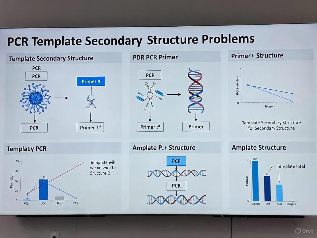
Figure 1: Structural problems in PCR and their specific manifestations.
Troubleshooting FAQ: Addressing Common Experimental Challenges
Frequently Asked Questions
Q1: My PCR results show either no product or a DNA smear when amplifying GC-rich templates. What should I investigate first?
Begin by evaluating your polymerase choice and reaction additives. Standard Taq polymerase often struggles with GC-rich templates due to stalling at secondary structures. Switch to a polymerase specifically optimized for GC-rich amplification, such as OneTaq or Q5 High-Fidelity DNA Polymerase, which are formulated with specialized GC buffers and enhancers [15] [17]. Additionally, incorporate additives like DMSO, betaine, or glycerol at optimized concentrations to reduce secondary structure formation [15] [16].
Q2: How can I prevent non-specific amplification and primer-dimer formation in difficult amplifications?
Non-specific amplification frequently results from insufficient primer annealing specificity. Increase your annealing temperature in 1-2°C increments using a gradient thermal cycler to determine the optimal stringency [3] [20]. Additionally, implement hot-start polymerases to prevent spurious initiation during reaction setup [3] [4]. For primer-dimer issues, carefully review primer design to minimize complementarity at 3' ends and optimize primer concentrations, typically between 0.1-1 μM [3] [4].
Q3: What specific steps can I take to reduce homopolymer-associated errors in sequencing applications?
Homopolymer errors originate from both PCR amplification and sequencing processes. For PCR, ensure balanced dNTP concentrations and adequate Mg2+ levels to minimize misincorporation [3]. For downstream sequencing, consider implementing error-correcting unique molecular identifiers (UMIs) designed with homotrimeric nucleotide blocks. These specialized UMIs enable a "majority vote" correction method that significantly improves sequencing accuracy in both Illumina and Nanopore platforms [21].
Q4: How does magnesium concentration specifically affect GC-rich amplification, and how should I optimize it?
Magnesium (Mg2+) serves as an essential cofactor for polymerase activity and facilitates primer binding by reducing electrostatic repulsion between primer and template [15]. For GC-rich templates, the standard MgCl2 concentration of 1.5-2 mM may be suboptimal. Implement a concentration gradient from 1.0-4.0 mM in 0.5 mM increments to identify the optimal concentration that maximizes yield while minimizing non-specific products [15] [17].
Q5: My amplification efficiency varies dramatically between templates in multi-template PCR. How can I reduce this bias?
Recent research demonstrates that specific sequence motifs near primer binding sites cause significant amplification bias independent of GC content [19]. To mitigate this, consider using deep learning prediction tools (e.g., 1D-CNN models) to identify sequences with inherently poor amplification efficiency during assay design. Additionally, minimize PCR cycle numbers and optimize denaturation times, as extended denaturation (up to 80 seconds) significantly improves representation of GC-rich templates [22].
Table 1: Optimal Reaction Component Ranges for Challenging Templates
| Component | Standard Recommendation | GC-Rich/Homopolymer Optimization | Effect |
|---|---|---|---|
| Polymerase Type | Standard Taq | Specialty polymerases (OneTaq, Q5) with GC enhancers | Improves processivity through secondary structures [15] |
| Mg2+ Concentration | 1.5-2.0 mM | 1.0-4.0 mM (gradient recommended) | Optimizes enzyme processivity and primer binding [15] [17] |
| Denaturation Time | 10-30 seconds | Up to 80 seconds for GC-rich templates | Improves strand separation of stable templates [22] |
| Annealing Temperature | 3-5°C below Tm | Gradient testing 1-2°C increments | Increases specificity, reduces mispriming [3] [20] |
| DMSO | 0% | 3-10% (v/v) | Reduces secondary structure formation [15] [16] |
| Betaine | 0M | 1-2M | Equalizes Tm of AT and GC base pairs [22] |
| PCR Cycles | 25-35 | Minimum necessary (increased if yield low) | Redplicates errors and biases [3] [21] |
Table 2: Additives and Their Mechanisms of Action
| Additive | Recommended Concentration | Primary Mechanism | Considerations |
|---|---|---|---|
| DMSO | 3-10% (v/v) | Disrupts secondary structures, lowers Tm | High concentrations can inhibit polymerase [15] [16] |
| Betaine | 1-2M | Equalizes base pair stability, reduces secondary structure | Can depress mid-GC fragments at high concentrations [22] |
| Glycerol | 5-10% (v/v) | Stabilizes enzymes, reduces secondary structure | Increases stringency, may require Ta adjustment [15] |
| 7-deaza-dGTP | Partial substitution for dGTP | Analog that reduces base stacking | Incompatible with ethidium bromide staining [15] [16] |
| Formamide | 1-5% (v/v) | Increases primer stringency | Reduces non-specific amplification [15] |
Experimental Protocols: Methodologies for Reliable Amplification
Optimized Workflow for GC-Rich Amplification
The following protocol provides a systematic approach for amplifying GC-rich targets that resist standard amplification conditions. This methodology incorporates best practices from commercial kit formulations and published optimization strategies [15] [22].
Figure 2: GC-rich template optimization workflow.
Reagents and Equipment:
- GC-optimized DNA polymerase (e.g., OneTaq with GC Buffer or Q5 High-Fidelity)
- MgCl2 stock solution (for concentration adjustment)
- PCR additives: DMSO, betaine, or commercial GC enhancer
- Gradient thermal cycler
- Standard PCR reagents: dNTPs, primers, template DNA
Step-by-Step Procedure:
Initial Setup: Prepare a master mix containing 1X specialized GC buffer, 200 μM of each dNTP, 0.5 μM of each primer, and 1-2 units of GC-optimized polymerase per reaction.
Additive Incorporation: Supplement reactions with 1X final concentration of GC enhancer or 3-5% DMSO. For extremely challenging templates (>80% GC), include 1M betaine as an additional enhancer [15] [17].
Magnesium Optimization: Prepare a dilution series of MgCl2 from 1.0 mM to 4.0 mM in 0.5 mM increments if using a standalone polymerase system.
Thermal Cycling Parameters:
- Initial denaturation: 98°C for 3 minutes (extended from standard 30 seconds)
- Cycling parameters (35 cycles):
- Denaturation: 98°C for 60-80 seconds (extended from standard 10-30 seconds)
- Annealing: Temperature gradient from 5°C below to 5°C above calculated Tm
- Extension: 72°C for 30 seconds per kb
- Final extension: 72°C for 5 minutes [15] [22]
Analysis: Resolve PCR products on an agarose gel. Successful amplification should show a single, clear band of expected size. For quantitative applications, verify amplification efficiency using qPCR methods.
Protocol for Minimizing Amplification Bias in Multi-Template PCR
This protocol addresses the sequence-specific amplification efficiency variations that complicate quantitative applications such as metabarcoding and multiplex target amplification [19] [22].
Reagents and Equipment:
- High-fidelity polymerase with bias-reduction properties (e.g., AccuPrime Taq HiFi)
- Betaine (5M stock solution)
- Standard PCR reagents
- Controlled-ramp-rate thermal cycler
Step-by-Step Procedure:
Polymerase Selection: Use a polymerase blend specifically formulated for uniform amplification across diverse sequences. The AccuPrime Taq HiFi system has demonstrated reduced GC bias in comparative studies [22].
Reaction Assembly: Prepare master mix containing 1X manufacturer's buffer, 200 μM dNTPs, 0.3 μM primers, and 1M betaine as a homogenizing agent.
Thermal Cycling Optimization:
- Initial denaturation: 95°C for 2 minutes
- Cycling parameters (25-30 cycles):
- Denaturation: 95°C for 40 seconds (extended time)
- Annealing: 60°C for 30 seconds
- Extension: 68°C for 60 seconds per kb
- Use slow ramp rates (2-3°C/second) between annealing and extension phases
Cycle Limitation: Use the minimum number of PCR cycles required for adequate product yield to prevent differential amplification from becoming pronounced [21] [22].
Validation: For quantitative applications, spike in control templates with varying GC content to verify uniform amplification efficiency across the sequence composition spectrum.
Research Reagent Solutions: Essential Materials for Success
Table 3: Key Reagents for Troubleshooting Challenging Templates
| Reagent Category | Specific Examples | Function & Application |
|---|---|---|
| Specialized Polymerases | OneTaq DNA Polymerase with GC Buffer (NEB #M0480), Q5 High-Fidelity DNA Polymerase (NEB #M0491), AccuPrime Taq HiFi | Enhanced processivity through secondary structures; improved fidelity for complex templates [15] [22] |
| GC Enhancers | OneTaq High GC Enhancer, Q5 High GC Enhancer | Proprietary formulations that disrupt secondary structures and increase primer stringency [15] [17] |
| Chemical Additives | DMSO, betaine, glycerol, formamide | Reduce secondary structure formation (DMSO, betaine, glycerol) or increase primer stringency (formamide) [15] [16] |
| Modified Nucleotides | 7-deaza-2'-deoxyguanosine | dGTP analog that improves yield of GC-rich regions by reducing base stacking interactions [15] [16] |
| Hot-Start Enzymes | Hot-start Taq, Hot-start Q5 | Prevent non-specific amplification during reaction setup by requiring thermal activation [3] [4] |
| Bias-Reduction Kits | AccuPrime Taq HiFi, KAPA HiFi HotStart ReadyMix | Specialized formulations that provide more uniform amplification across diverse sequences in multi-template PCR [22] |
Amplification bias in multi-template Polymerase Chain Reaction (PCR) has long been a significant challenge in molecular biology, compromising the accuracy of results in fields ranging from genomic research to clinical diagnostics. Traditional explanations often centered on factors like GC content and primer-template interactions. However, recent breakthroughs in deep learning have identified sequence-specific motifs as a fundamental and previously underappreciated driver of this bias. This technical support center equips researchers with the knowledge and tools to identify, troubleshoot, and mitigate these newly characterized biases in their experiments, framing them within the well-known context of template secondary structure problems.
Frequently Asked Questions (FAQs)
1. What is the new, deep-learning-informed understanding of PCR amplification bias? Traditional views attributed PCR bias primarily to broad sequence characteristics like high GC content. Recent research using convolutional neural networks (CNNs) has revealed that specific, short nucleotide sequences (motifs) within the DNA template are a major cause of unequal amplification. These motifs can induce specific physical problems, most notably adaptor-template self-priming, a mechanism previously disregarded in PCR. In this scenario, a sequence within the template itself acts as an unintended primer, leading to inefficient and skewed amplification [23].
2. How does this sequence-specific bias relate to the classic problem of template secondary structure? The problem of sequence-specific motifs is a direct and mechanistically detailed extension of the traditional template secondary structure problem. While secondary structure refers generally to hairpins and other folds that can block polymerase progression, deep learning has now pinpointed the exact nucleotide patterns that cause these issues. For instance, the motif CGTG, when located near primer binding sites, is strongly associated with poor amplification efficiency, likely because it facilitates the self-priming that leads to unproductive secondary structures [23]. Therefore, what was once a broad, hard-to-predict problem (secondary structure) can now be understood and predicted at a precise, sequence-based level.
3. What is the CluMo framework and how does it help diagnose bias? CluMo is a comprehensive bioinformatics framework that combines deep learning with systematic motif discovery. It was specifically developed to interpret deep learning models and uncover the sequence motifs responsible for PCR amplification bias. Its workflow is as follows [23]:
- Step 1: Importance Attribution. A trained deep learning model analyzes a DNA sequence and attributes an importance score to each nucleotide based on its contribution to the predicted amplification efficiency.
- Step 2: k-mer Analysis. The sequence is broken down into smaller subsequences (k-mers), and the significant k-mers linked to poor amplification are identified.
- Step 3: Motif Clustering. Similar significant k-mers are clustered together to reveal common, recurring motifs that negatively impact PCR performance.
4. Can deep learning accurately predict which sequences will amplify poorly? Yes. Studies have trained one-dimensional convolutional neural networks (1D-CNNs) on synthetic DNA pools to predict the PCR amplification efficiency of individual templates based solely on their sequence information. These models have demonstrated high predictive power, achieving an Area Under the Receiver Operating Characteristic Curve (AUROC) of up to 0.88, significantly outperforming baseline models that relied only on GC content and nucleotide frequency [23].
5. What are the practical implications for my experiment design? These insights enable a more proactive approach to PCR experiment design:
- Informed Primer Design: Beyond checking for self-complementarity, you can now screen your primer and template sequences for newly identified problematic motifs, such as CGTG, especially near the priming sites [23].
- Template Optimization: For critical applications like DNA data storage, where accurate representation is paramount, template sequences can be designed or modified to avoid motifs known to cause severe amplification bias [23].
- Pre-experiment Screening: Before wet-lab experiments, you can use in silico PCR tools to predict potential amplification problems, saving time and resources [24].
Troubleshooting Guides
Guide 1: Diagnosing and Resolving Sequence-Specific Amplification Bias
| Step | Action | Key Technical Details | Interpretation & Solution |
|---|---|---|---|
| 1. Initial Symptom Check | Observe amplification results (e.g., gel electrophoresis, qPCR curves, NGS data) for signs of bias. | Look for failed reactions, smeared bands, low yield, or inconsistent amplification between similar templates [25]. | Indicates a potential amplification issue. Proceed to sequence analysis. |
| 2. Sequence Analysis | Run your primer and template sequences through analysis software. | Use tools like OligoAnalyzer to check for traditional secondary structure and self-dimers [25]. Also, perform a manual or script-based search for the motif CGTG and other motifs identified by CluMo near primer binding sites [23]. | If problematic motifs are found, they are a likely cause. This provides a specific target for re-design. |
| 3. In Silico Validation | Use bioinformatics tools to predict PCR products and efficiency. | Utilize FastPCR or similar in silico PCR software to virtually amplify your template. These tools can predict amplicon size, specificity, and potential alternative products [24]. | Helps confirm that the suspected sequence issue leads to a failed or inefficient virtual amplification. |
| 4. Wet-Lab Redesign & Validation | Re-design primers or templates based on findings. | Solution A: Re-design primers to bind to a different region, avoiding areas with problematic motifs.Solution B: For synthetic templates, use synonymous codon replacement to eliminate the bias-inducing motif without altering the encoded information [23]. | The ultimate validation. Successful amplification after re-design confirms the diagnosis of sequence-specific bias. |
Guide 2: Differentiating Between Common PCR Artifacts
This guide helps distinguish sequence-specific bias from other common issues.
| Symptom | Possible Cause | Diagnostic Experiment | Solution |
|---|---|---|---|
| No amplification or very weak band | Low template concentration, poor quality DNA, PCR inhibitors, or a template with a high density of problematic motifs [25] [12]. | Check DNA purity (A260/280 ratio ≥1.8). Run a positive control with a known, well-amplifying template. Analyze sequence for poor-efficiency motifs [25] [23]. | Re-purify DNA. Optimize template concentration. Re-design primers to avoid "poor amplifier" sequences. |
| Multiple non-specific bands | Non-specific primer binding (traditional view) or amplification bias favoring non-target sequences in a multi-template reaction (new view) [25] [23]. | BLAST primer sequence for specificity. Use a gradient PCR to optimize annealing temperature. Analyze sequence with CluMo-like framework for bias [25] [23]. | Increase annealing temperature. Use a hot-start polymerase. Re-design primers for greater specificity and to avoid bias-inducing motifs. |
| Sequence read ends abruptly (hard stop) | Classic template secondary structure (e.g., hairpins) blocking polymerase progression [12]. | The chromatogram shows good quality data that suddenly terminates. Check for regions of high self-complementarity. | Use a polymerase mixture or chemistry designed for difficult templates (e.g., with DMSO). Design a primer that sequences through the structure from the other direction [12]. |
| Double peaks in chromatogram (mixed sequence) | Colony contamination (multiple clones) or a toxic sequence causing deletions in the plasmid during cloning [12]. | The mixed sequence appears from the beginning. Re-streak from original stock and re-pick a single colony. | Sequence a single colony. Use a low-copy vector. Grow cells at a lower temperature (30°C) [12]. |
Experimental Protocols & Data
Key Experimental Workflow: From Data to Discovery
The following diagram outlines the core methodology used in recent research to uncover sequence-specific biases.
Quantitative Performance of Deep Learning Models
The table below summarizes the performance metrics of deep learning models in predicting PCR-related outcomes, as demonstrated in recent studies.
Table 1: Performance Metrics of Deep Learning Models in PCR Analysis
| Application Context | Model Type | Key Performance Metric | Result | Reference |
|---|---|---|---|---|
| Predicting poor-amplifying templates in multi-template PCR | 1D-CNN | AUROC (Area Under the ROC Curve) | 0.88 | [23] |
| Predicting poor-amplifying templates in multi-template PCR | 1D-CNN | AUPRC (Area Under the Precision-Recall Curve) | 0.44 (at 2% prevalence) | [23] |
| Early COVID-19 diagnosis from RT-PCR cycles | LSTM (Long Short-Term Memory) | Sensitivity / Specificity (at 24 cycles) | 90.00% / 92.54% | [26] |
| Classifying carbapenem-resistant genes with digital PCR | Amplification and Melting Curve Analysis (AMCA) | Overall Accuracy | 99.6% | [27] |
The Scientist's Toolkit: Research Reagent Solutions
Table 2: Essential Materials and Tools for Investigating PCR Amplification Bias
| Item | Function / Application | Specific Example / Note |
|---|---|---|
| Synthetic DNA Pools | Provides a controlled, known template mixture for training and validating deep learning models without biological variability. | gBlocks Gene Fragments [27] |
| One-Dimensional Convolutional Neural Networks (1D-CNNs) | The primary deep learning architecture used to learn and predict PCR amplification efficiency directly from DNA sequence data. | Achieves AUROC of 0.88 for identifying poor amplifiers [23] |
| CluMo Framework | A computational framework for interpreting deep learning models to discover the specific sequence motifs that cause amplification bias. | Combines importance attribution, k-mer analysis, and clustering [23] |
| In Silico PCR Software | Bioinformatic tool for predicting PCR products and potential issues from sequence data before wet-lab experiments. | FastPCR, Primer-BLAST, UCSC In-Silico PCR [24] |
| Polymerase for Difficult Templates | Specialized enzyme blends often containing additives or engineered polymerases to overcome secondary structures and GC-rich regions. | Not specified in results, but commonly available from major suppliers. |
| Digital PCR (dPCR) | Provides absolute quantification and generates thousands of data points from amplification curves, ideal for feeding machine learning classifiers. | Fluidigm’s Biomark HD system [27] |
| DMSO or other destabilizers | Chemical additives that help disrupt secondary structures in GC-rich templates, a traditional but still relevant solution. | Used to improve amplification of difficult templates [25] |
Proactive Strategies: Predictive Tools and Primer Design to Mitigate Secondary Structures
Frequently Asked Questions (FAQs)
Q1: My homology model has poor stereochemistry. How can I identify and fix these errors? Poor stereochemistry, indicated by unusual bond angles or lengths, is a common issue in template-based modeling. To address this:
- Use Validation Tools: Run your model through specialized validation software like MolProbity. This tool analyzes your structure and provides detailed reports, including Ramachandran plots, which show the allowed and disallowed regions for protein backbone dihedral angles [28].
- Refine the Model: Use molecular modeling software such as MODELLER to perform energy minimization and refine the structure. This process adjusts atomic coordinates to resolve steric clashes and improve geometric quality [29].
- Check the Template: Ensure your initial template structure is of high quality, as errors in the template can propagate to your model.
Q2: What does a "low-quality template" error mean, and what are my options? This error signifies that the template structure used for modeling has low sequence identity to your target protein, which often leads to an unreliable model [29].
- Find a Better Template: Expand your database search or use more sensitive, iterative search methods like PSI-BLAST to find a homolog with higher sequence identity [30] [28].
- Use Multiple Templates: If no single high-quality template exists, use several templates to model different domains of your target protein. This approach can combine the most reliable regions from multiple structures [28].
- Consider Advanced Tools: For very difficult targets, consider using AlphaFold, a novel machine learning approach that can regularly predict protein structures with atomic accuracy even when no similar structure is known [31].
Q3: How accurate are automated template-based predictions compared to experimental structures? The accuracy is highly dependent on the sequence similarity between the target and the template. The table below summarizes a systematic assessment of automated metaserver predictions compared to subsequently determined experimental structures [29].
Table 1: Accuracy of Automated Template-Based Structure Prediction
| Sequence Identity to Template | Probability of a "Correct" Model* | Typical Model Quality |
|---|---|---|
| >30% | High | Often high-quality; can be suitable for many applications. |
| ~25-30% | Moderate | Useful for guiding experiments, but requires careful validation. |
| <25% | Low | Model may have significant errors; use with caution. |
*A "correct" model was defined as having >70% of its Cα atoms within 2 Å of their true positions in the experimental structure [29].
Q4: I have a PCR target with a complex secondary structure. How can bioinformatics help? Bioinformatics tools can predict regions of stable secondary structure in your DNA or RNA template that may hinder PCR amplification [3].
- Identify Problematic Regions: Use nucleic acid folding tools (e.g., mFold, UNAFold) to predict GC-rich regions or stem-loops.
- Choose a Robust Polymerase: Select a DNA polymerase with high processivity, which displays high affinity for DNA templates and is more suitable for amplifying difficult targets [3] [4].
- Optimize Reaction Conditions: Use a PCR additive or co-solvent, such as betaine or DMSO, to help denature GC-rich DNA and sequences with secondary structures [3] [4].
Troubleshooting Guide: Common Errors and Solutions
Table 2: Troubleshooting Common Bioinformatics Workflow Errors
| Problem Area | Common Error | Potential Solution |
|---|---|---|
| Sequence Analysis & Alignment | Poor alignment or wrong domain hits. | Choose the proper substitution matrix (e.g., BLOSUM, PAM) and adjust the E-value cutoff and query coverage [28]. |
| Low sequence identity to known templates. | Expand your database search or use iterative search methods like PSI-BLAST [30] [28]. | |
| Homology Modeling | Structure gaps or missing loops. | Model missing loops manually or use refinement functions in software like MODELLER [28] [29]. |
| Unstable model during simulation. | Use molecular dynamics pre-relaxation to stabilize the model before running full simulations [28]. | |
| Molecular Dynamics | Simulation becomes unstable ("exploding" coordinates). | Verify restraints and box setup. Ensure you have performed a proper minimization and equilibration (NVT/NPT) phase [28]. |
| Scripting & Data Automation | Script crashes or inconsistent results. | Verify library versions (e.g., Python, Biopython) and use absolute paths. For reproducibility, seed random number generators [28]. |
Experimental Protocols for Key Tasks
Protocol 1: Basic Workflow for Template-Based Structure Prediction
This protocol outlines the standard steps for creating a homology model using an automated metaserver, which is accessible to non-experts [29].
- Input Target Sequence: Provide the amino acid sequence of your target protein in FASTA format.
- Template Identification: The metaserver automatically sends your sequence to multiple prediction servers. These servers search databases (like the PDB) for potential template structures using sequence alignment, profile alignment, or threading algorithms [29].
- Model Generation: The server uses the target-to-template alignment to build a 3D model. Programs like MODELLER derive spatial restraints from the template and build the model by satisfying these restraints [29].
- Model Selection and Validation: The metaserver returns a list of the best models ranked by a consensus score (e.g., 3D-Jury, Pcons5). Always validate the final model using tools like MolProbity to check stereochemical quality [28] [29].
Protocol 2: Overcoming PCR Amplification Issues Caused by Template Secondary Structure
This protocol provides a methodological framework for troubleshooting PCR experiments where the DNA template's secondary structure is a suspected issue [3] [4].
- In Silico Analysis:
- Use bioinformatics tools to analyze your template sequence for high GC-content or predicted stable secondary structures.
- Wet-Lab Optimization:
- Polymerase Selection: Use a hot-start DNA polymerase with high processivity to prevent non-specific amplification and improve efficiency on complex templates [3] [4].
- PCR Additives: Include additives like betaine (1-1.5 M) or DMSO (1-10%) in the reaction mix to destabilize secondary structures [3].
- Thermal Cycling Adjustments:
- Verify Template Quality: Ensure the template DNA is of high purity and integrity to rule out degradation as the cause of failure [3].
The Scientist's Toolkit: Essential Research Reagents & Software
Table 3: Key Resources for In Silico Prediction and Troubleshooting
| Item Name | Category | Function / Application |
|---|---|---|
| MODELLER | Software | A program for comparative or homology modeling of protein 3D structures [29]. |
| MolProbity | Software | A structure-validation tool that analyzes stereochemistry, including Ramachandran plots [28]. |
| PSI-BLAST | Software | An iterative, sensitive sequence-search tool for detecting distant homologs [30]. |
| AlphaFold | Software | A deep learning system for highly accurate protein structure prediction [31]. |
| Hot-Start DNA Polymerase | Lab Reagent | Reduces non-specific amplification by remaining inactive until a high-temperature activation step [3] [4]. |
| Betaine | Lab Reagent | A PCR additive that helps denature GC-rich templates and minimize secondary structure formation [4]. |
Workflow and Relationship Diagrams
The following diagram illustrates the logical workflow for a bioinformatics-driven PCR experiment, from in silico analysis to wet-lab validation.
The following diagram outlines the key steps and decision points in a standard template-based protein structure prediction pipeline.
FAQ: Addressing Template Secondary Structure in PCR
1. Why does my PCR fail or produce low yield even with well-designed primers? Stable intramolecular secondary structures within your DNA template are a common cause of PCR failure. These structures form faster than primer-template binding and can physically block the polymerase, causing stalling, premature dissociation, or even enzymatic cleavage of the template, leading to reduced yield or complete amplification failure [32].
2. How can I identify if my template has problematic secondary structures? You can use bioinformatics tools to predict secondary structure formation. The Mfold web server is commonly used for this purpose [32]. Additionally, if your template is known to be GC-rich or contains palindromic sequences (like AAV inverted terminal repeats), you should suspect stable secondary structures [32].
3. What are the standard reagent solutions for difficult templates, and what are their limitations? Standard additives like DMSO and betaine are often used to mitigate secondary structure. However, their effectiveness is variable and can be template-dependent. In some cases, such as with recombinant AAV ITR sequences, they may provide no improvement at all [32]. The table below summarizes common solutions.
Table: Research Reagent Solutions for Templates with Secondary Structures
| Reagent / Solution | Function | Limitations |
|---|---|---|
| DMSO | Reduces thermal stability of DNA secondary structures [32]. | Variable effectiveness; can inhibit Taq polymerase activity [32]. |
| Betaine | Equalizes the contribution of GC and AT base pairs, destabilizing secondary structures [32]. | Variable effectiveness; may not work on ultra-stable structures [32]. |
| 7-deaza-dGTP | A modified nucleotide that reduces hydrogen bonding strength in GC-rich regions [32]. | Requires complete substitution of dGTP in the PCR mix [32]. |
| Disruptor Oligonucleotides | Novel class of oligos that bind template and actively unwind secondary structures [32]. | Requires custom design of a three-component oligonucleotide [32]. |
4. What is the mechanism of the novel "Disruptor" technology? Disruptors are specially designed oligonucleotides that actively unwind secondary structures. They consist of three functional parts [32]:
- Anchor: Binds to a single-stranded region of the template to initiate the process.
- Effector: Binds adjacent to the structured region and mediates strand displacement to unwind the hairpin.
- 3' Blocker: A chemical modification that prevents the disruptor itself from being extended by the DNA polymerase.
Diagram: Disruptor Oligonucleotide Mechanism
5. Where should I place my primers to avoid secondary structures? Whenever possible, design primers to target accessible, single-stranded regions. Use tools like RNAstructure to predict and avoid regions of the template that are prone to forming stable intra-molecular bonds [33]. For mRNA templates, designing primers to span an exon-exon junction can also help, as this forces specificity to the processed transcript and may avoid structured intronic regions [34].
6. What are the critical primer design rules to prevent self-complementarity issues? Self-complementarity leads to primer-dimer formation and hairpins, which drastically reduce PCR efficiency. Adhere to the following rules [33] [35] [20]:
- Avoid Self-Complementarity: Primers should not have sequences complementary to themselves or to the other primer in the pair.
- Check ΔG Values: The free energy (ΔG) of any predicted self-dimers or hairpins should be weaker (more positive) than -9.0 kcal/mol [20].
- Prevent Runs of a Single Base: Avoid sequences with runs of 4 or more of the same base (e.g., ACCCC) or dinucleotide repeats (e.g., ATATATAT), as these promote mispriming [35].
- Ensure Random Base Distribution: The sequence should have a random distribution of bases and avoid regions rich only in purines or pyrimidines [33].
7. How significant is the impact of a single base mismatch, and where is the worst location for it? A single primer-template mismatch can have a severe impact on PCR efficiency, especially in quantitative applications. The effect is highly dependent on the type of mismatch and its position. Mismatches at the 3'-terminal end of the primer (the last 1-5 nucleotides) have the most detrimental effect because they can disrupt the polymerase's active site [36]. The table below quantifies this effect for different mismatch types.
Table: Impact of 3'-End Single Base Mismatches on PCR Efficiency
| Mismatch Type | Example Pairing | Impact on PCR (Cycle Threshold, Ct) |
|---|---|---|
| High Impact | A-A, G-A, A-G, C-C | >7.0 Ct delay (Severe reduction) [36] |
| Low Impact | A-C, C-A, T-G, G-T | <1.5 Ct delay (Minor reduction) [36] |
8. What are the general golden rules for primer design to ensure robustness? Follow these consolidated guidelines for optimal primer design [33] [20] [37]:
- Length: 18-30 nucleotides.
- Melting Temperature (Tm): 55-70°C; forward and reverse primers should be within 5°C of each other.
- GC Content: 40-60%.
- GC Clamp: Include a G or C at the 3' end to promote binding, but avoid more than 3 G/C bases in the last 5 nucleotides.
- Specificity Verification: Always perform a BLAST search to ensure primers are unique to your intended target [33] [20].
Diagram: Primer Design and Secondary Structure Troubleshooting Workflow
FAQs: Addressing Common Primer Design Challenges
Q1: What are the core principles for designing a specific and efficient PCR primer?
The core principles for effective primer design involve optimizing several key sequence properties to ensure specific binding and efficient amplification [35] [38]:
- Length: Primers should typically be 18–30 nucleotides long. This provides a balance between specificity and efficient hybridization [35] [38].
- Melting Temperature (Tm): The Tm for both forward and reverse primers should be between 65°C and 75°C, and within 5°C of each other to work under a single annealing temperature [35] [39].
- GC Content: Aim for a GC content of 40–60%. This ensures stable binding without promoting non-specific interactions [35] [38] [39].
- GC Clamp: Include a G or C base at the 3' end of the primer (a GC clamp) to strengthen binding, but avoid more than 3 G or C bases in a row at the 3' end [35] [38].
- Specificity: Avoid repeated sequences and long self-complementary regions within or between primers to prevent primer-dimer formation and hairpins [35].
Q2: How does template secondary structure cause unexpected PCR results, and how can I overcome it?
Template secondary structures, such as hairpins caused by inverted repeats, can cause polymerases to "jump," leading to shorter, unexpected amplicons instead of the desired product [40]. To overcome this [41]:
- Choose a Robust Enzyme: Use DNA polymerases with high processivity, as they have a stronger affinity for the template and are better at amplifying difficult targets.
- Modify Thermal Cycling: Increase the denaturation temperature and/or time to help break apart the secondary structures before the annealing step.
- Use Additives: Incorporate PCR co-solvents like betaine or DMSO to help denature GC-rich DNA and sequences with stable secondary structures.
Q3: My PCR shows multiple non-specific bands. What is the first parameter I should optimize?
The most common first step is to increase the annealing temperature [4] [41]. A low annealing temperature is a frequent cause of non-specific binding. Optimize the temperature in 1–2°C increments using a gradient thermal cycler. The optimal annealing temperature is typically 3–5°C below the calculated Tm of the primers [41] [39]. Additionally, using hot-start DNA polymerases can prevent enzyme activity during reaction setup and reduce non-specific amplification [4] [41].
Q4: What is the most reliable method to check the specificity of my designed primers for my target sequence?
The NCBI Primer-BLAST tool is the gold standard for checking primer specificity [34]. It combines primer design with a BLAST search, checking your primer pairs against a selected database (e.g., RefSeq mRNA) to ensure they will amplify only your intended target and not other unrelated sequences. This step is crucial for confirming that your primers are gene-specific [34].
Troubleshooting Guide: Primer-Related PCR Failures
The following table outlines common primer-related PCR issues, their causes, and solutions.
| Problem | Possible Causes | Recommended Solutions |
|---|---|---|
| No Amplification or Low Yield [4] [41] | - Primer Tm too high- Poor primer design (e.g., secondary structure)- Primer concentration too low- Primer binding site obscured by template secondary structure | - Re-design primers with a Tm of 65–75°C [35].- Check for hairpins and re-design [38].- Optimize final primer concentration (typically 0.1–0.5 µM) [39].- Use a polymerase with high processivity or a PCR additive [41]. |
| Non-Specific Bands / Multiple Bands [4] [41] | - Annealing temperature too low- Primers bind to non-target sequences- Mg2+ concentration too high- Primer concentration too high | - Increase annealing temperature in 1–2°C increments [41].- Verify specificity with NCBI Primer-BLAST [34].- Optimize Mg2+ concentration (typically 1.5–2.0 mM for Taq) [39].- Lower primer concentration [41]. |
| Primer-Dimer Formation [4] [38] | - High complementarity between primers (especially at 3' ends)- Long annealing times- Excessively low annealing temperature | - Re-design primers to minimize inter-primer homology [35].- Use a primer design tool that checks for dimers [42].- Shorten annealing time and/or increase temperature [4]. |
| Smear of Products on Gel [4] | - Gradual accumulation of amplifiable DNA contaminants from previous runs- Degraded DNA template- Excessively long extension time | - Switch to a new set of primers with a different sequence.- Use separate pre- and post-PCR lab areas to prevent contamination.- Purify template DNA and optimize extension time [39]. |
Quantitative Data for Primer Design
Optimal Primer Properties
The table below summarizes the target ranges for key primer parameters to guide your initial design [35] [38] [39].
| Parameter | Optimal Range | Critical Notes |
|---|---|---|
| Length | 18–30 nucleotides | Shorter primers hybridize faster but may lack specificity; longer primers are less efficient [38]. |
| GC Content | 40–60% | A higher GC content leads to a higher Tm. Avoid long runs of a single base [35]. |
| Melting Temperature (Tm) | 65–75°C (within 5°C for a pair) | Calculate using a reliable tool. The annealing temperature (Ta) is typically 3–5°C below the Tm [41] [39]. |
| GC Clamp | 1-2 G/C bases at the 3' end | Strengthens binding. Avoid >3 consecutive G/C bases at the 3' end to prevent non-specific binding [35] [38]. |
Standard PCR Component Concentrations
This table provides standard working concentrations for a 50 µL PCR reaction using Taq DNA Polymerase [39].
| Component | Final Concentration | Notes |
|---|---|---|
| DNA Template | 1 pg–10 ng (plasmid)1 ng–1 µg (genomic) | Higher concentrations can reduce specificity [39]. |
| Primers | 0.1–0.5 µM each | Higher concentrations may promote primer-dimer formation [39]. |
| MgCl2 | 1.5–2.0 mM | Optimize in 0.5 mM increments. Critical for polymerase activity [39]. |
| dNTPs | 200 µM each | Higher concentrations can reduce fidelity but may help with long PCR [39]. |
| Taq Polymerase | 1.25 units/50 µL | Follow the manufacturer's specific recommendations [39]. |
Experimental Protocol: A Step-by-Step Primer Design and Validation Workflow
Objective: To design and validate target-specific PCR primers that are robust against template secondary structure.
Materials:
- Target DNA sequence (FASTA format or Accession ID)
- Computer with internet access
- Primer design software (e.g., NCBI Primer-BLAST, PrimerQuest [IDT])
- Thermostable DNA polymerase (standard and high-processivity)
- Standard PCR reagents: buffer, MgCl2, dNTPs, nuclease-free water
- Thermal cycler with gradient functionality
Methodology:
Sequence Acquisition and Analysis:
- Obtain the target DNA sequence from a reliable database (e.g., NCBI GenBank).
- Analyze the sequence for regions of high GC content (>60%) and potential secondary structures using bioinformatics tools. This helps in identifying potentially problematic regions to avoid.
Primary Primer Design:
- Use an automated tool (e.g., PrimerQuest [IDT] [42]) to generate initial candidate primer pairs.
- Input the target sequence and set the parameters to the optimal ranges listed in Table 3.1. Ensure the "self-complementarity" and "self 3'-complementarity" scores are low to minimize secondary structures [38].
Specificity Verification with NCBI Primer-BLAST:
- Input your candidate primer sequences into the NCBI Primer-BLAST tool [34].
- Set the parameters as follows:
- PCR Template: Your target sequence.
- Database: RefSeq mRNA or a specific organism's genome.
- Organism: Specify your target organism to limit off-target checking.
- Run the tool. Select a primer pair that shows a single, specific amplicon only for your intended target.
Empirical PCR Optimization:
- Prepare a master mix with your selected primers and template.
- Use a gradient thermal cycler to test a range of annealing temperatures (e.g., from 3°C to 10°C below the calculated Tm).
- If amplification fails or is non-specific, consider:
Validation:
- Analyze PCR products by gel electrophoresis. A single, sharp band of the expected size indicates success.
- For definitive confirmation, purify the PCR product and perform Sanger sequencing.
Workflow and Signaling Pathways
The following diagram illustrates the logical workflow for designing and troubleshooting primers, with a focus on overcoming secondary structure issues.
Primer Design and Troubleshooting Workflow
The Scientist's Toolkit: Research Reagent Solutions
This table details essential reagents and their specific functions in optimizing PCR, particularly for challenging templates.
| Item | Function in PCR | Key Consideration |
|---|---|---|
| High-Processivity DNA Polymerase | Enzyme with high affinity for template; essential for amplifying through GC-rich regions and secondary structures [41]. | More effective than standard Taq for long or complex targets. |
| Hot-Start DNA Polymerase | Polymerase inactive at room temperature; prevents non-specific priming and primer-dimer formation during reaction setup [4] [41]. | Crucial for improving specificity and yield. |
| Betaine | PCR additive that destabilizes secondary structures by acting as a kosmotrope; especially useful for GC-rich templates [41]. | Typical working concentration is 0.5–1.5 M. |
| DMSO | Co-solvent that helps denature DNA strands by disrupting base pairing; aids in amplifying structured templates [41]. | Use at low concentrations (1–10%) as it can inhibit polymerase at high levels. |
| Mg2+ Solution | Essential cofactor for DNA polymerase activity. Concentration must be optimized for each primer-template system [39]. | Excess Mg2+ reduces specificity; too little prevents amplification. |
| dNTP Mix | Provides the nucleotide building blocks (dATP, dCTP, dGTP, dTTP) for the synthesis of new DNA strands [39]. | Use balanced, equimolar concentrations to maintain polymerase fidelity. |
Troubleshooting Guides
Guide 1: Addressing False Negatives and Poor Sensitivity
Q: Why is my multi-template PCR showing inconsistent amplification or complete failure (false negatives) for some targets?
A: False negatives are often caused by factors that prevent primers from binding to their templates or that deplete essential reaction components.
| Problem Cause | Underlying Mechanism | Recommended Solution |
|---|---|---|
| Target Secondary Structure [43] | Template folding (hairpins) hides primer-binding sites, creating an energetic barrier to hybridization. | Use predictive software to model secondary structure [43]. Incorporate PCR co-solvents like DMSO (1-10%) or formamide (1.25-10%) to destabilize structures [3] [6]. |
| Primer-Dimer Formation [43] | Accidental 3' complementarity between primers leads to extension, depleting dNTPs and primers. | Optimize primer concentrations (0.1-1 µM). Use hot-start DNA polymerases to prevent low-temperature activity [3]. Redesign primers to avoid 3' complementarity. |
| Primer-Amplicon Interactions [43] | A primer for one target binds to and extends off an amplicon from a different target, generating shorter, non-detected products. | Use specialized multiplex design software to check for cross-hybridization. Physically separate highly homologous targets into different reaction tubes [44]. |
| Sequence Variation [43] | Unknown SNPs, particularly in the 3' end of the primer-binding site, prevent primer annealing (allele dropout). | Design primers against conserved regions. Use database checks (e.g., BLAST) to avoid known polymorphisms in the last three 3' bases [44]. |
Guide 2: Managing False Positives and Non-Specific Amplification
Q: What causes non-specific bands, smears, or false positives in my multiplex assay?
A: False positives often arise from non-specific primer binding or the formation of reaction artifacts.
| Problem Cause | Underlying Mechanism | Recommended Solution |
|---|---|---|
| Low Annealing Temperature [3] | Primers bind imperfectly to non-target sequences, leading to mis-priming and spurious amplification. | Optimize annealing temperature, typically 3–5°C below the primer Tm. Use a thermal cycler with a gradient function for empirical testing [3] [25]. |
| Primer Homology to Non-Target Sites [3] | Primers are not unique to the intended target and amplify homologous regions in the background. | Perform a BLAST search against the host genome to ensure primer specificity before ordering [25] [44]. |
| Formation of Heteroduplexes [45] | In multi-template reactions, single-stranded amplicons from different, but similar, templates cross-hybridize, creating hybrid molecules that can be misinterpreted. | Increase final extension time. Treat post-PCR products with a single-strand-specific exonuclease [45]. |
| Contamination [46] | Carryover of amplicons from previous runs or cross-contamination between samples generates false signals. | Use separate pre- and post-PCR workstations. Include UDG/dUTP systems. Use automated, closed-system sample prep to reduce handling [47] [46]. |
Guide 3: Solving Coverage Bias and Uneven Amplification
Q: How can I achieve uniform coverage across all targets in a multi-template PCR for NGS?
A: Coverage bias occurs when some templates amplify more efficiently than others, skewing the representation in the final library.
| Problem Cause | Underlying Mechanism | Recommended Solution |
|---|---|---|
| Differential Amplification Efficiency [45] | Templates vary in their innate "amplifiability" due to factors like GC content and secondary structure, leading to competition for reagents. | Limit the number of PCR cycles to reduce bias accumulation. Use high-processivity DNA polymerases better suited for complex templates [3] [45]. |
| Variable Tm and Primer Performance [44] | Primers with significantly different melting temperatures (Tm) will anneal with varying efficiencies in the same reaction. | Design all primers to have a tightly matched Tm (within 0.5°C of each other and the optimum) [44]. |
| Chimeric Amplicon Formation [45] | Partially extended primers "switch" to a different template during cycling, creating recombinant sequences that do not represent the original sample and disrupt coverage. | Reduce the number of PCR cycles and extension times. Use polymerases with high processivity and fidelity to minimize incomplete products [45]. |
| Inhibitor Carryover [3] [45] | Contaminants from the sample (phenol, salts, EDTA) inhibit the polymerase, but may affect amplification of different templates to varying degrees. | Re-purify the input DNA. Include a cleanup step post-fragmentation. Use polymerases known for high inhibitor tolerance [3]. |
Frequently Asked Questions (FAQs)
Q: What are the best practices for primer design in complex multi-template PCR? A: Beyond standard rules (length of 18-30 bp, GC content 40-60%), multiplex design requires stricter constraints [6] [25] [44].
- Tm Uniformity: Ensure all primers have a Tm within a very narrow range (e.g., 0.5°C) [44].
- Specificity Checking: Use BLAST to verify primers are unique to their intended target [44].
- Avoiding SNPs: Check that the 3' end of the primer (last 3 bases) does not contain known polymorphisms [44].
- Interaction Analysis: Use software to screen for primer-dimer and hetero-dimer formation, ensuring the interaction ΔG is greater than -3 kcal/mol [44].
- Overlapping Coverage: Design schemes where primer-binding regions are covered by another amplicon to mitigate allele dropout [44].
Q: My template has high GC content (>65%). How can I improve amplification? A: GC-rich regions form stable secondary structures that impede polymerase progression [25].
- Additives: Incorporate PCR co-solvents such as DMSO (1-10%), formamide (1.25-10%), or commercial GC enhancers. These help destabilize secondary structures [3] [6].
- Polymerase Choice: Select a polymerase with high processivity, which has a stronger affinity for the template and is less likely to dissociate from structured regions [3].
- Thermal Cycling: Increase the denaturation temperature or use a longer denaturation time to ensure templates are fully single-stranded [3].
Q: What is the difference between multiplex PCR and multi-template PCR? A: This is a critical distinction [45].
- Multiplex PCR involves amplifying multiple, different, non-homologous target sequences in the same reaction tube, each with its own specific primer pair. The products are often different sizes and correspond to known sequences (e.g., detecting multiple pathogens) [45].
- Multi-template PCR involves amplifying a mixture of homologous sequences (like the 16S rRNA gene from a microbial community) using a single, universal primer pair. The exact sequences of the individual templates are unknown beforehand [45].
Q: How does automated sample preparation improve NGS library quality? A: Automation addresses several key sources of error in manual library prep [47]:
- Reduced Human Error: Eliminates pipetting inaccuracies and protocol deviations between technicians.
- Enhanced Reproducibility: Improves consistency across experiments and operators, reducing batch effects.
- Lower Contamination Risk: Closed systems minimize environmental exposure and cross-contamination between samples.
- Increased Throughput and Efficiency: Allows for faster processing of many samples, freeing up researcher time [47].
Experimental Protocols
Protocol 1: A Standard Workflow for Multi-Template NGS Library Preparation
This protocol outlines the key steps for creating a sequencing library from multiple, homologous templates, incorporating troubleshooting measures.
Diagram Title: NGS Library Prep and Troubleshooting Workflow
Detailed Methodology:
Input DNA Quality Control (QC):
- Quantification: Use a fluorometric method (e.g., Qubit) for accurate DNA concentration measurement. Avoid spectrophotometry (NanoDrop) alone, as it overestimates concentration in the presence of contaminants [46].
- Purity: Check absorbance ratios (A260/280 ~1.8, A260/230 >1.8) to detect residual salts or organics [46].
- Integrity: Run a sample on an agarose gel or BioAnalyzer to confirm high molecular weight and lack of degradation [3].
Fragmentation & Size Selection:
- Method: Use enzymatic (tagmentation) or acoustic shearing to fragment DNA to the desired size (e.g., 200-500bp).
- Troubleshooting: Analyze fragment size distribution post-fragmentation using a BioAnalyzer. Over- or under-shearing will lead to poor library complexity and yield [46].
- Cleanup: Perform magnetic bead-based cleanup with a optimized bead-to-sample ratio to remove small fragments and reagents. Avoid over-drying beads, which leads to poor elution [46].
Adapter Ligation:
- Procedure: Incubate fragmented, end-repaired DNA with sequencing adapters and DNA ligase.
- Optimization: Titrate the adapter-to-insert molar ratio to maximize ligation efficiency while minimizing the formation of adapter dimers (visible as a ~70-90 bp peak on a BioAnalyzer trace) [46].
Indexing PCR:
- Cycling: Use the minimum number of PCR cycles necessary to incorporate indices and amplify the library (e.g., 8-12 cycles). Over-cycling increases duplicates, bias, and chimera formation [45] [46].
- Polymerase: Use a high-fidelity, hot-start polymerase to maintain sequence accuracy and reduce non-specific amplification [3] [6].
Final Library QC:
- Quantification: Use qPCR for the most accurate quantification of amplifiable library molecules.
- Quality: Run the final library on a BioAnalyzer or TapeStation. A clean, single peak at the expected size without a adapter-dimer peak indicates a high-quality library ready for sequencing [46].
Protocol 2: Validating Primer Specificity for Multiplex Panels
Objective: To computationally and empirically verify that primers in a multiplex pool bind only to their intended genomic targets.
Procedure:
In Silico Specificity Check:
- Perform a BLAST search for each primer sequence against the relevant reference genome (e.g., hg38 for human) [44].
- Acceptance Criterion: The primer should have a single, exact match only at the intended target locus. Discard primers with significant off-target homology.
In Vitro Validation by Single-Plex PCR:
- Before pooling, run each primer pair in a separate (single-plex) PCR reaction using the same DNA template that will be used in the multiplex.
- Analysis: Visualize PCR products on an agarose gel.
- Acceptance Criterion: A single, clean band of the expected size for each reaction. The presence of multiple bands or smears indicates non-specific binding and requires primer redesign.
Cross-Reaction Check (If Possible):
- For diagnostic panels, test each primer pair against individual, pure templates of non-target organisms or sequences that are likely to be present in the sample.
- Acceptance Criterion: No amplification product should be generated from non-target templates.
The Scientist's Toolkit: Research Reagent Solutions
| Reagent / Material | Function in Multi-Template PCR/NGS | Key Considerations |
|---|---|---|
| High-Processivity DNA Polymerase [3] | Extends primers efficiently on difficult templates with secondary structures or high GC content. Essential for even amplification. | Look for enzymes marketed for amplifying complex or GC-rich targets. Often lacks proofreading activity. |
| Hot-Start DNA Polymerase [3] [6] | Remains inactive at room temperature, preventing non-specific priming and primer-dimer formation during reaction setup. | Critical for multiplex assays. Can be antibody-mediated or chemically modified. |
| PCR Additives (DMSO, Formamide) [3] [6] | Destabilizes DNA secondary structure by interfering with base pairing. Improves yield and specificity for structured templates. | Use at optimized concentrations (e.g., DMSO at 1-10%). High concentrations can inhibit the polymerase. |
| Magnetic Beads (SPRI) [46] | For post-reaction cleanup and size selection. Binds DNA to separate it from enzymes, salts, and short unwanted fragments like adapter dimers. | The bead-to-sample ratio is critical. It determines the size range of fragments retained. |
| NGS Adapters [46] | Short, double-stranded oligonucleotides ligated to fragmented DNA. Contain sequences necessary for binding to the flow cell and for indexing samples. | Must have a balanced molar ratio with the insert DNA during ligation to maximize yield and minimize adapter-dimer formation. |
| dNTPs [3] [6] | The building blocks (dATP, dCTP, dGTP, dTTP) for DNA synthesis by the polymerase. | Use balanced equimolar concentrations. Unbalanced dNTPs increase PCR error rates and can inhibit the reaction. |
| Magnesium Salts (MgCl₂) [3] [6] | An essential cofactor for all DNA polymerases. Concentration directly affects enzyme activity and fidelity. | Requires optimization (typically 1.5-4.0 mM). Excess Mg²⁺ can reduce specificity and fidelity. |
FAQs on Template Secondary Structures in PCR
Q1: What are the specific negative effects of template secondary structures on my PCR efficiency? Template secondary structures can severely hinder PCR by preventing primers from effectively binding to their target sites [48]. When the primer binding sites are located within regions of the template DNA that form stable secondary structures, the amplification efficiency can drop significantly. Experimental data has shown that such pitfalls can lead to a decrease in PCR efficiency of over 20% [48]. Furthermore, these structures can cause the DNA polymerase to pause or fall off, leading to incomplete or failed amplification, particularly for GC-rich templates [3].
Q2: How can I experimentally verify if my PCR failure is due to template secondary structures? You can verify this through a combination of bioinformatics and experimental methods. First, use software tools to predict secondary structures around your primer binding sites [48]. Experimentally, try using PCR additives or co-solvents, such as GC Enhancers, DMSO, or betaine, which can help denature stable structures [3]. A successful amplification after adding these reagents is a strong indicator that secondary structures were the problem. Additionally, comparing amplification efficiency with a control template that lacks predicted structures can provide clear evidence [48].
Q3: Which predictive models or software are most effective for designing amplicons that avoid structured regions? A combination of utilities is often most effective. Tools like Primer Express (Applied Biosystems), Oligo, and web-based resources like Primer-BLAST can be used to design primers with optimal characteristics [48] [49]. When designing, select primer binding sites that avoid regions with thermodynamically stable secondary structures. Furthermore, assess the internal stability (−ΔG) of your primer sequences, choosing those with a decrease in 5′–3′ sense to prevent nonspecific hybridization [48].
Q4: Beyond primer design, what wet-lab strategies can mitigate secondary structure issues? Several wet-lab strategies can be highly effective:
- PCR Additives: Incorporate co-solvents like betaine, DMSO, or formamide. These additives help denature GC-rich templates and destabilize secondary structures [4] [3].
- Specialized Polymerases: Use DNA polymerases with high processivity, as they display higher affinity for templates and are better suited for amplifying difficult targets [3].
- Thermal Cycling Adjustments: Increase the denaturation temperature and/or time to more efficiently separate double-stranded DNA templates [3]. Using a temperature gradient cycler to optimize the annealing temperature can also help [49].
Q5: My amplicon sequencing data shows inconsistent results. Could template secondary structure be a factor? Yes, absolutely. Secondary structures can cause biased amplification during the library preparation PCR step. This means some sequences amplify more efficiently than others, leading to a distorted representation of the true community in your final sequencing data [50]. This is a critical consideration when interpreting amplicon sequencing data, as the initial PCR is foundational to all downstream results.
Troubleshooting Guide: Template Secondary Structures
The following table summarizes the core problems, their symptoms, and the recommended solutions.
| Problem | Observed Symptoms | Recommended Solutions |
|---|---|---|
| Stable Secondary Structures | Low amplification yield, high Cq values, failed reactions [48] [3] | Use predictive software to design primers away from structured regions [48]; Employ PCR additives (e.g., Betaine, DMSO) [3]; Increase denaturation temperature/time [3] |
| GC-Rich Templates | Smeared bands, no amplification, non-specific products [3] | Use a polymerase with high processivity [3]; Incorporate a GC-enhancer solution [3]; Optimize Mg2+ concentration [4] |
| Biased Amplicon Sequencing | Inconsistent community profiles, low diversity in final data [50] | Re-design primers to target less-structured regions; Use a high-fidelity, processive polymerase; Validate with a mock community |
Experimental Protocol: Validating Primer Performance and Overcoming Secondary Structures
This protocol provides a detailed methodology for testing primer efficiency and diagnosing secondary structure issues, as referenced in the case study [48].
Objective: To determine the optimal primer concentration and evaluate the impact of secondary structures on PCR efficiency.
Materials:
- Purified DNA template
- Designed forward and reverse primers
- Hot-start DNA polymerase and corresponding buffer (e.g., EXPRESS SYBR GreenER qPCR SuperMix)
- PCR additives (e.g., Betaine, DMSO, GC Enhancer)
- Real-time PCR instrument (e.g., ABI PRISM 7500 HT)
- Standard curve dilutions of a control template
Methodology:
- Primer Concentration Optimization:
- Prepare a series of PCR reactions with varying primer concentrations (e.g., 50 nM, 100 nM, 200 nM, 300 nM).
- Use a constant, optimal amount of DNA template.
- Run the reactions under standard cycling conditions.
- Plot a standard curve for each concentration. The optimal concentration produces a slope of -3.3, corresponding to 100% efficiency. Suboptimal concentrations can cause efficiency drops of over 10% [48].
Testing for Secondary Structure Interference:
- Set up two sets of identical PCR reactions with the optimized primer concentration.
- To the test set, add a PCR additive like betaine (at a final concentration of 1 M) or a proprietary GC enhancer.
- Run both sets (with and without additive) simultaneously on the real-time PCR instrument.
- Compare the Cq values and amplification curves. A significant decrease in Cq and a steeper amplification curve in the test set indicate that secondary structures were inhibiting the original reaction.
Data Analysis:
- Calculate PCR efficiency (E) using the formula from the standard curve: E = [10^(-1/slope) - 1] [48].
- An ideal reaction has an efficiency between 90-105%. Compare the efficiencies of reactions with and without additives to quantify the improvement.
The Scientist's Toolkit: Research Reagent Solutions
| Item | Function/Benefit |
|---|---|
| Hot-Start DNA Polymerase | Prevents non-specific amplification and primer degradation at room temperature, increasing yield and specificity [4] [3]. |
| Betaine | PCR additive that equalizes the stability of AT and GC base pairs, helping to denature GC-rich secondary structures [4]. |
| DMSO | A co-solvent that disrupts secondary DNA structures by reducing the melting temperature of the DNA, facilitating primer binding [3]. |
| GC Enhancer | Proprietary solutions (e.g., from Invitrogen) specifically formulated to aid in the amplification of difficult, GC-rich templates [3]. |
| Chimeric Plasmid DNA (cpDNA) | A non-pathogenic positive control that can be used to validate the sensitivity of diagnostic methods and avoid the risks of handling live pathogens [51]. |
Workflow for Troubleshooting Secondary Structures
The following diagram illustrates a logical workflow for diagnosing and resolving PCR issues related to template secondary structure.
Practical Solutions: A Step-by-Step Troubleshooting and Optimization Protocol
In polymerase chain reaction (PCR) research, the amplification of DNA templates with high GC-content poses a significant challenge due to the formation of stable secondary structures and intramolecular folding. These structures hinder efficient amplification, leading to poor yield, non-specific products, or complete amplification failure. Chemical additives such as DMSO and betaine are pivotal in mitigating these issues by destabilizing secondary structures, thereby facilitating the successful amplification of difficult templates. This guide provides detailed troubleshooting and methodological support for researchers confronting these common experimental obstacles.
Understanding PCR Enhancers and Their Mechanisms
PCR enhancers are chemical additives that improve the amplification efficiency of difficult DNA targets, such as those with high GC-content, stable secondary structures, or long amplicons. They function through distinct mechanisms, primarily by facilitating DNA denaturation and preventing the re-formation of secondary structures during the cycling process.
The table below summarizes the primary enhancers, their mechanisms, and typical working concentrations:
Table 1: Common PCR Enhancers and Their Properties
| Additive | Primary Mechanism | Typical Working Concentration | Key Applications |
|---|---|---|---|
| Betaine | Equilibrates GC and AT base-pair stability; reduces secondary structure formation [52] [53] | 0.5 M - 2.5 M [54] [55] | GC-rich templates, long-range PCR, inhibitor tolerance [56] [57] |
| DMSO (Dimethyl Sulfoxide) | Disrupts secondary DNA structures by interfering with hydrogen bonding [52] [54] | 2% - 10% (v/v) [54] [56] | GC-rich templates, reduces non-specific binding |
| Formamide | Destabilizes DNA double helix by binding to major/minor grooves, lowering melting temperature [54] [53] | 1.25% - 10% (v/v) [5] [53] | GC-rich templates, enhances specificity |
| 7-Deaza-dGTP | Nucleotide analog that reduces hydrogen bonding in GC-rich regions [58] | Used to partially replace dGTP | Refractory GC-rich sequences |
| BSA (Bovine Serum Albumin) | Binds to PCR inhibitors (e.g., phenols), neutralizes their effects [54] [4] | 10 - 100 µg/ml [5] [54] | Reactions with inhibitor carryover (e.g., from blood, plants) |
The following diagram illustrates the logical workflow for selecting and applying these additives to overcome template secondary structure problems:
Research Reagent Solutions
The following table details essential reagents for experiments targeting GC-rich templates, along with their specific functions in overcoming structural challenges.
Table 2: Essential Research Reagents for GC-Rich PCR
| Reagent | Specific Function in GC-Rich PCR | Example Formulation/Notes |
|---|---|---|
| Betaine (N,N,N-Trimethylglycine) | Isostabilizing agent; equalizes the melting temperature (Tm) disparity between GC and AT base pairs, preventing the formation of secondary structures like hairpins [52] [53] | Use betaine or betaine monohydrate, NOT betaine HCl [54]. Final concentration: 1.0-1.7 M. |
| DMSO (Dimethyl Sulfoxide) | Polar solvent that disrupts hydrogen bonding and base stacking, leading to lowered DNA Tm and denaturation of stable secondary structures [52] [54] | Final concentration of 2-10% (v/v). Can reduce Taq polymerase activity at higher concentrations [54] [56]. |
| 7-Deaza-dGTP | Guanine analog that lacks the N-7 position, reducing the number of hydrogen bonds in GC pairs. This destabilizes secondary structures without compromising base-pairing fidelity [58] | Used to partially or fully replace dGTP in the dNTP mix. Often used in a powerful cocktail with betaine and DMSO [58]. |
| Ammonium Sulfate (NH₄)₂SO₄-based Buffer | Substitutes KCl to enhance specificity; ammonium ions disrupt non-specific primer-template interactions, which is particularly useful for complex templates [55] [53] | Often included in specialized PCR buffers. Can be used as 10x PCR buffer AMS [55]. |
| Mg²⁺ Ions | Essential cofactor for DNA polymerase. Optimal concentration is critical for enzyme processivity and fidelity, especially for difficult templates [3] [54] | Concentration typically ranges from 1.5-4.0 mM. Must be optimized, as excess Mg²⁺ can reduce fidelity and increase non-specific products [3] [55]. |
Quantitative Data and Optimization
Systematic comparison of enhancers is crucial for protocol optimization. The following table synthesizes quantitative data from recent studies on the effectiveness of various additives in amplifying targets with different GC content, as measured by Cycle Threshold (Ct) values in real-time PCR. Lower Ct values indicate more efficient amplification [57].
Table 3: Performance of PCR Enhancers on Targets with Varying GC Content
| Additive | Concentration | Ct for 53.8% GC (Moderate) | Ct for 68.0% GC (High) | Ct for 78.4% GC (Super High) |
|---|---|---|---|---|
| Control (No Additive) | - | 15.84 | 15.48 | 32.17 |
| DMSO | 5% | 16.68 | 15.72 | 17.90 |
| Formamide | 5% | 18.08 | 15.44 | 16.32 |
| Betaine | 0.5 M | 16.03 | 15.08 | 16.97 |
| Sucrose | 0.4 M | 16.39 | 15.03 | 16.67 |
| Trehalose | 0.4 M | 16.43 | 15.15 | 16.91 |
Key Insights from the Data:
- For moderate GC targets, most additives slightly inhibit amplification (Ct increases), suggesting they should be reserved for difficult templates [57].
- For high GC targets, all shown additives provide a modest improvement.
- For super high GC targets, all additives cause a dramatic improvement in amplification efficiency (Ct decreases by over 15 cycles in some cases). Betaine, sucrose, and trehalose are among the most effective [57].
- Synergistic effects of combining additives can be powerful. For example, a mixture of betaine, DMSO, and 7-deaza-dGTP has been reported as essential for amplifying sequences with 67-79% GC content [58].
Detailed Experimental Protocols
Protocol 1: Amplification of a GC-Rich Putative Promoter Using Betaine and DMSO
This protocol is adapted from a study that successfully amplified the GC-rich (71%) promoter region of the mouse PeP gene [55].
Materials:
- Template DNA: Genomic DNA (e.g., 50-100 ng).
- Primers: Forward and reverse primers specific to the target.
- PCR Buffer: 10x PCR buffer AMS (composed of 750 mM Tris-HCl pH 8.8, 200 mM (NH₄)₂SO₄, 0.1% Tween 20).
- MgCl₂: 50 mM stock.
- Additives: 5 M Betaine stock, 100% DMSO.
- Enzymes & dNTPs: High-fidelity DNA polymerase (e.g., Pfu), 10 mM dNTP mix.
Method:
- Prepare Reaction Mix (on ice):
- 10x PCR buffer AMS: 5 µl
- MgCl₂ (50 mM): 1.6 µl (Final concentration 4 mM)
- dNTPs (10 mM): 1 µl
- Forward Primer (20 µM): 0.5 µl
- Reverse Primer (20 µM): 0.5 µl
- Betaine (5 M): 10 µl (Final concentration 1 M)
- DMSO: 2.5 µl (Final concentration 5% v/v)
- DNA Polymerase: 1 U (e.g., 0.5 µl)
- Template DNA: 50-100 ng
- Nuclease-free H₂O: to 50 µl final volume
- Thermal Cycling Conditions:
- Initial Denaturation: 95°C for 5 min
- 35-40 Cycles of:
- Denaturation: 94°C for 10-30 sec
- Annealing: Temperature gradient (e.g., 66°C to 56°C in touchdown) or 3-5°C below primer Tm for 30 sec
- Extension: 72°C for 1 min/kb
- Final Extension: 72°C for 10 min
- Hold: 4°C
Protocol 2: LCR-Based Assembly of GC-Rich Constructs
For de novo gene synthesis of GC-rich fragments, the Ligase Chain Reaction (LCR) method has proven superior to Polymerase Chain Assembly (PCA) in generating stable templates for subsequent PCR amplification [52].
Method:
- Oligonucleotide Preparation:
- Design overlapping 40-mer oligonucleotides with 20 bp overlaps.
- Pool oligonucleotides into separate plus and minus strands.
- Phosphorylate oligonucleotides using T4 Polynucleotide Kinase in provided buffer. Incubate at 37°C for 30 min, then heat-inactivate at 60°C for 20 min.
Ligation Assembly:
- Combine phosphorylated oligonucleotide pools.
- Prepare LCR reaction:
- Phosphorylated ODN pool: ~12 pmoles
- Ampligase 10x Reaction Buffer: 5 µl
- Ampligase (10 U): 2 µl
- Nuclease-free H₂O: to 50 µl
- Cycle the LCR reaction: 21 cycles of [95°C for 1 min, 70°C for 4 min], decreasing the temperature by 1°C per cycle (ramp-down).
PCR Amplification of Assembled Product:
- Use 1-2 µl of the LCR product as a template for a standard PCR, including 1 M betaine or 5% DMSO in the reaction mix to efficiently amplify the full-length GC-rich construct [52].
Troubleshooting Guide and FAQs
FAQ 1: Why does my PCR fail with a GC-rich template, and how can additives like betaine and DMSO help?
Answer: GC-rich DNA templates (typically >60% GC content) tend to form stable intra-strand secondary structures (e.g., hairpins) due to strong triple-hydrogen bonding between G and C bases. These structures can cause the polymerase to stall or fall off, resulting in truncated products or no amplification [52] [3]. Betaine and DMSO work as destabilizing agents through different mechanisms:
- Betaine is an isostabilizer that reduces the differential in thermal stability between GC and AT base pairs. This equalizes the melting temperature (Tm) across the template, preventing the localized, premature formation of these stable secondary structures [52] [53].
- DMSO interferes with hydrogen bonding between nucleic acid bases, effectively lowering the overall Tm of the DNA and helping to denature secondary structures that would otherwise persist during the cycling process [52] [54].
FAQ 2: I am getting no amplification or very low yield from my GC-rich target. What is my systematic troubleshooting strategy?
Answer:
- Step 1: Verify Template Quality and Quantity. Ensure your DNA is intact and free of inhibitors. Check concentration and purity (A260/A280 ratio) [3] [4].
- Step 2: Optimize Additives. Start with a single additive:
- Test betaine at a final concentration of 0.5 M, 1.0 M, and 1.5 M.
- In parallel, test DMSO at 2%, 5%, and 10%.
- Step 3: Combine Additives. If single additives show partial improvement, try a combination of 1 M betaine and 5% DMSO. For extremely refractory sequences, consider adding 7-deaza-dGTP to the dNTP mix [58].
- Step 4: Adjust Thermal Cycling Parameters. Increase the denaturation temperature to 98°C and/or extend the denaturation time [3]. Use a higher annealing temperature and a "touchdown" protocol to improve specificity.
- Step 5: Optimize Mg²⁺ Concentration. Titrate Mg²⁺ from 1.5 mM to 4.0 mM in 0.5 mM increments, as Mg²⁺ is a critical cofactor and its optimal concentration can shift in the presence of additives [3] [55].
FAQ 3: My PCR produces multiple non-specific bands or a smear with my GC-rich template, even though I get a product. How can I improve specificity?
Answer: Non-specific amplification is often due to reduced stringency.
- Increase Annealing Temperature: Optimize the annealing temperature by using a gradient thermal cycler. Increase the temperature in 1-2°C increments [3].
- Use Hot-Start DNA Polymerase: This prevents enzyme activity during reaction setup at lower temperatures, thereby reducing primer-dimer formation and non-specific priming [3] [4].
- Reduce Primer Concentration: High primer concentrations can promote mis-priming. Titrate primer concentrations between 0.1-1 µM [3].
- Adjust Additive Concentration: High concentrations of DMSO or betaine can sometimes reduce specificity by lowering the Tm too much. Try lowering the concentration of your additive slightly [54] [56].
- Switch Buffer Systems: Use a buffer containing ammonium sulfate ((NH₄)₂SO₄) instead of KCl, as ammonium ions can enhance specificity by destabilizing non-specific primer binding [55] [53].
FAQ: Addressing Common Researcher Questions on Polymerase Selection
1. What is the fundamental difference between a high-processivity polymerase and a proofreading polymerase?
The core difference lies in their primary function:
- High-Processivity Polymerases are characterized by their ability to add a large number of nucleotides to a growing DNA strand per single binding event. This makes them ideal for amplifying long targets, as they can synthesize DNA efficiently without frequently dissociating from the template [3] [59].
- Proofreading Polymerases possess 3'→5' exonuclease activity, which allows them to recognize and remove misincorporated nucleotides during DNA synthesis. This "proofreading" function drastically reduces error rates, making these enzymes essential for applications like cloning and sequencing where sequence accuracy is paramount [3] [60].
2. When should I prioritize a proofreading enzyme, and what is a key pitfall to avoid?
You should prioritize a proofreading enzyme for downstream applications that require high sequence fidelity, such as cloning, site-directed mutagenesis, and next-generation sequencing [3] [60]. A key pitfall is that the 3'→5' exonuclease activity of proofreading enzymes can sometimes degrade the 3' ends of the primers themselves. To prevent this, you can add phosphorothioate bonds to the 3' end of your primers, which resist nuclease activity. Additionally, always add the enzyme to your PCR mix after the dNTPs, as the presence of nucleotides can minimize this undesirable exonuclease activity [61].
3. My target is a long, GC-rich region. What polymerase characteristics should I look for?
For such challenging templates, you need a polymerase that combines multiple robust features. You should select an enzyme with high processivity to handle the long amplicon and maintain synthesis efficiency. The enzyme should also have high thermal stability to withstand the elevated denaturation temperatures often needed for GC-rich templates. Furthermore, using a hot-start polymerase is recommended to prevent nonspecific amplification and primer-dimer formation that can deplete reaction reagents [3] [62] [6].
4. Can I mix polymerases to achieve both high processivity and high fidelity?
Yes, this is a common and effective strategy. Many commercial "long-range" or "high-fidelity" PCR systems use a blend of polymerases. A typical blend might include a small amount of a proofreading enzyme (like Pfu) with a larger amount of a high-processivity enzyme (like Taq). This combination leverages the strong DNA-binding and synthesis power of one enzyme with the error-correcting capability of the other, enabling the accurate amplification of long and difficult targets [6].
Troubleshooting Guide: PCR Failures with Difficult Templates
This guide addresses common issues when amplifying complex templates like those with secondary structures, high GC-content, or long amplicons.
No Amplification Product
| Observation | Possible Cause | Recommended Solution |
|---|---|---|
| No Product | Polymerase inhibited by template secondary structures | Use a high-processivity polymerase for better strand displacement. Incorporate additives like DMSO (1-10%) or formamide (1.25-10%) to destabilize secondary structures [3] [6]. |
| Enzyme denatured during reaction setup | Use a hot-start polymerase. Set up reactions on ice and use a pre-heated thermal cycler to avoid non-specific activity [3] [62]. | |
| Excessively high annealing temperature | Recalculate primer Tm and optimize the annealing temperature using a gradient cycler. Start 5°C below the lower primer Tm [3] [62]. | |
| Insufficient enzyme activity for complex template | Increase the amount of DNA polymerase, as additives or sample inhibitors can require more enzyme for effective amplification [3]. |
Non-Specific Bands and Multiple Products
| Observation | Possible Cause | Recommended Solution |
|---|---|---|
| Non-Specific Bands | Primer annealing temperature is too low | Increase the annealing temperature in 1-2°C increments to improve specificity [3] [62]. |
| Premature polymerase activity before cycling | Use a hot-start DNA polymerase to inhibit activity until the first high-temperature denaturation step [3] [62]. | |
| Excess primers or magnesium | Optimize primer concentrations (typically 0.1-1 µM). Adjust Mg2+ concentration in 0.2-1 mM increments [3] [62]. | |
| Polymerase jumping due to template structure | For templates with strong inverted repeats, use polymerases with high processivity and increase denaturation time/temperature [40]. |
Sequence Errors and Low Fidelity
| Observation | Possible Cause | Recommended Solution |
|---|---|---|
| Sequence Errors | Use of a low-fidelity polymerase | Switch to a high-fidelity, proofreading polymerase (e.g., Q5, Pfu) for applications requiring accurate sequencing [3] [62] [60]. |
| Unbalanced dNTP concentrations | Ensure equimolar concentrations of all four dNTPs in the reaction mix. Prepare fresh dNTP stocks if necessary [3] [62]. | |
| Excessive number of PCR cycles | Reduce the number of amplification cycles to minimize the accumulation of stochastic errors over time [3] [62]. | |
| High magnesium concentration | Lower Mg2+ concentration, as excess Mg2+ can reduce fidelity by stabilizing mismatched nucleotides [3] [62]. |
Experimental Protocol: Overcoming Template Secondary Structures
The following methodology is adapted from proven approaches for sequencing and amplifying difficult templates, incorporating a critical heat denaturation step [63].
Objective: To amplify a DNA target with known or suspected high GC-content or strong secondary structures. Rationale: A controlled heat denaturation in low-salt buffer before PCR cycling can effectively melt stable secondary structures that standard denaturation steps cannot, providing a single-stranded template that is more accessible to the polymerase and primers.
Materials and Reagents
- Template DNA: 1-100 ng of your target DNA in a low-salt buffer (e.g., 10 mM Tris-HCl, pH 8.0) or nuclease-free water. Avoid buffers containing high concentrations of MgCl2 at this stage [63].
- Primers: Forward and reverse primers, designed according to standard guidelines, resuspended and diluted in nuclease-free water.
- DNA Polymerase: A high-processivity, thermostable polymerase (e.g., a specialized blend for GC-rich or long templates), preferably with hot-start capability.
- PCR Buffer: The 10X concentration buffer supplied with your polymerase.
- dNTP Mix: A solution containing all four dNTPs at 10 mM total concentration.
- Magnesium Salt: Usually MgCl2 or MgSO4, as required by your polymerase.
- PCR Additives: DMSO, formamide, or a commercial PCR enhancer solution.
- Nuclease-free Water.
Workflow: Modified PCR with Pre-Denaturation
Step-by-Step Procedure
Preliminary Mix: In a thin-walled PCR tube, combine the following components on ice:
- Template DNA (in low-salt buffer or water)
- Forward and Reverse Primers (to final concentration 0.1-1 µM)
- Nuclease-free water (to adjust final volume)
- Additive (e.g., DMSO to final 3-5%)
- Important: Do not add Mg2+, dNTPs, or DNA polymerase at this stage.
Controlled Heat Denaturation:
- Place the tube in a thermal cycler and run the following program: 98°C for 5 minutes.
- This extended denaturation in low-salt conditions helps convert complex, structured DNA into a single-stranded form, making it more accessible [63].
Master Mix Addition:
- After the heat step, briefly centrifuge the tube to collect condensation.
- Prepare a Master Mix on ice containing the remaining components:
- 10X PCR Buffer
- dNTP Mix (to final 200 µM each)
- Magnesium Salt (to optimal final concentration, e.g., 1.5-2.5 mM)
- DNA Polymerase (0.5-2.5 units per 50 µL reaction)
- Add the complete Master Mix to the heat-treated template and primer mixture. Pipette gently to mix.
Standard PCR Amplification:
- Immediately transfer the tube to the thermal cycler and start the standard PCR program. A typical 3-step protocol is shown below.
- Initial Denaturation: 94-98°C for 1-2 minutes.
- Cycling (25-35 cycles):
- Denature: 94-98°C for 10-30 seconds.
- Anneal: Temperature optimized for your primers (e.g., 5°C below Tm) for 30 seconds.
- Extend: 68-72°C for 1 minute per 1 kb of product. For long targets, extend the time.
- Final Extension: 68-72°C for 5-10 minutes.
- Hold: 4°C.
The Scientist's Toolkit: Essential Reagents for Difficult PCRs
| Reagent / Material | Function / Rationale |
|---|---|
| High-Processivity Polymerase Blends | Engineered to remain bound to the template for longer, enabling amplification of long targets and those with secondary structures by improving strand displacement [3] [59]. |
| Proofreading Polymerases (e.g., Pfu, Q5) | Essential for high-fidelity amplification due to their 3'→5' exonuclease activity, which corrects misincorporated nucleotides, reducing error rates [62] [60]. |
| Hot-Start Polymerases | Inactive at room temperature, preventing non-specific priming and primer-dimer formation before the first denaturation step, thereby increasing yield and specificity [3] [6]. |
| DMSO (Dimethyl Sulfoxide) | A common PCR additive that disrupts base pairing, helping to denature GC-rich sequences and destabilize secondary structures in the template DNA [5] [6]. |
| Betaine | A stabilizing co-solvent that can help amplify GC-rich templates by reducing the gap between the melting temperatures of GC-rich and AT-rich regions, promoting uniform amplification [5]. |
| Phosphorothioate-Modified Primers | Primers with a sulfur atom substituted for a non-bridging oxygen in the phosphate backbone at the 3' end. This modification protects against degradation by the 3'→5' exonuclease activity of proofreading polymerases [61]. |
Troubleshooting Guides
Guide 1: Addressing Non-Specific Amplification and Low Yield
| Problem & Symptoms | Possible Cause | Recommended Solution |
|---|---|---|
| Non-specific bands or multiple incorrect products on gel [3] [64]. | Low annealing temperature leading to mispriming [3]. | Increase annealing temperature in 2-3°C increments [65] [3]. Use a hot-start DNA polymerase to prevent activity at room temperature [3] [13]. |
| Excessive template or primer concentration [3] [66]. | Lower DNA template input to 10-100 ng (genomic) or 1 pg-10 ng (plasmid). Optimize primer concentration between 0.1-0.5 µM [66] [64]. | |
| No product or very faint band [4] [64]. | Denaturation temperature too low or time too short, especially for GC-rich templates [65] [3]. | Increase denaturation temperature to 98°C or extend denaturation time to 2-3 minutes [65] [67]. |
| Annealing temperature too high [64]. | Lower annealing temperature 3-5°C below the primer Tm and optimize [65] [66]. Verify primer design and ensure they are specific to the target [64]. | |
| Insufficient number of cycles for low-copy targets [65]. | Increase cycle number to 35-40 for low-abundance targets [65] [3]. |
Guide 2: Overcoming Challenges with Complex Templates
| Problem & Symptoms | Possible Cause | Recommended Solution |
|---|---|---|
| Smeared bands or incomplete amplification [4]. | Complex template secondary structures (e.g., GC-rich sequences) preventing complete denaturation or extension [3] [67]. | Use a higher denaturation temperature (e.g., 98°C) [67]. Include PCR additives like DMSO (1-10%), betaine (0.5-2.5 M), or GC enhancer solutions [3] [13] [5]. |
| DNA polymerase stalling on complex structures [13]. | Switch to a highly processive DNA polymerase, which has higher affinity for templates and is better for difficult targets [3] [13]. | |
| Failure to amplify long targets (>5 kb) [3] [67]. | DNA template degradation or depurination due to suboptimal pH or excessive denaturation times [67]. | Use high-quality, intact DNA. Resuspend DNA in TE buffer (pH 8.0) or water to prevent degradation [3] [67]. Minimize denaturation time [67]. |
| Extension time is too short [65] [66]. | Increase extension time according to polymerase speed (e.g., 1-2 min/kb). For long targets, consider reducing the extension temperature to 68°C to maintain enzyme stability [3] [67]. |
Frequently Asked Questions (FAQs)
Q1: How do I determine the correct denaturation temperature and time for my PCR protocol?
The standard denaturation temperature is 94-95°C for 15-30 seconds per cycle [65] [66]. However, this requires optimization based on your template. For GC-rich templates (>65% GC), use a higher temperature of 98°C for more efficient strand separation [67] [13]. The initial denaturation at the start of PCR is critical and often longer (1-3 minutes) to ensure complete separation of complex DNA and activation of the enzyme [65]. Always balance temperature and time to avoid excessive loss of polymerase activity [67].
Q2: What is Touchdown PCR and when should I use it?
Touchdown PCR (TD-PCR) is a technique that enhances amplification specificity by starting with an annealing temperature higher than the primer's calculated Tm and gradually decreasing it in subsequent cycles until the optimal temperature is reached [68] [13]. You should use it when you observe non-specific amplification products or primer-dimer formation under standard PCR conditions. It is particularly useful when the precise optimal annealing temperature for a primer set is unknown, as it selectively enriches for the correct product in the initial cycles [68].
Q3: How do I implement a Touchdown PCR protocol?
A typical protocol has two stages [68]:
- Touchdown Phase: Start with an annealing temperature 10°C above the primer's calculated Tm. Lower the annealing temperature by 1°C per cycle for 10-15 cycles.
- Amplification Phase: Continue for another 20-25 cycles using the final, optimal annealing temperature reached at the end of the touchdown phase.
Table: Example Touchdown PCR Protocol based on a primer Tm of 57°C [68].
| Step | Temperature (°C) | Time | Stage & Cycles |
|---|---|---|---|
| Initial Denaturation | 95 | 3:00 | 1 cycle |
| Denaturation | 95 | 0:30 | Stage 1: 10 cycles |
| Annealing | 67 (Tm +10) | 0:45 | (Temperature decreases by 1°C/cycle) |
| Extension | 72 | 0:45 | |
| Denaturation | 95 | 0:30 | Stage 2: 15-20 cycles |
| Annealing | [Final TD temp] | 0:45 | (At constant temperature) |
| Extension | 72 | 0:45 | |
| Final Extension | 72 | 5:00 | 1 cycle |
Q4: My template has a very high GC content. What specific thermal cycling adjustments can help?
GC-rich templates form strong secondary structures that are difficult to denature. Employ the following adjustments [67] [13]:
- Denaturation: Use a higher temperature (98°C) for both the initial and cyclic denaturation steps.
- Annealing: Use shorter annealing times and primers with a higher Tm (>68°C) to allow binding at a higher, more specific temperature.
- Additives: Incorporate co-solvents like DMSO (2.5-5%) or specialized GC enhancers to help denature stable duplexes [67].
- Polymerase: Choose a highly processive and hyperthermostable DNA polymerase robust enough to withstand the stringent conditions.
Experimental Protocols
Protocol 1: Optimizing Denaturation for GC-Rich Templates
This protocol is designed to overcome the challenges of amplifying DNA sequences with high guanine-cytosine (GC) content, which form stable secondary structures.
Materials & Reagents
- DNA Polymerase: Highly thermostable (e.g., derived from Archaea) and processive enzyme [65] [13].
- 10X PCR Buffer: As supplied with the polymerase.
- dNTP Mix: 10 mM aqueous solution.
- Primers: Forward and reverse, resuspended to a working concentration.
- Template DNA: 10-100 ng of high-quality DNA.
- PCR Additive: DMSO, Betaine, or a commercial GC Enhancer solution [13] [5].
- Nuclease-free Water.
Methodology
- Reaction Setup: Assemble the following in a thin-walled PCR tube on ice:
- Nuclease-free Water: to a final volume of 50 µL
- 10X PCR Buffer: 5 µL
- dNTP Mix (10 mM): 1 µL
- Forward Primer (20 µM): 1 µL
- Reverse Primer (20 µM): 1 µL
- Template DNA: variable (e.g., 2 µL)
- PCR Additive (e.g., DMSO): 2.5 µL (5% final concentration)
- DNA Polymerase: 0.5-2.5 units
- Thermal Cycling: Run the following program in a thermal cycler:
Protocol 2: Standard Touchdown PCR for Enhanced Specificity
This protocol is used to increase the specificity of amplification, reducing non-specific products and primer-dimers.
Materials & Reagents
- DNA Polymerase: Hot-Start DNA polymerase is highly recommended [68] [13].
- 10X PCR Buffer: As supplied with the polymerase.
- dNTP Mix: 10 mM aqueous solution.
- Primers: Forward and reverse, resuspended.
- Template DNA.
- Nuclease-free Water.
Methodology
- Reaction Setup: Assemble the master mix on ice, including all standard components. The use of a hot-start polymerase is critical to prevent non-specific activity during setup [68].
- Thermal Cycling: Run the following program, using the calculated Tm of your primers (example uses a primer Tm of 60°C):
- Initial Denaturation: 95°C for 3 minutes.
- Touchdown Phase (10 cycles):
- Denaturation: 95°C for 30 seconds.
- Annealing: Start at 70°C (Tm +10) for 45 seconds. Reduce the annealing temperature by 1°C per cycle over the next 9 cycles (i.e., cycle 2 at 69°C, cycle 3 at 68°C, etc.) [68].
- Extension: 72°C for 1 min/kb.
- Amplification Phase (20-25 cycles):
- Denaturation: 95°C for 30 seconds.
- Annealing: 60°C (final Tm) for 45 seconds.
- Extension: 72°C for 1 min/kb.
- Final Extension: 72°C for 5 minutes.
- Hold: 4°C.
Workflow and Decision Diagrams
Research Reagent Solutions
The following table details key reagents essential for optimizing thermal cycling parameters to overcome template secondary structure problems.
| Reagent / Material | Function in Optimization |
|---|---|
| Hot-Start DNA Polymerase | Prevents enzymatic activity during reaction setup at low temperatures, drastically reducing non-specific amplification and primer-dimer formation [3] [13]. |
| Highly Thermostable Polymerase (e.g., from Archaea) | Withstands prolonged high-temperature denaturation (98°C+) required for efficient separation of GC-rich templates without significant loss of activity [65] [13]. |
| GC Enhancer / Additives (DMSO, Betaine, Formamide) | Co-solvents that help denature stable DNA secondary structures by interfering with hydrogen bonding. They lower the melting temperature of DNA, facilitating the amplification of GC-rich regions [65] [13] [5]. |
| Magnesium Salts (MgCl₂, MgSO₄) | A necessary cofactor for DNA polymerase activity. Its concentration must be optimized, as excess Mg²⁺ can reduce fidelity and increase non-specific products, while insufficient Mg²⁺ results in low or no yield [66] [67]. |
| Optimized Primer Pairs | Well-designed primers (Tm within 5°C of each other, 40-60% GC content, no self-complementarity) are the foundation for specific amplification and are critical for the success of techniques like touchdown PCR [66] [5]. |
Troubleshooting Guide: Common PCR Buffer Issues and Solutions
This guide addresses common PCR problems related to buffer composition, with a specific focus on challenges arising from template secondary structures.
| Observation | Primary Related Buffer Cause | Recommended Solution |
|---|---|---|
| No PCR product | Mg2+ concentration too low; essential cofactor missing [69] [70]. | Optimize Mg2+ concentration by testing in 0.2-1.0 mM increments, starting from a low baseline [70] [71]. |
| Multiple or nonspecific bands | Mg2+ concentration too high or suboptimal annealing stringency [69] [70] [3]. | Increase annealing temperature; optimize Mg2+ by testing lower concentrations; use a hot-start polymerase [70] [3]. |
| PCR smear on gel | Often due to mispriming from excessive Mg2+ or presence of contaminants interacting with reagents [4] [72]. | Increase annealing temperature; reduce Mg2+ and number of cycles; use clean, new primers to avoid primer-contaminant interactions [4] [72]. |
| Low reaction yield | Imbalanced dNTP:Mg2+ ratio; dNTPs chelate Mg2+, making it unavailable for the polymerase [37] [71]. | Ensure Mg2+ concentration is higher than total dNTP concentration; prepare fresh dNTP stocks [37] [71]. |
| Errors in sequence (low fidelity) | High Mg2+ concentration can reduce base-pairing specificity, increasing misincorporation [3] [71]. | Use a high-fidelity polymerase; reduce Mg2+ concentration; ensure balanced dNTPs [3] [71]. |
| Failure with GC-rich templates | Standard buffer cannot disrupt stable secondary structures formed by GC-rich templates [16]. | Use a specialized polymerase/buffer; include additives like DMSO or betaine [3] [16]. |
FAQs on Buffer Composition and Mg2+ Optimization
Q1: Why is Mg2+ concentration so critical for PCR success? Mg2+ is an essential cofactor for DNA polymerase activity. It facilitates the binding of the enzyme to the DNA template and catalyzes the incorporation of dNTPs into the growing DNA strand [37]. If the concentration is too low, the enzyme is inactive, leading to no product. If it is too high, the polymerase loses specificity, resulting in non-specific amplification and reduced fidelity [69] [71].
Q2: How do I systematically optimize the Mg2+ concentration for a new assay? The optimal Mg2+ concentration depends on the specific template, primers, and buffer composition. Begin with the recommended concentration for your polymerase (often 1.5 mM for Taq) and perform a titration series, testing concentrations in 0.2 mM to 0.5 mM increments, typically from 0.5 mM up to 4-5 mM [69] [71]. Analyze the results by gel electrophoresis to identify the concentration that yields the strongest specific product with the least background.
Q3: How do template secondary structures, common in GC-rich regions, affect buffer requirements? GC-rich templates form stable secondary structures (e.g., hairpins) that do not denature efficiently at standard temperatures. This prevents primers and polymerase from accessing the template [16]. To overcome this, the buffer system often requires higher denaturation temperatures and additives like DMSO, glycerol, or betaine, which help to lower the melting temperature of DNA and destabilize these secondary structures [6] [16].
Q4: What is the relationship between dNTPs and Mg2+? dNTPs chelate Mg2+. Therefore, the concentration of free Mg2+ available for the polymerase is the total Mg2+ minus the amount bound to dNTPs [37]. It is critical that the Mg2+ concentration is always higher than the total dNTP concentration. A change in dNTP concentration may necessitate re-optimization of Mg2+ [71].
Q5: When should I consider using PCR additives? Additives are particularly useful for challenging templates:
- GC-rich templates: Use DMSO (typically 1-10%), formamide (1.25-10%), or betaine (1-2 M) to help denature secondary structures [6] [16].
- Inhibitor presence: Use Bovine Serum Albumin (BSA, ~400 ng/µL) to bind and neutralize inhibitors commonly found in biological samples [4] [6].
- Long amplicons: Betaine can help homogenize the melting temperature across the template, improving the amplification of long targets [71].
Experimental Protocol: Mg2+ Concentration Titration
Objective: To empirically determine the optimal Mg2+ concentration for a specific PCR assay.
Materials:
- Taq DNA Polymerase with supplied 10X Reaction Buffer (without MgCl2)
- 25 mM MgCl2 stock solution
- 10 mM dNTP Mix
- 20 µM Forward and Reverse Primers
- Template DNA
- Nuclease-free Water
Method:
- Prepare a master mix for all reactions to ensure consistency. Calculate for n (number of Mg2+ conditions) + 1 (negative control) reactions.
- Per 50 µL Reaction:
- 5.0 µL — 10X Reaction Buffer (without MgCl2)
- 1.0 µL — 10 mM dNTP Mix (200 µM final)
- 1.0 µL — 20 µM Forward Primer (0.4 µM final)
- 1.0 µL — 20 µM Reverse Primer (0.4 µM final)
- 0.5 µL — Taq DNA Polymerase (e.g., 2.5 U)
- 1.0 µL — Template DNA (e.g., 10-100 ng genomic DNA)
- Variable — MgCl2 Stock (see table below)
- Variable — Nuclease-free Water to a final volume of 50 µL
- Per 50 µL Reaction:
Aliquot the master mix into n PCR tubes. Add the appropriate volume of MgCl2 to each tube to create the desired concentration gradient.
For the negative control, add a volume of water equal to the highest MgCl2 volume used.
Run the PCR using the following "Typical Cycling Conditions" or a protocol suited to your amplicon [69]:
- Initial Denaturation: 95°C for 2 minutes
- 25-35 Cycles:
- Denature: 95°C for 15-30 seconds
- Anneal: 5°C below primer Tm for 15-30 seconds
- Extend: 68°C for 1 minute per 1 kb
- Final Extension: 68°C for 5 minutes
- Hold: 4°C
Analyze the results by agarose gel electrophoresis. The optimal condition is the one that produces the brightest specific band with the least background or nonspecific products.
Example Mg2+ Titration Table:
| Tube | Desired [Mg2+] (mM) | Volume of 25 mM MgCl2 Stock (µL) | Final Reaction Volume (µL) |
|---|---|---|---|
| 1 | 1.0 | 2.0 | 50 |
| 2 | 1.5 | 3.0 | 50 |
| 3 | 2.0 | 4.0 | 50 |
| 4 | 2.5 | 5.0 | 50 |
| 5 | 3.0 | 6.0 | 50 |
| 6 (Control) | 0.0 | 0.0 (add 6.0 µL H2O) | 50 |
Research Reagent Solutions for Secondary Structure Problems
The following reagents are essential for troubleshooting PCR experiments hampered by template secondary structures.
| Reagent | Function in PCR Buffer | Consideration for Template Secondary Structure |
|---|---|---|
| Magnesium Chloride (MgCl2) | Essential cofactor for DNA polymerase; stabilizes primer-template binding [69] [37]. | Optimal concentration is critical. Excess Mg2+ can promote non-specific priming on inaccessible, structured templates [71]. |
| DMSO (Dimethyl Sulfoxide) | Additive that disrupts base pairing, lowering the melting temperature (Tm) of DNA [6]. | Highly effective for denaturing stable GC-rich secondary structures; use at 2-10% (v/v) [16]. |
| Betaine | Additive that equalizes the stability of GC and AT base pairs [71]. | Homogenizes the DNA template, preventing pausing of polymerase at GC-rich regions; typical concentration is 1-2 M. |
| dNTP Mix | Provides the nucleotide building blocks for new DNA synthesis [69]. | Unbalanced dNTPs can increase error rates. Total dNTP concentration chelates Mg2+, so [Mg2+] must be higher [37] [71]. |
| High-Fidelity Polymerase | Enzyme with 3'→5' exonuclease (proofreading) activity for high accuracy [6] [71]. | Some high-fidelity polymerases (e.g., Pfu) are more robust for amplifying complex templates and are often paired with optimized buffers. |
| Specialized GC Buffer | Commercially optimized buffers that often include proprietary additives [16]. | Designed specifically to overcome challenges of GC-rich and highly structured templates, often providing the most straightforward solution. |
Workflow Diagram: Mg2+ and Buffer Optimization Strategy
Systematic PCR Buffer Optimization
Frequently Asked Questions
What are the primary causes of no PCR amplification? The most common causes include incorrect annealing temperature, poor primer design (e.g., self-complementarity or low Tm), poor template quality (degraded or containing inhibitors), insufficient reaction components (e.g., Mg2+, polymerase, or primers), or an insufficient number of cycles [3] [73]. Always verify your thermocycler program and ensure all reagents are added.
How can I reduce non-specific amplification and primer dimers? To enhance specificity, use a hot-start DNA polymerase to prevent activity during setup [3] [73]. Increase the annealing temperature in 1-2°C increments [3] and optimize primer concentrations to prevent excess primers that can form dimers [3] [74]. Additionally, reduce the number of PCR cycles and ensure the Mg2+ concentration is not too high [3] [73].
My template has a high GC-content or complex secondary structure. What can I do? GC-rich templates and those with stable secondary structures are a common cause of amplification failure, a phenomenon directly related to template secondary structure problems in PCR research. To resolve this, use a DNA polymerase with high processivity [3] and incorporate PCR additives or co-solvents such as DMSO, formamide, or Betaine [3] [5]. You can also increase the denaturation temperature and/or time to help melt these stubborn structures [3].
Why did my PCR yield a product of the wrong size? An incorrect product size typically results from mispriming, where primers bind to non-target sequences [73]. This can be due to an annealing temperature that is too low, excessive Mg2+ concentration, or poorly designed primers that have complementary regions elsewhere in the template [73]. Recalculate primer Tm and use a temperature gradient to find the optimal annealing temperature.
How do I improve the fidelity of my PCR reaction? To minimize sequencing errors, use a high-fidelity DNA polymerase (e.g., Q5 or Phusion) [73]. Reduce the number of cycles to decrease the cumulative chance of errors, use balanced dNTP concentrations, and avoid excessive Mg2+, which can reduce fidelity [3] [73].
Troubleshooting Guide: Common Problems and Solutions
The table below summarizes quantitative data and specific solutions for frequent PCR issues.
| Observation | Possible Cause | Recommended Solution | Key Reagent Adjustments |
|---|---|---|---|
| No Product [73] | Incorrect annealing temperature | Test a temperature gradient starting 5°C below the lower primer Tm [73]. | Optimize Mg2+ in 0.2-1 mM increments [73]. |
| Poor template quality or inhibitors | Repurify template (e.g., ethanol precipitation); dilute sample to reduce inhibitors [3] [73]. | Use a polymerase with high inhibitor tolerance [3]. | |
| Insufficient number of cycles | Increase cycles to 25-40; use up to 40 cycles for low copy number (<10) [3]. | Ensure DNA polymerase is added at 0.5-2.5 units/50 µL reaction [5]. | |
| Multiple or Non-Specific Bands [3] [73] | Annealing temperature too low | Increase annealing temperature stepwise by 1-2°C [3]. | Lower Mg2+ concentration in 0.2-1 mM increments [73]. |
| Premature replication / mispriming | Use a hot-start DNA polymerase; set up reactions on ice [3] [73]. | Reduce primer concentration (optimal range 0.1–1 µM) [3] [73]. | |
| Excess primer or Mg2+ | Optimize primer and Mg2+ concentrations [3] [73]. | Use proofreading/high-fidelity polymerases for complex templates [73]. | |
| Primer-Dimers & Smears [3] [74] | High primer concentration | Lower primer concentration to the minimum within the 0.1–1 µM range [3] [74]. | Use hot-start polymerase to prevent low-temperature activity [3] [74]. |
| Degraded primers or template | Use fresh aliquots of primers; re-purify template DNA [3] [74]. | For smears, dilute DNA extract to reduce self-priming [74]. | |
| Incorrect Product Size [73] | Mispriming | Verify primer specificity; check for complementary regions in the template [73]. | Adjust Mg2+ concentration [73]. |
| Suboptimal Mg2+ | Optimize Mg2+ concentration in 0.2–1 mM increments [73]. | Recalculate primer Tm; use a polymerase with high specificity [73]. | |
| Low Fidelity / Sequence Errors [3] [73] | Low-fidelity polymerase | Switch to a high-fidelity polymerase (e.g., Q5, Phusion) [73]. | Use balanced dNTP concentrations; reduce Mg2+ [3] [73]. |
| Unbalanced dNTPs / Excess Mg2+ | Use fresh, equimolar dNTP mixes; optimize Mg2+ down [3] [73]. | Reduce number of cycles to minimize misincorporation [3] [73]. |
Experimental Protocols for Troubleshooting
Protocol 1: Systematic Optimization of Annealing Temperature and Mg2+
Purpose: To empirically determine the optimal annealing temperature and Mg2+ concentration for a new primer set or template, mitigating issues caused by template secondary structures [3] [73].
Materials:
- DNA template (e.g., 1-100 ng genomic DNA)
- Forward and reverse primers (e.g., 20 µM stock each)
- 10X PCR Buffer (with and without Mg2+)
- MgCl2 or MgSO4 stock solution (e.g., 25 mM)
- dNTP mix (e.g., 10 mM)
- DNA Polymerase (e.g., 5 U/µL)
- Sterile distilled water
- Thermal cycler with gradient functionality
Method:
- Prepare a Master Mix for all shared components. For a 50 µL reaction, combine [5]:
- 5.0 µL of 10X PCR Buffer (without Mg2+)
- 1.0 µL of 10 mM dNTP mix
- 1.0 µL of 20 µM Forward Primer
- 1.0 µL of 20 µM Reverse Primer
- 0.5 µL DNA Polymerase
- X µL MgCl2 stock (for a matrix, test 0.0, 1.5, 2.0, 3.0, 4.0 µL)
- Y µL DNA template
- Z µL sterile water to a final volume of 50 µL.
- Aliquot the Master Mix into multiple PCR tubes.
- Program the thermal cycler with a denaturation step (e.g., 98°C for 30 sec), followed by an annealing step with a temperature gradient (e.g., spanning 50°C to 65°C) for 30 sec, and an extension step (e.g., 72°C for 1 min/kb) for 25-35 cycles [3] [5].
- Analyze the PCR products via agarose gel electrophoresis. The optimal conditions will show a single, bright band of the expected size.
Protocol 2: Amplification of GC-Rich Templates
Purpose: To overcome amplification failure due to stable secondary structures in GC-rich regions by using specialized additives [3] [5].
Materials: (In addition to standard PCR reagents)
- PCR enhancers (e.g., DMSO, Formamide, Betaine, GC Enhancer specific to your polymerase)
- DNA polymerase recommended for difficult templates (high processivity)
Method:
- Set up multiple 50 µL reactions identical to your standard protocol.
- To individual tubes, add one of the following additives [5]:
- DMSO: 1-10% final concentration (e.g., 0.5 - 5.0 µL)
- Formamide: 1.25-10% final concentration
- Betaine: 0.5 M to 2.5 M final concentration
- Include a control reaction with no additive.
- Run the PCR with an increased denaturation temperature (e.g., from 95°C to 98°C) and/or a longer denaturation time (e.g., from 30 sec to 1 min) [3].
- Analyze results by gel electrophoresis. The successful condition will show a clear, specific band where the control may fail or show smearing.
The Scientist's Toolkit: Research Reagent Solutions
| Reagent / Material | Function / Explanation |
|---|---|
| Hot-Start DNA Polymerase | Remains inactive until a high-temperature activation step, preventing nonspecific amplification and primer-dimer formation during reaction setup [3] [73]. |
| High-Fidelity DNA Polymerase | Polymerases with proofreading (3'→5' exonuclease) activity drastically reduce error rates during amplification, essential for cloning and sequencing [73]. |
| PCR Additives (DMSO, Betaine) | Act as denaturants that destabilize DNA secondary structures, crucial for amplifying GC-rich templates or those with strong hairpins [3] [5]. |
| dNTP Mix | The building blocks for new DNA strands. A fresh, equimolar mix of dATP, dCTP, dGTP, and dTTP is critical for high yield and fidelity [3] [73]. |
| Magnesium Salts (MgCl₂/MgSO₄) | Mg2+ is a essential cofactor for DNA polymerase activity. Its concentration must be optimized, as it affects enzyme activity, specificity, and fidelity [3] [5] [73]. |
Systematic PCR Troubleshooting Workflow
The following flowchart provides a logical pathway for diagnosing and resolving common PCR problems, from complete amplification failure to non-specific products. This systematic approach is particularly effective for addressing challenges arising from template secondary structures.
Troubleshooting Template Secondary Structures
The challenge of template secondary structure is a central focus in PCR research, as these structures can halt polymerase progression and cause failure. The flowchart below details a targeted approach for diagnosing and resolving these specific issues, which are often the root cause of poor amplification efficiency and specificity.
Ensuring Accuracy: Validation Techniques and Comparative Analysis of PCR Systems
Within the broader context of research on template secondary structure problems, benchmarking PCR kits for their error rates and amplification bias is not merely a procedural task—it is a fundamental requirement for data integrity. The choice of DNA polymerase directly influences the susceptibility of the amplification process to errors introduced by stable secondary structures in the DNA template. These structures, such as hairpins and stem-loops, can form transiently during the rapid temperature changes of a PCR cycle and have been demonstrated to significantly hamper amplification efficiency and fidelity [75] [76]. In fact, the adverse effects of stem-loop structures on PCR performance are directly correlated with their thermal stability [76]. This technical support center provides a structured guide to help researchers select and optimize PCR kits, with a specific focus on mitigating these pervasive challenges.
Troubleshooting Guides
PCR Kit Selection and Benchmarking
Problem: Inconsistent community representation in DNA metabarcoding studies. This issue often arises from amplification bias, where different templates in a mixed sample amplify at non-uniform rates, skewing the final results.
- Possible Causes & Recommendations
- Cause: Inherent bias of the DNA polymerase. Different polymerases exhibit varying degrees of amplification bias in multi-template PCR [77] [78].
- Recommendation: Select a polymerase proven to minimize bias. A comparative study of 14 kits showed that those containing KOD plus Neo and HotStart Taq DNA polymerase performed better in parameters like chimeras and deletions [77].
- Cause: Sequence-specific amplification efficiency. The sequence of the template itself, independent of GC content, can cause drastic under-representation due to mechanisms like adapter-mediated self-priming [19].
- Recommendation: For specialized applications like library preparation, consider using deep learning prediction tools (e.g., 1D-CNNs) to identify and avoid template sequences with poor predicted amplification efficiency [19].
- Cause: Reaction entering the plateau phase. When a reaction saturates, templates that stop amplifying early will be underrepresented compared to those that are still amplifying [78].
- Recommendation: Use a high concentration of template DNA and perform fewer amplification cycles to keep the reaction out of the plateau phase [78].
- Cause: Inherent bias of the DNA polymerase. Different polymerases exhibit varying degrees of amplification bias in multi-template PCR [77] [78].
Managing Errors Linked to Template Structure
Problem: High error rates and sequence mutations in amplified products. Stable secondary structures in the template can induce errors by interfering with the polymerase, sometimes even triggering an endonuclease activity in enzymes like Taq polymerase, leading to truncated products [76].
- Possible Causes & Recommendations
- Cause: Low-fidelity DNA polymerase.
- Recommendation: Use a high-fidelity polymerase (e.g., Q5 or Phusion) which often possesses proofreading (3'-5' exonuclease) activity to correct misincorporated bases [79].
- Cause: Suboptimal reaction conditions exacerbating secondary structures.
- Cause: Unbalanced dNTP or excess Mg²⁺ concentrations. These can increase the misincorporation rate of nucleotides [3] [79].
- Recommendation: Use fresh, equimolar dNTP mixes and optimize Mg²⁺ concentration for each primer-template set [79].
- Cause: Low-fidelity DNA polymerase.
Frequently Asked Questions (FAQs)
Q1: My PCR yield is very low for a specific amplicon, but my primers are well-designed. What could be the issue? This is a classic symptom of template secondary structure interference. Stable hairpins or stem-loops, especially those formed near the primer-binding sites, can competitively inhibit primer binding and block polymerase progression [75]. To resolve this:
- Analyze the template sequence: Use software to check for secondary structures in at least 60-base sequences both inside and outside the amplicon, focusing on regions near the primer-binding sites [75].
- Use a high-processivity polymerase: These enzymes have a stronger affinity for the template and are better at unwinding difficult structures [3].
- Optimize thermal cycling: Increase the denaturation temperature and/or time, and consider using a slow cooling ramp to allow more time for primers to bind before secondary structures re-form [3].
Q2: In multi-template PCR (e.g., for metabarcoding), how can I reduce amplification bias between different sequences? Bias in multi-template PCR is a complex issue, but several strategies can mitigate it.
- Kit Selection: As highlighted in comparative studies, the choice of polymerase kit is critical. Kits based on KOD plus Neo have shown lower bias for several parameters [77].
- Reaction Parameters: Using a high starting template concentration and minimizing the number of PCR cycles prevents the reaction from reaching the plateau phase, which can artificially skew product ratios [78].
- Replicate Reactions: Mixing the products of several independent replicate PCR preparations can average out stochastic "PCR drift" that occurs in the early cycles [78].
Q3: I suspect secondary structures are causing truncated products. Is there a mechanistic explanation? Yes. Recent research provides a mechanism beyond simple polymerase stalling. When Taq DNA polymerase encounters a stable stem-loop structure during extension, its 5'-3' exonuclease activity can act as an endonuclease, cleaving the template strand within the duplex region. This cleavage unwinds the structure but results in a truncated product that is then amplified [76]. Switching to a polymerase with minimal or no 5'-3' exonuclease activity may help in such cases.
Experimental Data & Protocols
Quantitative Comparison of PCR Kits
The following table summarizes key findings from a study that compared 14 different PCR kits using a mock eukaryotic community DNA sample, focusing on parameters critical for metabarcoding accuracy [77].
Table 1: Comparative Performance of Selected PCR Kits in DNA Metabarcoding
| PCR Kit (DNA Polymerase) | Chimera Formation | Blast Top Hit Accuracy | Deletion Errors | Overall Performance Note |
|---|---|---|---|---|
| KOD plus Neo (TOYOBO) | Low | High | Low | Statistically superior for parameters associated with chimeras, similarity, and deletions. |
| HotStart Taq (BiONEER) | Low | High | Low | Displayed better results at an annealing temperature of 65°C. |
| Other Tested Kits | Variable (Statistically significant differences, p<0.05) | Variable (Statistically significant differences, p<0.05) | Variable (Statistically significant differences, p<0.05) | Performance varied significantly across all seven tested parameters. |
Key Experimental Protocol: Measuring Amplification Bias
This protocol is adapted from methods used to quantify bias in multi-template PCR [78] [19].
Objective: To evaluate the amplification bias introduced by a PCR kit when amplifying a complex mixture of templates.
Materials:
- Mock Community DNA: A defined mixture of genomic DNA or plasmid DNA from multiple species (e.g., 40 microalgal species [77]).
- Test PCR Kits: The kits to be benchmarked.
- Universal Primers: Degenerate primers targeting a conserved gene (e.g., 16S rRNA for bacteria, 18S rRNA for eukaryotes).
- High-Through Sequencing Platform.
Method:
- Sample Preparation: Prepare the mock community DNA with known, equal starting quantities for each template.
- PCR Amplification: Amplify the mock community using each PCR kit to be tested, strictly following the manufacturers' protocols. It is critical to use a high template concentration and low cycle number to avoid the plateau phase [78].
- Library Preparation and Sequencing: Prepare sequencing libraries from the PCR products and sequence using a high-throughput platform.
- Bioinformatic Analysis:
- Process the sequencing data to map reads back to the individual templates in the mock community.
- Calculate the relative abundance of each template in the sequencing data.
- Compare the measured relative abundance to the known starting abundance.
- Statistical Evaluation: Quantify bias by calculating the degree of over- or under-representation for each template. The kit that produces product ratios closest to the known template ratios has the lowest amplification bias.
Visualization of Workflows and Relationships
Diagram Title: Troubleshooting PCR Issues from Template Secondary Structure
The Scientist's Toolkit: Essential Reagents for Reliable PCR
Table 2: Key Research Reagent Solutions for PCR Benchmarking and Optimization
| Reagent / Material | Function / Explanation | Considerations for Selection |
|---|---|---|
| High-Fidelity DNA Polymerase | Enzymes with proofreading (3'→5' exonuclease) activity to reduce replication errors and increase sequence accuracy. | Essential for cloning, sequencing, and applications requiring high sequence fidelity. Examples include Q5 and Phusion [79]. |
| Bias-Reduced Polymerase Kits | Kits specifically validated for low amplification bias in multi-template reactions. | Look for kits with polymerases like KOD plus Neo, which demonstrated superior performance in comparative studies [77]. |
| Hot-Start DNA Polymerase | Engineered to be inactive at room temperature, preventing non-specific amplification and primer-dimer formation during reaction setup. | Improves specificity and yield of the desired product. Available in various formats (antibody-mediated, chemical modification) [3] [79]. |
| PCR Additives (e.g., DMSO, GC Enhancers) | Co-solvents that help denature GC-rich DNA and disrupt template secondary structures (e.g., hairpins), improving amplification efficiency. | Concentration must be optimized, as excess can inhibit the polymerase. Specific commercial enhancers are often supplied with polymerase kits [3] [79]. |
| Synthetic DNA Mock Communities | Defined mixtures of DNA templates from known species, used as a calibrated standard to benchmark PCR bias and error rates. | Allows for quantitative comparison of different PCR kits and protocols by providing a known "ground truth" [77] [19]. |
| Optimized Mg²⁺ Solution | Magnesium ions are essential cofactors for DNA polymerase activity. The type (MgCl₂ vs. MgSO₄) and concentration critically affect fidelity, specificity, and yield. | The optimal concentration is template- and primer-specific. Excess Mg²⁺ can reduce fidelity and increase non-specific binding [3] [79]. |
FAQ: What is PCR Fidelity and Why Does it Matter?
Q1: What does "fidelity" mean in the context of PCR? Fidelity refers to the accuracy with which a DNA polymerase can replicate a template DNA sequence. A high-fidelity enzyme introduces fewer errors (nucleotide misincorporations) during amplification. This is crucial for applications where the DNA sequence must be correct after amplification, such as cloning, sequencing, and SNP analysis [80].
Q2: How does template secondary structure relate to PCR fidelity? Complex template sequences, such as those with high GC-content that form stable secondary structures, can challenge the DNA polymerase during amplification. These structures can cause the enzyme to stall, which may increase the likelihood of misincorporating a nucleotide, thereby reducing fidelity [3] [9]. Using polymerases with high processivity and fidelity is key to accurately amplifying these difficult templates.
Quantitative Fidelity Data for Common Polymerases
The fidelity of different DNA polymerases can vary by an order of magnitude. Fidelity is often expressed relative to Taq DNA polymerase or as an error rate. The following table summarizes the fidelity of several commercially available high-fidelity enzymes [81].
Table 1: Fidelity Comparison of High-Fidelity DNA Polymerases
| Product Name (Supplier) | Polymerase Fidelity (Relative to Taq) | Maximum Amplicon Length | Key Features / Notes |
|---|---|---|---|
| Q5 High-Fidelity DNA Polymerase (NEB) | ~280X | 20 kb (simple); 10 kb (complex) | One of the highest fidelities available. |
| Phusion High-Fidelity DNA Polymerase (Thermo Fisher) | 39X | 20 kb (simple); 10 kb (complex) | High thermostability. |
| AccuPrime Pfx (Life Technologies) | 26X | 12 kb | |
| PfuUltra II Fusion HS (Agilent) | 20X | 19 kb | |
| PfuUltra High-Fidelity DNA Polymerase (Agilent) | 19X | 17 kb (simple); 6 kb (complex) | |
| KOD DNA Polymerase (EMD) | 12X | 6 kb (simple); 2 kb (complex) | |
| Platinum Taq HiFi (Life Technologies) | 6X | 20 kb |
Experimental Protocol: Quantifying PCR Error Rates
Accurate measurement of polymerase error rates is essential for selecting the right enzyme for sensitive applications. Below is a detailed methodology for a high-throughput sequencing-based assay that uses Unique Molecular Identifiers (UMIs) to precisely quantify errors [82].
Workflow: High-Throughput PCR Error Quantification
Step-by-Step Procedure:
- Tag Template Molecules (Linear Amplification): Tag each input template DNA molecule with a random 14-nucleotide Unique Molecular Identifier (UMI). This UMI will allow all subsequent copies to be traced back to their original molecule [82].
- First PCR Amplification: Perform a PCR with the test polymerase for 20 cycles (or 25 for low-efficiency polymerases like Phusion). Errors introduced during this step will be carried forward [82].
- Create a Dilution Bottleneck: Perform a limiting dilution of the first PCR product. The goal is to sample so few molecules that it is statistically likely each sampled molecule originated from a different original template. This ensures that subsequent amplification does not create duplicates that mask errors from the first PCR [82].
- Second PCR Amplification: Use a second PCR (22-29 cycles) to amplify the diluted samples for sequencing. This step adds the necessary sequencing adapters [82].
- High-Throughput Sequencing: Sequence the final library on a platform like Illumina [82].
Data Analysis and Error Rate Calculation:
- Group all sequencing reads by their UMI tag.
- For each UMI group, generate a consensus sequence. This corrects for errors introduced during the second PCR and sequencing steps.
- Compare the consensus sequence for each UMI to the known original template sequence to identify errors introduced during the first PCR.
Calculate the error rate using the following formula [82]:
Error Rate = (Number of Errors in Consensus Sequences) / (Total UMI Tags × Template Length × Number of Cycles in First PCR)
The Scientist's Toolkit: Essential Reagents for Fidelity Assays
Table 2: Key Research Reagent Solutions
| Reagent | Function in the Experiment |
|---|---|
| High-Fidelity DNA Polymerase | The enzyme being tested. Chosen for its proofreading (3'→5' exonuclease) activity, which corrects misincorporated nucleotides [9]. |
| UMI Adapters | Short, random nucleotide sequences used to uniquely tag each original input DNA molecule, enabling error correction and accurate tracing [82]. |
| dNTPs | Deoxynucleotide triphosphates (dATP, dCTP, dGTP, dTTP) are the building blocks for new DNA strands. Must be provided in equimolar concentrations to prevent unbalanced incorporation that can increase error rates [3]. |
| Mg2+ Solution | An essential cofactor for DNA polymerase activity. Its concentration must be optimized, as excess Mg2+ can promote misincorporation of nucleotides [3]. |
| NGS Library Prep Kit | A commercial kit used to prepare the final PCR products for high-throughput sequencing, typically by adding platform-specific adapters and indices [82]. |
FAQ: Troubleshooting Fidelity and Amplification Issues
Q3: My PCR yield is low when using a complex template. How can I improve it without sacrificing fidelity? Low yield with complex templates (e.g., GC-rich sequences) is often due to secondary structures that hinder polymerase progression. To address this:
- Use a specialized polymerase: Choose a high-fidelity DNA polymerase with high processivity (the number of nucleotides added per enzyme binding event) and high thermostability, as these are better suited for difficult templates [3] [9].
- Employ PCR additives: Reagents like DMSO (1-10%), formamide (1.25-10%), or betaine can help denature GC-rich secondary structures and improve yield and accuracy [3] [6].
- Optimize cycling conditions: Increase the denaturation temperature or time to ensure template strands separate completely [3].
Q4: My sequencing results show a high error rate. What are the most common causes? A high error rate can stem from several factors related to the polymerase and reaction conditions:
- Low-fidelity polymerase: Verify you are using a high-fidelity, proofreading polymerase. Standard Taq polymerase has a significantly higher error rate than engineered enzymes like Q5 or Phusion [81] [9].
- Excessive cycling: High numbers of PCR cycles (>30) increase the cumulative probability of errors. Use the minimum number of cycles necessary to obtain sufficient product [3].
- Unbalanced dNTPs or Mg2+: Ensure all four dNTPs are present at equimolar concentrations. Excessive Mg2+ concentration can also increase the error rate by reducing the enzyme's stringency [3].
- Polymerase-specific substitution preferences: Different polymerases have characteristic error profiles (e.g., some favor C>T transitions, while others favor A>G) [82].
How do template secondary structures negatively impact PCR quality control metrics?
Template secondary structures, such as hairpins and stable intramolecular folds, adversely affect PCR quality by several mechanisms. They can physically block polymerase progression, leading to stalling and the production of incomplete or truncated products [32]. These structures form preferentially during the annealing step, competitively inhibiting the primers from binding to their target sites [75]. This directly reduces amplification efficiency and yield. Furthermore, stable secondary structures can promote polymerase "jumping" or mis-priming, which is a direct cause of chimeric product formation and can significantly reduce the specificity of the reaction [32]. The thermal stability of these structures is a key factor; longer stems and smaller loops in hairpins create more stable structures that cause greater suppression of amplification [75].
Table 1: Impact of Hairpin Structures on qPCR Amplification Efficiency [75]
| Hairpin Location | Stem Length | Loop Size | Effect on Amplification |
|---|---|---|---|
| Inside the amplicon | Increases | Decreases | Notable suppression; effect increases with longer stem and smaller loop |
| Inside the amplicon | 20 bp | N/S | Drastic suppression; no targeted amplification products formed |
| Outside the amplicon | Increases | Decreases | Notable suppression; effect increases with longer stem and smaller loop |
Table 2: Effects of GC-rich Templates and Common Mitigation Strategies [25] [3] [83]
| Feature/Factor | Impact on PCR | Recommended Mitigation |
|---|---|---|
| High GC Content (>60%) | Resists denaturation; forms stable secondary structures; promotes primer-dimer formation | Use polymerases optimized for GC-rich templates; include additives like DMSO or betaine |
| Mg2+ Concentration | Too little reduces polymerase activity; too much promotes non-specific binding | Optimize with a gradient (e.g., 1.0-4.0 mM in 0.5 mM increments) |
| Annealing Temperature (Ta) | Too low causes non-specific binding; too high prevents primer hybridization | Optimize Ta 3-5°C below primer Tm; use a temperature gradient |
Experimental Protocol for Evaluating Secondary Structure Impact
This protocol systematically evaluates how secondary structures in a DNA template affect key QC metrics: specificity, yield, and chimerism.
1. Design Templates with Predicted Secondary Structures
- Template Design: Using software like Mfold, design a series of DNA templates that contain hairpin structures with varying stem lengths (e.g., from 5 bp to 20 bp) and loop sizes [75] [32].
- Control Template: Design a control template with minimal predicted secondary structure for comparison.
- Primer Design: Design primer pairs that bind both inside and outside the region of the secondary structure. Ensure primers meet standard design criteria (length 18-22 bp, Tm 52-58°C, GC content 40-60%) [25] [35].
2. PCR Amplification and QC Assessment
- Reaction Setup: Perform separate PCR reactions for each template-primer combination. Use a hot-start DNA polymerase to enhance specificity [3].
- Gel Electrophoresis: Analyze PCR products on an agarose gel.
- Yield: Compare the band intensity of the target product to the control.
- Specificity: Check for the presence of a single, clean band versus multiple non-specific bands or smears.
- qPCR Analysis: If using qPCR, compare the amplification efficiency and Cq values between structured and control templates [75].
- Sequencing for Chimeras: Clone the PCR products from reactions with structured templates. Sequence multiple clones and analyze the sequences for unexpected rearrangements or junctions that indicate chimeric formation [32].
3. Mitigation Using Disruptor Oligonucleotides
- Disruptor Design: For a problematic template, design "disruptor" oligonucleotides. These should be reverse-complementary to the template sequence and partially overlap the duplex region of the secondary structure. A disruptor typically consists of [32]:
- An anchor sequence to initiate template binding.
- An effector sequence to mediate strand displacement and unwind the structure.
- A 3' blocker (e.g., C3 spacer) to prevent elongation by the polymerase.
- Re-test: Repeat the PCR with the addition of the disruptor oligonucleotides and re-evaluate yield, specificity, and chimerism as described above [32].
The Scientist's Toolkit: Research Reagent Solutions
Table 3: Key Reagents for Managing Template Secondary Structures
| Reagent / Material | Function / Explanation |
|---|---|
| Specialized DNA Polymerases (e.g., Q5, OneTaq) | Polymerases with high processivity are better at navigating through complex secondary structures. Some are supplied with proprietary GC buffers [3] [83]. |
| GC Enhancer / Betaine | Reduces the thermal stability of secondary structures by acting as a destabilizing agent, facilitating the denaturation of GC-rich templates [32] [83]. |
| DMSO (Dimethyl Sulfoxide) | A co-solvent that helps denature GC-rich DNA and prevents the re-formation of secondary structures by interfering with base pairing [83]. |
| Disruptor Oligonucleotides | Novel reagents designed to bind the template and actively unwind stable intramolecular secondary structures via strand displacement, preventing polymerase stalling [32]. |
| 7-deaza-dGTP | A modified nucleotide analog that can be substituted for dGTP. It reduces the strength of hydrogen bonding in GC pairs, making secondary structures less stable [32]. |
| Mg2+ Solution | A critical cofactor for polymerase activity. Its concentration must be optimized, as both too little and too much can negatively impact reactions with difficult templates [3] [83]. |
Frequently Asked Questions (FAQs)
Q1: My PCR for a GC-rich promoter region shows a smear on the gel. What should I optimize first? A: A smear often indicates non-specific amplification or incomplete products due to secondary structures. Your first steps should be:
- Polymerase Choice: Switch to a polymerase specifically engineered for GC-rich and structured templates, such as those supplied with a GC Enhancer [83].
- Annealing Temperature: Increase the annealing temperature in a gradient (e.g., 1-2°C increments) to improve primer binding stringency [3] [83].
- Additives: Incorporate a destabilizing agent like betaine or DMSO into your reaction mix [83].
Q2: How can I proactively design a PCR assay to avoid problems from template secondary structures? A: During the in-silico design phase:
- Sequence Analysis: Use software to analyze at least 60 base pairs of sequence surrounding the primer-binding sites (both inside and outside the amplicon) to check for potential stable secondary structures [75].
- Primer Placement: If possible, design primers so they do not bind to regions involved in stable secondary structures.
- Tool Assistance: Use tools like NCBI's Primer-BLAST to check for specificity and OligoAnalyzer to predict secondary structure formation in the primers themselves [34] [25].
FAQs: Addressing Key Experimental Challenges
Q1: What are the primary advantages of using synthetic DNA pools to quantify amplification efficiency?
Synthetic DNA pools provide a controlled and defined system to study amplification biases without the confounding variables present in biological samples. Key advantages include:
- Defined Composition: They consist of known, random sequences, preventing bias from enriched biological motifs and allowing for precise tracking of individual sequence abundance [84].
- Reproducibility: Experiments show that sequences identified as having low amplification efficiency in one pool reproducibly under-perform in new, independent pools and in single-template qPCR validation, confirming the sequence-specific nature of the bias [84].
- Mechanistic Insight: Using these pools, researchers can link poor amplification efficiency to specific sequence motifs, such as those causing adapter-mediated self-priming, challenging long-standing PCR design assumptions [84].
Q2: How can I experimentally track amplification efficiency across multiple PCR cycles?
A serial amplification protocol followed by sequencing can precisely quantify how amplification skews over time. The core methodology is as follows [84]:
- Library Construction: Synthesize a pool of DNA sequences (e.g., 12,000 random sequences) with common terminal primer binding sites.
- Serial PCR: Perform multiple consecutive PCR reactions (e.g., 6 reactions of 15 cycles each).
- Sequencing Sampling: After each PCR round, take a sample for high-throughput sequencing to quantify the amplicon composition.
- Data Fitting: Fit the sequencing coverage data for each sequence to an exponential PCR amplification model. This fit yields two key parameters per sequence: the initial synthesis bias and its sequence-specific amplification efficiency (εi) [84].
Q3: My multi-template PCR shows a progressive skew in coverage and some sequences drop out entirely. What is the likely cause?
This is a classic symptom of sequence-specific differences in amplification efficiency. Even small, reproducible differences in how efficiently each template is amplified are exponentially compounded over multiple PCR cycles. For example, a template with an amplification efficiency just 5% below the average will be underrepresented by a factor of two after only 12 cycles [84]. This is independent of GC content and is driven by intrinsic sequence properties [84].
Q4: How can I improve the homogeneity of amplification in my multi-template assay?
- In-Silico Prediction: Employ deep learning models, such as 1D-CNNs, trained on synthetic DNA pool data to predict sequences with poor amplification efficiency directly from their sequence, allowing for pre-selection of better-performing templates [84].
- Library Design: Use predictive tools to design inherently homogeneous amplicon libraries by avoiding sequences with motifs linked to poor amplification [84].
- Wet-Lab Optimization: Follow a structured development process for multiplex assays, which includes in-silico design, wet-lab validation with positive controls, and final implementation on sample matrices to validate specificity and sensitivity [85].
Troubleshooting Guide: Common Issues and Solutions
| Observation | Possible Cause | Recommended Solution |
|---|---|---|
| No or Low Amplification | PCR inhibitors present in template (e.g., phenol, salts, heparin) [3] [86] | Dilute template 100-fold; re-purify using silica-column cleanup or ethanol precipitation; use polymerases with high inhibitor tolerance [3] [86]. |
| Suboptimal cycling conditions [6] [3] | Increase cycle number (up to 40); lower annealing temperature in 2°C increments; increase extension time [3] [86]. | |
| Non-specific amplification due to adapter-mediated self-priming [84] | Redesign adapter/primers using in-silico tools to avoid self-complementary motifs identified by models like CluMo [84]. | |
| Non-Specific Bands or Smearing | Reaction conditions not stringent enough [87] [86] | Increase annealing temperature; use touchdown PCR; reduce number of cycles; use hot-start DNA polymerase to prevent primer-dimer formation [87] [3] [86]. |
| Primer-dimer formation or mispriming [4] | Optimize primer concentration (0.1-1 µM); verify primer specificity using BLAST; redesign primers to avoid 3' end complementarity [6] [86]. | |
| Contamination from previous PCR products [86] | Use separate pre- and post-PCR work areas; equip pipettes with aerosol filter tips; use UV irradiation and bleach to decontaminate surfaces [86]. | |
| Skewed Coverage / Sequence Dropout | Sequence-specific amplification bias [84] | Use synthetic DNA pools to pre-validate amplification efficiency; employ balanced primer design; utilize deep learning models to predict and filter poor amplifiers [84]. |
| High Error Rate / Sequence Mutations | Low-fidelity DNA polymerase [87] [3] | Switch to a high-fidelity polymerase with 3'→5' proofreading exonuclease activity (e.g., Q5, Pfu) [87] [6] [3]. |
| Unbalanced dNTP concentrations or excess Mg2+ [3] [86] | Use fresh, equimolar dNTP mixtures; optimize Mg2+ concentration to the lowest effective level [3] [86]. |
Experimental Protocol: Quantifying Amplification Efficiency with Synthetic DNA
This protocol details the method for using synthetic DNA pools to track and calculate sequence-specific amplification efficiencies, as pioneered in recent research [84].
I. Materials and Equipment
- Synthetic DNA Pool: A custom-designed oligonucleotide library comprising thousands of random sequences flanked by constant adapter/primer binding regions. A GC-content constrained pool (e.g., 50% GC) is recommended as a control [84].
- High-Fidelity, Hot-Start DNA Polymerase: To minimize non-specific amplification and polymerase-introduced errors [6] [3].
- PCR Reagents: dNTPs, MgCl2 or MgSO4, and appropriate reaction buffer.
- Primers: Complementary to the constant adapter regions on the synthetic DNA pool.
- Thermal Cycler
- Next-Generation Sequencing (NGS) Platform: e.g., Illumina.
II. Step-by-Step Procedure
Library Preparation and Initial Sampling:
- Resuspend the synthetic DNA pool in molecular-grade water or TE buffer to a defined concentration.
- T0 Sample: Remove an aliquot of the pool before any PCR amplification. This sample will be used to determine the initial coverage bias introduced during synthesis. Prepare this sample for sequencing (e.g., using a PCR-free library protocol if possible) [84].
Serial PCR Amplification:
- Set up a large-volume PCR reaction with the synthetic DNA pool as the template.
- Run the first round of PCR for a defined number of cycles (e.g., 15 cycles).
- T1 Sample: After the first round, remove a small aliquot of the PCR product. Purify this aliquot using a PCR clean-up kit to remove enzymes and dNTPs.
- Use the remaining PCR product as the template for the next round of amplification.
- Repeat this process of amplification and sampling for multiple rounds (e.g., 6 rounds of 15 cycles each, for a total of 90 cycles), collecting samples T2, T3, ... Tn [84].
Sequencing Library Preparation and Sequencing:
- Prepare all collected samples (T0 through Tn) for next-generation sequencing using a standard library prep kit, adding the required sequencing adapters and indices.
- Pool the libraries and perform high-throughput sequencing to obtain high-coverage data for each sample.
III. Data Analysis and Calculation of Amplification Efficiency
Bioinformatic Processing:
- Demultiplex the sequencing data and map reads back to the reference list of sequences in the original synthetic pool.
- Count the number of reads for each unique sequence in each sample (T0, T1, T2, ... Tn).
Model Fitting and Efficiency Calculation:
- For each individual sequence (i), model its read count over the PCR cycles as an exponential growth process.
- The read count (C) at cycle number (n) can be fit to the equation:
( C{i,n} = C{i,0} \times (1 + \epsiloni)^n )
where:
- ( C{i,0} ) is the initial bias for sequence i (derived from the T0 sample data).
- ( \epsilon_i ) is the sequence-specific amplification efficiency per cycle [84].
- Fit this model to the time-series data for each sequence to extract its ( \epsiloni ). Sequences with low ( \epsiloni ) values are those with poor amplification efficiency.
Workflow for Quantifying Amplification Efficiency
The following table summarizes quantitative findings on amplification efficiency distribution from a systematic analysis using synthetic DNA pools.
Table 1: Quantification of Amplification Bias in Multi-Template PCR
| Parameter | Value | Experimental Context |
|---|---|---|
| Subset of Sequences with Very Poor Efficiency | ~2% of pool | Synthetic oligo pools with random sequences (GCall) and 50% GC-constrained sequences (GCfix) [84]. |
| Lowest Estimated Efficiency | ~80% relative to population mean | Equivalent to a halving in relative abundance every 3 PCR cycles [84]. |
| Predictive Model Performance (1D-CNN) | AUROC: 0.88 | Model trained to predict sequence-specific amplification efficiency from sequence data alone [84]. |
| Sequencing Depth Improvement | 4-fold reduction to recover 99% of amplicons | Achieved by using models to design inherently homogeneous amplicon libraries [84]. |
| Effect of 5% Lower Efficiency | 2-fold under-representation after 12 cycles | Demonstrates the exponential impact of small efficiency differences [84]. |
The Scientist's Toolkit: Essential Research Reagents
This table lists key materials and their functions for developing and validating multi-template assays.
Table 2: Research Reagent Solutions for Multi-Template Assay Development
| Item | Function / Application in Validation |
|---|---|
| Synthetic DNA Pool (Custom Oligo Library) | Serves as a defined, reproducible standard for quantifying sequence-specific amplification biases and validating assay performance [84]. |
| High-Fidelity Hot-Start DNA Polymerase | Reduces error rates and prevents non-specific amplification during reaction setup, crucial for maintaining accuracy in complex multiplex reactions [6] [3]. |
| PCR Additives (DMSO, BSA, Betaine) | DMSO helps denature GC-rich secondary structures; BSA can bind inhibitors; betaine helps amplify GC-rich targets and destabilize secondary structures [6] [3]. |
| One-Dimensional Convolutional Neural Network (1D-CNN) Model | A deep learning tool to predict amplification efficiency from sequence data, enabling the design of better-performing amplicons and libraries [84]. |
| CluMo (Clustering and Motif Discovery) Framework | An interpretation framework that identifies specific sequence motifs (e.g., those causing adapter self-priming) responsible for poor amplification [84]. |
Relationship Between Sequence, Efficiency, and Model Prediction
In the context of PCR research, challenges such as template secondary structures, GC-rich sequences, and the presence of inhibitors can significantly compromise experimental results. These factors can lead to false negatives, reduced amplification efficiency, and unreliable data. Implementing a robust system of controls is not merely a best practice but a fundamental requirement for validating PCR outcomes. Controls serve as critical diagnostic tools, allowing researchers to distinguish between true negative results and assay failure, confirm the specificity of amplification, and ensure the entire experimental system is functioning correctly. This guide details the essential controls required to troubleshoot and validate PCR experiments, with a particular focus on overcoming challenges related to template quality and structure.
The Control Toolkit: Essential Validation Components
A comprehensive control strategy is vital for interpreting PCR results accurately. The table below summarizes the key controls, their components, and the specific experimental failures they help diagnose.
Table 1: Essential PCR Controls for Experimental Validation
| Control Type | Purpose | Composition | Interpretation of Results |
|---|---|---|---|
| No-Template Control (NTC) [88] | Detects contamination in PCR reagents or amplicon carryover. | All reaction components except the template DNA, replaced with sterile water or buffer. | A positive signal (amplification) in the NTC indicates the reagents are contaminated with the target nucleic acid. |
| Positive Control [88] | Verifies that the entire PCR process, from primer binding to amplification, is working correctly. | Contains all reaction components including a template known to amplify efficiently with the primer set. | A negative result in the positive control indicates a fundamental failure in the reaction setup, such as inactive enzyme, incorrect buffer, or faulty thermal cycling. |
| No RT Control (for RT-PCR) [88] | Assesses RNA sample purity by detecting contaminating genomic DNA. | All RT-PCR components but without the reverse transcriptase (RT) enzyme. | Amplification in the no-RT control indicates the presence of contaminating DNA, which could lead to false positive results in gene expression or viral load assays. |
| Internal Positive Control (IPC) [88] | Tests for the presence of PCR inhibitors in the sample. | A control sequence (exogenous or endogenous) amplified in the same tube as the target. | If the IPC amplifies but the target does not, it suggests the target is absent or below the detection limit, but the reaction is not inhibited. Failure of both indicates PCR inhibition. |
The relationship between these controls and the problems they diagnose can be visualized in the following troubleshooting workflow:
Troubleshooting Guide: FAQs on Controls and Template-Related Issues
Q1: My positive control is amplifying, but my sample with a GC-rich template is not. All my other controls (NTC, IPC) are correct. What does this indicate?
This scenario strongly suggests that the problem is specific to the sample's template, likely due to its secondary structure or high GC content, and not a general assay failure [3].
- Problem: The positive control confirms the reagents and thermal cycler are functional. The clean NTC rules out contamination. The failure is isolated to the challenging template.
- Solutions:
- Optimize Denaturation: Increase the denaturation temperature or use a longer denaturation time to help melt apart the stubborn secondary structures [3] [89].
- Use PCR Additives: Incorporate co-solvents like DMSO (1-10%), formamide (1.25-10%), or GC enhancers specifically formulated for your polymerase. These additives help destabilize secondary structures and lower the melting temperature of the DNA [4] [6].
- Choose a Specialized Polymerase: Switch to a DNA polymerase with high processivity, which has a stronger affinity for the template and is more effective at amplifying difficult targets [3].
Q2: My no-RT control shows amplification in my RT-qPCR experiment. What is the cause, and how can I resolve it?
Amplification in the no-RT control is a clear indicator of genomic DNA (gDNA) contamination in your RNA sample [88].
- Problem: The primers are amplifying residual gDNA, meaning your quantitative results for gene expression will be inaccurate.
- Solutions:
- DNase Treatment: Treat your RNA samples with DNase I before proceeding with the reverse transcription reaction. This enzyme will degrade any contaminating DNA [88].
- Improved Primer Design: Design your PCR primers to span an exon-exon junction. This ensures that the amplicon can only be generated from spliced cDNA, not from gDNA, as the intron in the gDNA template will be too large to amplify under standard cycling conditions [88].
Q3: I get no amplification in my test samples, and my internal positive control also fails. What is the most likely cause?
The simultaneous failure of both the target and the IPC is a classic symptom of PCR inhibition [88].
- Problem: Substances carried over from the sample (e.g., phenol, EDTA, heparin, or salts from soil or blood) are directly inhibiting the DNA polymerase enzyme [4] [3].
- Solutions:
- Purify the Template: Re-purify your DNA template. Methods like alcohol precipitation or using a spin-column cleanup kit can effectively remove inhibitors [3] [89].
- Use Inhibitor-Resistant Enzymes: Choose a DNA polymerase known for high tolerance to common inhibitors [3].
- Add BSA: Include Bovine Serum Albumin (BSA) in your reaction at a final concentration of 10-100 μg/mL. BSA can bind to inhibitors and prevent them from inactivating the polymerase [4] [6].
Research Reagent Solutions
The following table lists key reagents that are essential for implementing effective controls and for troubleshooting amplification issues related to template secondary structures.
Table 2: Key Reagents for PCR Validation and Troubleshooting
| Reagent | Function | Application Notes |
|---|---|---|
| Hot-Start DNA Polymerase [3] [6] | Prevents non-specific amplification and primer-dimer formation by remaining inactive at room temperature. | Essential for improving assay specificity and yield. Activated by a high-temperature step. |
| PCR Additives (DMSO, Betaine) [4] [6] | Destabilizes DNA secondary structures, homogenizes the melting temperature of GC-rich regions. | Use at optimized concentrations (e.g., DMSO at 1-10%, Betaine at 0.5-2.5 M) for GC-rich templates. |
| Bovine Serum Albumin (BSA) [4] [6] | Binds to and neutralizes common PCR inhibitors found in biological samples. | Critical for amplifying targets from complex samples like blood, soil, or plant tissues. |
| dNTP Mix [89] [6] | Provides the building blocks (A, T, C, G) for DNA synthesis. | Use a balanced, high-quality mix to prevent incorporation errors. Unbalanced concentrations increase the error rate. |
| Absolute Standard [88] | A template of known concentration used in a positive control for quantitative PCR. | Enables the creation of a standard curve for absolute quantification of the target. |
Experimental Protocol: Validating a PCR Assay with Controls
This protocol provides a step-by-step methodology for setting up a validated PCR reaction, incorporating the essential controls discussed.
1. Preparation and Primer Design [5]
- Primer Design: Design primers with a length of 15-30 nucleotides, GC content of 40-60%, and closely matched melting temperatures (Tm within 5°C). Verify specificity using tools like NCBI Primer-BLAST.
- Reconstitution: Prepare fresh working stocks of all reagents, including primers, dNTPs, and buffer. Keep all reagents on ice during setup.
2. Master Mix Preparation [5] [6]
- For multiple reactions, prepare a Master Mix in a sterile microcentrifuge tube to minimize pipetting errors and ensure consistency.
- A typical 50 μL reaction mixture includes:
- Sterile Water: Q.S. to 50 μL
- 10X PCR Buffer: 5 μL
- 10 mM dNTP Mix: 1 μL (final 200 μM each)
- 25 mM MgCl₂: Variable, e.g., 1.5-3 μL (optimize between 1.5-5.0 mM final)
- 20 μM Forward Primer: 1 μL (final 0.4 μM)
- 20 μM Reverse Primer: 1 μL (final 0.4 μM)
- DNA Polymerase: 0.5-1 μL (follow manufacturer's recommendations for units)
3. Aliquot and Add Templates
- Aliquot the Master Mix into individual PCR tubes.
- Add template DNA to the respective sample tubes.
- For the No-Template Control (NTC), add sterile water instead of template.
- For the Positive Control, add a known, amplifiable template.
4. Thermal Cycling [6]
- Run the PCR using the following standardized cycling conditions, adjusting the annealing temperature based on the primer Tm:
- Initial Denaturation: 94-98°C for 1-5 minutes
- Denaturation: 94-98°C for 10-60 seconds
- Annealing: 45-65°C (typically 5°C below Tm) for 30 seconds
- Extension: 70-80°C (time based on polymerase and product length) for 25-40 cycles
- Final Extension: 70-80°C for 5-15 minutes
- Hold: 4°C
5. Endpoint Analysis and Validation [5]
- Analyze the PCR products using agarose gel electrophoresis.
- Validation Criteria:
- Assay Valid: The positive control shows a band of the expected size. The NTC shows no bands. Sample results can now be trusted.
- Contamination Detected: The NTC shows a band. Results are invalid; the source of contamination must be identified and eliminated.
- Assay Failure: The positive control shows no band. Results are invalid; the reaction components and thermal cycler program must be troubleshooted.
Conclusion
Effectively managing template secondary structure is not a single step but an integrated process spanning from initial in silico design to final experimental validation. A proactive approach, combining predictive bioinformatics with optimized wet-lab protocols—including specialized polymerases, chemical additives, and precise thermal cycling—is essential for success. The advent of deep learning models offers a transformative path forward, enabling the prediction of sequence-specific amplification issues before experiments begin. For biomedical and clinical research, mastering these strategies is paramount for achieving the reproducibility and accuracy required in sensitive applications like rare variant detection, quantitative metabarcoding, and the development of robust molecular diagnostics, ultimately ensuring that PCR results are a true reflection of the underlying biology.
References
- 1. Detection of alternative DNA structures and its implications ... [https://www.sciencedirect.com/science/article/pii/S1097276523006548]
- 2. A predicted hairpin cluster correlates with barriers to PCR ... [https://www.nature.com/articles/srep00106]
- 3. PCR Troubleshooting Guide | Thermo Fisher Scientific - US [https://www.thermofisher.com/us/en/home/life-science/cloning/cloning-learning-center/invitrogen-school-of-molecular-biology/pcr-education/pcr-reagents-enzymes/pcr-troubleshooting.html]
- 4. PCR Troubleshooting: Common Problems and Solutions [https://www.mybiosource.com/learn/pcr-troubleshooting-common-problems-and-solutions/]
- 5. Polymerase Chain Reaction: Basic Protocol Plus ... [https://pmc.ncbi.nlm.nih.gov/articles/PMC4846334/]
- 6. Optimizing PCR: Proven Tips and Troubleshooting Tricks [https://www.the-scientist.com/optimizing-pcr-proven-tips-and-troubleshooting-tricks-71660]
- 7. ViennaRNA Tutorial [https://www.tbi.univie.ac.at/RNA/tutorial/]
- 8. The effect of hairpin loop on the structure and gene ... [https://pmc.ncbi.nlm.nih.gov/articles/PMC8501965/]
- 9. DNA Polymerase—Four Key Characteristics for PCR [https://www.thermofisher.com/us/en/home/life-science/cloning/cloning-learning-center/invitrogen-school-of-molecular-biology/pcr-education/pcr-reagents-enzymes/dna-polymerase-characteristics.html]
- 10. Anatomy of a Polymerase - How Function and Structure are ... [https://www.neb.com/en-us/tools-and-resources/feature-articles/anatomy-of-a-polymerase-how-structure-effects-function?srsltid=AfmBOop7bEhYLBfpzBUfwjW857ePoHISNyYCGkULgjvJFDLZogrfmbvL]
- 11. Secondary structure prediction and structure-specific ... [https://pmc.ncbi.nlm.nih.gov/articles/PMC55824/]
- 12. Sanger DNA Sequencing: Troubleshooting [https://dnacore.mgh.harvard.edu/new-cgi-bin/site/pages/sequencing_pages/seq_troubleshooting.jsp]
- 13. PCR Methods - Top Ten Strategies [https://www.thermofisher.com/us/en/home/life-science/cloning/cloning-learning-center/invitrogen-school-of-molecular-biology/pcr-education/pcr-reagents-enzymes/pcr-methods.html]
- 14. PCR Troubleshooting Guide [https://www.neb.com/en-us/tools-and-resources/troubleshooting-guides/pcr-troubleshooting-guide?srsltid=AfmBOoqt0S4pDcdHc6ObUMPSOdHhQWHhtmRvzniEjOVDE54MQzxMkub-]
- 15. Four tips for PCR amplification of GC-rich sequences [https://www.neb.com/en-us/nebinspired-blog/four-tips-for-pcr-amplification-of-gc-rich-sequences?srsltid=AfmBOorB7PdnrJYorN7KNETSqLB8HdcN1y4y3vKUXqx9FJG3k3hGQtvc]
- 16. 5 easy tips to address problems amplifying GC-rich regions [https://bitesizebio.com/24002/problems-amplifying-gc-rich-regions-problem-solved/]
- 17. Having trouble when amplifying GC-rich sequences? [https://bionordika.no/science-hub/blog-articles/having-trouble-when-amplifying-gc-rich-sequences-here-are-four-tips-on-how-to-troubleshoot]
- 18. Homopolymers and Other Recurring Topics in Pore Taste [http://omicsomics.blogspot.com/2016/11/homopolymers-and-other-recurring-topics.html]
- 19. Predicting sequence-specific amplification efficiency in ... [https://www.nature.com/articles/s41467-025-64221-4]
- 20. Rules and Tips for PCR & qPCR Primer Design | IDT [https://www.idtdna.com/page/support-and-education/decoded-plus/how-to-design-primers-and-probes-for-pcr-and-qpcr/]
- 21. Correcting PCR amplification errors in unique molecular ... [https://pmc.ncbi.nlm.nih.gov/articles/PMC10927542/]
- 22. Analyzing and minimizing PCR amplification bias in Illumina ... [https://genomebiology.biomedcentral.com/articles/10.1186/gb-2011-12-2-r18]
- 23. Improving DNA Analysis with CluMo Framework - Simple Science [https://scisimple.com/en/articles/2025-07-25-improving-dna-analysis-with-clumo-framework--a3ox278]
- 24. In silico PCR analysis: a comprehensive bioinformatics tool ... [https://www.frontiersin.org/journals/bioinformatics/articles/10.3389/fbinf.2024.1464197/full]
- 25. Our Tips to Help You Survive a Difficult PCR [https://bitesizebio.com/37205/how-to-survive-a-difficult-pcr/]
- 26. The application of a deep learning system developed to ... [https://www.nature.com/articles/s41598-022-05069-2]
- 27. Coupling Machine Learning and High Throughput ... [https://pmc.ncbi.nlm.nih.gov/articles/PMC8650054/]
- 28. Bioinformatics_Troubleshooting | PDF | Sequence Alignment [https://www.scribd.com/document/946122469/Bioinformatics-Troubleshooting]
- 29. Assessing the Accuracy of Template-Based Structure ... [https://pmc.ncbi.nlm.nih.gov/articles/PMC4105012/]
- 30. The Rough Guide to In Silico Function Prediction, or How ... [https://pmc.ncbi.nlm.nih.gov/articles/PMC2518264/]
- 31. Highly accurate protein structure prediction with AlphaFold [https://www.nature.com/articles/s41586-021-03819-2]
- 32. Disruptors, a new class of oligonucleotide reagents ... [https://www.sciencedirect.com/science/article/abs/pii/S0003269721000701]
- 33. Mastering PCR Primer Design: The 12 Golden Rules for Su... [https://www.sbsgenetech.com/blog/mastering-pcr-primer-design-the-12-golden-rules-for-success?srsltid=AfmBOor11L48-9AsT35nyTuUykQXkxWVeAP-KY9yT_OD63kHW0BloXhz]
- 34. Primer designing tool - NCBI - NIH [https://www.ncbi.nlm.nih.gov/tools/primer-blast/]
- 35. PCR Primer Design Tips - Behind the Bench [https://www.thermofisher.com/blog/behindthebench/pcr-primer-design-tips/]
- 36. The Effect of Primer-Template Mismatches on the Detection ... [https://pmc.ncbi.nlm.nih.gov/articles/PMC2797725/]
- 37. PCR Setup—Six Critical Components to Consider [https://www.thermofisher.com/us/en/home/life-science/cloning/cloning-learning-center/invitrogen-school-of-molecular-biology/pcr-education/pcr-reagents-enzymes/pcr-component-considerations.html]
- 38. Primer design guide - 5 tips for best PCR results [https://the-dna-universe.com/2022/09/05/primer-design-guide-the-top-5-factors-to-consider-for-optimum-performance/]
- 39. Guidelines for PCR Optimization with Taq DNA Polymerase [https://www.neb.com/en-us/tools-and-resources/usage-guidelines/guidelines-for-pcr-optimization-with-taq-dna-polymerase?srsltid=AfmBOoohaD3Ll-0J5ECG92BFIZGQcMeqHeSmmtnh59r8UP3e1qP7ZNGC]
- 40. Template secondary structure promotes polymerase jumping ... [https://pubmed.ncbi.nlm.nih.gov/10489610/]
- 41. PCR Troubleshooting Guide | Thermo Fisher Scientific - FR [https://www.thermofisher.com/fr/en/home/life-science/cloning/cloning-learning-center/invitrogen-school-of-molecular-biology/pcr-education/pcr-reagents-enzymes/pcr-troubleshooting.html]
- 42. PrimerQuest PCR & qPCR Primer Design Tool | IDT [https://www.idtdna.com/pages/tools/primerquest]
- 43. The Four Most Common Problems in Multiplex pcr Design [https://www.dnasoftware.com/common-problems-in-multiplex-panel-design/]
- 44. A multiplex primer design algorithm for target amplification of ... [https://bmcbioinformatics.biomedcentral.com/articles/10.1186/s12859-017-1716-7]
- 45. Multi-template polymerase chain reaction - PMC [https://pmc.ncbi.nlm.nih.gov/articles/PMC5121205/]
- 46. How to Troubleshoot Sequencing Preparation Errors (NGS ... [https://www.cd-genomics.com/resource-sequencing-preparation-troubleshooting.html]
- 47. Overcoming Challenges in NGS Workflows with Automated ... [https://dispendix.com/blog/overcoming-challenges-in-ngs-workflows-with-automated-sample-prep]
- 48. Troubleshooting fine‐tuning procedures for qPCR system ... [https://pmc.ncbi.nlm.nih.gov/articles/PMC6647679/]
- 49. Common Mistakes in PCR Experiments and How to Avoid Them [https://shop.cgenomix.com/archives/54051/common-mistakes-in-pcr-experiments-and-how-to-avoid-them/?srsltid=AfmBOopzG6HmV39YQF2ol_nxGQ4Gz4rZtkOq61C934OkY1ZRrZofMCla]
- 50. A critical perspective on interpreting amplicon sequencing ... [https://www.sciencedirect.com/science/article/pii/S0038071721002303]
- 51. Development of a novel strategy to reduce diagnostic ... [https://www.nature.com/articles/s41598-024-78654-2]
- 52. DMSO and Betaine Greatly Improve Amplification of GC- ... [https://pmc.ncbi.nlm.nih.gov/articles/PMC2883997/]
- 53. Types, mechanisms, and applications in long-range PCR [https://www.sciencedirect.com/science/article/abs/pii/S0300908422000499]
- 54. Just What Do All These Additives Do? [https://bitesizebio.com/19420/just-what-do-all-these-additives-do/]
- 55. Amplification of GC-rich Putative Mouse PeP Promoter ... [https://www.ajmb.org/Article?id=101]
- 56. Sweet enhancers of polymerase chain reaction | PLOS One [https://journals.plos.org/plosone/article?id=10.1371/journal.pone.0311939]
- 57. Sweet enhancers of polymerase chain reaction - PMC [https://pmc.ncbi.nlm.nih.gov/articles/PMC11521273/]
- 58. Betaine, Dimethyl Sulfoxide, and 7-Deaza-dGTP, a ... [https://www.sciencedirect.com/science/article/abs/pii/S1525157810603431]
- 59. Choosing the Right DNA Polymerase for Your PCR ... [https://synapse.patsnap.com/article/choosing-the-right-dna-polymerase-for-your-pcr-application]
- 60. Mind Your P's And Q's: A Short Primer On Proofreading ... [https://bitesizebio.com/8080/mind-your-ps-and-qs-a-short-primer-on-proofreading-polymerases/]
- 61. Primers deleted 3' bases in the amplified fragment with a ... [https://eu.idtdna.com/pages/support/faqs/why-do-my-primers-have-deleted-3'-bases-in-the-amplified-fragment-when-i-use-a-proofreading-enzyme-for-pcr-]
- 62. PCR Troubleshooting Guide [https://www.neb.com/en-us/tools-and-resources/troubleshooting-guides/pcr-troubleshooting-guide?srsltid=AfmBOopPMvscUHMBz71NCPoF8pJvrW8NO4BM3hz2qgifRVA6IpSOGQI0]
- 63. Fundamentals of Sequencing of Difficult Templates—An ... [https://pmc.ncbi.nlm.nih.gov/articles/PMC2291785/]
- 64. PCR Troubleshooting Guide [https://www.neb.com/en-us/tools-and-resources/troubleshooting-guides/pcr-troubleshooting-guide?srsltid=AfmBOopXffO0Qtaf0FcvAn-MU65-Y5-YkKKDrQOudaFgkJph97UcyIdd]
- 65. PCR Cycling Parameters—Six Key Considerations for ... [https://www.thermofisher.com/us/en/home/life-science/cloning/cloning-learning-center/invitrogen-school-of-molecular-biology/pcr-education/pcr-reagents-enzymes/pcr-cycling-considerations.html]
- 66. Guidelines for PCR Optimization with Taq DNA Polymerase [https://www.neb.com/en-us/tools-and-resources/usage-guidelines/guidelines-for-pcr-optimization-with-taq-dna-polymerase?srsltid=AfmBOorRzqpVVaaywlr0OpYLELbBhpzA5auRsh9tuS7zcMf5VENNVxcr]
- 67. Optimizing your PCR [https://www.takarabio.com/learning-centers/pcr/faq/optimization?srsltid=AfmBOoqIrG7F-M3FqC_2MTHpceO75vf42MW2nJsMxuuW5exFFSnEDErp]
- 68. Touchdown PCR: Easy Intro Plus 5 Expert Tips [https://bitesizebio.com/2203/touchdown-pcr-a-primer-and-some-tips/]
- 69. Guidelines for PCR Optimization with Taq DNA Polymerase [https://www.neb.com/en-us/tools-and-resources/usage-guidelines/guidelines-for-pcr-optimization-with-taq-dna-polymerase?srsltid=AfmBOordsEpYHLDd0j96__48FxFXAtQQzBWCHy6g1kgAbvfwMgPK0ker]
- 70. PCR Troubleshooting Guide [https://www.neb.com/en-us/tools-and-resources/troubleshooting-guides/pcr-troubleshooting-guide?srsltid=AfmBOoqjQrRUIu_PF2_FTmwTUE-Qk0tZQrIgLUR_3Od4K6NI_bioEeJM]
- 71. Optimizing PCR Reaction Conditions for High Fidelity and ... [https://www.labx.com/resources/optimizing-pcr-reaction-conditions-for-high-fidelity-and-yield/5579]
- 72. Troubleshooting your PCR [https://www.takarabio.com/learning-centers/pcr/faq/troubleshooting?srsltid=AfmBOoqhJK1iMfNm7qg42l6nFJHa8gVs8UcEA50rK5tS2dVfA3NkaFwd]
- 73. PCR Troubleshooting Guide [https://www.neb.com/en-us/tools-and-resources/troubleshooting-guides/pcr-troubleshooting-guide?srsltid=AfmBOorUDO6k2pBIF-JFbcjqDN3EsFccVSwPbzCCSOvUHUTn4ifWIUg1]
- 74. Troubleshooting Non-Specific Amplification [https://bento.bio/resources/bento-lab-advice/troubleshooting-non-specific-amplification-with-bento-lab/]
- 75. Effects of secondary structures of DNA templates on the ... [https://pubmed.ncbi.nlm.nih.gov/30101656/]
- 76. Transient stem-loop structure of nucleic acid template may ... [https://www.sciencedirect.com/science/article/abs/pii/S0378111920307642]
- 77. Comparing PCR-generated artifacts of different ... [https://www.sciencedirect.com/science/article/pii/S2534970822000163]
- 78. Bias in Template-to-Product Ratios in Multitemplate PCR [https://pmc.ncbi.nlm.nih.gov/articles/PMC106531/]
- 79. PCR Troubleshooting Guide [https://www.neb.com/en-us/tools-and-resources/troubleshooting-guides/pcr-troubleshooting-guide?srsltid=AfmBOoouus2fAnXL-9PmmVH1o8W7yj2fkdWnBrMM1Ntk50jG4GCqFjxb]
- 80. For What Applications is Fidelity Important? - PCR [https://www.neb.com/en-us/applications/dna-amplification-pcr-and-qpcr/pcr/high-fidelity-pcr/for-what-applications-is-fidelity-important?srsltid=AfmBOoowrBOJOMrIn3UKMFwc_PcgUigFvNiC_QfL79DwZJ4cz37zbUyu]
- 81. How is Fidelity Measured? [https://www.neb.com/en-us/applications/dna-amplification-pcr-and-qpcr/pcr/high-fidelity-pcr/how-is-fidelity-measured?srsltid=AfmBOoraQKWS5L9kn2MRjLKvRkEdJbW60PBEA8NcK9h3gud0oOTGDmlS]
- 82. A high-throughput assay for quantitative measurement of ... [https://www.nature.com/articles/s41598-017-02727-8]
- 83. Four tips for PCR amplification of GC-rich sequences [https://www.neb.com/en-us/nebinspired-blog/four-tips-for-pcr-amplification-of-gc-rich-sequences?srsltid=AfmBOoo4xhJ54kvq4QhAhbk-1-kLC8wI8kvoBvOZkuUsmVUlf8aDcgy8]
- 84. Predicting sequence-specific amplification efficiency in ... [https://pmc.ncbi.nlm.nih.gov/articles/PMC12533003/]
- 85. Design, validation, and implementation of multiplex digital ... [https://pubmed.ncbi.nlm.nih.gov/39701810]
- 86. Troubleshooting your PCR [https://www.takarabio.com/learning-centers/pcr/faq/troubleshooting?srsltid=AfmBOorOKJmjwfjxF1ogZvQI8O_yxD2MMBkN-Hl0fOsib6MbVA4UGLrM]
- 87. PCR Troubleshooting Guide [https://www.neb.com/en-us/tools-and-resources/troubleshooting-guides/pcr-troubleshooting-guide?srsltid=AfmBOooqnU_HEYRRSE2jA3ZqrIX8hE8yBRV2smQpjSBXB-w9JAVN7o9z]
- 88. PCR controls [https://www.qiagen.com/us/knowledge-and-support/knowledge-hub/bench-guide/pcr/reference-genes-and-controls/pcr-controls]
- 89. PCR Troubleshooting Guide [https://www.neb.com/en-us/tools-and-resources/troubleshooting-guides/pcr-troubleshooting-guide?srsltid=AfmBOorMZwHV0t6p416j2A1NtevnVWtZpzkM0U0jkNKUuUyCF1Wzvjte]