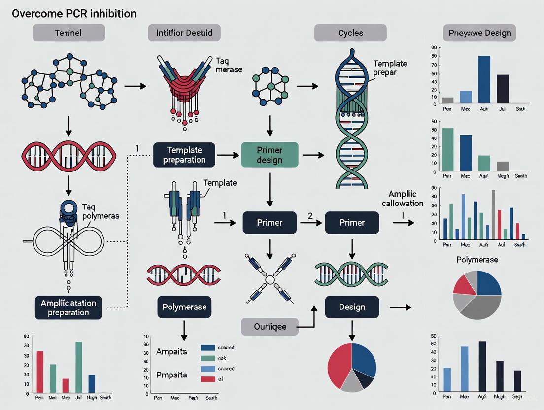
Overcoming PCR Inhibition: A Comprehensive Guide for Robust Nucleic Acid Amplification
PCR inhibition remains a significant hurdle in molecular diagnostics and research, leading to false negatives, reduced sensitivity, and unreliable data.
Overcoming PCR Inhibition: A Comprehensive Guide for Robust Nucleic Acid Amplification
Abstract
PCR inhibition remains a significant hurdle in molecular diagnostics and research, leading to false negatives, reduced sensitivity, and unreliable data. This article provides a systematic framework for researchers and drug development professionals to understand, troubleshoot, and overcome PCR inhibition. Drawing on the latest research and methodologies, we explore the fundamental mechanisms of inhibition, from hydrogel monomers to biological sample contaminants. We detail practical strategies for sample purification, reaction optimization, and the use of specialized additives and enzymes. The guide also covers advanced troubleshooting protocols and evaluates innovative technologies like digital PCR that offer enhanced inhibitor tolerance. By synthesizing foundational knowledge with applied solutions, this resource aims to empower scientists to achieve accurate and reproducible amplification results across diverse and challenging sample types.
Understanding the Enemy: A Deep Dive into the Sources and Mechanisms of PCR Inhibition
Defining PCR Inhibition and Its Impact on Diagnostic Accuracy and Research Reproducibility
What is PCR Inhibition?
PCR inhibition occurs when substances in a reaction interfere with the polymerase chain reaction, preventing the efficient amplification of nucleic acids. These inhibitors can affect various components of the PCR, primarily through interactions with the DNA polymerase enzyme or the DNA template itself [1] [2].
Inhibitory substances can originate from the original sample (such as blood, tissues, or soil) or be introduced during sample processing and DNA extraction [2]. The interference mechanisms are diverse: some inhibitors bind directly to the DNA polymerase, preventing enzymatic activity, while others crosslink with the DNA template, preventing strand separation during denaturation [3]. Additional mechanisms include chelation of essential cofactors like Mg²⁺ ions or interference with fluorescence signaling in real-time PCR applications [1] [4].
The consequences of undetected PCR inhibition are particularly severe in diagnostic and research settings, potentially leading to false-negative results, reduced sensitivity, and inaccurate quantification [5] [3]. This directly compromises diagnostic accuracy and undermines research reproducibility, making understanding and addressing PCR inhibition a critical component of reliable molecular analysis.
PCR inhibitors represent a diverse group of molecules with different mechanisms of action. The table below categorizes common inhibitors, their typical sources, and their primary mechanisms of interference.
Table 1: Common PCR Inhibitors, Sources, and Mechanisms
| Inhibitor | Common Sources | Mechanism of Inhibition |
|---|---|---|
| Hemoglobin/Hemin [6] | Blood, tissue samples | Binds to DNA polymerase, interfering with its activity [1] |
| Humic and Fulvic Acids [1] [3] | Soil, sediment, environmental water | Bind to DNA polymerase and may interact with nucleic acids [1] |
| Polysaccharides [3] | Feces, plants, bacteria | Can interfere with nucleic acid isolation and enzymatic reactions [3] |
| Polyphenolics (Tannins) [3] | Plants, fabrics (dyes), wastewater | Chelate Mg²⁺ ions or crosslink with DNA [3] |
| Heparin/EDTA [1] [6] | Blood (anticoagulants), laboratory reagents | Heparin binds to polymerase; EDTA chelates Mg²⁺ ions [1] [6] |
| Ionic Detergents (SDS) [2] [6] | Lysis buffers, extraction reagents | Disrupts enzyme activity at concentrations >0.005% [6] |
| Salts (KCl, NaCl) [2] | Extraction buffers, body fluids | High ionic strength disrupts primer annealing [2] |
| Urea [6] | Urine, some extraction protocols | Denatures enzymes at elevated concentrations (>20 mM) [6] |
| Phenol [2] [6] | Organic extraction methods | Interferes with enzymatic activity at concentrations >0.2% [6] |
| Ethanol/Isopropanol [2] [6] | DNA precipitation steps | Disrupts enzymatic activity at concentrations >1% [6] |
How Do I Detect PCR Inhibition in My Experiments?
Detecting PCR inhibition is a critical quality control step. The most common detection methods include the use of internal controls, sample dilution, and amplification controls.
Internal Amplification Controls (IACs)
IACs are exogenous, non-target DNA sequences added to the reaction mix. Their failure to amplify indicates the presence of inhibitors affecting the PCR. This method is highly relevant but requires careful design to avoid competition with the target [5] [1].
Pathogen-Specific Amplification Controls
This method involves spiking a known quantity of the target pathogen DNA into the patient's sample extract. A significant delay or failure in the amplification of this spike (measured by a shift in Cp or Cq value) indicates inhibition. This method directly tests inhibition for the specific assay but increases the risk of contamination [5].
Human Gene Amplification (e.g., Albumin, RNase P)
This method amplifies a ubiquitous human gene present in the sample. However, its reliability is debated because a high Cp value could indicate either inhibition or a genuinely low cellular/DNA content in the sample. One study found that human albumin gene amplification was not adequate for reliably identifying PCR inhibitors in microbiological assays [5].
Sample Dilution Test
This is a simple and effective practical approach.
- Prepare a 1:10 dilution of your DNA extract with nuclease-free water.
- Run both the undiluted and diluted samples in your qPCR assay.
- Compare the Cq values:
- No Inhibition: The diluted sample will have a higher Cq value (approximately 3.3 cycles higher for a 1:10 dilution of an efficient PCR) due to the lower template concentration.
- Inhibition Present: The diluted sample will have a lower or similar Cq compared to the undiluted sample. Diluting the sample also dilutes the inhibitors, restoring amplification efficiency [3].
A Systematic Workflow for Troubleshooting PCR Inhibition
The following diagram outlines a logical, step-by-step approach to identifying and resolving PCR inhibition in your experiments.
What Experimental Strategies Can Overcome PCR Inhibition?
Several well-established methodologies can be employed to mitigate the effects of PCR inhibitors. The strategies range from simple dilution to the use of specialized biochemical additives.
Physical and Chemical Removal of Inhibitors
- Sample Dilution: A 1:10 dilution of the DNA extract is a common and effective first step, as it reduces inhibitor concentration. The drawback is a concomitant reduction in target DNA concentration, which can be problematic for low-abundance targets [7].
- Advanced Nucleic Acid Purification: Using silica-membrane spin columns or magnetic beads designed for specific sample types (e.g., feces, soil) can effectively co-purify and remove inhibitors. Kits with specialized matrices can bind polyphenolic compounds like humic acids and tannins [3] [7].
- Precipitation and Wash Steps: Ethanol precipitation followed by a 70% ethanol wash can help remove salts and other small soluble inhibitors [8].
Biochemical Enhancement of PCR
The addition of specific enhancers to the PCR mix can counteract inhibitors. The table below summarizes key enhancers and their applications based on recent research.
Table 2: PCR Enhancers for Overcoming Inhibition
| Enhancer | Recommended Concentration | Mechanism of Action | Effectiveness & Notes |
|---|---|---|---|
| Bovine Serum Albumin (BSA) [2] [7] | 0.1 - 0.5 µg/µL | Binds to inhibitors, preventing them from interacting with the DNA polymerase [2]. | Effective against a range of inhibitors like humic acids and polyphenolics; widely used and cost-effective [7]. |
| T4 Gene 32 Protein (gp32) [7] | 0.2 µg/µL | Binds to single-stranded DNA, stabilizing the template and preventing the action of inhibitors [7]. | In one study, gp32 was the most significant method for removing inhibition in wastewater samples [7]. |
| Nonionic Detergents (Tween-20) [7] | Varies (e.g., 0.1-1%) | Counteracts inhibitory effects on Taq DNA polymerase [7]. | Can be effective, but concentration needs optimization. |
| Organic Solvents (DMSO, Formamide) [8] [7] | DMSO: 1-10% Formamide: 1-5% | Lowers the melting temperature (Tm) of DNA, destabilizes secondary structures, and can help denature GC-rich templates [8]. | Can be beneficial for complex templates, but high concentrations can inhibit the polymerase. Use the lowest effective concentration [8]. |
Strategic Use of Polymerase Enzymes and Digital PCR
- Inhibitor-Tolerant DNA Polymerases: Many modern DNA polymerases are enzyme blends specifically engineered or formulated for high resistance to common inhibitors found in blood, soil, and plants [1] [8]. Increasing the concentration of the DNA polymerase in the reaction can also help overcome inhibition [2].
- Digital PCR (dPCR): dPCR has been proven less affected by PCR inhibitors than quantitative PCR (qPCR). Because dPCR partitions a sample into thousands of individual reactions and uses end-point measurement, it is less reliant on amplification kinetics. This makes quantification more accurate in the presence of inhibitors that would normally skew qPCR Cq values [1].
Research Reagent Solutions for Inhibitor-Prone Samples
Selecting the right reagents is crucial for successful PCR with challenging samples. The following table lists essential materials and their functions.
Table 3: Key Reagents for Managing PCR Inhibition
| Reagent / Kit | Function / Purpose | Example Applications |
|---|---|---|
| Inhibitor-Tolerant DNA Polymerase [1] [8] | Enzyme blends with high resistance to inhibitors; often hot-start to improve specificity. | Direct PCR from crude samples; amplification from blood, soil, plant extracts. |
| Inhibitor Removal Kits [3] [7] | Specialized column matrices that bind common inhibitors (humic acids, tannins, melanin). | Cleaning DNA extracted from feces, soil, wastewater, or plant tissues. |
| PCR Enhancers (BSA, gp32) [2] [7] | Additives that bind to inhibitors or stabilize reaction components. | Added to the PCR mix when inhibiting substances are suspected. |
| Spin-Column DNA Purification Kits [5] [3] | Standardized methods for isolating and purifying DNA while removing salts, proteins, and other contaminants. | Routine DNA extraction from various sample types; included in many specialized kits for soil, feces, etc. |
Frequently Asked Questions (FAQs)
Q1: My PCR works with a clean control template but fails with my sample extract. Is this definitely inhibition? While this is a strong indicator of inhibition, other issues like poor DNA quality or quantity could be the cause. Always check the concentration and purity (A260/A280 ratio) of your DNA extract and perform a dilution test to confirm inhibition [8] [3].
Q2: Why can't I just use a human gene as an internal control for inhibition? Amplification of a human gene (e.g., albumin) is not a reliable indicator for pathogen detection PCRs. The Cp value is highly dependent on the sample's cellularity, which varies by matrix and patient physiology. A high Cp could mean inhibition OR a sample with low human DNA content. Studies have shown poor correlation between human gene controls and pathogen-specific inhibition controls [5].
Q3: Is digital PCR (dPCR) completely immune to inhibitors? No, dPCR is not immune, but it is generally more tolerant than qPCR. The partitioning step may reduce the local concentration of an inhibitor in positive droplets, and quantification is based on end-point presence/absence, which is less skewed than Cq-based quantification in qPCR. However, strong inhibitors can still prevent amplification entirely, leading to an underestimation of copy number [1].
Q4: What is the most critical step to prevent PCR inhibition? Optimizing the sample preparation and DNA extraction method for your specific sample type is the most critical proactive step. Using a validated, specialized extraction protocol that effectively removes inhibitors while efficiently recovering nucleic acids will prevent most problems downstream [4] [3].
Troubleshooting Guides
How do I identify which inhibitor is affecting my PCR assay?
The first step in troubleshooting is to recognize the specific symptoms of inhibition and link them to the common inhibitors found in your sample matrix. The table below summarizes the key indicators and their most likely causes.
Table 1: Identifying Common PCR Inhibitors
| Inhibitor | Common Sample Sources | Primary Mechanism of Action | Key Symptoms in qPCR/dPCR |
|---|---|---|---|
| Hemoglobin | Whole blood, red blood cells [9] | Direct inhibition of DNA polymerase activity; fluorescence quenching [9] | Increased Cq, reduced amplification efficiency, fluorescence quenching [9] |
| Heparin | Blood collected in heparinized tubes [10] | Interference with DNA polymerase; co-factor chelation [11] | Dose-dependent suppression of DNA amplification; can vary by DNA polymerase type [10] |
| Polysaccharides | Plant tissues, foods [12] | Interaction with nucleic acids; disruption of polymerization [2] | Failure to amplify; symptoms can often be reversed with additives [12] |
| Bile Salts | Feces, intestinal content [13] | Inhibition of DNA polymerase activity [14] | Reduced amplification capacity; sensitivity varies greatly between polymerases [14] |
What strategies can I use to overcome PCR inhibition?
Overcoming inhibition requires a multi-faceted approach, from sample preparation to reaction optimization. The following table provides a comparative overview of effective strategies.
Table 2: Strategies to Overcome PCR Inhibition
| Strategy | Typical Protocol | Effectiveness | Considerations |
|---|---|---|---|
| Silica Membrane Purification | Use commercial kits (e.g., QIAamp DNA Mini Kit) to bind DNA and wash away inhibitors [15]. | Reduced inhibition rates from 12.5% to 1.1% in clinical samples [15]. | Can lead to DNA loss; may not remove all inhibitors bound to DNA [1]. |
| Dilution of DNA Extract | Dilute the DNA template 1:10 or 1:100 in nuclease-free water or buffer. | Reduces inhibitor concentration; simple and cost-effective [11]. | Risk of diluting the target DNA below the detection limit [11]. |
| Additives & Buffer Optimization | Add BSA (0.1-0.5 μg/μL) or Tween 20 (0.1-0.5%) to the PCR master mix [12]. | BSA neutralizes inhibitors; Tween reverses polysaccharide inhibition [12]. | Requires optimization; may not be effective against all inhibitors. |
| Inhibitor-Resistant Polymerases | Use specialized enzyme blends (e.g., Phusion Flash, GoTaq Endure) [11]. | Enables direct PCR from challenging samples like blood and soil [11]. | Higher cost; performance may vary by inhibitor type. |
Frequently Asked Questions (FAQs)
Q1: What are the specific experimental protocols for evaluating inhibition from hemoglobin and IgG?
Answer: A detailed protocol based on peer-reviewed research is as follows [9]:
1. Inhibitor Preparation:
- Hemoglobin: Dissolve human hemoglobin in water to a stock concentration of 100 μg/μL.
- IgG: Dissolve IgG in water to a stock concentration of 80 μg/μL.
- Prepare subsequent dilutions with water for use in reactions.
2. PCR Setup:
- Use a standardized qPCR or dPCR assay (e.g., targeting a human genomic sequence like the retinoblastoma 1 gene, RB1).
- Keep the amount of DNA template constant across all reactions to isolate the effect of the inhibitors.
- Test a range of concentrations in the reaction mix:
- Hemoglobin: 0.1 to 620 μM.
- IgG: 1.7 to 53 μM.
- Whole Blood: 0.00005% to 20% (v/v).
3. Data Analysis:
- In qPCR: Monitor the quantification cycle (Cq), amplification efficiency, and fluorescence levels. A significant increase in Cq and a decrease in efficiency indicate inhibition.
- In dPCR: Analyze the number of positive partitions and the calculated DNA concentration. A decrease in both indicates inhibition.
- Fluorescence Quenching: Observe the baseline fluorescence of the passive reference dye or intercalating dye. Hemoglobin and its derivative hematin are known quenchers [9].
Q2: How does the mechanism of heparin inhibition differ from that of hemoglobin?
Answer: While both inhibit PCR, their molecular mechanisms are distinct [9] [10].
- Hemoglobin (and its component heme) primarily acts by directly inhibiting the DNA polymerase enzyme, reducing its activity. It also causes fluorescence quenching, which interferes with detection in qPCR and dPCR [9].
- Heparin is a highly sulfated polysaccharide that can chelates divalent cations like Mg²⁺, which are essential co-factors for DNA polymerase. This effectively starves the enzyme of a critical component needed for catalysis [10] [11]. The degree of interference is also affected by the type of Taq DNA polymerase used [10].
Q3: Which polysaccharides are most inhibitory and how can this be reversed?
Answer: Among various plant polysaccharides tested, the acidic polysaccharides dextran sulfate and gum ghatti were found to be particularly inhibitory [12]. The inhibition can often be reversed with specific buffer additives.
- For Dextran Sulfate: The inhibitory effect at a polysaccharide-to-DNA ratio of 50:1 can be reversed by adding:
- Tween 20 (0.25% or 0.5%)
- DMSO (5%)
- Polyethylene glycol 400 (5%) [12].
- For Gum Ghatti: The addition of 0.5% Tween 20 was sufficient to reverse the inhibition [12].
Q4: What is the most effective pre-PCR treatment for bile samples?
Answer: To optimize PCR detection from bile samples, a multi-pronged pre-PCR treatment is effective [14]:
- Dilution and Heating: Dilute the bile sample and heat it at 98°C.
- Reaction Mixture Optimization: Add casein and formamide to the PCR reaction mixture.
- Enzyme Selection: Use a robust DNA polymerase such as rTth, which was shown to have the lowest sensitivity to bile inhibitors compared to other enzymes like Taq [14].
This combined approach reduces the PCR inhibitory effect and enables efficient DNA amplification directly from bile.
Visualizing Inhibition Mechanisms and Solutions
The following diagram illustrates the points at which common inhibitors disrupt the PCR process and highlights the corresponding solutions.
The Scientist's Toolkit: Research Reagent Solutions
Table 3: Essential Reagents for Overcoming PCR Inhibition
| Reagent / Material | Function / Purpose | Example Use Case |
|---|---|---|
| Silica Membrane Columns | Binds nucleic acids, allowing impurities and inhibitors to be washed away [15]. | Purification of DNA from complex samples like sputum, lymph nodes, and stool [15]. |
| Bovine Serum Albumin (BSA) | Binds to and neutralizes a range of inhibitors, stabilizing the DNA polymerase [11]. | Added to PCR master mix to counteract inhibitors in blood (hemoglobin, IgG) [9]. |
| Tween 20 | A non-ionic detergent that can disrupt inhibitor interactions with DNA or polymerase [12]. | Reverses PCR inhibition caused by acidic polysaccharides like gum ghatti [12]. |
| Inhibitor-Resistant DNA Polymerase | Engineered enzymes or blends with enhanced tolerance to specific inhibitor classes [11]. | Direct PCR amplification from blood samples without prior DNA purification [11]. |
| Casein & Formamide | Additives that can reduce the inhibitory effect of complex matrices [14]. | Enabling PCR detection of Helicobacter DNA directly from bile samples [14]. |
Polymerase chain reaction (PCR) is a fundamental tool in molecular biology, but its sensitivity makes it vulnerable to inhibition by various substances commonly encountered in environmental and laboratory samples [16]. Inhibitors prevent the amplification of nucleic acids, leading to false-negative results, reduced sensitivity, and inaccurate quantification [16] [17]. This guide addresses the specific challenges posed by key contaminants: humic acids, phenols, tannins, and the detergent SDS (sodium dodecyl sulphate). Understanding their sources, mechanisms, and removal strategies is essential for developing robust, inhibitor-tolerant PCR protocols within a research context focused on overcoming PCR inhibition [7].
Understanding Common PCR Inhibitors
PCR inhibitors are a heterogeneous class of substances that can originate from the sample itself or be introduced during sample preparation and nucleic acid extraction [16] [17]. They interfere with the amplification process through diverse mechanisms.
Table 1: Common PCR Inhibitors, Their Sources, and Mechanisms of Action
| Inhibitor | Common Sample Sources | Primary Mechanism of PCR Inhibition |
|---|---|---|
| Humic and Fulvic Acids | Soil, sediment, decaying organic matter, water [16] [17] | Bind to DNA polymerase and template DNA, preventing the enzymatic reaction [16]. |
| Phenols | Plant tissues (e.g., berries, tomatoes), laboratory reagents [16] [17] | Denature proteins, including DNA polymerase and reverse transcriptase [16]. |
| Tannins | Plant tissues, tea-colored waters [18] | Deplete magnesium ions (Mg2+), an essential cofactor for DNA polymerase [16]. |
| SDS (Sodium Dodecyl Sulphate) | Laboratory reagent (detergent) from sample preparation [16] [17] | Degrades and inhibits DNA polymerase; highly inhibitory even at low concentrations [16] [17]. |
Troubleshooting Guide & FAQs
This section provides targeted solutions for overcoming PCR inhibition caused by the specified contaminants.
FAQ 1: My PCR from soil/water samples consistently fails. I suspect humic acids are the problem. What are my primary options for removal?
Humic acids are a prevalent and potent inhibitor in environmental samples. A multi-faceted approach is recommended:
- Improve Nucleic Acid Purification: Use specialized inhibitor removal kits. Silica column-based kits (e.g., NucleoSpin Inhibitor Removal Kit) or magnetic bead technologies (e.g., BcMag Kit) are explicitly designed to bind and remove polyphenolic compounds like humic acids [19] [17]. These can achieve >75% DNA recovery while efficiently removing contaminants [19].
- Employ Chemical Additives (Amplification Facilitators): Add Bovine Serum Albumin (BSA) or T4 gene 32 protein (gp32) to your PCR master mix. These proteins bind to inhibitory compounds present in the reaction, preventing them from interfering with the DNA polymerase [16] [7]. A study on wastewater found that adding gp32 at a final concentration of 0.2 μg/μl was particularly effective at removing inhibition and enhancing viral detection [7].
- Dilute the Template: A simple 10-fold dilution of the extracted nucleic acid can reduce inhibitor concentration below a critical threshold [16] [7]. The major drawback is a concomitant dilution of the target DNA, which may decrease assay sensitivity [16].
- Select a Robust DNA Polymerase: Choose a polymerase engineered for high inhibitor tolerance. Mutant Taq polymerases with greater resistance to inhibitors found in blood and soil are commercially available and can significantly improve performance in contaminated samples [16].
FAQ 2: My nucleic acid extraction uses phenol-chloroform. How can I prevent residual phenol from inhibiting my downstream PCR?
Residual phenol from extraction procedures is a common laboratory-introduced inhibitor.
- Ensure Complete Removal: During extraction, ensure proper phase separation and carefully avoid transferring any of the organic (phenol) phase. Perform an additional chloroform extraction step to remove any residual phenol traces.
- Ethanol Precipitation: Perform a thorough ethanol precipitation with subsequent washes with 70% ethanol. It is critical to allow the pellet to dry completely after washing to let all residual ethanol evaporate, as ethanol itself is a PCR inhibitor [16].
- Use Inhibitor Removal Kits: As with humic acids, post-extraction cleanup with a dedicated inhibitor removal kit will effectively remove residual phenol and other organic solvents [19].
- Verify with Spectrophotometry: Check the purity of your DNA using a spectrophotometer (A260/A280 ratio). A ratio significantly lower than 1.8 may indicate phenol contamination.
FAQ 3: I am working with plant extracts rich in tannins. What strategies can I use to mitigate their inhibitory effects?
Tannins act primarily by chelating magnesium ions. Strategies to counteract this include:
- Magnesium Optimization: Increase the concentration of MgCl2 in your PCR buffer. This provides excess Mg2+ to compensate for what is bound by tannins, ensuring sufficient ions remain available for the DNA polymerase [16].
- Use of Additives: Add BSA to your reactions. BSA can bind tannic acids, relieving the inhibition of the DNA polymerase [16]. The non-ionic detergent Tween-20 can also stimulate polymerase activity and reduce false terminations [16].
- Employ Compatible Storage Buffers: Research on environmental DNA (eDNA) from tannin-laden water has shown that using CTAB (Cetyl trimethylammonium bromide) as a short-term storage buffer prior to a phenol-chloroform-isoamyl alcohol (PCI) isolation resulted in the highest eDNA yields and most effective reduction of PCR inhibitors [18].
FAQ 4: I see SDS is a potent inhibitor. What are the best practices for removing it after nucleic acid extraction?
As an ionic detergent, SDS is highly inhibitory. Its removal is crucial.
- Avoid Ionic Detergents: Where possible, substitute SDS with non-ionic detergents like Tween-20, Triton X-100, or Nonidet P-40, which are only inhibitory at relatively high concentrations [16].
- Dialysis or Precipitation: For "homebrew" extraction protocols, dialysis or ethanol/isopropanol precipitation can effectively remove SDS.
- Commercial Cleanup Kits: Silica-based column cleanup kits are highly effective at removing detergents like SDS from DNA samples [17] [19]. Ensure the sample volume and composition are compatible with the kit's specifications.
Experimental Protocols for Inhibitor Removal
Protocol 4.1: Chemical Removal of Inhibitors using CTAB-PCI
This protocol, adapted from research on environmental DNA, is highly effective for removing organic inhibitors like humics and tannins [18].
- Lysis and Binding: Resuspend your sample or filter in a CTAB buffer. Incubate at 65°C for 10-30 minutes to lyse cells and allow CTAB to form complexes with inhibitors and polysaccharides.
- Organic Extraction: Add an equal volume of Phenol-Chloroform-Isoamyl alcohol (PCI) to the sample. Mix thoroughly by vortexing.
- Phase Separation: Centrifuge at >12,000 × g for 5 minutes. Carefully transfer the upper aqueous phase (containing the DNA) to a new tube.
- Precipitation: Add 0.7 volumes of isopropanol or 2 volumes of 100% ethanol to the aqueous phase to precipitate the DNA. Incubate at -20°C for at least 30 minutes.
- Pellet and Wash: Centrifuge at >12,000 × g for 15 minutes to pellet the DNA. Carefully decant the supernatant.
- Wash: Wash the pellet with 1 mL of 70% ethanol. Centrifuge again for 5 minutes and carefully remove all ethanol.
- Resuspension: Air-dry the pellet for 5-10 minutes and resuspend in nuclease-free water or TE buffer.
Protocol 4.2: Use of PCR Enhancers to Overcome Inhibition
This protocol outlines the incorporation of facilitator proteins into the PCR master mix [7].
- Prepare Master Mix: On ice, prepare a standard PCR master mix for all reactions, excluding the template DNA.
- Add Enhancer: Supplement the master mix with your chosen enhancer. Common effective options and concentrations are:
- T4 gp32 Protein: Final concentration of 0.2 μg/μL [7].
- Bovine Serum Albumin (BSA): Final concentration of 0.2-0.5 μg/μL.
- Aliquot and Add Template: Aliquot the enhanced master mix into PCR tubes. Then, add the template DNA to each tube.
- Run PCR: Proceed with your standard PCR cycling conditions.
Table 2: Research Reagent Solutions for Overcoming PCR Inhibition
| Reagent / Kit | Function / Application | Key Characteristics |
|---|---|---|
| OneStep PCR Inhibitor Removal Kit (Zymo Research) | Fast, one-step cleanup of DNA/RNA to remove polyphenolics, humic acids, tannins, etc. [20] [21] | Uses a column slurry/filter; ≥80% recovery; processes 50-200μl samples [20]. |
| NucleoSpin Inhibitor Removal Kit (Takara Bio) | Silica-membrane column for removing humic acids, heme, polyphenols, and tannins [19]. | Fast 15-minute protocol; >75% recovery; processes inputs up to 100 μl [19]. |
| BcMag One-Step PCR Inhibitor Removal Kit (BioClone) | Magnetic bead-based removal of a wide range of inhibitors using negative chromatography [17]. | Captures inhibitors, leaving pure DNA in solution; suitable for automation [17]. |
| T4 gene 32 Protein (gp32) | PCR additive that binds to single-stranded DNA and inhibitors, stabilizing replication [16] [7]. | Particularly effective in complex samples like wastewater; use at 0.2 μg/μL [7]. |
| Bovine Serum Albumin (BSA) | PCR additive that binds to a variety of inhibitory compounds [16] [7]. | Effective against phenolics, humic acids, and tannins; alleviates protease activity [16]. |
Workflow for Troubleshooting PCR Inhibition
The following diagram outlines a systematic decision-making process for diagnosing and resolving PCR inhibition.
Systematic troubleshooting workflow for PCR inhibition
Successfully navigating the challenges posed by PCR inhibitors such as humic acids, phenols, tannins, and SDS requires a systematic and informed approach. Key to this process is accurately diagnosing the problem through appropriate controls, understanding the specific inhibitors present in your sample matrix, and applying a combination of optimized nucleic acid cleanup, strategic use of PCR enhancers, and selection of robust enzymes. The protocols and workflows provided here offer a practical foundation for developing inhibitor-tolerant molecular assays, a critical capability for advancing research in fields ranging from clinical diagnostics to environmental monitoring.
Troubleshooting Guide: Overcoming PCR Inhibition in Hydrogel-Integrated Systems
Problem: My PCR amplification fails when I integrate hydrogel monomers into my diagnostic platform. What is the cause?
Answer: PCR failure is likely due to the structure-dependent inhibitory effects of certain hydrogel monomers on the Taq polymerase enzyme. Acrylamide and PEGDMA are particularly strong inhibitors, even at low concentrations. Research shows that these monomers contain α,β-unsaturated carbonyl groups that can covalently bind to nucleophilic amino acids in the polymerase's active site, permanently inactinating the enzyme [22]. This direct chemical interaction prevents DNA amplification.
Problem: How can I restore PCR functionality when using inhibitory monomers like PEGDMA?
Answer: For PEGDMA-rich conditions, adding nonionic surfactants with low critical micelle concentrations (CMC), such as Tween 20, Tween 80, or NP-40, can successfully restore PCR amplification [22]. These surfactants likely form micelles that sequester the inhibitory monomers, preventing them from interacting with the polymerase. In contrast, common additives like DMSO and Triton X-100 were found to be ineffective for this specific application [22].
Problem: My system uses acrylamide monomers and suffers from poor amplification efficiency. What solutions can I implement?
Answer: Acrylamide-induced inhibition can be competitively alleviated by using a significant excess of Taq polymerase in your reaction setup [22]. This approach provides enough active enzyme molecules to withstand the covalent interactions with acrylamide monomers while maintaining sufficient polymerase activity for amplification. Additionally, consider alternative monomers with lower inhibition potential, such as GelMA or EGDMA, if they are compatible with your hydrogel design requirements [22].
Problem: I work with complex sample matrices like wastewater, and my molecular assays show inhibition. What enhancement strategies are most effective?
Answer: For complex matrices, multiple enhancement strategies have been systematically evaluated. The most significant inhibition removal was achieved through:
- Addition of T4 gene 32 protein (gp32) at a final concentration of 0.2 μg/μL [7]
- 10-fold dilution of the extracted sample [7]
- Addition of Bovine Serum Albumin (BSA) [7]
- Using a commercial inhibitor removal kit [7] The gp32 protein binds to inhibitory substances like humic acids, preventing them from interfering with polymerase activity [7].
Experimental Protocols & Methodologies
Protocol 1: Evaluating Monomer Inhibition in PCR
This protocol is adapted from systematic evaluation methods used in recent studies [22].
Materials Needed:
- Standard PCR reagents (Taq polymerase, dNTPs, buffer, primers, template DNA)
- Hydrogel monomers to test (e.g., acrylamide, PEGDMA, EGDA, EGDMA, GelMA)
- PCR enhancers (Tween 20, Tween 80, NP-40, BSA, additional Taq polymerase)
- Agarose gel electrophoresis equipment
Procedure:
- Prepare a standard PCR master mixture according to your established protocol.
- Add hydrogel monomers to individual reaction tubes at final concentrations ranging from 0.5% to 5% (v/v).
- For mitigation tests, add potential enhancers:
- Nonionic surfactants: Tween 20, Tween 80, or NP-40 at 0.1-1% (v/v)
- Additional Taq polymerase: 1.5-2x the standard concentration
- BSA: 0.1-0.5 μg/μL
- Run PCR under standard thermal cycling conditions using a positive control template (e.g., lambda DNA).
- Analyze results using agarose gel electrophoresis to compare amplification yields.
- Quantify inhibition by comparing band intensity to monomer-free controls.
Expected Outcomes:
- Strong inhibition should be observed with PEGDMA and acrylamide at concentrations as low as 1-2%.
- GelMA and EGDMA should show minimal interference at equivalent concentrations.
- Surfactants should restore amplification in PEGDMA conditions, while excess polymerase should help with acrylamide inhibition.
Protocol 2: PCR Enhancement Strategy Screening
Adapted from comprehensive evaluations of PCR enhancement approaches [7].
Materials Needed:
- Inhibited sample (e.g., wastewater extract or monomer-spiked sample)
- PCR/qPCR reagents
- Enhancement agents:
- T4 gene 32 protein (gp32)
- BSA
- DMSO
- Formamide
- Tween-20
- Glycerol
- Inhibitor removal kit (commercial)
Procedure:
- Divide the inhibited sample into aliquots for each enhancement approach.
- Prepare reactions with each enhancer at multiple concentrations:
- gp32: 0.1, 0.2, 0.5 μg/μL
- BSA: 0.1, 0.2, 0.5 μg/μL
- DMSO: 1%, 3%, 5% (v/v)
- Tween-20: 0.1%, 0.5%, 1% (v/v)
- 10-fold and 100-fold sample dilutions
- Commercial inhibitor removal kit (follow manufacturer instructions)
- Run qPCR with all samples including uninhibited and inhibited controls.
- Compare quantification cycle (Cq) values and amplification curves to identify the most effective approach for your specific inhibition challenge.
Table 1: Inhibition Thresholds of Common Hydrogel Monomers in PCR
| Monomer | Chemical Class | Strong Inhibition Threshold | Minimal Interference Level | Inhibition Mechanism |
|---|---|---|---|---|
| PEGDMA | Methacrylate | <2% (v/v) | N/A | Covalent interaction with polymerase via α,β-unsaturated carbonyl groups |
| Acrylamide | Acrylamide | <2% (v/v) | N/A | Covalent interaction with polymerase via α,β-unsaturated carbonyl groups |
| GelMA | Methacryloyl | >5% (v/v) | Up to 5% (v/v) | Minimal interference due to high molecular weight and substitution |
| EGDMA | Methacrylate | >5% (v/v) | Up to 5% (v/v) | Minimal interference, structure-dependent |
| EGDA | Acrylate | ~5% (v/v) | <2% (v/v) | Moderate inhibition |
Data compiled from systematic evaluation of monomer inhibition [22]
Table 2: Effectiveness of PCR Enhancement Strategies
| Enhancement Strategy | Effective Concentration | Effectiveness for Monomer Inhibition | Effectiveness for Complex Matrices | Mechanism of Action |
|---|---|---|---|---|
| Tween 20 | 0.1-1% (v/v) | High (PEGDMA) | Moderate | Low CMC surfactant sequesters inhibitors |
| Tween 80 | 0.1-1% (v/v) | High (PEGDMA) | Moderate | Low CMC surfactant sequesters inhibitors |
| NP-40 | 0.1-1% (v/v) | High (PEGDMA) | Moderate | Low CMC surfactant sequesters inhibitors |
| Excess Taq Polymerase | 1.5-2x standard | High (Acrylamide) | Low | Competitive alleviation of enzyme inhibition |
| T4 gene 32 protein (gp32) | 0.2 μg/μL | Not tested | High | Binds inhibitory substances like humic acids |
| BSA | 0.1-0.5 μg/μL | Moderate | High | Binds inhibitors, stabilizes enzymes |
| Sample Dilution (10-fold) | 1:10 dilution | Moderate | High | Reduces inhibitor concentration |
| Inhibitor Removal Kit | Manufacturer protocol | Moderate | High | Physically removes inhibitory compounds |
Data compiled from studies on monomer inhibition [22] and wastewater analysis [7]
Mechanism Visualization
PCR Inhibition and Restoration Mechanisms
The Scientist's Toolkit: Essential Research Reagents
Table 3: Research Reagent Solutions for Hydrogel-PCR Compatibility
| Reagent | Function & Application | Specific Usage Notes |
|---|---|---|
| Nonionic Surfactants (Tween 20, Tween 80, NP-40) | Mitigate PEGDMA inhibition by forming micelles that sequester monomers | Use at 0.1-1% (v/v); effective due to low critical micelle concentration [22] |
| BSA (Bovine Serum Albumin) | Stabilizes polymerase, binds inhibitors in complex matrices | Use at 0.1-0.5 μg/μL; effective for various inhibition types [7] |
| T4 gene 32 protein (gp32) | Single-stranded DNA binding protein that counteracts inhibitors | Use at 0.2 μg/μL; particularly effective for humic substances [7] |
| Alternative Monomers (GelMA, EGDMA) | Replace inhibitory monomers while maintaining hydrogel properties | Show minimal PCR interference at concentrations up to 5% (v/v) [22] |
| Inhibitor-Tolerant Polymerases | Engineered enzymes with enhanced resistance to inhibitors | Screening different polymerase-buffer systems can increase tolerance 48-fold [23] |
| Commercial Inhibitor Removal Kits | Physically remove inhibitory compounds from samples | Follow manufacturer protocols; effective but adds processing step [7] |
Frequently Asked Questions (FAQs)
Q: Why are some hydrogel monomers inhibitory while others are not?
The inhibition potential depends on the chemical structure of the monomers. Monomers containing α,β-unsaturated carbonyl groups (particularly acrylates and methacrylates) can act as Michael acceptors, forming covalent bonds with nucleophilic residues in the polymerase enzyme. PEGDMA and acrylamide have highly accessible electrophilic sites, making them strong inhibitors. In contrast, GelMA has a larger molecular structure with different substitution patterns that reduce this reactivity [22].
Q: Are digital PCR (dPCR) methods less susceptible to monomer inhibition?
Yes, digital PCR generally shows higher tolerance to inhibitors compared to conventional qPCR. This is because dPCR partitions the reaction into thousands of nanoreactors, effectively diluting inhibitors and increasing the probability that some partitions will contain sufficient active polymerase for amplification [23]. However, strong inhibitors like high concentrations of PEGDMA and acrylamide can still affect dPCR efficiency.
Q: How do I select the right hydrogel monomer for my PCR-integrated diagnostic device?
Consider these factors when selecting monomers:
- Choose GelMA or EGDMA over PEGDMA or acrylamide when possible, as they show minimal interference at working concentrations [22]
- If using inhibitory monomers, incorporate compatible surfactants from the beginning
- Test monomer concentration series in your specific PCR assay to establish safe thresholds
- Consider hydrogel fabrication requirements alongside PCR compatibility for optimal device performance
Q: Can these inhibition issues affect other amplification methods like LAMP?
Yes, inhibition mechanisms can affect various nucleic acid amplification techniques, though the specific tolerance levels may differ. LAMP (Loop-Mediated Isothermal Amplification) generally shows different inhibitor tolerance profiles compared to PCR due to its isothermal nature and different enzyme requirements. However, the fundamental chemical interactions between inhibitory monomers and enzymatic components remain a concern across amplification methodologies [24].
Q: What concentration of surfactants should I test initially for mitigating monomer inhibition?
Begin with 0.1% (v/v) Tween 20, Tween 80, or NP-40 and test up to 1% if needed. These concentrations have been shown effective for PEGDMA-rich conditions without significantly interfering with PCR components [22]. Always include controls without surfactants to verify they don't introduce new issues in your specific system.
Frequently Asked Questions (FAQs) on PCR Inhibition
FAQ 1: What are the primary biochemical mechanisms through which PCR inhibitors act? PCR inhibitors disrupt amplification through three core biochemical mechanisms:
- Enzyme Inactivation: Inhibitors can directly degrade, denature, or sterically block the DNA polymerase. For example, proteases in fecal samples degrade the enzyme, while humic acid or melanin can form complexes with the polymerase, preventing its function [1] [16] [25].
- Cofactor Chelation: Many inhibitors sequester magnesium ions (Mg²⁺), which are essential cofactors for DNA polymerase activity. Substances like EDTA, humic substances, and tannic acids bind Mg²⁺, making it unavailable for the enzymatic reaction [16] [2] [25].
- Template Modification: Inhibitors can bind directly to the nucleic acid template, preventing denaturation or primer annealing. For instance, humic acids interact with DNA, and polysaccharides can mimic DNA structures, interfering with the polymerization process [1] [16].
FAQ 2: Why is my digital PCR (dPCR) assay less affected by inhibitors than my quantitative PCR (qPCR) assay? dPCR is generally more tolerant to inhibitors than qPCR because of its fundamental methodology. qPCR relies on the efficiency of amplification kinetics for quantification; any delay caused by an inhibitor (seen as a higher Cq value) directly skews the quantitative result. In contrast, dPCR uses end-point measurements, counting the absolute number of positive and negative partitions. While inhibitors can reduce the amplification efficiency within some partitions, as long as amplification occurs sufficiently to be detected as "positive," the quantification remains accurate [1]. The partitioning of the sample itself may also reduce the local concentration of inhibitors in reaction droplets, mitigating their effect [1].
FAQ 3: How can I confirm that my PCR failure is due to inhibition and not another issue? The most reliable method is to use an internal positive control (IPC). This involves spiking a known, non-target DNA sequence into your reaction mixture. If amplification of both the IPC and your target fails, inhibition is likely. If the IPC amplifies successfully but your target does not, the issue is likely related to your template or target-specific primers [2] [25]. Spectrophotometric analysis (A260/280 and A260/230 ratios) can also indicate common contaminants like phenol or carbohydrates [25].
FAQ 4: Which DNA polymerases are most resistant to PCR inhibitors? Different DNA polymerases exhibit varying degrees of resistance. While the standard Taq polymerase is highly susceptible, other enzymes show superior performance. For instance, polymerases isolated from Thermus thermophilus (rTth) and Thermus flavus (Tfl) are significantly more resistant to blood inhibitors than Taq [16]. Furthermore, engineered mutant versions of Taq polymerase, such as OmniTaq and recently identified variants like Taq C-66 and Klentaq1 H101, have been specifically selected for high resistance to a broad spectrum of inhibitors found in blood, soil, and food [26].
Troubleshooting Guide: Identifying and Overcoming PCR Inhibition
Step 1: Recognize the Symptoms
Common signs of PCR inhibition in your results include:
- Complete amplification failure (no product) despite a confirmed DNA template [27] [16].
- Reduced amplification efficiency, visible as a higher quantification cycle (Cq) value in qPCR or a lower yield in conventional PCR [1] [27].
- Inconsistent or inaccurate quantification in qPCR and dPCR [1].
- "Smeared" or non-specific bands on an agarose gel [27].
Step 2: Identify the Source of the Inhibitor
Inhibitors can originate from the sample itself or be introduced during preparation.
- Common Sample-Derived Inhibitors:
- Common Preparation-Derived Inhibitors:
Step 3: Apply Strategic Solutions
The table below summarizes the primary solutions for overcoming PCR inhibition.
Table 1: Strategic Solutions to Overcome PCR Inhibition
| Strategy | Method | Mechanism of Action | Key Considerations |
|---|---|---|---|
| Sample Purification | Silica columns, magnetic beads, phenol-chloroform extraction, dialysis [1] [16] [25]. | Physically removes inhibitory substances from the nucleic acid extract. | Can lead to DNA loss; method efficiency depends on the inhibitor type [1] [16]. |
| Sample Dilution | Diluting the DNA template before PCR [16] [2]. | Reduces the concentration of the inhibitor below its effective threshold. | Simple but reduces target sensitivity; may not work for potent inhibitors [16]. |
| Polymerase Selection | Using inhibitor-resistant DNA polymerases (e.g., engineered Taq variants, rTth, Tfl) [1] [16] [26]. | The enzyme is less susceptible to degradation or blocking by inhibitors. | A direct and powerful solution; various commercial options available. |
| Chemical Additives | Adding Bovine Serum Albumin (BSA), betaine, DMSO, or formamide to the reaction mix [28] [16] [29]. | BSA binds inhibitors; betaine/DMSO destabilize DNA secondary structure, facilitating polymerization. | Concentration must be optimized; can enhance specificity and yield for difficult templates [16] [29]. |
| Reaction Optimization | Increasing Mg²⁺ concentration, using a hot-start polymerase, optimizing annealing temperature [27] [16] [29]. | Counteracts chelation, prevents non-specific priming, and increases reaction stringency. | A fundamental step in any PCR optimization protocol. |
Experimental Protocols for Inhibition Research
Protocol 1: Evaluating PCR Inhibitor Resistance Using a Spike-In Control
This protocol allows researchers to quantify the extent of inhibition in a sample [2].
- Prepare Test Reactions: Set up two parallel PCR reactions.
- Reaction A (Test): Contains the investigational DNA template with potential inhibitors.
- Reaction B (Control): Contains a known, purified DNA template (the spike) in water or a known inhibitor-free buffer.
- Add Internal Positive Control (IPC): Spike both Reaction A and Reaction B with an identical, low copy number of a control DNA sequence that is different from the target. This control should be amplified with its own set of primers.
- Amplify: Run both reactions simultaneously on a qPCR instrument.
- Analyze: Compare the Cq values for the IPC between the two reactions.
- ΔCq = Cq(IPC in Reaction A) - Cq(IPC in Reaction B)
- A significant ΔCq (e.g., > 2-3 cycles) indicates the presence of inhibitors in the investigational sample.
Protocol 2: Live Culture-Based Screening for Inhibitor-Resistant Polymerase Variants
This advanced, high-throughput protocol describes a method for discovering new inhibitor-resistant DNA polymerases, as demonstrated in recent research [26].
Workflow Title: Screening for Resistant Polymerases
Detailed Methodology:
- Library Creation: Create a library of randomly mutagenized Taq or Klentaq1 DNA polymerase genes using error-prone PCR and clone them into an expression vector [26].
- Cell Culture & Induction: Transform a bacterial host (e.g., E. coli) with the library. Plate to obtain single colonies. Grow and induce individual clones in a 96-well format with IPTG to express the polymerase variants [26].
- Live Culture PCR Screening: Directly transfer a small aliquot (e.g., 5 µL) of the induced bacterial culture (which serves as both the source of the polymerase variant and the DNA template, e.g., via 16S rRNA genes) into a PCR plate. The master mix contains SYBR Green, a PCR enhancer, and a challenging concentration of a target inhibitor (e.g., chocolate or black pepper extract) [26].
- Real-Time PCR & Selection: Immediately subject the plates to real-time PCR. Clones expressing polymerase variants that are resistant to the inhibitor will produce a fluorescence signal and a low Cq value. Sensitive variants will show no signal or a high Cq [26].
- Validation: Isolate the promising clones, purify the mutant enzymes, and re-test their resistance against a panel of inhibitors to confirm the phenotype [26].
The Scientist's Toolkit: Research Reagent Solutions
Table 2: Essential Reagents for Research on PCR Inhibition
| Reagent | Function in Inhibition Research | Example Use Case |
|---|---|---|
| Inhibitor-Resistant DNA Polymerases (e.g., engineered Taq variants, rTth polymerase) | Core enzyme with enhanced tolerance; allows amplification directly from crude samples [16] [26]. | Amplifying target DNA from blood or soil extracts without extensive purification. |
| Chemical Additives (e.g., BSA, Betaine, DMSO) | Amplification facilitators that counteract specific inhibitors or stabilize the reaction [16] [29]. | Adding 10-100 μg/mL BSA to neutralize inhibitors in plant or fecal samples. Using DMSO (2.5-5%) to assist with GC-rich templates [29]. |
| Standardized Inhibitor Stocks (e.g., Humic Acid, Hematin, IgG) | Provide a consistent and quantifiable challenge for testing inhibition resistance [1] [26]. | Creating a dose-response curve to compare the performance of different polymerases against a known inhibitor. |
| Silica-Based Purification Kits | Standard method for comparative studies; baseline for evaluating "direct" PCR methods [1] [16]. | Purifying DNA from complex samples to establish a benchmark for comparison with inhibitor-tolerant direct PCR approaches. |
| Internal Positive Control (IPC) Assays | A spiked, non-target DNA sequence to distinguish between true target absence and PCR failure due to inhibition [2] [25]. | Quantifying the level of inhibition in an unknown sample by comparing IPC Cq values with a control reaction. |
FAQ: How can I detect the presence of a PCR inhibitor in my reaction?
PCR inhibition can be detected through several key indicators in your qPCR data. The most common signs include delayed quantification cycle (Cq) values, poor amplification efficiency, and abnormal amplification curve morphology [11]. When inhibitors are present, you may observe that all samples, including controls, exhibit increased Cq values [11]. Another clear indicator is when the calculated amplification efficiency falls outside the optimal range of 90-110% [11] [30]. Additionally, the shape of the amplification curves may appear abnormal—flattened, inconsistent, or lacking a clear exponential growth phase [11] [31].
Systematic detection approaches include using an internal PCR control (IPC) to differentiate between true inhibition and low target concentration. If the IPC shows delayed Cq values, inhibition is likely present [11]. Another method involves performing a dilution series of your sample; if inhibition is concentration-dependent, you may observe a return to expected efficiency in more diluted samples where inhibitors fall below effective concentrations [30].
PCR inhibitors can originate from various sources, including biological samples, laboratory reagents, and environmental contaminants. The table below summarizes common inhibitors and their effects on PCR:
Table: Common qPCR Inhibitors and Their Effects
| Source | Examples | Effect on qPCR |
|---|---|---|
| Biological Samples | Hemoglobin (blood), heparin (tissues), polysaccharides (plants) | Polymerase inhibition, co-factor chelation [11] |
| Environmental Contaminants | Humic acids (soil), phenols (water), tannins (food) | DNA degradation, fluorescence interference [11] |
| Laboratory Reagents | SDS, ethanol, salts from extraction kits | Template precipitation, primer binding disruption [11] |
| Fluorescent Interference | Excessive background fluorescence, quenching compounds | Reduced probe/fluorophore signal [11] |
In clinical samples, common inhibitors include hemoglobin from blood, heparin from anticoagulated tissues, and immunoglobulin G [4]. For environmental samples, humic acids from soil, tannins from plants, and polysaccharides represent frequent challenges [11] [4]. Laboratory-derived inhibitors may include phenol, ethanol, SDS, or proteinase K carried over from nucleic acid extraction procedures [8] [11]. Even the sample collection method can introduce inhibitors; for example, heparinized blood collection tubes are known to inhibit PCR [4].
FAQ: My amplification efficiency is above 110%. Is this caused by inhibition?
Yes, amplification efficiency exceeding 110% can indeed be caused by inhibition [30]. While theoretically PCR efficiency should not exceed 100% (representing perfect doubling each cycle), efficiency calculations above 110% often indicate the presence of polymerase inhibitors in your concentrated samples [30].
This paradoxical effect occurs because inhibitors are more problematic in concentrated samples. When inhibitors are present in concentrated samples, more cycles are needed to cross the detection threshold compared to samples without inhibitors. As samples are diluted, inhibitors become less concentrated and their effect diminishes, causing ΔCt values between dilutions to be smaller than theoretically predicted, which flattens the standard curve slope and results in calculated efficiency values over 100% [30].
Table: Interpretation of qPCR Efficiency Values
| Efficiency Range | Interpretation | Recommended Action |
|---|---|---|
| 90-110% | Optimal | None needed |
| <90% | Poor efficiency | Check primer design, reagent concentrations, or reaction conditions [11] |
| >110% | Potential inhibition or dilution errors | Dilute template, improve sample purification, or exclude concentrated samples from efficiency calculation [30] |
To address this issue, we recommend using highly diluted samples or excluding the most concentrated samples from efficiency calculations [30]. Additionally, analyze nucleic acid purity by spectrophotometric measurement (A260/A280 ratios should be >1.8 for DNA or >2.0 for RNA) and consider additional purification steps if needed [30].
FAQ: What specific characteristics of amplification curves suggest inhibition?
Inhibition can manifest in amplification curves in several distinct ways:
Delayed Cq Values: All samples show increased Cq values compared to expected results [11] [31]. The internal positive control (if used) will also show this delay [11].
Abnormal Curve Morphology: Curves may appear flattened, show inconsistent growth, or fail to cross the detection threshold properly [11]. The curves might lack a clear exponential phase or show irregular shapes that deviate from the characteristic sigmoidal pattern [31].
Reduced Plateau Height: The plateau phase may appear much lower than expected, potentially indicating limiting reagents or enzyme inhibition [31].
Sloped Baselines: Unusual baseline drift before exponential amplification may indicate probe degradation or the presence of interfering substances [31].
The diagram below illustrates the key features of normal and inhibited amplification curves:
Diagram: Comparison of normal and inhibited amplification curves showing key differences including delayed Cq and reduced plateau phase.
FAQ: What experimental protocols can I use to confirm and overcome PCR inhibition?
Protocol 1: Dilution Series to Confirm Inhibition
This protocol helps confirm whether observed issues are due to inhibition by testing template dilution.
Prepare Dilutions: Create a 5-10 fold dilution series of your template DNA/cDNA in nuclease-free water, typically spanning 3-4 dilution points [11] [30].
Run qPCR: Amplify all dilutions using your standard qPCR conditions.
Analyze Results: Calculate amplification efficiency from the dilution series. If efficiency improves with dilution and approaches 100% in more diluted samples, inhibition is confirmed [30]. The ΔCt between dilutions should be approximately 3.32 for 10-fold dilutions with 100% efficiency; smaller values indicate inhibition in concentrated samples [30].
Protocol 2: Sample Purification for Inhibitor Removal
This protocol provides methods to remove common inhibitors from nucleic acid samples.
Additional Purification: After standard nucleic acid extraction, perform additional purification using column-based clean-up kits or ethanol precipitation [11].
Ethanol Precipitation:
- Add 0.1 volume of 3M sodium acetate (pH 5.2) and 2 volumes of 100% ethanol to your DNA/RNA sample [8].
- Incubate at -20°C for 30 minutes.
- Centrifuge at maximum speed (>12,000 × g) for 15 minutes.
- Wash pellet with 70% ethanol [8].
- Centrifuge again for 5 minutes.
- Air-dry pellet and resuspend in nuclease-free water or TE buffer [8].
Quality Assessment: Measure A260/A280 ratios to verify purity (ideal: 1.8-2.0) [30].
Protocol 3: Reaction Optimization to Overcome Inhibition
This protocol adjusts reaction conditions to mitigate the effects of inhibitors.
Enhance Reaction Robustness:
Use Inhibitor-Resistant Enzymes:
Modify Thermal Cycling Conditions:
The following workflow diagram illustrates the systematic approach to addressing PCR inhibition:
Diagram: Systematic workflow for identifying and overcoming PCR inhibition.
FAQ: What reagent solutions are specifically designed to overcome PCR inhibition?
Several specialized reagents and kits are available to help overcome PCR inhibition. The table below summarizes key solutions:
Table: Research Reagent Solutions for Overcoming PCR Inhibition
| Reagent Type | Function | Examples/Applications |
|---|---|---|
| Inhibitor-Resistant Master Mixes | Specially formulated to maintain activity in presence of common inhibitors | GoTaq Endure qPCR Master Mix [11]; DNA polymerases with high processivity [8] |
| Polymerase Enhancers | Stabilize enzyme, counter inhibitors | BSA (10-100 μg/mL) [28]; trehalose [11] |
| PCR Additives | Improve amplification of difficult templates | DMSO (1-10%) [28]; formamide (1.25-10%) [28]; betaine (0.5-2.5 M) [28] |
| Hot-Start DNA Polymerases | Prevent non-specific amplification, improve yield | Reduce primer-dimer formation; enhance specificity [8] |
| Enhanced Nucleic Acid Purification Kits | Remove inhibitors during extraction | Column-based clean-up; inhibitor removal resins [11] |
When selecting reagents for inhibition-prone samples, consider that inhibitor-resistant master mixes are specifically designed to deliver consistent, sensitive amplification even with challenging samples such as blood, soil, and plant-derived nucleic acids [11]. These specialized formulations often include polymerases with high processivity that display high tolerance to common PCR inhibitors [8] [11]. Additionally, using double-quenched probes can reduce background fluorescence and increase sensitivity, which helps maintain detection capability even when inhibitor presence necessitates sample dilution [32].
Practical Strategies: Sample Preparation and Reaction Optimization to Counteract Inhibition
In the field of molecular biology, particularly in research dedicated to overcoming PCR inhibition, the quality of nucleic acid templates is paramount. Inhibitors co-purified from biological samples or laboratory reagents can severely disrupt enzyme activity, primer binding, and fluorescent detection, leading to inaccurate quantification, reduced sensitivity, or complete amplification failure [11]. Enhanced sample purification protocols, primarily column-based clean-up and ethanol precipitation, serve as critical first-line strategies to remove these contaminants. This guide provides detailed troubleshooting and methodological support for these essential techniques, enabling researchers to obtain the high-purity DNA necessary for reliable and reproducible PCR results, even from challenging sample types.
Troubleshooting Guides
Troubleshooting Column-Based Clean-Up
Column-based clean-up kits are widely used for their speed and efficiency. The table below outlines common problems, their causes, and solutions.
Table 1: Troubleshooting Guide for Column-Based Clean-Up
| Problem | Possible Cause | Recommended Solution |
|---|---|---|
| Low DNA Yield | Incomplete cell lysis or buffer mixing.Overloaded column.Incomplete elution. | - For plasmid preps, ensure the pellet is fully resuspended before lysis; the solution should change from light to dark pink [33].- Do not exceed the recommended sample or cell mass [33].- Deliver elution buffer directly to the center of the membrane. Use larger volumes, pre-heat elution buffer to 50°C for large fragments (>10 kb), and/or extend incubation time to 5 minutes [33]. |
| Low DNA Quality (Inhibition or Contamination) | Carryover of ethanol, salts, or carbohydrates.RNA or genomic DNA contamination.Plasmid degradation or denaturation. | - Centrifuge the final wash for an extra minute and ensure the column does not contact the flow-through [33].- For plasmid preps, incubate in neutralization buffer for the full time and avoid vortexing after lysis to prevent genomic DNA shearing [33].- Avoid host strains with high endogenous nuclease activity. Limit lysis time with alkaline buffers to 2 minutes [33]. |
| Agarose Gel Extraction Failures | Gel slice not fully dissolved.Incorrect buffer-to-gel ratio. | - Incubate gel slice at the specified temperature (37-55°C) until completely dissolved. Higher temperatures can denature DNA [33].- Use 300 µL buffer per 100 mg gel for <2% agarose; use 600 µL for >2% agarose [34]. |
Troubleshooting Ethanol Precipitation
Ethanol precipitation is a foundational technique for concentrating and desalting nucleic acids. The following table addresses its specific challenges.
Table 2: Troubleshooting Guide for Ethanol Precipitation
| Problem | Possible Cause | Recommended Solution |
|---|---|---|
| Low or No Recovery | Insufficient incubation or incorrect salt type.DNA concentration too low.Incomplete resuspension of dried pellet. | - Incubate for at least 30 minutes to 1 hour at -20°C or use 15-30 minutes on ice [35]. Use the appropriate salt for your sample (e.g., sodium acetate for routine DNA, NaCl for samples containing SDS) [35].- For low concentrations or small fragments (<100 nt), add MgCl₂ to 0.01 M and increase incubation time on ice to 1 hour [35].- Do not over-dry the pellet. Resuspend in a suitable buffer (e.g., TE or water) once the pellet appears translucent. Overdried pellets may require extended incubation at 4°C or 37°C with periodic pipetting [34]. |
| Salt or Inhibitor Carryover | Incomplete washing of the pellet. | - Wash the pellet thoroughly with room-temperature 70% ethanol to remove residual salts [35] [36]. This step is crucial as residual salts can inhibit downstream PCR [8]. |
| Inhibited Downstream PCR | Residual phenol or other contaminants. | - If phenol smell is present, repeat the ethanol precipitation. A second precipitation can also remove excess salt [34]. |
Frequently Asked Questions (FAQs)
Q1: How can I detect if my DNA sample still contains PCR inhibitors after purification? Inhibition can be detected during qPCR setup by several indicators: delayed quantification cycle (Cq) values across samples and controls; poor amplification efficiency (standard curve slope outside -3.1 to -3.6); and abnormal amplification curves that are flattened or fail to reach the threshold [11]. Using an internal positive control (IPC) is highly recommended—if the IPC Cq is also delayed, inhibition is likely present [11].
Q2: What is the mechanism by which ethanol precipitation works? Ethanol precipitation reduces the solubility of nucleic acids in two ways. First, the added salt (e.g., sodium acetate) neutralizes the negative charge on the phosphate backbone of DNA, making the molecule less hydrophilic. Second, ethanol dramatically lowers the solution's dielectric constant, which strengthens the electrostatic attraction between the Na+ ions and the PO4– groups, effectively shielding the charge and causing the DNA to fall out of solution [35].
Q3: My DNA pellet is difficult to resuspend after ethanol precipitation. What should I do? This is often caused by over-drying the pellet. It is important not to dry the pellet for more than 5 minutes, and vacuum suction devices should be avoided as they almost always cause over-drying [34]. To salvage an overdried pellet, you can try incubating it in TE buffer or 8 mM NaOH at 4°C or 37°C overnight, with periodic pipetting to aid rehydration [34].
Q4: When should I choose column-based clean-up over ethanol precipitation, and vice versa? Column-based clean-up is generally faster, more convenient for high-throughput processing, and better at removing a wide range of inhibitors like salts and enzymes. Ethanol precipitation is more scalable, cost-effective for large volumes, and allows for greater control over the final resuspension volume and buffer. It is also the preferred method when dealing with high concentrations of SDS, which can be kept soluble using NaCl in the precipitation mix [35].
Q5: My sample has a low A260/A280 ratio after purification. What does this indicate? A low A260/A280 ratio typically indicates protein contamination. For column-based protocols, this can happen if the viscous supernatant is not carefully pipetted away from the DNA pellet [34]. For samples in water, a low ratio can also be an artifact of the acidic pH of the water itself. Re-purifying the sample or re-precipitating it can help remove the protein contaminant [34].
Experimental Protocols
Detailed Protocol: Standard Ethanol Precipitation
This protocol is adapted from common laboratory manuals and manufacturer guidelines [35] [36].
- Measure Volume: Determine the volume of your aqueous DNA sample.
- Add Salt: Add 1/10 volume of 3 M sodium acetate (pH 5.2). Alternative salts can be used for specific applications (e.g., 0.2 M final concentration of NaCl for samples containing SDS) [35].
- Add Ethanol: Add 2 to 2.5 volumes of ice-cold 100% ethanol. Mix thoroughly by vortexing.
- Precipitate: Incubate the mixture at -20°C for at least 1 hour. For concentrations above 20 ng/µL, a 15-30 minute incubation on ice can be sufficient [35].
- Pellet DNA: Centrifuge at full speed (>12,000 x g) in a microcentrifuge for 15-20 minutes at 4°C. Carefully decant the supernatant without disturbing the pellet.
- Wash Pellet: Add 500 µL to 1 mL of room-temperature 70% ethanol to the pellet. Centrifuge again at full speed for 5 minutes. Carefully decant the supernatant.
- Dry Pellet: Air-dry the pellet for approximately 5 minutes or until it appears translucent. Avoid over-drying.
- Resuspend DNA: Resuspend the purified DNA in a suitable volume of nuclease-free water, TE buffer, or your desired elution buffer. Ensure complete resuspension by pipetting up and down.
Detailed Protocol: Column-Based Clean-Up of PCR Products
This protocol outlines the general workflow for silica membrane-based columns [33].
- Bind DNA: Combine the PCR reaction or dissolved gel slice with the appropriate volume of binding buffer (often provided as a concentrate). Mix thoroughly. The buffer conditions are optimized to allow DNA to bind to the silica membrane in the spin column.
- Apply to Column: Transfer the mixture to the spin column and centrifuge for 30-60 seconds. Discard the flow-through. The DNA is now bound to the membrane.
- Wash: Add the recommended wash buffer (typically containing ethanol) to the column. Centrifuge for 30-60 seconds and discard the flow-through. Repeat this wash step as specified by the protocol. A final 1-minute centrifugation with an empty column ensures all ethanol is removed [33].
- Elute DNA: Place the column in a clean collection tube. Apply DNA Elution Buffer or nuclease-free water (pre-heated to 50°C for eluting large fragments) directly to the center of the membrane [33]. Let it stand for 1-5 minutes, then centrifuge to elute the purified DNA.
Workflow and Decision Pathway
The following diagram illustrates the logical decision process for selecting and applying the appropriate purification method within a research workflow aimed at overcoming PCR inhibition.
Diagram 1: Nucleic Acid Purification Decision Pathway
The Scientist's Toolkit: Essential Reagents and Materials
Table 3: Research Reagent Solutions for DNA Purification
| Item | Function & Application |
|---|---|
| Silica Membrane Spin Columns | The core of most commercial kits; bind DNA in high-salt conditions for efficient capture and washing of nucleic acids [33]. |
| Sodium Acetate (pH 5.2) | A commonly used salt for ethanol precipitation that neutralizes the DNA backbone's charge, facilitating precipitation [35] [36]. |
| Bovine Serum Albumin (BSA) | A PCR additive that can stabilize DNA polymerases and counteract the effects of inhibitors that may remain after purification [11]. |
| Ethanol (100% and 70%) | The precipitating agent (100%) and the key component of the wash solution (70%) used to remove salts without dissolving the DNA pellet [35] [36]. |
| Inhibitor-Resistant Polymerase | Specialized enzymes in master mixes (e.g., GoTaq Endure) designed to maintain activity in the presence of common inhibitors, providing a robust solution for challenging samples [11]. |
| TE Buffer (pH 8.0) | A stable, slightly alkaline resuspension buffer (Tris-EDTA) that protects DNA from acidic degradation and chelates divalent cations to prevent nuclease activity [8] [34]. |
Strategic Template Dilution to Reduce Inhibitor Concentration While Maintaining Detectability
FAQs on Strategic Template Dilution
What is the primary goal of strategic template dilution in PCR? The primary goal is to reduce the concentration of PCR inhibitors present in a sample to a level that no longer interferes with the amplification reaction, while still maintaining a sufficient concentration of the target DNA template to allow for reliable detection [37].
How does dilution help overcome PCR inhibition? Many PCR inhibitors function in a concentration-dependent manner [37]. Compounds such as humic acids (in soil), hemoglobin (in blood), or bilirubin (in stool) can interfere with the DNA polymerase or bind to nucleic acids. Diluting the sample decreases the concentration of these inhibitors, effectively reducing their interference below a critical threshold, which can restore polymerase activity and enable amplification [38] [37].
What is the most common dilution factor to start with? A 1:5 or 1:10 dilution is a frequently used and effective starting point for troubleshooting inhibition [39] [37]. For instance, one study on fecal samples found that a five-fold dilution successfully relieved inhibition, increasing the test sensitivity of a quantitative PCR (qPCR) for Mycobacterium avium subspecies paratuberculosis from 55% to 80% compared to fecal culture [37]. Another study on wastewater also identified a 10-fold dilution as an effective method for eliminating false-negative results [39].
What are the potential drawbacks of diluting my template? The main risk is over-dilution. If the sample is diluted too much, the concentration of the target DNA may fall below the detection limit of your assay, leading to a false negative result [37]. This is particularly critical for samples with a low initial concentration of the target organism or nucleic acid.
How can I determine the optimal dilution factor for my sample? The optimal dilution is best determined empirically by testing a dilution series. This involves preparing a range of dilutions (e.g., 1:2, 1:5, 1:10, 1:20) of your extracted DNA and running them in your PCR or qPCR assay alongside the undiluted extract. The optimal dilution is the one that yields a positive amplification signal (e.g., a lower Cq value in qPCR) where the undiluted sample may have failed or shown significant inhibition [37].
Besides dilution, what other strategies can be combined to combat inhibition? Strategic dilution can be effectively combined with other approaches:
- PCR Enhancers: Adding enhancers like Bovine Serum Albumin (BSA) or T4 gene 32 protein (gp32) to the reaction mix can bind inhibitors and increase polymerase tolerance [39].
- Inhibitor-Tolerant Polymerases: Using DNA polymerases specifically engineered or formulated for high tolerance to inhibitors present in blood, soil, or feces provides a more robust solution [8] [38].
- Digital PCR (dPCR): This technology has been shown to be less affected by some inhibitors compared to qPCR because it relies on end-point, rather than kinetic, measurements [40].
Troubleshooting Guide: No Amplification or Reduced Sensitivity
| Problem Description | Possible Cause | Recommended Solution |
|---|---|---|
| No amplification in PCR or high Cq (quantification cycle) in qPCR | High concentration of PCR inhibitors from complex sample matrices (e.g., feces, soil, blood, wastewater) [37] [39]. | Perform a template dilution series (e.g., 1:5, 1:10). If amplification appears, the optimal dilution factor has been found [37]. |
| False negative results despite confirmed presence of target | Co-purified inhibitors are completely suppressing the PCR reaction [38]. | Implement strategic dilution as a standard check for inhibition. Use an Internal Amplification Control (IAC) to distinguish true negatives from inhibition [37]. |
| Low yield of specific PCR product | Partial inhibition of the DNA polymerase, reducing amplification efficiency [8]. | Test a 1:5 and 1:10 dilution of the template. Combine dilution with the use of a hot-start, inhibitor-tolerant DNA polymerase [8] [38]. |
Experimental Protocol: Determining the Optimal Dilution Factor
This protocol provides a step-by-step method to empirically determine the best dilution factor to overcome PCR inhibition in a problematic sample.
Objective: To identify the dilution factor that relieves PCR inhibition while maintaining detectable amplification of the target sequence.
Materials:
- Inhibited DNA template extract
- Nuclease-free water or the elution buffer used in DNA extraction (e.g., AVE buffer) [37]
- PCR master mix (including buffer, dNTPs, MgCl₂)
- Forward and reverse primers
- DNA polymerase
- Pipettes and sterile, aerosol-resistant tips
- PCR tubes or plates
Method:
- Prepare Dilution Series: Create a serial dilution of the inhibited DNA extract in nuclease-free water or elution buffer. A recommended series is: Neat (undiluted), 1:2, 1:5, 1:10, and 1:20.
- Set Up Reactions: Prepare a PCR master mix sufficient for all reactions, including controls. Aliquot the mix into PCR tubes and add each template dilution to its respective tube. Include a positive control (known, uninhibited DNA template) and a negative control (nuclease-free water).
- Perform Amplification: Run the PCR or qPCR using your standard cycling conditions.
- Analyze Results:
- For qPCR, analyze the Cq values. A significant decrease in Cq (e.g., >2-3 cycles) in a diluted sample compared to the neat sample is a clear indicator that inhibition has been relieved. The optimal dilution is the one with the lowest Cq value.
- For conventional PCR, analyze the PCR products on an agarose gel. Look for the dilution where the intensity of the specific band is strongest, or where a band first appears after being absent in the neat sample.
Diagram 1: Workflow for determining the optimal template dilution factor to overcome PCR inhibition.
Research Reagent Solutions
The following table lists key reagents and tools referenced in research for overcoming PCR inhibition through dilution and complementary strategies.
| Reagent / Tool | Function in Overcoming Inhibition | Example Context / Citation |
|---|---|---|
| Inhibitor-Tolerant Polymerase Blends | Engineered DNA polymerases or blends with high resistance to specific inhibitors found in blood, soil, and feces. | Phusion Blood Direct PCR Kit, Phire Hot Start II DNA Polymerase, KAPA Blood PCR Kit [38]. |
| PCR Enhancers (BSA, gp32) | Additives that bind to inhibitors, preventing them from interfering with the DNA polymerase. | BSA and T4 gene 32 protein (gp32) were effective in removing inhibition in wastewater samples [39]. |
| Digital PCR (dPCR) | A technology less susceptible to inhibition due to end-point measurement and sample partitioning, which reduces inhibitor concentration in positive partitions. | Provides more accurate quantification than qPCR in the presence of inhibitors like humic acid [40]. |
| Internal Amplification Control (IAC) | A non-target DNA sequence co-amplified with the target to distinguish true negative results from false negatives caused by inhibition. | Essential for diagnostic PCR assays on complex samples like feces to confirm reaction validity [37]. |
Polymersse Chain Reaction (PCR) is a cornerstone technique in molecular biology, yet its efficiency is frequently compromised by the presence of inhibitors. These inhibitors are substances that prevent the amplification of nucleic acids through various mechanisms, including interaction with DNA or interference with the DNA polymerase [2]. They may be present in the original sample (e.g., blood, fabrics, tissues, soil) or introduced during sample processing and DNA extraction [2]. The challenge of PCR inhibition is particularly acute in fields like forensics, clinical diagnostics, and environmental microbiology, where samples often contain low amounts of nucleic acids in a challenging matrix [1].
PCR enhancers such as Bovine Serum Albumin (BSA), betaine, and trehalose provide powerful strategies to counteract these inhibitory effects. These compounds work through distinct molecular mechanisms to facilitate successful amplification. BSA primarily functions by binding and neutralizing inhibitors present in the reaction mixture [41]. Betaine, an osmoprotectant, reduces the formation of DNA secondary structures, which is especially beneficial for amplifying GC-rich templates [42] [41]. Trehalose, a disaccharide, acts as a thermostabilizing agent for the DNA polymerase enzyme [42]. Understanding and applying these enhancers is crucial for developing robust PCR assays, particularly when dealing with complex or impure samples. This guide provides a detailed technical resource for researchers aiming to overcome PCR inhibition through the strategic use of these key enhancers.
FAQ: Understanding PCR Enhancers
What are PCR inhibitors and where do they come from? PCR inhibitors are diverse organic and inorganic compounds that can obstruct the amplification process. They work either by directly inhibiting the DNA polymerase (e.g., by causing enzyme degradation or blocking its active center) or indirectly by interfering with essential cofactors like magnesium ions [27]. Common sources include:
- Biological Samples: Hemoglobin in blood, immunoglobulins, lactoferrin, and anticoagulants like EDTA and heparin [1].
- Environmental Samples: Humic and fulvic acids in soil and sediment [1].
- Sample Processing Reagents: Phenol, salts, ionic detergents, and alcohols [2]. The impact of these inhibitors can be profound, leading to reduced amplification efficiency, false negative results, and inaccurate quantification [1] [7].
How do BSA, betaine, and trehalose specifically help overcome PCR inhibition?
- BSA: Acts as a "scavenger" protein. It binds to a wide range of PCR-inhibitory substances, such as polyphenolic compounds and humic acids, preventing them from interacting with the DNA polymerase or the nucleic acids [41]. This protective role is well-documented in challenging samples like buccal swabs and wastewater [43] [7] [44].
- Betaine: Also known as trimethylglycine, betaine equalizes the buoyant densities of DNA bases. It disrupts the stable secondary structures formed by GC-rich regions, which have a higher melting temperature (Tm) [42] [41]. This action ensures thorough denaturation of the template DNA during the PCR cycling, facilitating primer binding and polymerase extension.
- Trehalose: This sugar acts as a thermoprotectant. It helps to stabilize the DNA polymerase enzyme against heat-induced denaturation during the high-temperature steps of PCR, thereby maintaining high enzyme activity throughout the cycling process [42].
Are there any downsides or inhibitory effects of using these PCR enhancers? Yes, while beneficial in overcoming inhibition, these enhancers can have negative effects if used at inappropriate concentrations. Generally, enhancers like betaine and trehalose can reduce PCR efficiency when used at high concentrations [42]. For instance, a systematic study found that while PCR enhancers improved the amplification of GC-rich fragments, they more or less reduced the amplification efficiency of DNA fragments with moderate GC-content [42]. Therefore, optimization of enhancer concentration is critical to maximize benefits while minimizing any potential inhibitory effects on the specific PCR assay.
Troubleshooting Guide: Common PCR Problems and Enhancer Solutions
Table 1: Troubleshooting Common PCR Issues with Enhancers
| Problem | Possible Cause | Recommended Solution | Role of Enhancer |
|---|---|---|---|
| No/Low Amplification | Presence of PCR inhibitors from complex samples (e.g., blood, soil). | - Purify DNA template.- Add BSA (e.g., 0.8 mg/mL) [41] or use inhibitor-tolerant polymerases [1]. | BSA binds to inhibitors, shielding the polymerase [43] [44]. |
| Failure with GC-rich Targets | Stable secondary structures and high Tm preventing DNA denaturation. | - Add betaine (1 M final concentration) [42] [45].- Use a specialized GC-enhancer buffer. | Betaine destabilizes DNA secondary structures, promoting even denaturation [42] [41]. |
| Inconsistent Results | Enzyme instability, especially during long cycling programs. | - Add trehalose (e.g., 0.2-0.4 M) to the reaction mix [42]. | Trehalose thermostabilizes the DNA polymerase, maintaining activity [42]. |
| Non-specific Amplification | Low reaction stringency leading to primer binding to non-target sites. | - Optimize annealing temperature.- Use hot-start polymerases.- Add formamide (1-5%) or TMAC (15-100 mM) [41]. | Formamide and TMAC increase stringency, reducing off-target binding [41]. |
Experimental Protocols: Incorporating Enhancers
Protocol 1: Using BSA to Overcome Inhibition in Buccal Swab Samples
Buccal swabs are a common non-invasive DNA source but can contain sporadic inhibitors. This protocol, adapted from a high-throughput genotyping study, demonstrates the use of BSA to ensure robust amplification [43] [44].
Methodology:
- DNA Extraction: Extract genomic DNA from buccal swabs using a silica-based magnetic bead purification method to maximize yield and purity.
- PCR Reaction Setup:
- Control Reaction: Set up a standard PCR master mix containing buffer, dNTPs, primers, DNA polymerase, and template DNA.
- BSA-Supplemented Reaction: Prepare an identical master mix, but supplement it with Bovine Serum Albumin (BSA) to a final concentration of 0.8 mg/mL [41].
- Thermal Cycling: Run both reactions using the laboratory's standard thermal cycling conditions for genotyping.
- Analysis: Compare amplification success rates and signal intensities (e.g., from gel electrophoresis or qPCR Cq values) between the control and BSA-supplemented reactions.
Expected Outcome: The study demonstrated that incorporating BSA significantly improved robustness, lowering PCR failure rates to 0.1% across a vast number of samples in a high-throughput setting [43] [44].
Protocol 2: Amplifying GC-Rich Targets with Betaine and Trehalose
GC-rich regions and sequences with stable secondary structures are notoriously difficult to amplify. This protocol utilizes betaine and trehalose to improve efficiency [42].
Methodology:
- Template and Primers: Design primers targeting a GC-rich region (e.g., >70% GC content).
- Experimental Setup: Prepare a series of PCR reactions with the following enhancer conditions:
- Condition A: No enhancer (control).
- Condition B: 1 M Betaine.
- Condition C: 0.4 M Trehalose.
- Condition D: A combination of 0.5 M Betaine and 0.2 M Sucrose (a related sugar enhancer) [42].
- Thermal Cycling: Use standard cycling conditions. Note that betaine may lower the effective Tm; a slight reduction (e.g., 3°C) in the annealing temperature can be tested if necessary [45].
- Analysis: Evaluate results using real-time PCR (comparing Cq values) or end-point gel electrophoresis to assess yield and specificity.
Expected Outcome: Research shows that betaine outperforms other enhancers in amplifying GC-rich DNA fragments. Combinations like 0.5 M betaine + 0.2 M sucrose can effectively promote amplification while minimizing negative effects on simpler templates [42].
Table 2: Optimal Concentrations and Mechanisms of Action
| Enhancer | Typical Working Concentration | Key Mechanism of Action | Primary Use Case |
|---|---|---|---|
| BSA | 0.1 - 0.8 mg/mL [7] [41] | Binds to and neutralizes PCR inhibitors (e.g., phenolics, humic acids). | Complex samples: blood, buccal swabs, soil, wastewater. |
| Betaine | 0.5 - 1.3 M [42] [45] | Equalizes base-pair stability; disrupts DNA secondary structures. | GC-rich templates (>60% GC). |
| Trehalose | 0.2 - 0.4 M [42] | Thermally stabilizes DNA polymerase enzyme. | Improving enzyme longevity; inhibitor tolerance. |
| DMSO | 2 - 10% (v/v) [45] [41] | Reduces DNA secondary structure by interfering with hydrogen bonding. | GC-rich templates; sequences with strong secondary structure. |
The Scientist's Toolkit: Essential Research Reagents
Table 3: Key Reagent Solutions for Overcoming PCR Inhibition
| Reagent | Function/Benefit | Example Application |
|---|---|---|
| Inhibitor-Tolerant DNA Polymerases | Engineered enzymes or blends with inherent resistance to common inhibitors. | Direct PCR from crude samples; forensic analysis [1] [8]. |
| Bovine Serum Albumin (BSA) | A non-specific protein that binds inhibitors, preventing their interaction with the polymerase. | Relieving inhibition in blood, buccal swab, and wastewater samples [43] [7] [44]. |
| Betaine (Monohydrate) | Osmolyte that destabilizes secondary DNA structures, homogenizing the melting temperature. | Amplification of high-GC content genomic regions and promoter sequences [42] [45]. |
| Trehalose | Disaccharide that stabilizes proteins against heat denaturation and desiccation. | Enhancing polymerase thermostability and reaction robustness [42]. |
| T4 Gene 32 Protein (gp32) | A single-stranded DNA-binding protein that prevents renaturation and blocks inhibitor action. | Found to be highly effective in improving viral detection in wastewater [7]. |
Workflow and Mechanism Diagrams
Diagram 1: Strategic selection of PCR enhancers to overcome inhibition. Different enhancers address inhibition through unique mechanisms, leading to successful amplification.
Diagram 2: BSA mechanism of action. BSA acts as a decoy, binding inhibitors and preventing them from inactivating the DNA polymerase.
Within the critical field of PCR inhibition research, optimizing reaction chemistry is paramount for obtaining reliable and reproducible results. Two of the most powerful strategies for overcoming inhibition and enhancing amplification specificity are the precise adjustment of magnesium chloride (MgCl2) concentration and the implementation of hot-start DNA polymerases. This guide provides detailed troubleshooting and methodological frameworks to help researchers systematically address common PCR challenges, enabling successful DNA amplification even from complex and inhibitor-rich samples.
FAQs and Troubleshooting Guides
How does MgCl2 concentration specifically affect PCR efficiency and specificity?
MgCl2 serves as an essential cofactor for DNA polymerase activity and a key modulator of nucleic acid interactions. Its concentration is a critical determinant of PCR success [46].
- Molecular Mechanism: The Mg²⁺ ion facilitates the catalytic activity of DNA polymerase by binding to dNTPs at the alpha phosphate group, enabling the removal of beta and gamma phosphates and the formation of phosphodiester bonds with the 3' OH group of the adjacent nucleotide [46].
- Primer Binding: Mg²⁺ influences the melting temperature (Tm) of the primer-template duplex. It binds to the negatively charged phosphate backbone of DNA, reducing electrostatic repulsion between strands and thereby stabilizing the duplex and promoting proper annealing [46].
- Concentration Effects: The optimal concentration range for MgCl2 in standard PCR is typically 1.5 mM to 3.0 mM [47]. A meta-analysis revealed a strong logarithmic relationship between MgCl2 concentration and DNA melting temperature, with every 0.5 mM increase within this range associated with a 1.2 °C increase in Tm [47].
- Too little MgCl2 ( < 1.5 mM): Results in poor polymerase activity and inefficient primer annealing, leading to weak or non-existent amplification [8] [46].
- Too much MgCl2 ( > 3.0 mM): Promotes non-specific primer binding, resulting in spurious amplification products, primer-dimer formation, and increased error rates due to nucleotide misincorporation [8] [48] [46].
Table 1: Effects of MgCl2 Concentration on PCR Outcomes
| MgCl2 Status | Primary Effect on Polymerase | Primary Effect on Primers | Observed Result |
|---|---|---|---|
| Too Little | Greatly reduced catalytic activity | Failure to stably anneal to template | Low yield or PCR failure [46] |
| Optimal (1.5-3.0 mM) | Maximal enzymatic activity [47] | Specific and stable binding [46] | High yield of specific product [8] |
| Too Much | Increased misincorporation errors [8] | Non-specific binding and primer-dimer formation [8] [46] | Multiple bands or smearing on gel [8] |
What is the mechanism behind hot-start polymerases, and how do they improve PCR?
Hot-start PCR is a technique designed to suppress non-specific amplification during reaction setup before the initial denaturation step.
- Mechanism of Action: Hot-start DNA polymerases are rendered inactive at room temperature through antibody-mediated inhibition, chemical modification, or aptamer binding. This inactivation prevents enzymatic activity during reaction preparation. The polymerase is activated only after a high-temperature incubation step (typically >90°C), which releases the inhibitor or modifies the enzyme, allowing amplification to proceed specifically at the intended cycling temperatures [49].
- Benefits: This method significantly enhances specificity and sensitivity by preventing:
- Primer-dimer formation: Extension of primers that have bound to each other or to non-target sequences at low temperatures.
- Mis-priming: Extension of primers bound to partially homologous, non-target sites before the first cycle.
- Degradation of primers: For proofreading enzymes with 3'→5' exonuclease activity, which can degrade primers at low temperatures [8] [49].
My PCR shows no product. What steps should I take?
A complete lack of amplification can be due to issues with multiple components. Follow this systematic approach:
- Verify Reagents and Program: Ensure all reaction components were added and check thermocycler programming for correct temperatures and times [48] [50].
- Optimize MgCl2 Concentration: Titrate MgCl2 in 0.2–1.0 mM increments. Remember that the presence of EDTA or high dNTP concentrations can chelate Mg²⁺, making it unavailable, which may require a higher MgCl2 concentration [8] [48].
- Check Template Quality and Quantity:
- Purity: Assess the 260/280 ratio and re-purify the template if necessary to remove inhibitors like phenol, EDTA, or proteins. Precipitate with 70% ethanol to remove residual salts [8] [48].
- Integrity: Run template DNA on a gel to check for degradation [8].
- Amount: Increase the amount of input DNA or use a DNA polymerase with high sensitivity. For very low copy numbers ( <10 copies), increase cycles up to 40 [8] [48].
- Optimize Thermal Cycling:
My PCR results in multiple bands or a smear. How can I increase specificity?
Non-specific amplification is a common issue, often addressed by enhancing reaction stringency.
- Employ a Hot-Start Polymerase: This is one of the most effective steps. Using a hot-start enzyme prevents pre-amplification activity that leads to non-specific products [8] [49] [48].
- Increase Annealing Temperature: Raise the temperature in 1-2°C increments. The optimal annealing temperature is typically 3-5°C below the calculated Tm of the primers. Use a gradient cycler if available [8] [48] [50].
- Optimize MgCl2 Concentration: High MgCl2 reduces stringency. Decrease the concentration in 0.2–1.0 mM increments to discourage non-specific primer binding [8] [48].
- Check Primer Design and Concentration:
- Reduce Cycle Number: High cycle numbers can lead to accumulation of non-specific products. Reduce the number of cycles without drastically compromising yield [8] [50].
- Reduce Template Amount: Too much template can cause high background. Reduce the amount by 2–5 fold [48] [50].
The following diagram illustrates a logical workflow for troubleshooting non-specific amplification:
How do PCR inhibitors work, and what are the solutions?
PCR inhibitors are substances that co-purify with the template DNA and impair amplification through various mechanisms. Understanding these mechanisms is key to overcoming them.
Common Inhibitors and Their Mechanisms:
- Humic Acid/Humic Substances (from soil): Interact with the DNA polymerase and template DNA, preventing the enzymatic reaction. They can also cause fluorescence quenching in qPCR/dPCR [23].
- Hemoglobin/Haemin (from blood): Negatively impact DNA polymerase activity and can also quench fluorescence [23].
- Immunoglobulin G (IgG from blood): Binds to single-stranded genomic DNA, preventing primer annealing [23].
- Polysaccharides: Mimic the structure of nucleic acids, interfering with primer binding [50].
- EDTA: Chelates Mg²⁺ ions, making them unavailable for the polymerase [8] [50].
Strategies to Overcome Inhibition:
- Dilute the Template: A simple 10- to 100-fold dilution can reduce inhibitor concentration to a non-inhibitory level [50].
- Re-purify the Template: Use commercial cleanup kits, ethanol precipitation, or drop dialysis to remove inhibitors [8] [48].
- Use Inhibitor-Tolerant Enzymes: Select DNA polymerases engineered for high processivity and inhibitor tolerance. Research shows that switching to a robust DNA polymerase-buffer system can increase tolerance to humic acid by 48 times [8] [23].
- Add Bovine Serum Albumin (BSA): BSA can bind to and neutralize certain inhibitors, improving performance in inhibitor-rich samples [23].
- Adjust MgCl2 Concentration: Increase MgCl2 concentration to compensate for inhibitors that chelate Mg²⁺ or compete for binding sites [8] [46].
Experimental Protocols
Protocol 1: MgCl2 Concentration Optimization Titration
This protocol is essential for empirically determining the optimal MgCl2 concentration for a new primer set or template type.
Materials:
- 10X PCR Buffer (without MgCl2)
- 25 mM MgCl2 stock solution
- DNA Template (e.g., genomic DNA)
- Forward and Reverse Primers
- dNTP Mix
- Hot-Start DNA Polymerase
- Nuclease-free Water
Method:
- Prepare a master mix for n+1 reactions, containing all components except MgCl2 and the DNA template. Calculate the total volume for one reaction as follows, then multiply by (n+1):
- Aliquot the master mix into n PCR tubes.
Add MgCl2 from the 25 mM stock to each tube to create a titration series. A recommended range is 0.5 mM to 4.0 mM.
Table 3: MgCl2 Titration Series Setup
Tube Volume of 25 mM MgCl2 (µL) Final [MgCl2] (mM) 1 0.5 0.5 2 1.0 1.0 3 1.5 1.5 4 2.0 2.0 5 2.5 2.5 6 3.0 3.0 7 3.5 3.5 8 4.0 4.0 Add DNA template to each tube (e.g., 5 µL per tube).
- Adjust the final volume in each tube to 25 µL with nuclease-free water.
- Run the PCR with an optimized thermal cycling protocol.
- Analyze results via agarose gel electrophoresis to identify the MgCl2 concentration that yields the strongest specific product with the least background.
Protocol 2: Implementing Hot-Start PCR for High-Sensitivity Applications
This protocol utilizes a commercial hot-start polymerase to maximize specificity and yield, ideal for challenging samples.
Materials:
- Platinum II Taq Hot-Start DNA Polymerase (or equivalent) [49]
- 2X Master Mix or separate buffer components
- Template DNA
- Primers
- Nuclease-free Water
Method:
- Reaction Setup: Assemble the reaction on ice. For a 50 µL reaction:
- 2X Platinum II Taq Master Mix: 25 µL
- Forward Primer (10 µM): 1 µL
- Reverse Primer (10 µM): 1 µL
- Template DNA: 1-100 ng (volume variable)
- Nuclease-free Water: to 50 µL
- Thermal Cycling: Place tubes in a pre-heated thermocycler and run the following program:
- Initial Activation/Denaturation: 95°C for 2-5 minutes (This step is critical to activate the enzyme).
- Amplification (30-40 cycles):
- Denature: 95°C for 15-30 seconds.
- Anneal: Use a universal 60°C (for Platinum II Taq) or 3-5°C below primer Tm for others, for 15-30 seconds [49].
- Extend: 68°C for 15-60 seconds/kb.
- Final Extension: 68°C for 5 minutes.
- Hold: 4°C.
- Analysis: Analyze 5-10 µL of the PCR product by agarose gel electrophoresis.
The Scientist's Toolkit: Research Reagent Solutions
Table 4: Essential Reagents for Overcoming PCR Inhibition
| Reagent / Tool | Function / Rationale | Example Use Case |
|---|---|---|
| Hot-Start DNA Polymerase | Prevents non-specific amplification and primer-dimer formation during reaction setup by requiring thermal activation [8] [49]. | Essential for all high-specificity PCRs, especially with low-copy-number targets or complex templates. |
| MgCl2 Stock Solution | A titratable source of Mg²⁺ ions, allowing fine-tuning of polymerase activity and primer-template stability [8] [46]. | Required for optimization titrations; crucial when using non-optimized buffers or inhibitor-containing samples. |
| PCR Additives (e.g., GC Enhancer, BSA) | GC Enhancer helps denature GC-rich templates; BSA binds to and neutralizes specific inhibitors in the sample [8] [23]. | Add GC Enhancer for >65% GC targets [8]. Add BSA (0.1-0.5 µg/µL) for samples from blood or soil. |
| Inhibitor-Tolerant Polymerase Blends | Engineered enzyme-buffer systems with high processivity and robustness against common PCR inhibitors [49] [23]. | Amplification from direct samples (e.g., soil extracts, blood, plant tissue) without extensive purification. |
| dNTP Mix | Balanced equimolar solutions of dATP, dCTP, dGTP, and dTTP. Unbalanced concentrations increase error rates [8] [50]. | Use a pre-mixed, quality-controlled dNTP solution to ensure fidelity and reproducibility. |
| Nucleic Acid Cleanup Kit | For post-amplification purification or template cleanup to remove salts, enzymes, and PCR inhibitors [48]. | Purify template DNA prior to PCR if inhibition is suspected. Clean up PCR product for downstream applications. |
Advanced Concepts: Predictive Modeling for PCR Optimization
Recent research has moved beyond purely empirical optimization towards predictive modeling. A 2025 study developed a thermodynamic model using a third-order multivariate Taylor series expansion to predict optimal MgCl2 concentration and hybridization temperature based on reaction components and primer properties [51].
The key predictive equation for MgCl2 concentration is: (MgCl2) ≈ 1.5625 + (-0.0073 × Tm) + (-0.0629 × GC) + (0.0273 × L) + (0.0013× dNTP) + (-0.0120 × Primers) + (0.0007 × Polymerase) + (0.0012 × log (L)) + (0.0016 × TmGC) + (0.0639 × dNTPPrimers) + (0.0056 ×pH_Polymerase) [51]
This model, which achieved an R² of 0.9942, highlights the significant interaction between dNTP and primer concentrations, underscoring the complex, interdependent nature of PCR components and providing a roadmap for more intelligent, data-driven optimization [51].
Why are GC-rich DNA templates so challenging to amplify by PCR?
Amplifying GC-rich DNA sequences (typically defined as those with a guanine-cytosine content >60%) is a common obstacle in PCR. The primary reasons for this difficulty are:
- Thermal and Structural Stability: The G-C base pair is stabilized by three hydrogen bonds, compared to the two bonds in an A-T base pair. This makes GC-rich double-stranded DNA more thermostable and resistant to denaturation at standard temperatures (e.g., 94–95°C) [52]. This stability is primarily due to base-stacking interactions, not just hydrogen bonding [52].
- Formation of Secondary Structures: GC-rich sequences have a high propensity to form stable and complex secondary structures, such as hairpin loops, knots, and tetraplexes, which can block the progress of the DNA polymerase enzyme or prevent primers from annealing correctly to the template [53] [54]. These structures are very stable and often do not melt well at standard PCR denaturation temperatures [52].
- Impaired Primer Binding: The high melting temperature ((T_m)) of the template can lead to reduced primer annealing efficiency. Furthermore, primers designed for GC-rich regions may themselves form secondary structures like self-dimers or cross-dimers, which can further impede the PCR process [53].
How do DMSO and Formamide help in amplifying GC-rich regions?
DMSO (Dimethyl Sulfoxide) and Formamide are polar solvents used as PCR additives to overcome the challenges of GC-rich templates. They function through the following mechanisms:
- DMSO: It interferes with the formation of hydrogen bonds between DNA strands, thereby reducing the melting temperature ((T_m)) of the DNA. This promotes easier strand separation and helps to denature stable secondary structures that would otherwise block the polymerase [53] [55].
- Formamide: Like DMSO, formamide weakens base pairing by disrupting hydrogen bonds, which increases primer annealing specificity and enhances PCR amplification for templates with high GC content [55]. It is also noted for increasing PCR specificity when working with GC-rich targets [53].
The following workflow outlines a strategic approach to selecting and optimizing these additives for your experiment:
What are the recommended concentrations and how do I optimize them?
Optimization is critical, as the effects of additives are highly dependent on the specific template, primers, and polymerase used. The table below summarizes typical working concentrations and their impacts.
Table 1: Optimization Guide for DMSO and Formamide
| Additive | Typical Working Concentration | Key Function | Optimization Tips & Cautions |
|---|---|---|---|
| DMSO | 1–10% (v/v); commonly 3–5% [56] [57] | Disrupts hydrogen bonding, reduces DNA (T_m), and helps denature secondary structures [53] [55]. | - Start with 5% and titrate in 1-2% increments [56].- Concentrations >5% can reduce DNA polymerase activity; 10% is often inhibitory [58].- Can influence the error rate of the PCR [58]. |
| Formamide | 1.25–10% (v/v) [55] | Weakens base pairing, increasing primer annealing specificity [55]. | - Often used to increase specificity for GC-rich targets [53].- Optimization via concentration gradient is recommended. |
Detailed Optimization Protocol
A study optimizing the amplification of an extremely GC-rich EGFR promoter region provides a clear methodological example [56]:
Reaction Setup:
- The 25 µL reaction contained: 1 µL genomic DNA, 0.2 µM of each primer, 0.25 mM of each dNTP, 0.625 U of Taq DNA polymerase, and 1X PCR buffer.
- MgCl₂ concentration was tested in a range from 0.5 to 2.5 mM, with an optimum found at 1.5 mM [56].
- DMSO concentration was tested at 1%, 3%, and 5%. The final concentration of 5% DMSO was necessary and sufficient to provide the desired amplicon yield without nonspecific amplification [56].
Thermal Cycling Conditions:
- Initial Denaturation: 94°C for 3 minutes.
- Amplification (45 cycles):
- Denaturation: 94°C for 30 seconds.
- Annealing: A gradient from 61°C to 69°C was tested. The optimal temperature was found to be 63°C, which was 7°C higher than the calculated (T_m) of the primers [56].
- Extension: 72°C for 60 seconds.
- Final Extension: 72°C for 7 minutes.
What other strategies can I use alongside additives?
A multi-pronged approach is often necessary for the most challenging templates. The table below lists key reagents and their roles in overcoming GC-rich amplification challenges.
Table 2: Research Reagent Solutions for GC-Rich PCR
| Reagent / Solution | Function in GC-Rich PCR |
|---|---|
| Betaine | An amino acid analog that decreases the energy required for DNA strand denaturation, homogenizes the (T_m) of the DNA, and prevents secondary structure formation [53] [54]. |
| Commercial GC Enhancers | Specially formulated solutions (e.g., from NEB, Thermo Fisher) that often contain a proprietary mix of detergents and additives like DMSO to inhibit secondary structure formation [58] [59]. |
| 7-deaza-dGTP | A dGTP analog that can be incorporated into DNA, reducing the stability of secondary structures and improving polymerase processivity. Note:它可以挑战一些染色剂嵌入 [52] [59]. |
| High-Fidelity/GC-Rich Polymerases | Specialized enzyme blends (e.g., PrimeSTAR GXL, Q5, GC-RICH System) with high processivity and affinity for difficult templates, often paired with optimized buffers [53] [58] [54]. |
| Mg²⁺ | An essential cofactor for DNA polymerases. Its concentration must be optimized, as both too little and too much can lead to failed or non-specific amplification [56] [59]. |
Other effective strategies include:
- Touchdown PCR: Starts with an annealing temperature higher than the calculated (T_m) and gradually decreases it in subsequent cycles, favoring the accumulation of the specific target early on [53].
- Slow-down PCR: Uses lower temperature ramp rates and additional cycles to facilitate amplification through difficult regions [53] [52].
- Two-Step PCR: Combines the annealing and extension steps at a higher temperature (e.g., 68°C), creating a more favorable environment for the polymerase to navigate through secondary structures [53].
- Hot-Start PCR: Employing a hot-start DNA polymerase prevents non-specific amplification and primer-dimer formation during reaction setup, which is especially beneficial for complex templates [60] [59].
Frequently Asked Questions (FAQs)
Q: Can I use DMSO and formamide together? A: While possible, combining multiple additives should be approached with caution as they can have synergistic or inhibitory effects on the DNA polymerase. It is generally better to optimize one additive at a time. For complex challenges, consider using a commercially pre-mixed "GC Enhancer" which is formulated for compatibility and effectiveness [54] [59].
Q: What is the optimal DNA template concentration for GC-rich PCR? A: Sufficient template concentration is crucial. One study found that for a difficult GC-rich target, a DNA concentration of at least 2 µg/mL was necessary for successful amplification. Samples with lower concentrations failed to yield a product under otherwise optimal conditions [56].
Q: My PCR still isn't working after trying these. What should I do next? A: If optimization of additives, Mg²⁺, and cycling conditions fails, consider:
- Redesigning your primers to avoid regions of extreme GC content, if possible.
- Switching to a DNA polymerase specifically engineered for high GC content. These enzymes often have superior processivity and are supplied with specialized buffers and enhancers that can be more effective than individual additive optimization [53] [59].
- Using a dedicated GC-Rich PCR System, such as those offered by Roche or other suppliers, which include a specialized enzyme mix, buffer, and resolution solution designed for this purpose [58].
Polymersse Chain Reaction (PCR) is a cornerstone technique in molecular biology, but its sensitivity makes it highly susceptible to inhibition. PCR inhibitors are a heterogeneous class of substances that can originate from the biological sample itself (e.g., blood, plants, soil) or be introduced during the sample preparation process [16]. These inhibitors exert their effects through various mechanisms, including degrading or denaturing the DNA polymerase, sequestering essential co-factors like magnesium ions, or interfering with the primer annealing to the template DNA [11] [16]. The consequences range from reduced amplification efficiency and underestimation of target nucleic acids to complete amplification failure, leading to false-negative results [61] [16]. This is particularly critical in clinical diagnostics, environmental testing, and food safety applications, where accurate detection is paramount. Selecting robust reagents, including inhibitor-resistant master mixes and high-processivity enzymes, is therefore a fundamental strategy to ensure reliable and reproducible results across diverse and challenging sample types.
Troubleshooting Guide: Identifying and Overcoming PCR Inhibition
FAQ 1: How can I tell if my PCR reaction is inhibited?
Inhibition can be detected through several key indicators in your amplification data, especially in quantitative PCR (qPCR) [11].
- Delayed Quantification Cycle (Cq) Values: A consistent increase in Cq values across samples and controls suggests the presence of an inhibitor. Using an Internal PCR Control (IPC) can help distinguish between true inhibition and simply low target concentration; a delayed IPC is a strong indicator of inhibition [11].
- Poor Amplification Efficiency: In an optimal qPCR, amplification efficiency should be 90–110%, corresponding to a standard curve slope between -3.1 and -3.6. A steeper or shallower slope often signals inhibition affecting polymerase function or primer binding [11].
- Abnormal Amplification Curves: Flattened curves, a lack of a clear exponential growth phase, or a failure to cross the detection threshold all suggest interference with the enzymatic reaction or fluorescence detection [11].
- No Product or Unusual Banding Patterns: In endpoint PCR, complete reaction failure, smeared bands, or multiple nonspecific products on a gel can also point to inhibition or suboptimal conditions exacerbated by inhibitors [8] [62].
FAQ 2: What are the most common sources of PCR inhibitors?
PCR inhibitors are found in a wide variety of sample types. The table below summarizes common inhibitors and their origins.
Table 1: Common PCR Inhibitors and Their Sources
| Source | Examples of Inhibitors | Primary Mechanism of Inhibition |
|---|---|---|
| Blood & Tissues | Hemoglobin, Heparin, Lactoferrin, IgG | Binds to DNA polymerase or single-stranded DNA; Heparin chelates Mg²⁺ [11] [16]. |
| Stool & Fecal Samples | Bilirubin, Bile Salts, Complex Polysaccharides | Degrades or denatures DNA polymerase [61] [16]. |
| Plants & Food | Polyphenols, Pectin, Xylan, Tannins | Cross-link nucleic acids, mimic DNA structure, or deplete Mg²⁺ [62] [16]. |
| Soil & Environment | Humic Acid, Fulvic Acid, Humic Compounds | Interacts with both template DNA and polymerase, preventing the enzymatic reaction [61] [62] [16]. |
| Laboratory Reagents | Phenol, EDTA, Ethanol, SDS, Salts | Denatures enzymes (Phenol, SDS); Chelates Mg²⁺ (EDTA); Precipitates DNA (Ethanol) [8] [62] [16]. |
FAQ 3: My PCR is inhibited. What are my first steps to resolve this?
A systematic approach is key to overcoming PCR inhibition.
- Improve Sample Purification: Use high-quality nucleic acid extraction kits designed for your specific sample type. Consider additional purification steps like ethanol precipitation or column-based clean-up [8] [11] [63].
- Dilute the Template: Diluting your DNA sample can reduce the concentration of inhibitors to a non-inhibitory level. A trade-off is that the target DNA is also diluted, which may reduce sensitivity [62] [16].
- Use a Robust DNA Polymerase: Switch to an inhibitor-resistant DNA polymerase or master mix. Enzymes with high processivity or those specifically engineered for tolerance (e.g., those fused with DNA-binding domains) often perform better in the presence of inhibitors [61] [64] [65].
- Optimize the Reaction: Add facilitators like Bovine Serum Albumin (BSA) at 0.4-4 mg/ml to bind inhibitors, or use betaine and DMSO to assist with difficult templates [16] [66]. Adjust Mg²⁺ concentration to counteract chelators, but avoid excess which can promote non-specific amplification [8] [63].
The following diagram illustrates a logical workflow for diagnosing and addressing PCR inhibition.
Experimental Protocols for Evaluating Inhibitor Resistance
This section provides a detailed methodology, adapted from published research, for systematically evaluating the performance of different PCR reagents in the presence of inhibitors [61].
Protocol: Comparative Evaluation of PCR Reagents in Inhibitory Matrices
Objective: To determine the limit of detection (LOD) and amplification efficiency of various inhibitor-resistant PCR master mixes and enzymes across a panel of complex sample matrices.
Materials:
- Tested Chemistries: A selection of inhibitor-resistant PCR reagents. In the cited study, these included Phusion Blood Direct PCR Kit, Phire Hot Start DNA Polymerase, KAPA Blood PCR Kit, and others [61].
- Sample Matrices: Prepare stocks of inhibitory matrices such as whole blood (collected in EDTA), sputum, stool, soil, and sand. For solid matrices like soil and stool, create a 10% (w/v) suspension in PBS with 0.05% Tween-20. For sputum, add a mucolytic agent like 0.15% dithiothreitol [61].
- Target DNA: Purified genomic DNA from a target organism (e.g., Francisella tularensis SCHU S4). Prepare serial dilutions in both a non-inhibitory buffer (e.g., PBS) and in dilutions of the sample matrices [61].
- qPCR Assay: A validated real-time PCR assay with defined primers and probe.
Method:
- Matrix and DNA Preparation: Generate a dilution series of the target DNA covering a broad range (e.g., from 10 pg to 0.002 pg per reaction). Spike these DNA concentrations into dilutions of the sample matrices (e.g., 2%, 10%, 20% final matrix concentration in the PCR reaction) [61].
- PCR Setup: For each chemistry being tested, prepare PCR reactions according to the manufacturer's instructions, using the DNA-matrix mixtures as template.
- Thermal Cycling: Run the real-time PCR assay using the recommended cycling conditions for the chemistry. Include a minimum of 20 replicates per DNA concentration to statistically determine the LOD [61].
- Data Analysis:
- Calculate the LOD for each chemistry in each matrix, defined as the lowest concentration of DNA where ≥95% of the replicates are positive.
- Compare the Cq values and amplification efficiencies at different DNA concentrations and matrix concentrations.
- No single chemistry performs best across all matrices. The optimal reagent must be selected based on the primary sample type you work with [61].
Table 2: Example LOD Data from a Comparative Study (in femtograms) [61]
| PCR Chemistry / Matrix | Buffer (PBS) | Whole Blood | Sputum | Soil |
|---|---|---|---|---|
| Phusion Blood Direct | ~2 fg | ~20 fg | ~200 fg | >10,000 fg |
| Phire Hot Start + STR Boost | ~2 fg | ~20 fg | ~200 fg | ~20 fg |
| KAPA Blood PCR Kit | ~20 fg | ~200 fg | ~2,000 fg | >10,000 fg |
| Omni Klentaq | ~200 fg | ~2,000 fg | ~20,000 fg | >10,000 fg |
The Scientist's Toolkit: Key Reagent Solutions
Selecting the right reagents is crucial for robust PCR. The following table details key characteristics and solutions for overcoming inhibition.
Table 3: Research Reagent Solutions for Inhibitor-Resistant PCR
| Reagent / Solution | Function / Key Characteristic | Example Use Cases |
|---|---|---|
| High-Processivity DNA Polymerase | Engineered to incorporate more nucleotides per binding event, providing higher affinity for the template and better tolerance to inhibitors [64] [65] [67]. | Amplification of long targets, GC-rich sequences, and samples with carryover inhibitors from blood, plants, or soil [8] [64]. |
| Inhibitor-Resistant Master Mix | Specialized formulations often include facilitator molecules and engineered polymerases designed to function in the presence of common inhibitors [61] [11]. | Direct PCR from crude samples (e.g., blood, tissue lysates) without extensive nucleic acid purification [61]. |
| Hot-Start DNA Polymerase | Remains inactive until a high-temperature activation step, preventing non-specific amplification and primer-dimer formation at lower temperatures, which improves specificity and yield [8] [64]. | Essential for high-throughput setups and for maximizing specificity in all PCR applications, especially with complex templates [8] [64]. |
| Bovine Serum Albumin (BSA) | Acts as a chemical facilitator by binding to a wide range of inhibitory compounds (e.g., phenolics, humic acid, heparin), neutralizing their effect [16] [66]. | Added to reactions (0.4-4 mg/ml) to relieve inhibition from complex biological samples like blood, stool, and plants [66]. |
| Betaine & DMSO | PCR facilitators that reduce the formation of secondary structures by equalizing the melting temperatures of GC- and AT-rich regions [8] [16]. | Amplification of GC-rich templates and sequences with strong secondary structures [8]. |
The relationship between DNA polymerase engineering, its enhanced characteristics, and the resulting benefits in PCR is summarized in the following diagram.
Systematic Troubleshooting: A Step-by-Step Protocol for Resolving PCR Failure
In polymerase chain reaction (PCR) experiments, low or no amplification is a frequent challenge that can halt research progress. This issue often stems from problems within three core components: the template DNA, primers, and reagents [27] [68]. Within the broader context of research on overcoming PCR inhibition, understanding and diagnosing these failures is the first step toward developing robust, reliable assays. This guide provides a systematic checklist to help researchers identify the root cause of amplification failure.
Initial Quick Checks
Before delving into complex troubleshooting, confirm these common oversights:
- Reagent Omission: Verify that every component (water, buffer, dNTPs, MgCl₂, primers, template DNA, and polymerase) was added to the reaction [69] [68]. Using a pre-mixed master mix can reduce this risk.
- Thermal Cycler Settings: Confirm that the programmed temperatures and times on the thermal cycler align with the experimental design [69].
Troubleshooting Template DNA
The quality, quantity, and characteristics of the template DNA are a primary source of amplification problems.
Problem: No or Low Amplification
| Possible Cause | Detailed Checkpoints & Methodologies |
|---|---|
| Low Purity / Inhibitors | Check: Presence of phenol, EDTA, heparin, hemoglobin, humic substances, or other impurities [70] [23] [8].Methodology:1. Measure the A260/A280 and A260/A230 ratios via spectrophotometry. Pure DNA should have an A260/A280 ratio of ~1.8 and A260/A230 >2.0.2. Perform a 10-fold template dilution test. If amplification improves, inhibitors were likely present [68].3. Purify template using silica-column kits, ethanol precipitation, or chloroform extraction [70] [8]. |
| Insufficient Quantity | Check: Too few copies of the target sequence [55].Methodology:1. Quantify DNA using fluorometry for accuracy over spectrophotometry.2. For standard PCR, use 10-200 ng of genomic DNA in a 50 µL reaction. For low-copy targets, increase input DNA or cycle number to 34-40 [69] [55]. |
| Degraded Template | Check: Sheared or nicked DNA, appearing as a smear on a gel.Methodology:1. Analyze template integrity by agarose gel electrophoresis. Intact genomic DNA should appear as a tight, high-molecular-weight band.2. Re-isolate DNA, minimizing shearing forces, and store in TE buffer (pH 8.0) or molecular-grade water to prevent nuclease degradation [8]. |
| Complex Secondary Structures | Check: GC-rich regions (>60%) that form stable secondary structures [8] [55].Methodology:1. Increase denaturation temperature (up to 98°C) and/or time [8].2. Include additives like DMSO (1-10%), formamide (1.25-10%), or glycerol in the reaction mix to help denature stable structures [69] [55]. |
Troubleshooting Primers
The design and quality of primers are critical for specific and efficient amplification.
Problem: No or Low Amplification
| Possible Cause | Detailed Checkpoints & Methodologies |
|---|---|
| Suboptimal Design | Check: Primers not following design rules.Methodology:1. Melting Temperature (Tm): Ensure Tm is between 60-64°C, with forward and reverse primer Tms within 2°C of each other [71].2. GC Content: Aim for 35-65%, ideally 50% [71]. Avoid runs of 4 or more G residues [71].3. 3'-End Specificity: The 3' end should be a G or C base for strong binding (GC clamp) but avoid 3'-end complementarity between primers to prevent primer-dimer formation [55].4. Use tools like IDT's OligoAnalyzer to check for self-dimers, heterodimers, and hairpins (ΔG > -9.0 kcal/mol) [71]. |
| Incorrect Annealing Temperature | Check: Temperature is too high for primer binding.Methodology:1. Calculate Tm using a nearest-neighbor method in tools like OligoAnalyzer, inputting your specific reaction conditions (e.g., 50 mM K+, 3 mM Mg2+) [71].2. Empirically determine the optimal temperature using a gradient thermal cycler. Test a range from the lowest primer Tm to 10°C below it in 1-2°C increments [69] [8]. The ideal Ta is typically 3-5°C below the primer Tm [71] [8]. |
| Low Quality or Concentration | Check: Degraded primers or insufficient amount.Methodology:1. Aliquot primers after resuspension to avoid repeated freeze-thaw cycles [8].2. Optimize primer concentration, typically between 0.1-1 µM. High concentrations can cause primer-dimer, while low concentrations yield no product [8] [55]. |
Diagram 1: A systematic workflow for diagnosing the root cause of PCR amplification failure, focusing on the three core reaction components.
Troubleshooting Reagents and Reaction Components
The integrity and concentration of core reagents directly govern PCR efficiency.
Problem: No or Low Amplification
| Possible Cause | Detailed Checkpoints & Methodologies |
|---|---|
| Inactive or Wrong DNA Polymerase | Check: Enzyme degraded by improper storage or mishandling.Methodology:1. Use a hot-start polymerase to prevent non-specific amplification and primer degradation at room temperature [27] [8].2. For problematic amplifications, try a different polymerase. Taq polymerase can sometimes succeed where proofreading enzymes fail. A mixture of Taq and a proof-reading enzyme can also be effective [69].3. Ensure the polymerase is appropriate for the template (e.g., use high-processivity enzymes for long or GC-rich targets) [8]. |
| Insufficient Mg²⁺ Concentration | Check: Mg²⁺ is a crucial cofactor for polymerase activity. Its concentration is critical.Methodology: Optimize Mg²⁺ concentration in a range of 0.5-5.0 mM [55]. Note that EDTA or high dNTP concentrations can chelate Mg²⁺, necessitating a higher concentration [8]. |
| Degraded or Unbalanced dNTPs | Check: dNTPs can degrade after repeated freeze-thaw cycles.Methodology:1. Make a fresh dNTP solution [69].2. Ensure all four dNTPs are at equimolar concentrations, typically 20-200 µM each [8] [55]. Unbalanced dNTPs increase error rates and can reduce yield. |
| Lack of Additives | Check: Reaction requires enhancement for difficult templates.Methodology: For GC-rich templates or samples with known inhibitors, include additives:1. DMSO (1-10%): Disrupts DNA secondary structures [69] [55].2. BSA (400 ng/µL): Proven to counteract PCR inhibition by binding impurities, significantly improving success rates in inhibited samples like buccal swabs [44] [55].3. Betaine: Can help amplify GC-rich sequences [27]. |
The Scientist's Toolkit: Key Research Reagent Solutions
This table outlines essential reagents for overcoming PCR inhibition and failure, as evidenced by recent research.
| Reagent | Function in Overcoming Inhibition/Amplification Failure | Example & Evidence |
|---|---|---|
| Bovine Serum Albumin (BSA) | Binds to and neutralizes a wide range of PCR inhibitors (e.g., from blood, soil, buccal swabs), relieving inhibition of the DNA polymerase [44] [23]. | A 2025 study on buccal swabs demonstrated that incorporating BSA lowered PCR failure rates to 0.1% across 1,000,000 samples [44]. |
| Hot-Start DNA Polymerase | Remains inactive until a high-temperature activation step, preventing non-specific priming and primer-dimer formation at lower temperatures during reaction setup [27] [8]. | Available in various formats (antibody-mediated, chemically modified). Essential for improving specificity and yield in standard and quantitative PCR [27] [55]. |
| Alternative Polymerase-Buffer Systems | Different polymerases have varying innate tolerances to specific inhibitors. Switching systems can dramatically improve results in challenging samples [23]. | Research showed that using an alternative DNA polymerase-buffer system increased tolerance to humic acid (a soil inhibitor) by 48-fold in digital PCR [23]. |
| Dimethyl Sulfoxide (DMSO) | A co-solvent that aids in the denaturation of DNA templates with high GC-content or strong secondary structures by lowering the melting temperature [69] [55]. | Widely used for amplifying difficult targets at recommended final concentrations of 1-10% [55]. |
Successful PCR amplification relies on a delicate balance between template quality, primer design, and reagent integrity. This checklist provides a systematic methodology for diagnosing the most common causes of failure. When troubleshooting, always change one variable at a time to accurately identify the solution. The strategic use of additives like BSA and the selection of robust, inhibitor-tolerant polymerase systems are proven methods to enhance assay robustness and reliability, directly addressing the critical need to overcome PCR inhibition in research and diagnostics.
Eliminating Non-Specific Products and Primer-Dimers through Cycling Condition Optimization
Within the broader research on overcoming PCR inhibition, the optimization of thermal cycling conditions stands as a critical and cost-effective strategy. A significant challenge in this domain is the formation of non-specific amplification products and primer-dimers, which are often exacerbated by the presence of inhibitors in complex sample matrices such as clinical, environmental, or wastewater specimens. These unwanted artifacts compete for precious reaction reagents, reduce the yield of the desired amplicon, and can severely compromise the accuracy of downstream analyses, including quantitative measurements and sequencing. This guide details precise methodological adjustments to thermal cycling parameters, providing researchers and drug development professionals with targeted protocols to suppress non-specific amplification and enhance the specificity and robustness of their PCR assays, even in the presence of common inhibitors.
FAQs and Troubleshooting Guides
FAQ 1: What are the primary cycling-related causes of non-specific bands and primer-dimers?
Non-specific amplification and primer-dimer formation are frequently traced to conditions that allow primers to bind to non-target sequences or to each other. The key cycling-related factors include:
- Low Annealing Stringency: An annealing temperature that is too low is a predominant cause [8] [72]. It reduces the stringency of primer binding, permitting annealing to sequences with partial complementarity.
- Excessive Cycle Number: Performing too many PCR cycles can lead to the accumulation of non-specific products that become detectable in later cycles, even if they were initially minimal [8] [73].
- Premature Enzyme Activity: When using non-hot-start DNA polymerases, enzymatic activity at low temperatures (during reaction setup) can extend misprimed products and primer-dimers before the cycling begins [74] [55].
- Insufficient Denaturation: Incomplete denaturation of complex DNA templates, particularly those with high GC content, can create secondary structures that promote non-specific priming [8].
FAQ 2: How can I optimize my PCR cycling conditions to improve specificity?
A systematic approach to adjusting thermal cycling parameters can dramatically enhance amplification specificity. The following protocol is designed to be tested using a thermal cycler with a gradient function.
Experimental Protocol: Gradient PCR for Annealing Temperature Optimization
This protocol provides a method to empirically determine the optimal annealing temperature for a primer set.
- Prepare a Master Mix: Create a standardized master mix for all reactions to ensure consistency. Include all components: buffer, dNTPs, primers (at a concentration of 0.1–1 μM, typically 0.4–0.5 μM is a good starting point [73]), template DNA (avoid excess [72]), and a hot-start DNA polymerase [74] [55].
- Aliquot the Master Mix: Dispense equal volumes of the master mix into the PCR tubes or plates.
- Set Up Gradient Annealing: Program your thermal cycler with a gradient across the annealing step. Set the range to span approximately 5–7°C, with the calculated Tm of your primers at the center [8].
- Execute the PCR Run: Initiate the cycling program. A standard three-step protocol is recommended for this optimization [55].
- Analyze Results: Separate the PCR products using agarose gel electrophoresis. The optimal annealing temperature is the highest one that produces a strong, specific target band with minimal to no non-specific bands or primer-dimer smears [8] [72].
Table 1: Summary of Cycling Parameters for Specificity Optimization
| Parameter | Common Pitfall | Optimization Strategy | Expected Outcome |
|---|---|---|---|
| Annealing Temperature | Too low, leading to mispriming [8] [75]. | Increase in 1–2°C increments; use a gradient cycler. Start at 3–5°C below primer Tm [8] [55]. | Increased specificity of primer binding; reduction of non-specific bands. |
| Number of Cycles | Too many cycles, leading to plateau-phase artifacts [8] [73]. | Reduce number of cycles (typically 25–35 is sufficient) [8]. | Reduced accumulation of non-specific products and primer-dimers. |
| Denaturation | Incomplete, especially for GC-rich templates [8]. | Increase denaturation temperature (up to 98°C) or time [8] [55]. | Better strand separation, reducing mispriming on secondary structures. |
| Annealing Time | Excessively long, increasing chance of off-target binding [72]. | Shorten to 10–30 seconds for most applications [72]. | Reduced opportunity for non-specific annealing events. |
| Polymerase Type | Use of standard (non-hot-start) polymerases [74]. | Switch to a hot-start polymerase [74] [8] [55]. | Elimination of pre-PCR enzymatic activity, minimizing primer-dimer formation. |
FAQ 3: My PCR is still producing a smear or primer-dimers after optimization. What advanced techniques can I use?
If basic parameter adjustments are insufficient, consider these advanced techniques:
- Touchdown PCR: This method starts with an annealing temperature higher than the estimated Tm and gradually decreases it in subsequent cycles. The early, high-stringency cycles favor the accumulation of the specific target, which then outcompetes non-specific products in later cycles [72].
- Two-Step PCR: Combine the annealing and extension steps into a single temperature (typically 68–72°C). This is effective when the primers have a high Tm and can work with the polymerase's extension temperature, further increasing stringency [72].
- Additives for Complex Templates: For templates with high GC content or strong secondary structures, additives like DMSO (1–10%), formamide (1.25–10%), or specialized GC enhancers can be included in the reaction mix to facilitate denaturation and improve specificity [8] [73] [55].
The following workflow diagram illustrates the decision-making process for troubleshooting these issues:
The Scientist's Toolkit: Research Reagent Solutions
The following reagents and kits are essential for implementing the optimization strategies discussed in this guide.
Table 2: Key Reagents for PCR Optimization and Inhibition Management
| Item | Function/Benefit | Example Use Case |
|---|---|---|
| Hot-Start DNA Polymerase | Prevents enzymatic activity until initial high-temperature denaturation step, drastically reducing primer-dimer formation [74] [8] [55]. | Essential for all PCR setups, especially with low-copy-number targets or complex templates. |
| PCR Additives (DMSO, BSA) | DMSO helps denature GC-rich templates; BSA can bind and neutralize inhibitors present in the sample [55]. | Adding 1-10% DMSO for amplifying genomic regions with high secondary structure. Using BSA with inhibitors from blood or feces. |
| PCR Inhibitor Removal Kit | Silica membrane-based columns or specific resins (e.g., DAX-8, Zymo PIR kit) remove humic acids, polyphenols, and other organic/inorganic inhibitors [76] [15] [77]. | Processing environmental water, wastewater, or clinical samples (e.g., sputum, feces) known to contain PCR inhibitors. |
| Gradient Thermal Cycler | Allows for empirical determination of the optimal annealing temperature by running multiple temperatures in a single experiment [8]. | Initial primer validation and optimization of any new PCR assay. |
| Specialized PCR Master Mixes | Formulations designed for specific challenges, such as high GC content, long amplicons, or rapid cycling, often include optimized buffers and enhancers [73]. | Hieff Ultra-Rapid II HotStart PCR Master Mix for fast and efficient colony PCR or difficult templates [73]. |
Troubleshooting Guide: Resolving Smeared Bands in Nucleic Acid Electrophoresis
Smeared bands on an agarose gel appear as diffused, fuzzy lines rather than sharp, distinct bands, complicating result interpretation and indicating suboptimal amplification or separation conditions [78]. The causes and solutions are systematically outlined in the table below.
Table 1: Troubleshooting Smeared Bands in Gel Electrophoresis
| Problem Category | Specific Cause | Recommended Solution |
|---|---|---|
| Sample Preparation | DNA degradation [79] [78] | Use molecular biology grade reagents and nuclease-free labware. Re-isolate DNA if degraded [79]. |
| Too much starting template [79] [80] | Reduce the amount of template DNA; perform serial dilutions of the stock template [80]. | |
| High salt concentration in sample [78] | Dilute sample in nuclease-free water or purify/precipitate to remove excess salt [78]. | |
| High protein content in sample [78] | Purify the sample or use a loading dye with SDS and heat the sample before loading [78]. | |
| PCR Amplification | Too many PCR cycles [79] [80] | Reduce the number of cycles, typically keeping within 20-35 cycles [79]. |
| Suboptimal Mg2+ concentration [80] | Titrate Mg2+ concentration (e.g., 1.5–5.0 mM in 0.5 mM steps) to find the optimum [80]. | |
| Primer issues (degraded or concentration) [80] | Use fresh primer aliquots; optimize primer concentration (e.g., 0.1–0.5 µM) [80]. | |
| Enzyme concentration too high [80] | Use the recommended enzyme amount per reaction volume (e.g., 2.5 units per 100 µl reaction for HotStarTaq) [80]. | |
| Gel Electrophoresis | High voltage [81] | Run the gel at a lower voltage (e.g., 110-130V instead of >150V) [81]. |
| Overloaded gel well [78] | Do not exceed 0.1–0.2 µg of DNA per millimeter of gel well width [78]. | |
| Incorrect gel percentage [78] | Ensure the gel percentage is appropriate for the DNA fragment size; smaller fragments require higher percentages [78]. | |
| Incompatible loading buffer [78] | For double-stranded DNA, avoid denaturing agents; for single-stranded nucleic acids like RNA, use a denaturing loading dye [78]. |
Frequently Asked Questions (FAQs)
Q1: My negative control shows a smear or bands, indicating contamination. What should I do immediately?
If your negative control (No Template Control or NTC) shows amplification, this signifies contamination, potentially leading to false positives [82] [83]. Take these immediate actions:
- Discard Reagents: Dispose of all reagents suspected of contamination, including master mixes, primers, and buffers. Use fresh aliquots for a new experiment [83] [80].
- Decontaminate Surfaces: Thoroughly clean your workspace and equipment with a 10% bleach solution (sodium hypochlorite), followed by ethanol or deionized water to remove the bleach residue [82] [84].
- Replace Consumables: Switch to new, uncontaminated boxes of pipette tips and tubes [83].
- Review Workflow: Re-examine your laboratory's physical separation of pre- and post-PCR areas and ensure a unidirectional workflow to prevent carryover [82] [84].
Q2: Besides contamination, what else can cause smearing in my PCR gel?
Beyond contamination, several amplification and sample handling issues can cause smearing:
- PCR Components: Excessive template DNA, too many thermal cycling cycles, suboptimal magnesium ion (Mg2+) concentration, or a primer concentration that is too high can all lead to non-specific amplification and smearing [80].
- DNA Degradation: Degraded DNA template will appear as a continuous smear on the gel. Ensure proper isolation and handling techniques to maintain DNA integrity [79] [78].
- Inhibitors in the Sample: Substances co-purified with DNA from complex matrices like blood or soil can inhibit the DNA polymerase, leading to partial amplification and smearing. The inhibition mechanisms can include enzyme disruption or fluorescence quenching [23].
Q3: How can I proactively prevent PCR product carryover contamination in my lab?
Preventing contamination is more effective than remediating it. Key strategies include:
- Physical Separation: Establish physically separate, dedicated areas for pre-amplification (reagent preparation, sample setup) and post-amplification (product analysis) activities. Ideally, these should be in different rooms with dedicated equipment, lab coats, and consumables [82] [84].
- Use of UNG: Incorporate the enzyme uracil-N-glycosylase (UNG) and dUTP into your PCR master mix. UNG selectively degrades PCR products from previous reactions (which contain uracil) before the new amplification cycle begins, effectively sterilizing the reaction of carryover contamination [82] [84].
- Good Pipetting Practices: Use aerosol-resistant filter pipette tips and positive-displacement pipettes to minimize the creation and transfer of aerosols [82] [83].
- Aliquoting Reagents: Divide all stock reagents and primers into single-use aliquots to prevent contamination of the entire stock [82] [83].
Experimental Protocol: Implementing a Contamination Control Workflow Using UNG
This protocol details the use of Uracil-N-glycosylase (UNG) to prevent amplification carryover contamination, a critical technique for robust PCR experiments [84].
Principle: UNG enzymatically degrades any uracil-containing DNA (such as PCR amplicons from previous runs where dUTP was substituted for dTTP) present in the reaction mix before thermal cycling. The UNG is then permanently inactivated by the high temperatures of the initial PCR denaturation step, allowing the new amplification with dUTP to proceed uncontaminated [82] [84].
Materials:
- PCR reagents: DNA polymerase, buffer, dNTP mix including dUTP, primers, nuclease-free water.
- Uracil-N-Glycosylase (UNG).
- Template DNA.
- Standard PCR tubes/plate and thermal cycler.
Procedure:
- Prepare Master Mix: On ice, prepare a master mix containing all standard PCR components, but substitute the dTTP in the dNTP mix with dUTP. Add UNG to the recommended concentration [84].
- Add Template: Aliquot the master mix into PCR tubes and add your template DNA.
- UNG Incubation: Incubate the closed reaction tubes at room temperature (20-25°C) for 10 minutes. During this step, UNG will actively degrade any contaminating uracil-containing DNA [82] [84].
- Thermal Cycling: Transfer the tubes to a thermal cycler and start the PCR program. The initial denaturation step (typically 95°C for 2-5 minutes) will irreversibly inactivate the UNG enzyme. The PCR then proceeds with standard cycling parameters [82].
Note: UNG is most effective for thymine-rich amplification products and may have reduced activity for guanine/cytosine (G+C)-rich targets [82] [84].
Research Reagent Solutions for Inhibition and Contamination
Table 2: Essential Reagents for Overcoming PCR Inhibition and Contamination
| Reagent | Function/Benefit | Application Note |
|---|---|---|
| UNG (Uracil-N-glycosylase) | Enzymatically destroys carryover PCR products from previous reactions that contain uracil [82] [84]. | Most effective when dUTP is fully substituted for dTTP in the PCR mix. Inactivated by high heat. |
| BSA (Bovine Serum Albumin) | Acts as a stabilizer; can bind to inhibitors present in the sample, reducing their interference with the DNA polymerase [23]. | Particularly useful for neutralizing inhibitors in challenging samples like blood and soil [23]. |
| Alternative Polymerases | Some DNA polymerase-buffer systems are inherently more robust to specific inhibitors (e.g., humic acid) than others [23]. | Screening different polymerase systems for your sample type can dramatically improve success. |
| Non-ionic Surfactants (Tween 20, NP-40) | Can restore PCR amplification efficiency in the presence of certain inhibitors, like PEGDMA, by mitigating their inhibitory effect [22]. | Effectiveness is surfactant-specific; for example, Triton X-100 was found ineffective against some inhibitors [22]. |
Workflow Diagram: Contamination Control in the PCR Laboratory
The following diagram illustrates the critical practice of maintaining a unidirectional workflow to prevent amplicon carryover contamination, a cornerstone of reliable PCR diagnostics and research [82] [84].
Frequently Asked Questions (FAQs)
1. What are the most critical thermal cycler parameters to optimize to overcome PCR inhibition?
The most critical parameters are the denaturation temperature and time, annealing temperature, and extension time and temperature. Inhibitors often interfere with enzyme activity and DNA denaturation. Optimizing these parameters helps to ensure complete DNA melting, specific primer binding, and efficient polymerase activity despite the presence of inhibitory substances [85] [8]. Using a gradient thermal cycler to test a range of annealing temperatures in a single run is one of the most effective strategies [85].
2. How does PCR inhibition specifically affect different amplification techniques like qPCR and dPCR?
PCR inhibitors affect techniques differently. In qPCR, inhibitors skew the quantification cycle (Cq), leading to inaccurate quantification. Digital PCR (dPCR) is generally more tolerant because it uses end-point measurement and partitions the sample, effectively diluting inhibitors. However, some inhibitors can still cause complete amplification failure in both methods if strong enough [40]. Furthermore, certain inhibitors can also quench fluorescence, affecting any method reliant on fluorescent detection [40].
3. My PCR yield is low, but I see a single, correct band. Should I focus on thermal parameters or reaction components?
Start with thermal parameters. A correct but faint band often indicates suboptimal amplification efficiency rather than outright failure. First, try increasing the number of cycles (e.g., by 3-5 cycles, up to 40 total) and extending the extension time to ensure complete synthesis, especially for longer amplicons [85] [86]. If this does not suffice, then investigate reaction components like template purity or DNA polymerase amount [8].
4. What is the purpose of a final extension step, and how long should it be?
The final extension step (typically 5-15 minutes at the extension temperature) ensures that all amplicons are fully synthesized to their full length. This is crucial for obtaining good yields of the target DNA, particularly for complex or GC-rich templates. A longer final extension is also recommended (e.g., 30 minutes) if you plan to clone the PCR product using TA cloning vectors, as it ensures proper addition of adenine (A) overhangs by certain DNA polymerases [85].
Troubleshooting Guides
No Amplification Product
| Possible Cause | Recommendations & Experimental Protocols |
|---|---|
| Incomplete Denaturation | • Increase initial denaturation: 94-98°C for 1-3 minutes [85].• For GC-rich templates (>65%): Use higher denaturation temperatures (98°C) or longer incubation times (3-5 minutes) [85] [8].• Add co-solvents: Include PCR enhancers like DMSO, formamide, or betaine (typically 5-10%) to help denature stable secondary structures [85] [8]. |
| Overly Stringent Annealing | • Lower the annealing temperature: Start 3-5°C below the calculated Tm of your primers [85] [86].• Use a gradient thermal cycler: Systematically test a range of annealing temperatures (e.g., 50-68°C) in a single experiment to find the optimal one [85].• Apply a touchdown PCR protocol: Start with an annealing temperature higher than the expected Tm and gradually decrease it in subsequent cycles to enhance specificity and yield [8] [86]. |
| Inefficient Extension | • Increase extension time: Standard is 1-2 minutes per kb; longer times may be needed for complex genomic DNA or in the presence of inhibitors [85] [86].• Ensure correct extension temperature: Typically 70-75°C for thermostable polymerases. For long targets (>10 kb), a slightly reduced temperature (e.g., 68°C) can help maintain enzyme activity [85] [8]. |
| PCR Inhibitors in Sample | • Dilute the template: A 10-fold dilution can reduce inhibitor concentration sufficiently for amplification [7] [86].• Add PCR enhancers: Bovine Serum Albumin (BSA) or T4 gene 32 protein (gp32) can bind to inhibitors. gp32 at 0.2 μg/μl has been shown to be particularly effective in complex samples like wastewater [7].• Use inhibitor-tolerant polymerases: Select polymerases known for high processivity and tolerance to common inhibitors found in your sample type (e.g., soil, blood) [4] [8] [40]. |
Non-Specific Bands or Primer-Dimers
| Possible Cause | Recommendations & Experimental Protocols |
|---|---|
| Low Annealing Temperature | • Increase annealing temperature: Raise in increments of 2-3°C. The optimal temperature is usually no less than 3-5°C below the lowest primer Tm [85] [8].• Shorten annealing time: Use 15-30 seconds to minimize non-specific binding [86].• Switch to a two-step PCR: Combine annealing and extension into one step if the annealing temperature is within 3°C of the extension temperature, which can improve specificity [85] [86]. |
| Excessive Cycle Number | • Reduce the number of cycles: Perform 25-35 cycles; more than 45 cycles often leads to accumulation of non-specific products [85] [8]. |
| Too Much Template or Enzyme | • Reduce template amount: Lower the quantity by 2-5 fold [86].• Use hot-start DNA polymerases: These enzymes remain inactive at room temperature, preventing non-specific amplification and primer-dimer formation during reaction setup [85] [8]. |
Smearing or High Background
| Possible Cause | Recommendations & Experimental Protocols |
|---|---|
| Overcycling | • Reduce the number of PCR cycles as described above [86]. |
| Excessively Long Extension | • Shorten the extension time, especially when using "fast" DNA polymerases. For some enzymes, long extensions can cause smearing [86]. |
| Contamination | • Physically separate pre- and post-PCR areas. Never bring amplified products back to the setup area [87] [86].• Set up reactions in a dedicated biosafety cabinet with HEPA filtration and UV decontamination to remove airborne contaminants [87] [88].• Use aerosol-filter pipette tips and dedicated equipment for PCR setup [86].• Include negative controls (no template) to detect contamination [86]. |
Quantitative Data for Parameter Optimization
Denaturation Temperature and Time Guidelines
| Template Type | Recommended Temperature | Recommended Time | Key Considerations |
|---|---|---|---|
| Standard DNA | 94–95°C | 30 sec – 2 min | Suitable for most plasmids and PCR products [85]. |
| Complex Genomic DNA | 94–98°C | 1–3 min | Required for initial strand separation; longer times help inactivate nucleases [85]. |
| GC-Rich DNA (>65%) | 98°C | Up to 5 min | Higher temperatures and/or longer times are critical for full denaturation. Can be combined with additives like DMSO [85] [8]. |
Annealing Temperature Optimization
| Method | Formula / Approach | When to Use |
|---|---|---|
| Basic Rule of Thumb | ( Ta = (Tm \text{ of primer}) - (3-5^\circ C) ) | Quick starting point for primers with simple composition [85]. |
| Salt-Adjusted Calculation | ( Tm = 81.5 + 16.6(log{10}[Na^+]) + 0.41(\%GC) - 675/\text{primer length} ) | More accurate, accounts for buffer conditions [85]. |
| Nearest Neighbor Method | Uses thermodynamic stability of dinucleotide pairs; implemented in online tools. | Most accurate method, essential for primers with modified bases or when using co-solvents [85]. |
| Universal Annealing | Use a specially formulated buffer to enable a fixed temperature (e.g., 60°C) for various primers. | Saves optimization time and is highly reproducible [85]. |
Extension Time and Temperature
| DNA Polymerase Type | Standard Rate | Example: 1.5 kb Amplicon | Notes |
|---|---|---|---|
| "Fast" Enzymes (e.g., SpeedSTAR) | 10–30 sec/kb | ~15–45 seconds | Shorter times reduce overall run time and can improve yield [85] [86]. |
| Standard Enzymes (e.g., Taq) | 1 min/kb | ~1.5 minutes | The conventional benchmark for extension [85]. |
| Proofreading Enzymes (e.g., Pfu) | 1–2 min/kb | ~1.5–3 minutes | Slower polymerization rate necessitates longer extension times [85]. |
Experimental Workflow and Visualization
The following workflow provides a structured protocol for systematically optimizing thermal cycler parameters to overcome PCR inhibition.
Systematic PCR Optimization Workflow
The Scientist's Toolkit: Research Reagent Solutions
The following table lists key reagents and their functions for developing inhibitor-tolerant PCR protocols.
| Reagent / Material | Function in Overcoming Inhibition | Example Usage & Concentration |
|---|---|---|
| T4 Gene 32 Protein (gp32) | Binds to single-stranded DNA, preventing denaturation by inhibitors and stabilizing the polymerase [7]. | Add at 0.2 μg/μl to reverse inhibition in complex samples like wastewater [7]. |
| Bovine Serum Albumin (BSA) | Binds to inhibitors like polyphenols and humic acids, preventing them from interacting with the DNA polymerase [7]. | Commonly used at 0.1-0.5 μg/μl in reactions with inhibitory samples [7]. |
| Non-Ionic Surfactants (Tween 20, Tween 80, NP-40) | Counteract inhibition by adsorbing to hydrophobic inhibitor molecules or by stabilizing the polymerase [7] [22]. | Effective at low concentrations (e.g., 0.1-0.5% v/v) to restore PCR in presence of PEGDMA monomers [22]. |
| Polymerase Blends / High-Processivity Enzymes | Engineered enzymes with enhanced affinity for DNA templates and higher tolerance to a broad range of inhibitors [8] [40]. | Use according to manufacturer's instructions for direct PCR from blood, soil, or plant tissues without extensive purification [8]. |
| Betaine | Equalizes the thermodynamic stability of GC and AT base pairs, aiding in the denaturation of GC-rich templates that are prone to form secondary structures [85] [8]. | Typically used at a concentration of 1-1.5 M [8]. |
| DMSO | Destabilizes DNA helix, lowering the melting temperature and helping denature GC-rich sequences and secondary structures [85] [8]. | Often used at 3-10% (v/v). Note: High concentrations can inhibit some DNA polymerases [8]. |
Validating Reagent Integrity and Preventing Cross-Contamination in the Workflow
FAQs: Addressing Common Challenges in PCR Workflows
1. What are the most common signs of PCR inhibition in complex samples like wastewater? PCR inhibition is often indicated by a complete failure of amplification (false negative results), a significant delay in quantification cycle (Cq) values, underestimation of target concentration, or inconsistent replicate measurements [7]. In samples with high inhibitor concentrations, the reaction may not produce any detectable signal.
2. How can I quickly validate that my reagents are free of contamination? Implement routine quality control checks by running negative controls and reagent blanks with each batch of experiments [89]. These controls should contain all reaction components except the template DNA. Any amplification in these controls indicates potential contamination of your reagents, requiring investigation before proceeding with sample analysis.
3. What are the most effective methods to prevent cross-contamination in the pre-analytical phase? Establish dedicated workspaces for different workflow stages (sample preparation, reagent handling, amplification), use aerosol-resistant pipette tips, and regularly decontaminate surfaces with appropriate solutions [90]. Implement unidirectional workflow practices where personnel and materials move from "clean" to "dirty" areas without backtracking.
4. Which PCR enhancers are most effective against inhibitors found in wastewater? Research shows that T4 gene 32 protein (gp32) at 0.2 μg/μl final concentration, Bovine Serum Albumin (BSA), 10-fold sample dilution, and commercial inhibitor removal kits can effectively mitigate inhibition [7]. Among these, gp32 demonstrated particularly significant improvement in detection and recovery of viral targets.
5. How does digital PCR compare to real-time PCR for inhibitor-tolerant detection? Droplet digital PCR (ddPCR) typically shows higher tolerance to inhibitors present in complex matrices like wastewater due to sample partitioning into thousands of individual reactions [7]. Studies have found that while both methods achieve 100% detection frequency for targets like SARS-CoV-2, ddPCR generally yields higher viral concentration measurements and better correlation when inhibitor levels are variable.
Troubleshooting Guides
Problem: Inconsistent Amplification in Complex Matrices
Potential Causes and Solutions:
| Cause | Diagnostic Signs | Solution |
|---|---|---|
| High inhibitor concentration | Delayed Cq, failed amplification, or reduced sensitivity [7] | Dilute sample 10-fold; Add enhancers (BSA or gp32); Use inhibitor-tolerant polymerases [7] |
| Reagent degradation | Gradual performance decline across multiple runs [89] | Implement reagent quality control testing; Proper storage conditions; Use fresh aliquots |
| Cross-contamination | False positives in negatives controls; Unexpected results [90] | UV treatment of consumables; Separate pre-and post-PCR areas; Use filtered tips |
Problem: Poor Sample Integrity Yielding Variable Results
Potential Causes and Solutions:
| Cause | Diagnostic Signs | Solution |
|---|---|---|
| Improper specimen handling | Degraded nucleic acids; Hemolysis; Inconsistent results [91] | Standardize collection protocols; Control temperature during transport; Minimize processing time |
| Insufficient personnel training | High sample rejection rates; Technique variation [91] | Document training; Regular competency assessments; Detailed SOPs for all pre-analytical processes |
Experimental Protocols for PCR Inhibition Research
Protocol 1: Evaluating PCR Enhancers for Inhibitor-Rich Samples
This protocol evaluates different additives for relieving PCR inhibition, adapted from methodologies used in wastewater analysis [7].
Materials:
- Inhibitor-rich sample (e.g., wastewater extract, soil DNA, complex biological fluid)
- Standard PCR reagents (polymerase, buffer, dNTPs, primers, probes)
- Tested enhancers: T4 gp32, BSA, DMSO, formamide, Tween-20, glycerol
- Real-time PCR instrument
Procedure:
- Prepare a master mix containing all standard PCR components
- Aliquot the master mix into separate tubes for each enhancer condition
- Add enhancers at the following final concentrations:
- T4 gp32: 0.2 μg/μl
- BSA: 0.1-0.5 μg/μl
- DMSO: 1-5%
- Formamide: 1-5%
- Tween-20: 0.1-1%
- Glycerol: 1-10%
- Include a no-enhancer control and a 10-fold diluted sample control
- Add template DNA and run PCR with optimized cycling conditions
- Compare Cq values, amplification efficiency, and endpoint fluorescence across conditions
Expected Outcomes: Effective enhancers will show reduced Cq values, improved amplification efficiency, and better correlation with expected target concentrations compared to the no-enhancer control [7].
Protocol 2: Quality Control Testing of Reagents and Consumables
This protocol establishes routine quality control measures for PCR reagents [89].
Materials:
- New reagent lots (polymerases, buffers, water)
- Consumables (tips, tubes, plates)
- Sensitive PCR assay with known positive control
- Real-time PCR instrument
Procedure:
- Reagent Testing:
- Prepare PCR reactions using new reagent lots with no-template controls
- Include a known low-copy positive control to verify sensitivity
- Run amplification and analyze for any non-specific amplification in negatives
Consumables Testing:
- Select a percentage of consumables from each lot number
- Add PCR-grade water to tubes/plates and let sit for 15 minutes
- Use this water as template in sensitive PCR assays
- Any amplification indicates potential contamination
Documentation:
- Record all quality control results with lot numbers and dates
- Establish acceptance criteria for new lots
- Maintain records for troubleshooting future issues
Quantitative Data on PCR Enhancement Strategies
The table below summarizes experimental data on the effectiveness of various approaches for reducing PCR inhibition in complex matrices, enabling evidence-based selection of mitigation strategies [7].
Table 1: Effectiveness of PCR Inhibition Reduction Strategies in Wastewater Samples
| Method | Mechanism of Action | Optimal Concentration | Reduction in Cq Value | Effect on Recovery | Limitations |
|---|---|---|---|---|---|
| T4 gp32 | Binds inhibitory substances like humic acids [7] | 0.2 μg/μl | Most significant reduction [7] | Marked improvement [7] | Cost may be prohibitive for high-throughput applications |
| BSA | Binds inhibitors; stabilizes polymerase [7] | 0.1-0.5 μg/μl | Substantial reduction | Consistent improvement | May require optimization for different sample types |
| 10-fold Dilution | Reduces inhibitor concentration [7] | 1:10 | Eliminated false negatives [7] | Good, but reduces sensitivity [7] | Not suitable for low-target samples |
| Inhibitor Removal Kit | Column-based removal of polyphenolics, humics [7] | As manufacturer's protocol | Eliminated false negatives [7] | Good improvement | Additional time and cost; potential sample loss |
| DMSO | Lowers DNA melting temperature [7] | 1-5% | Moderate reduction | Moderate improvement | Requires optimization; can inhibit at high concentrations |
The Scientist's Toolkit: Research Reagent Solutions
Table 2: Essential Materials for PCR Inhibition Research and Their Functions
| Item | Function | Application Notes |
|---|---|---|
| T4 gene 32 protein (gp32) | Single-stranded DNA binding protein that neutralizes PCR inhibitors [7] | Particularly effective against humic acids in environmental samples; use at 0.2 μg/μl final concentration [7] |
| Bovine Serum Albumin (BSA) | Competes with polymerase for binding of inhibitors [7] | Effective against a broad range of inhibitors; cost-effective for high-throughput applications |
| Inhibitor-Tolerant Polymerases | Engineered enzymes resistant to common inhibitors | Reduce but do not eliminate need for additional mitigation strategies |
| Digital PCR Systems | Partition samples to dilute inhibitors across reactions [7] | Provides absolute quantification without standard curves; higher tolerance to inhibitors than qPCR [7] |
| Commercial Inhibitor Removal Kits | Column-based removal of specific inhibitor classes [7] | Effective but adds cost and processing time; evaluate recovery rates for your target |
Workflow Diagrams
PCR Inhibition Management Strategy
Sample Integrity Maintenance Workflow
The integration of hydrogel materials into polymerase chain reaction (PCR) systems represents a significant advancement in diagnostic technology, enabling compartmentalized, high-throughput analysis at the microscale. However, this integration introduces a critical challenge: specific hydrogel monomers can severely inhibit the PCR process, leading to amplification failure and false-negative results. This case study, framed within a broader thesis on overcoming PCR inhibition, systematically investigates the inhibitory effects of common hydrogel monomers and provides evidence-based troubleshooting solutions for researchers, scientists, and drug development professionals. Our findings reveal that inhibition is structure-dependent, with monomers like poly(ethylene glycol) dimethacrylate (PEGDMA) and acrylamide causing significant suppression of amplification even at low concentrations [22] [92]. Conversely, gelatin methacryloyl (GelMA) and ethylene glycol dimethacrylate (EGDMA) demonstrate excellent biocompatibility with minimal interference [22]. The primary mechanism of inhibition involves covalent interactions between the electrophilic α,β-unsaturated carbonyl groups in certain monomers and nucleophilic amino acid residues in the DNA polymerase enzyme, effectively inactivating it [22]. This technical support document provides a comprehensive guide to diagnosing this inhibition and details practical strategies to restore robust PCR amplification in hydrogel-integrated systems, ensuring the development of reliable diagnostic platforms.
Mechanisms of PCR Inhibition by Hydrogel Monomers
Understanding the biochemical mechanisms through which hydrogel monomers interfere with PCR is fundamental to developing effective countermeasures. The inhibition is not universal across all hydrogel materials but is highly specific to their chemical structures.
- Chemical Structural Dependence: The presence of specific functional groups in the monomer directly correlates with its inhibitory potential. Monomers containing acrylate or methacrylate groups—particularly PEGDMA and acrylamide—exhibit strong inhibition. For instance, PEGDMA and acrylamide can completely inhibit PCR even at concentrations as low as 5% (v/v) [22]. In contrast, GelMA, which is also methacrylated, shows minimal interference with PCR, indicating that the inhibition is not solely determined by the presence of a methacrylate group but also by the overall molecular context and accessibility of the reactive group [22].
- Molecular Mechanism: The primary mechanism involves a Michael-type addition reaction. The electrophilic α,β-unsaturated carbonyl groups found in inhibitory monomers like acrylamide and PEGDMA covalently bind to nucleophilic residues (e.g., cysteine, lysine) in the Taq polymerase's active site or other critical regions [22] [92]. This covalent modification inactivates the enzyme, preventing DNA synthesis. This mechanism is supported by the finding that using an excess of Taq polymerase can competitively mitigate acrylamide-induced inhibition, confirming a direct monomer-enzyme interaction [22].
- Alternative Inhibition Pathways: While the direct inhibition of the polymerase is a primary pathway, hydrogel components could theoretically contribute to other common inhibition mechanisms, such as fluorescence quenching, as documented with other inhibitors like humic acid and hemoglobin in different sample matrices [23] [1]. Furthermore, the dense polymer network of a hydrogel could potentially sequester essential reagents like primers, dNTPs, or the DNA template itself, though the nanoporous structure of some hydrogels is designed to allow reagent diffusion [93].
The following diagram illustrates the key mechanisms by which hydrogel monomers inhibit the PCR process.
Quantitative Analysis of Monomer Inhibition
To guide the selection of hydrogel materials for PCR-integrated systems, a systematic evaluation of the inhibitory effects of commonly used monomers is essential. The data below quantitatively compares the performance of different monomers, providing a clear rationale for material choice.
Table 1: Inhibitory Effects of Common Hydrogel Monomers on PCR Amplification
| Hydrogel Monomer | Chemical Class | Inhibitory Potential | Key Observation |
|---|---|---|---|
| PEGDMA | Dimethacrylate | Strong | Complete inhibition at 5% (v/v) concentration [22]. |
| Acrylamide | Acrylamide | Strong | Complete inhibition at 5% (v/v) concentration [22]. |
| EGDA | Diacrylate | Moderate | Significant reduction in amplification yield [22]. |
| EGDMA | Dimethacrylate | Low | Minimal interference with PCR [22]. |
| GelMA | Methacryloyl Gelatin | Low | Minimal interference with PCR, suitable for integration [22]. |
The data in Table 1 demonstrates a clear structure-activity relationship. While acrylates and methacrylates can be problematic, the extent of inhibition varies. The strong inhibition by PEGDMA and acrylamide underscores the need for careful material selection or the implementation of the mitigation strategies outlined in the following sections.
Troubleshooting Guide: FAQs and Solutions
This section addresses the most common questions and problems researchers face when working with hydrogel-integrated PCR systems, offering practical, evidence-based solutions.
FAQ: My PCR amplification fails when performed in the presence of hydrogel monomers. What should I do?
Answer: PCR failure due to hydrogel monomers is a common issue. We recommend a systematic approach to diagnose and resolve the problem, beginning with the most straightforward solutions.
- Identify the Inhibitory Monomer: First, consult Table 1 to determine the inhibitory potential of your specific monomer. If you are using a strong inhibitor like PEGDMA or acrylamide, consider switching to a low-inhibition monomer like GelMA or EGDMA if your experimental design allows [22].
- Introduce PCR Enhancers: If changing the monomer is not feasible, incorporate a PCR enhancer into your reaction mix. The effectiveness of enhancers depends on the monomer.
- For PEGDMA-rich conditions: Add nonionic surfactants with a low critical micelle concentration (CMC), such as Tween 20, Tween 80, or NP-40, at a concentration of 0.1-1% (v/v) [22]. These surfactants successfully restore amplification, likely by forming micelles that sequester the inhibitory monomer.
- For acrylamide-induced inhibition: Increase the concentration of Taq DNA polymerase in the reaction (e.g., 1.5-2x the standard amount) [22]. This provides more enzyme molecules to competitively overcome the covalent inhibition.
- General enhancers: Bovine Serum Albumin (BSA) at 0.1-0.5 μg/μL can be effective by binding to inhibitors and stabilizing the polymerase [7]. Note that additives like DMSO and Triton X-100 were found to be ineffective against these specific monomer-based inhibitions [22].
- Optimize Thermal Cycling Conditions: Slight adjustments to your PCR protocol can sometimes improve yield. Consider increasing the denaturation temperature or time, and ensure an adequate number of cycles (e.g., up to 40 cycles) for low-abundance targets [8] [94].
FAQ: Which PCR enhancers are most effective against hydrogel monomer inhibition?
Answer: Not all PCR enhancers are equally effective. The choice of enhancer should be tailored to the specific hydrogel monomer causing the inhibition. The following table summarizes the efficacy of various additives based on experimental findings.
Table 2: Efficacy of PCR Enhancers Against Hydrogel Monomer Inhibition
| PCR Enhancer | Recommended Concentration | Effective Against | Mechanism of Action |
|---|---|---|---|
| Tween 20 | 0.1 - 1% (v/v) | PEGDMA, EGDA [22] | Nonionic surfactant; sequesters monomers in micelles [22]. |
| Tween 80 / NP-40 | 0.1 - 1% (v/v) | PEGDMA, EGDA [22] | Similar to Tween 20; low CMC surfactants [22]. |
| Excess Taq Polymerase | 1.5 - 2x standard | Acrylamide [22] | Competitive alleviation of enzyme inhibition [22]. |
| Bovine Serum Albumin (BSA) | 0.1 - 0.5 μg/μL | General inhibitor mitigation [7] | Binds inhibitors, stabilizes polymerase [7]. |
| T4 Gene 32 Protein (gp32) | 0.2 μg/μL | General inhibitor mitigation [7] | Binds to ssDNA, stabilizes nucleic acids [7]. |
| DMSO | 1 - 5% (v/v) | Not Effective | Ineffective against tested hydrogel monomers [22]. |
| Triton X-100 | 0.1 - 1% (v/v) | Not Effective | Ineffective against tested hydrogel monomers [22]. |
FAQ: My PCR works but the yield is low or I see nonspecific products. How can I optimize it?
Answer: Suboptimal amplification can be addressed by fine-tuning reaction components and conditions.
- Check Primer Design and Concentration: Ensure your primers are specific and do not form primer-dimers. Use primer design software and optimize the concentration, typically between 0.1–1 μM. High primer concentrations can promote nonspecific amplification [8].
- Optimize Mg²⁺ Concentration: Mg²⁺ is a essential cofactor for Taq polymerase. Excess Mg²⁺ can lead to nonspecific bands, while insufficient Mg²⁺ reduces yield. Perform a titration of Mg²⁺ concentration (e.g., 1.5 mM to 4 mM) to find the optimum for your system [8] [94].
- Use Hot-Start DNA Polymerase: Hot-start enzymes prevent nonspecific amplification and primer-dimer formation at low temperatures during reaction setup, greatly improving specificity and yield [8] [94].
- Employ Stringent Thermal Cycling: Increase the annealing temperature in 2°C increments to enhance specificity. Techniques like touchdown PCR can also help [8] [94]. Ensure your extension time is sufficient for the amplicon length.
Detailed Experimental Protocols
Protocol 1: Evaluating Monomer Inhibition and Enhancer Efficacy
This protocol is adapted from Tran et al. to systematically test the inhibitory effect of a new hydrogel monomer and screen for effective enhancers [22].
Materials:
- Standard PCR reagents: Taq DNA polymerase, dNTPs, reaction buffer, MgCl₂, primers, DNA template.
- Hydrogel monomer stock solution (e.g., PEGDMA, acrylamide, GelMA).
- Candidate enhancer stock solutions (e.g., 10% Tween 20, 10 mg/mL BSA).
- PCR tubes, thermal cycler, and agarose gel electrophoresis equipment.
Method:
- Prepare Master Mix: Create a standard PCR master mix excluding the monomer and enhancers.
- Set Up Reaction Conditions:
- Condition A (Control): PCR master mix + water.
- Condition B (Inhibition Test): PCR master mix + monomer (e.g., 5% v/v final concentration).
- Condition C (Enhancer Test): PCR master mix + monomer (5% v/v) + enhancer (e.g., 0.5% Tween 20).
- Run PCR: Perform amplification using standard thermal cycling conditions for your template.
- Analyze Results: Run the PCR products on an agarose gel. Compare the band intensity of Condition B and C to the Control (A). Effective enhancers will restore the amplification band.
Protocol 2: Performing Digital PCR in Hydrogel Capsules
This protocol, based on the work of Lin et al., describes a method to perform inhibition-free digital nucleic acid analysis within a hydrogel matrix, which is inherently resistant to inhibitors present in complex samples [93].
Materials:
- LAMP or PCR reagents.
- Sodium alginate solution (2% w/v in water).
- Barium chloride (20 mmol L⁻¹) in soybean oil or mineral oil.
- Soybean oil or mineral oil.
Method:
- Mix Reagents: Combine your nucleic acid amplification reaction mix (e.g., LAMP or PCR) with an equal volume of 2% sodium alginate solution.
- Form Droplets: Add this mixture dropwise into the barium chloride-soybean oil solution while stirring. The barium ions will cross-link the sodium alginate upon contact, forming solid hydrogel capsules that encapsulate the reaction mix.
- Amplify: Transfer the entire mixture (hydrogel capsules and oil) to a PCR tube or plate and run the amplification protocol (isothermal for LAMP or thermal cycling for PCR).
- Readout: After amplification, quantify the results by counting fluorescent capsules under a fluorescence microscope or using a microplate reader [95] [93]. The hydrogel matrix excludes inhibitors and confines the amplicons, leading to enhanced signals.
The workflow for this innovative method is illustrated below.
The Scientist's Toolkit: Key Research Reagent Solutions
Selecting the right reagents is critical for successfully integrating PCR with hydrogel platforms. The following table lists essential materials and their functions for developing robust, inhibition-free systems.
Table 3: Essential Reagents for Overcoming Hydrogel-Based PCR Inhibition
| Reagent / Material | Function / Application | Key Consideration |
|---|---|---|
| GelMA (Gelatin Methacryloyl) | Low-inhibition hydrogel monomer for creating biocompatible 3D matrices [22]. | Choose a degree of substitution that balances mechanical properties and PCR compatibility. |
| Tween 20 | Nonionic surfactant to counteract inhibition from PEG-based monomers [22]. | Use at 0.1-1%. Effective due to its low Critical Micelle Concentration (CMC). |
| Bovine Serum Albumin (BSA) | Protein-based additive that stabilizes polymerase and binds non-specifically to inhibitors [7]. | A versatile enhancer for various inhibition types; use at 0.1-0.5 μg/μL. |
| T4 Gene 32 Protein (gp32) | Single-stranded DNA binding protein that stabilizes nucleic acids and relieves inhibition [7]. | Particularly effective in complex samples; use at ~0.2 μg/μL. |
| Inhibitor-Tolerant Polymerase | Specialized enzyme blends with high tolerance to a broad range of inhibitors. | Ideal for direct PCR from crude samples without extensive purification [1]. |
| Sodium Alginate / Barium Chloride | Components for forming stable, cross-linked hydrogel capsules for digital PCR [95]. | Provides a stable, oil-free partitioning system that is cost-effective and robust. |
| Hot-Start DNA Polymerase | Enzyme engineered to be inactive at room temperature, preventing nonspecific amplification [8] [94]. | Crucial for maintaining specificity in complex reaction mixtures. |
Beyond Conventional PCR: Validating Results with Advanced and Inhibitor-Tolerant Technologies
FAQs: Core Principles and Advantages of Digital PCR
What is the fundamental principle that allows digital PCR (dPCR) to provide absolute quantification?
dPCR achieves absolute quantification by partitioning a PCR reaction into thousands of nanoscale reactions so that each contains zero, one, or a few nucleic acid molecules [96]. After end-point amplification, the fraction of positive partitions is counted, and the original target concentration is calculated using Poisson statistics, eliminating the need for a standard curve [96] [97].
Why is dPCR more robust to PCR inhibitors compared to real-time quantitative PCR (qPCR)?
This enhanced robustness is due to two main factors [98] [99]. First, inhibitors present in the sample are effectively diluted across the tens of thousands of individual partitions, reducing their local concentration and negative impact on the polymerase enzyme [98]. Second, because dPCR is an end-point measurement that relies on a simple positive/negative readout for each partition, it is less affected by the slower amplification kinetics that inhibitors cause, which severely compromise the Cycle quantification (Cq) values essential for qPCR accuracy [99].
For which applications is dPCR particularly superior to qPCR?
dPCR excels in applications that demand high sensitivity and precision [96] [97]. Key applications include:
- Detection of rare genetic variants and low-abundance targets, such as in liquid biopsies for oncology [100] [101].
- Absolute quantification of viral loads or pathogens in complex matrices like food, water, or clinical samples [102] [98] [103].
- Precise copy number variation (CNV) analysis, where it has been shown to be more accurate and precise than qPCR, especially at higher copy numbers [104].
Troubleshooting Guides: Addressing Common Experimental Challenges
Challenge 1: Inaccurate Quantification in Inhibitor-Prone Samples
Problem: When testing complex samples (e.g., soil, blood, food), co-purified inhibitors cause underestimation of the target concentration in qPCR, leading to unreliable data [99].
Solution: Implement a dPCR workflow, which is inherently more tolerant of inhibitors.
Detailed Protocol: Evaluating dPCR Performance in the Presence of Inhibitors
This protocol is adapted from experiments comparing the effect of humic acid and heparin on qPCR and dPCR [99].
- Objective: To compare the quantification accuracy of dPCR and qPCR against a known DNA standard in the presence of increasing concentrations of inhibitors.
- Key Research Reagent Solutions:
| Item | Function & Specification |
|---|---|
| Human Genomic DNA | Target template for quantification. |
| Primer/Probe Mix | Target-specific assays (e.g., for ALB, EGFR, BRAF genes). |
| Humic Acid | Model inhibitor common in environmental samples like soil. |
| Heparin | Model inhibitor common in clinical blood samples. |
| dPCR System | e.g., Crystal Digital PCR, Naica system, or droplet-based systems [99]. |
Methodology:
- Sample Preparation: Prepare a master mix containing a constant, known quantity of human genomic DNA, primers, probes, and PCR reagents.
- Spike Inhibitors: Aliquot the master mix and spike with a dilution series of the inhibitor (e.g., 0, 50, 100 pg/μL of humic acid; 0, 0.5, 1.0 U/μL of heparin).
- Parallel Analysis: Run each inhibited sample in parallel on both a qPCR instrument and a dPCR instrument according to manufacturers' protocols.
- Data Analysis: For each platform, calculate the percentage of inhibition by comparing the measured DNA concentration in inhibited samples to the concentration measured in the non-inhibited control.
Expected Outcome: As shown in the table below, dPCR will maintain accurate quantification at higher inhibitor concentrations where qPCR results begin to fail [99].
Table 1: Comparative Quantification Performance in Inhibitor Models
| Inhibitor Type | Concentration | qPCR Recovery | dPCR Recovery |
|---|---|---|---|
| Humic Acid | 0 pg/μL | ~100% | ~100% |
| 50 pg/μL | Significant under-quantification | ~100% | |
| 100 pg/μL | Near-complete failure | ~100% | |
| Heparin | 0 U/μL | ~100% | ~100% |
| 0.5 U/μL | Significant under-quantification | ~100% | |
| 1.0 U/μL | Near-complete failure | ~100% |
Challenge 2: Achieving Ultra-Sensitive Detection for Clinical Monitoring
Problem: Standard qPCR lacks the sensitivity to detect ultralow levels of disease biomarkers, which is critical for applications like monitoring measurable residual disease in cancer patients [101].
Solution: Use dPCR for its superior sensitivity and precision at very low target concentrations.
Detailed Protocol: Detection of BCR::ABL1 Transcripts in Chronic Myeloid Leukemia (CML)
This protocol is based on a clinical study that used dPCR to identify CML patients eligible for treatment discontinuation [101].
- Objective: To reliably detect and quantify the BCR::ABL1 fusion transcript at levels below the limit of detection of standard qPCR.
- Key Research Reagent Solutions:
| Item | Function & Specification |
|---|---|
| Patient RNA Samples | Extracted from peripheral blood. |
| Reverse Transcriptase | For cDNA synthesis. |
| FDA-approved BCR::ABL1 dPCR Assay | Commercially available, target-specific assay. |
| Droplet dPCR System | e.g., system capable of generating and analyzing thousands of droplets. |
- Methodology:
- Sample and Assay Setup: Convert patient RNA to cDNA. Set up the dPCR reaction using the commercial BCR::ABL1 assay.
- Partitioning and Amplification: Partition the reaction mix into thousands of droplets and run the PCR to endpoint.
- Droplet Reading and Analysis: Read the fluorescence of each droplet. Use Poisson statistics to calculate the absolute concentration of BCR::ABL1 transcripts, reported on the International Scale (IS).
- Expected Outcome: The study demonstrated that dPCR could accurately quantify BCR::ABL1 down to 0.0023% IS, a cutoff clinically relevant for predicting treatment-free remission. dPCR detected the transcript in 68% of samples that were below the detection limit of standard qPCR [101].
dPCR Workflow for Absolute Quantification
Technical Diagrams and Workflows
The following diagram illustrates the mechanism behind dPCR's superior tolerance to inhibitors, a core concept in overcoming PCR inhibition.
dPCR Inhibitor Tolerance Mechanism
Within the field of molecular diagnostics, the pervasive challenge of PCR inhibition significantly impedes the accuracy and reliability of nucleic acid amplification. This analysis directly addresses this problem by evaluating the comparative performance of digital PCR (dPCR) and quantitative real-time PCR (qPCR) when confronted with common inhibitors. The assessment is framed within a broader research thesis dedicated to developing robust methods for overcoming inhibition in PCR-based assays, a critical endeavor for researchers, scientists, and drug development professionals working with complex sample matrices. Through a systematic examination of foundational technologies, empirical performance data, and optimized experimental protocols, this technical support center provides a foundational resource for selecting and implementing the most appropriate PCR methodology to mitigate the effects of inhibitors in specific research contexts.
Fundamental Technological Differences
The inherent resilience of dPCR to inhibitors stems from its core technological principle: sample partitioning. Figure 1 illustrates the fundamental workflows of qPCR and dPCR, highlighting the critical difference—the division of the reaction into thousands of nanoscale partitions.
Figure 1: Comparative Workflows of qPCR and dPCR. In qPCR (top), amplification occurs in a single, bulk reaction, and quantification relies on comparing cycle threshold (Ct) values to a standard curve. In dPCR (bottom), the reaction mixture is partitioned into thousands of individual reactions before amplification. This partitioning naturally dilutes PCR inhibitors present in the sample, making dPCR more robust. Quantification is achieved by counting positive partitions after endpoint amplification and applying Poisson statistics [105] [98].
Unlike qPCR, which monitors amplification in real-time within a single reaction vessel, dPCR divides the sample into numerous partitions such that each contains zero, one, or a few target molecules. Following end-point amplification, partitions are analyzed as positive or negative for the target, enabling absolute quantification without a standard curve through the application of Poisson statistics [106] [98]. This partitioning effectively dilutes inhibitory substances present in the sample across thousands of reaction chambers, thereby reducing their local concentration and minimizing their impact on the amplification reaction. Furthermore, because dPCR is an end-point detection method and does not rely on the efficiency of the amplification curve to determine the initial template quantity, it is inherently less affected by factors that alter amplification kinetics [98].
Quantitative Performance Comparison
Analytical Performance Metrics
A direct comparative study of dPCR and qPCR for detecting periodontal pathobionts demonstrated clear advantages for dPCR in the presence of inhibitors commonly found in clinical samples. The key findings are summarized in Table 1.
Table 1: Analytical Performance of dPCR vs. qPCR in Periodontal Pathobiont Detection
| Performance Parameter | dPCR Performance | qPCR Performance | Experimental Context |
|---|---|---|---|
| Linearity (R²) | > 0.99 [105] | Not specified | Dynamic range using DNA standards. |
| Intra-assay Precision (Median CV%) | 4.5% [105] | Higher than dPCR (p=0.020) [105] | Repeated measurements of subgingival plaque samples. |
| Analytical Sensitivity | Superior; detected lower bacterial loads [105] | Inferior; false negatives at low concentrations [105] | Detection of P. gingivalis and A. actinomycetemcomitans. |
| Quantitative Accuracy at Low Concentrations | Good agreement with expected values [105] | 5-fold underestimation of A. actinomycetemcomitans prevalence [105] | Bland-Altman plots for concentrations < 3 log10Geq/mL. |
| Impact of Inhibitors | More robust; partitioning dilutes inhibitors [98] | More susceptible; inhibitors affect amplification efficiency [98] | Analysis of complex subgingival plaque samples. |
The data show that dPCR provides superior precision and sensitivity, particularly at low target concentrations where qPCR is prone to false-negative results and significant quantitative underestimation [105]. This enhanced performance is largely attributed to dPCR's resilience to PCR inhibitors, which are common in complex sample types like subgingival plaque.
Platform-Specific dPCR Performance
The performance of dPCR can also vary between different platforms. A 2025 study compared a droplet-based system (QX200 from Bio-Rad) and a nanoplate-based system (QIAcuity One from QIAGEN) using synthetic oligonucleotides and ciliate DNA [107]. The results, detailed in Table 2, provide critical metrics for platform selection in sensitive environmental or biological monitoring.
Table 2: Comparison of Two dPCR Platform Performance Parameters
| Performance Parameter | Nanoplate dPCR (QIAcuity One) | Droplet dPCR (QX200) |
|---|---|---|
| Limit of Detection (LOD) | 0.39 copies/µL input [107] | 0.17 copies/µL input [107] |
| Limit of Quantification (LOQ) | 1.35 copies/µL input (54 copies/reaction) [107] | 4.26 copies/µL input (85.2 copies/reaction) [107] |
| Precision (CV%) with EcoRI | 0.6% - 27.7% (depending on cell number) [107] | 2.5% - 62.1% (depending on cell number) [107] |
| Precision (CV%) with HaeIII | 1.6% - 14.6% (depending on cell number) [107] | < 5% (for all cell numbers) [107] |
| Accuracy (vs. Expected) | Consistently lower than expected [107] | Consistently lower than expected, but slightly better agreement [107] |
This study underscores that while dPCR platforms share general advantages over qPCR, their specific performance characteristics regarding sensitivity, precision, and susceptibility to factors like restriction enzyme choice differ and should be considered during experimental design [107].
Experimental Protocols for Inhibitor Resistance Testing
Protocol: Evaluating dPCR vs. qPCR with Inhibitor-Spiked Samples
This protocol provides a methodology for directly comparing the inhibitor tolerance of dPCR and qPCR, which is fundamental to the thesis research on overcoming PCR inhibition.
Step 1: Sample Preparation and Inhibition Model.
- Purify the target nucleic acid (e.g., from a bacterial culture or synthetic oligonucleotide) to a high degree of purity [8].
- Select a common PCR inhibitor (e.g., humic acids for environmental samples, hemoglobin for blood, or collagen for tissue) to create an inhibition model. Prepare a serial dilution of the inhibitor in the appropriate solvent.
Step 2: Reaction Setup with Inhibitors.
- Prepare master mixes for both qPCR and dPCR according to the manufacturer's instructions. Use identical primer/probe sets for both technologies.
- Aliquot the master mixes and spike them with the serial dilutions of the inhibitor. Include a no-inhibitor control for both technologies.
- Add a standardized quantity of the purified target nucleic acid to each reaction.
Step 3: Amplification and Quantification.
- Run the qPCR reactions using a standardized thermal cycling protocol. Ensure the protocol includes a dissociation curve analysis to check for amplification specificity [108] [109].
- Run the dPCR reactions following the manufacturer's partitioning and cycling guidelines. For the QIAcuity platform, this involves a priming/rolling step, thermocycling, and final imaging [105].
Step 4: Data Analysis.
- For qPCR, record the Ct values and the calculated concentrations from the standard curve. A significant increase in Ct or a drop in calculated concentration indicates inhibition.
- For dPCR, record the absolute copy number per reaction as calculated by the instrument's software using Poisson statistics.
- Compare the measured concentration or copy number across the inhibitor gradient for both technologies. dPCR is expected to maintain a more consistent quantification across a wider range of inhibitor concentrations than qPCR [98].
Protocol: Multiplex dPCR Assay for Complex Samples
Multiplexing in the presence of inhibitors is particularly challenging. The following protocol, adapted from a periodontal pathogen study, is designed for robust multiplex detection [105].
Step 1: DNA Extraction from Complex Matrices.
- Extract DNA from the complex sample (e.g., subgingival plaque, soil, biopsy tissue) using a kit designed for that specific sample type, such as the QIAamp DNA Mini Kit [105].
- Critical Step: To maximize yield and minimize co-purification of inhibitors, follow the manufacturer's instructions stringently. This may include using optional wash steps or specific incubation times [8].
Step 2: Multiplex dPCR Reaction Assembly.
- Assemble the 40 µL reaction mixture on ice. The mixture should contain:
- 10 µL of sample DNA.
- 10 µL of 4× Probe PCR Master Mix.
- Primers and probes for each target (e.g., 0.4 µM of each primer, 0.2 µM of each probe).
- A restriction enzyme (e.g., 0.025 U/µL of Anza 52 PvuII) to improve the accessibility of genomic DNA [105].
- Nuclease-free water to volume.
- Note: The use of a restriction enzyme can be critical for precise gene copy number estimation, especially in organisms with complex genomes or tandem repeats, as shown in Table 2 [107].
- Assemble the 40 µL reaction mixture on ice. The mixture should contain:
Step 3: Partitioning and Thermocycling.
- Transfer the reaction mixture to a nanoplate (e.g., QIAcuity Nanoplate 26k) and seal it.
- Load the plate into the dPCR instrument (e.g., QIAcuity Four).
- Run the workflow: (i) priming and partitioning, (ii) thermocycling (e.g., 2 min at 95°C; 45 cycles of 15 s at 95°C and 1 min at 58°C), and (iii) imaging [105].
Step 4: Multiplex Data Analysis.
- Analyze the data using the instrument's software suite. For multiplex assays, use a 2D plot to visualize and set thresholds for each fluorescent channel.
- The software will identify partitions positive for each target individually and those positive for multiple targets, providing absolute counts for each [98].
The Scientist's Toolkit: Essential Reagent Solutions
Table 3: Key Research Reagents for dPCR and qPCR Experiments
| Reagent / Material | Function | Considerations for Inhibitor-Prone Samples |
|---|---|---|
| DNA Purification Kits | Isolate nucleic acids from raw samples. | Select kits with inhibitor removal steps (e.g., specific wash buffers) designed for your sample type (e.g., soil, blood, plaque) [105] [8]. |
| Hot-Start DNA Polymerase | Reduces non-specific amplification and primer-dimer formation by requiring heat activation. | Essential for both qPCR and dPCR specificity. Some enzymes are engineered for higher tolerance to common inhibitors [8]. |
| Restriction Enzymes | Cleave genomic DNA to reduce viscosity and improve access to target sequences. | Can significantly improve precision and accuracy in dPCR, especially for targets in complex or repetitive genomic regions [105] [107]. |
| Double-Quenched Hydrolysis Probes | Provide target-specific detection with low background fluorescence. | Superior to single-quenched probes for multiplex assays, leading to clearer signal separation and more reliable partition calling in dPCR [105]. |
| Nuclease-Free Water | Serves as a pure solvent for reaction mixes. | Preances nuclease contamination that can degrade primers, probes, and templates. Essential for reproducible results [109]. |
Technical Support & FAQs
Q1: Why is dPCR more tolerant to PCR inhibitors than qPCR? dPCR's resilience is due to two main factors. First, the sample partitioning step effectively dilutes inhibitory substances across thousands of individual reactions, dramatically reducing their local concentration in any single partition. Second, dPCR uses end-point detection and Poisson statistics for quantification, making it independent of amplification efficiency. In contrast, qPCR relies on the efficiency of the amplification curve to calculate the initial template quantity; any delay or reduction in efficiency caused by an inhibitor leads to an inaccurate (usually underestimated) result [106] [98].
Q2: My qPCR results show high Ct values and low yield. Could inhibitors be the cause, and how can I confirm? Yes, these are classic signs of PCR inhibition. To confirm, you can:
- Dilute Your Template: Diluting the sample can dilute potential inhibitors. If the measured concentration increases or the Ct value shifts in a non-linear way with dilution, inhibition is likely [108].
- Check Reaction Efficiency: Poor reaction efficiency and a low R² value (<0.98) for your standard curve can indicate inhibition or pipetting errors [108].
- Use an Internal Control: Spiking a known amount of a control template into your reaction can reveal if inhibition is affecting amplification [8].
Q3: How do I choose between a nanoplate-based and a droplet-based dPCR system? The choice depends on your specific needs. As shown in Table 2, droplet-based systems may offer a slightly lower Limit of Detection (LOD), while nanoplate-based systems might have a better Limit of Quantification (LOQ) and more consistent precision across some assays [107]. Nanoplate systems can also offer a more streamlined, automated workflow with no risk of droplet emulsion breakdown. Consider running a pilot study with your specific samples and targets on both platforms if possible.
Q4: What are the critical steps to avoid contamination in PCR setups?
- Physical Separation: Use separate, clean areas for sample preparation, reaction mix preparation, and post-amplification analysis [109] [8].
- Meticulous Cleaning: Decontaminate work surfaces and pipettes with 70% ethanol or a dedicated DNA-decontaminating solution before starting [108] [109].
- Use of Aerosol Barrier Tips: Always use sterile pipette tips with filters to prevent aerosol contamination [109].
- Enzymatic Control: Incorporate Uracil-DNA Glycosylase (UDG/UNG) into your master mix to prevent carry-over contamination from previous PCR products [109].
Q5: When should I definitely choose qPCR over dPCR? qPCR remains the superior choice in several scenarios:
- High-Throughput Routine Screening: When the cost per reaction and throughput are the primary concerns, and the samples are not heavily inhibited [106].
- When Relative Quantification is Sufficient: For gene expression analysis (e.g., using the 2^–ΔΔCt method) where absolute copy number is not required [106].
- Dynamic Range Needs: qPCR typically has a wider dynamic range (up to 10 logs) compared to dPCR (5-6 logs) for quantifying very high-concentration targets without dilution [106].
Digital PCR (dPCR) represents a significant advancement in nucleic acid quantification by partitioning a sample into thousands of individual reactions, enabling absolute quantification without the need for a standard curve [110]. This technology offers superior sensitivity, precision, and tolerance to inhibitors compared to traditional quantitative PCR (qPCR) [7]. For researchers working with challenging samples—such as those from wastewater, clinical specimens, or complex food matrices—selecting the appropriate dPCR platform is critical for obtaining reliable results. Challenging samples often contain substances that inhibit PCR amplification, including humic acids, polysaccharides, hemoglobin, and heparin, which can compromise assay performance [4] [7] [11]. This technical evaluation compares two principal dPCR partitioning technologies: nanoplate-based systems and droplet-based systems, with a specific focus on their application for difficult sample types within the broader context of overcoming PCR inhibition.
Technical Comparison of dPCR Platforms
Core Technological Differences
The fundamental difference between nanoplate-based and droplet-based dPCR lies in their method of partition generation:
- Nanoplate-based dPCR utilizes a microfluidic digital PCR plate containing a fixed array of nanoliter-sized wells. The PCR mixture is evenly distributed into these partitions by a vacuum or pressure mechanism within a fully integrated instrument [111] [112]. This system typically consolidates partitioning, thermocycling, and imaging into a single device.
- Droplet-based dPCR (ddPCR) generates partitions by creating an emulsion of tens of thousands of picoliter-sized droplets in an oil phase using a droplet generator [111] [110]. The emulsion is then transferred to a PCR plate for thermocycling and subsequently to a droplet reader for fluorescence detection.
Quantitative Platform Comparison
The table below summarizes the key technical specifications and performance characteristics of nanoplate-based and droplet-based dPCR systems, particularly regarding their use with challenging samples.
Table 1: Comprehensive Comparison of dPCR Platforms for Challenging Samples
| Feature | Nanoplate-Based dPCR | Droplet-Based dPCR (ddPCR) |
|---|---|---|
| Partitioning Method | Microfluidic plate with fixed wells [111] | Water-in-oil emulsion droplets [111] [110] |
| Number of Partitions | 8,500 - 26,000 partitions per well [111] | 20,000 - Up to 80 million [111] |
| Partition Volume | ~10 nL [111] | 10 - 100 pL [111] |
| Workflow | Integrated, single instrument; similar to qPCR [111] [112] | Multiple instruments; requires droplet generation, transfer, and reading [111] |
| Risk of Contamination | Low (closed system) [112] | Higher (multiple transfer steps) [111] [113] |
| Hands-on Time | Minimal | More extensive [113] |
| Sample Turnaround Time | Faster (e.g., 8 hours for 1,248 samples) [111] | Slower (e.g., 21 hours for 480 samples) [111] |
| Tolerance to Inhibitors | High (fixed partitions, reduced handling) | Variable (droplets susceptible to coalescence) [111] |
| Data Quality Issues | Minimal | "Rain" droplets, droplet shearing/coalescence [111] |
| Multiplexing Capability | High (up to 5-plex reported) [111] | Moderate (typically 2-4 plex) [111] |
| Throughput | High (96-well or 384-well plates) [111] | Moderate (typically 96-well plates) [111] |
Practical Workflow Comparison
The following diagrams illustrate the core workflows for both platforms, highlighting key steps where challenges may arise with difficult samples.
Diagram Title: Nanoplate dPCR Workflow
Diagram Title: Droplet dPCR Workflow
Troubleshooting Guides for Challenging Samples
Addressing PCR Inhibition in dPCR
Inhibitors present in challenging samples can affect dPCR performance differently depending on the platform. The strategies below are specifically tailored for each system.
Table 2: Inhibition Mitigation Strategies for dPCR Platforms
| Inhibition Challenge | Nanoplate dPCR Solutions | Droplet dPCR Solutions |
|---|---|---|
| General Inhibition | - Use inhibitor-resistant polymerases [7]- Add PCR enhancers (BSA, gp32) [7] | - Dilute sample (1:10) [7]- Add T4 gp32 (0.2 μg/μL) [7] |
| Matrix Effects | - Pre-purify sample with inhibitor removal kits [7] | - Optimize surfactant concentration in oil [111] |
| Signal Interference | - Validate with internal controls | - Use probe-based detection with superior quenching |
| Partition Integrity | - Not applicable (fixed partitions) | - Optimize thermal cycling to prevent droplet coalescence [111] |
| Data Interpretation | - Standard threshold setting | - Adjust threshold to account for "rain" [111] |
Optimizing Assay Performance
For Nanoplate-based dPCR:
- Sample Preparation: Implement additional purification steps for complex matrices. For wastewater samples, the addition of T4 gene 32 protein (gp32) at 0.2 μg/μL has proven highly effective at counteracting inhibition [7].
- Reaction Setup: Ensure proper mixing before loading the nanoplate to avoid partition void formation.
- Volume Verification: Confirm that the partition volume is consistent across wells, as this affects quantification accuracy.
For Droplet-based dPCR:
- Emulsion Quality: Regularly validate droplet generator performance to ensure monodisperse droplets. Droplet variability in size and shape adversely affects robustness and reproducibility [111].
- Transfer Technique: Use careful pipetting techniques during emulsion transfer to minimize droplet shearing or cross-contamination between samples [111].
- Thermal Cycling Optimization: Implement gradual temperature transitions during PCR to prevent droplet coalescence caused by thermal oscillation [111].
Frequently Asked Questions (FAQs)
Q1: Which dPCR platform is more suitable for samples with known PCR inhibitors? Nanoplate-based systems generally offer advantages for inhibited samples due to their closed-system design, which minimizes handling errors and contamination risks [112]. However, droplet-based systems can also handle inhibitors effectively when optimized with additives like BSA or gp32 and appropriate sample dilution [7]. The choice depends on the specific inhibitor and its concentration.
Q2: How does "rain" in ddPCR affect results with challenging samples, and how can it be minimized? Rain refers to droplets with intermediate fluorescence that are difficult to classify as positive or negative [111]. This phenomenon is more pronounced in inhibited samples and can lead to quantification inaccuracies. To minimize rain: (1) optimize annealing temperature, (2) use high-quality probes with efficient quenching, (3) increase surfactant concentration in droplet oil, and (4) apply appropriate thresholding algorithms in analysis software.
Q3: Can dPCR completely eliminate the effects of PCR inhibitors? While dPCR is generally more tolerant to inhibitors than qPCR due to sample partitioning, it does not completely eliminate inhibition [7] [11]. Inhibitors can still affect amplification efficiency within partitions. A comprehensive approach combining optimized sample preparation, reaction additives, and platform selection is most effective for overcoming inhibition.
Q4: What is the optimal approach for validating dPCR assays for challenging samples? For method validation:
- Determine the dynamic range using spiked samples in the relevant matrix [112]
- Establish the limit of detection (LoD) and limit of quantification (LoQ) with statistical confidence [112]
- Assess precision and reproducibility across multiple runs [112]
- Compare results with a reference method when available [7]
- Include internal controls to monitor inhibition effects [11]
Q5: How does multiplexing capability differ between platforms for complex samples? Nanoplate-based systems generally offer higher multiplexing capabilities (up to 5-plex) due to their advanced imaging systems and filter sets [111]. Droplet-based systems typically support 2-4 plex depending on the instrument. For challenging samples, successful multiplexing may require more extensive optimization of primer-probe combinations and concentration balancing to maintain assay sensitivity.
The Scientist's Toolkit: Essential Reagents and Materials
Table 3: Key Research Reagent Solutions for dPCR with Challenging Samples
| Reagent/Material | Function | Application Notes |
|---|---|---|
| T4 Gene 32 Protein (gp32) | Binds to humic acids and other inhibitors [7] | Highly effective in wastewater samples; use at 0.2 μg/μL final concentration [7] |
| Bovine Serum Albumin (BSA) | Competes with polymerase for inhibitor binding [7] | Broad-spectrum inhibition relief; compatible with both platforms |
| Inhibitor-Resistant Polymerase | Engineered for tolerance to common inhibitors | Essential for highly inhibited samples; available in specialized master mixes |
| Sample Dilution | Reduces inhibitor concentration [7] | Simple but effective; may reduce sensitivity if target concentration is low |
| Inhibitor Removal Kits | Column-based removal of inhibitory compounds [7] | Effective for humic acids, polyphenolics; adds processing time |
| Surfactant-Optimized Oil | Stabilizes droplets against coalescence [111] | Critical for ddPCR with complex matrices |
| Digital PCR Plates | Fixed partitions for nanoplate-based systems | Ensure proper sealing to prevent evaporation |
- Inhibitor-Tolerant Master Mixes: Commercial master mixes specifically formulated for inhibitor tolerance can significantly improve performance in both platforms. These often contain proprietary enhancers and optimized buffer compositions [11].
Both nanoplate-based and droplet-based dPCR systems offer powerful solutions for analyzing challenging samples, with each platform presenting distinct advantages. Nanoplate-based systems provide a streamlined, contamination-resistant workflow particularly beneficial for routine analysis of inhibited samples in clinical [112] [114] and food authentication [113] contexts. Droplet-based systems offer exceptional partitioning power and flexibility, making them suitable for applications requiring ultra-high sensitivity, despite their more complex workflow. The optimal choice depends on specific application requirements, sample types, and available laboratory resources. By implementing the troubleshooting strategies and optimization protocols outlined in this guide, researchers can effectively overcome PCR inhibition challenges and leverage the full potential of digital PCR technology for even the most demanding samples.
Utilizing Internal PCR Controls (IPC) to Differentiate Between True Negatives and Inhibition
Frequently Asked Questions (FAQs)
1. What is the primary purpose of an Internal PCR Control (IPC) in a diagnostic assay? The primary purpose of an IPC is to distinguish between a true negative result (the target is absent) and a false negative result caused by PCR inhibition. It is a non-target nucleic acid sequence that is co-amplified with the sample DNA. If the IPC fails to amplify or shows a significantly delayed Ct (Cycle Threshold) value, it indicates the presence of inhibitors in the reaction, invalidating a negative result for the target [115] [116].
2. Where should an IPC be introduced into the reaction? For the most comprehensive monitoring, the IPC should be spiked into the sample at the beginning of the extraction process. This allows it to control for the entire workflow, including inefficient nucleic acid extraction and the presence of PCR inhibitors in the sample [116].
3. How do I interpret Ct values from an IPC? In an uninhibited reaction, the IPC should amplify within an expected range of Ct values. A significant increase in the IPC's Ct value (e.g., a delta of 3 or more cycles) in a test sample compared to the IPC's Ct in a negative control (water) is a strong indicator of PCR inhibition [116].
4. My IPC suggests inhibition. What are my next steps? If inhibition is detected, you can:
- Dilute the DNA template. This dilutes the inhibitors but also dilutes the target [16] [116].
- Clean up the nucleic acid extract using specialized kits designed to remove inhibitors, such as silica-column based cleanups or paramagnetic beads [117] [118] [116].
- Use a more robust master mix or DNA polymerase that is engineered to be tolerant of common inhibitors found in your sample type (e.g., blood, soil, plants) [16] [8] [116].
- Add PCR facilitators like Bovine Serum Albumin (BSA) or skim milk powder, which can bind to inhibitors and mitigate their effects [16] [116].
Troubleshooting Guide: Suspected PCR Inhibition
| Observation | Possible Cause | Recommended Action |
|---|---|---|
| No amplification of target or IPC | Severe PCR inhibition or failed reaction [16] [8] | Check reagent viability. Run a positive control. If positive control fails, troubleshoot reagents/thermal cycler. If positive control works, suspect severe inhibition and clean sample. |
| IPC Ct value is significantly higher than expected | Partial PCR inhibition. Reaction components are compromised, reducing amplification efficiency [116]. | Proceed with inhibitor removal strategies: dilute template, use a cleanup kit, or add facilitators like BSA. |
| Target is not detected, but IPC amplifies normally | True negative result for the specific target [115]. | The result is valid. The sample is negative for the target analyte. |
| High variation in IPC Ct values across samples | Variable inhibition across samples or pipetting errors [8]. | Ensure consistent pipetting and sample preparation. Investigate inconsistent sample composition. |
Experimental Protocol: Implementing an IPC to Detect Inhibition
Objective: To validate negative qPCR results by detecting the presence of co-extracted PCR inhibitors using an exogenous Internal PCR Control.
Materials:
- Test samples (e.g., soil, plant, clinical specimens)
- Nucleic acid extraction kit (e.g., kits with inhibitor removal technology [116])
- IPC: A known quantity of non-target DNA (e.g., a plasmid, synthetic DNA fragment, or from a bacterium/yeast not found in samples) [116]
- qPCR master mix
- Primers and probe specific to your target
- Primers and probe specific to the IPC
Methodology:
- Spike the IPC: Add a consistent, low concentration of the IPC material to the lysis buffer or directly to the sample at the start of the extraction process [116].
- Extract Nucleic Acids: Perform the nucleic acid extraction according to your optimized protocol. Using kits with built-in inhibitor removal technology is highly recommended for complex samples [117] [116].
- Set Up Multiplex qPCR: Prepare a multiplex qPCR reaction that contains:
- Primers and probe for your target gene.
- Primers and probe for the IPC.
- Ensure the fluorescent dyes for the target and IPC are spectrally distinct.
- Run qPCR and Analyze Data:
- Include a no-template control (NTC) and a negative control (e.g., water spiked with IPC) on every plate.
- After the run, record the Ct values for both the target and the IPC for all samples and controls.
Data Interpretation Workflow: The following diagram outlines the logical decision process for interpreting IPC results.
The Scientist's Toolkit: Research Reagent Solutions
| Item | Function & Application |
|---|---|
| Inhibitor-Removal DNA/RNA Kits | Specialized silica-column kits (e.g., PowerClean DNA Clean-Up Kit, DNA IQ System, Zymo OneStep PCR Inhibitor Removal Kit) designed to bind common inhibitors like humic acids, polyphenolics, and tannins, yielding PCR-ready nucleic acids [117] [118] [116]. |
| Inhibitor-Tolerant Polymerases | Robust DNA polymerases (e.g., rTth, Tfl) or proprietary master mixes (e.g., Environmental Master Mix, Perfecta qPCR Tough Mix) engineered for high resistance to inhibitors found in blood, soil, and plant tissues [16] [116]. |
| PCR Enhancers / Facilitators | Additives like Bovine Serum Albumin (BSA) or skim milk powder that bind to inhibitors (e.g., phenolics, humic acid). Organic solvents like DMSO help denature complex templates [16] [8] [116]. |
| Paramagnetic Beads | Beads (e.g., AMPure XP) used for post-extraction nucleic acid clean-up and size selection, effectively removing salts, proteins, and other impurities [116]. |
| Synthetic DNA IPC | A custom-designed, exogenous DNA sequence (gBlocks, GeneStrings) that is spiked into the sample. It is amplified with its own primers/probe, providing a reliable internal signal for reaction validity [116]. |
Assessing Assay Sensitivity and Limit of Detection in Complex Matrices
Frequently Asked Questions (FAQs)
1. What are the most common sources of PCR inhibition in complex matrices? PCR inhibitors are substances that interfere with the amplification process. Common sources include:
- Biological Samples: Hemoglobin in blood, heparin (an anticoagulant), immunoglobulins, bilirubin, and bile salts in stool [1] [119].
- Environmental Samples: Humic and fulvic acids in soil and sediment, and tannins in plants [1] [119].
- Laboratory Reagents: Detergents like SDS, and salts or ethanol carried over from DNA extraction kits [11] [120].
- Sample Collection Materials: Components of swabs, charcoal, or formalin used in transport media [13].
2. How can I detect the presence of PCR inhibitors in my assay? Inhibition can be detected through several indicators in your qPCR data [11]:
- Delayed Cq Values: A consistent increase in quantification cycle (Cq) values across samples and controls suggests general inhibition.
- Poor Amplification Efficiency: Assay efficiency falling outside the ideal 90-110% range (standard curve slope between -3.1 and -3.6) indicates inhibition affecting polymerase function or primer binding.
- Abnormal Amplification Curves: Flattened, inconsistent, or non-exponential curves signal interference.
- Use of an Internal Amplification Control (IAC): An IAC is a non-target DNA sequence spiked into the reaction. A delayed or absent signal from the IAC confirms the presence of inhibitors, helping to distinguish true target negativity from inhibition [13] [120].
3. Why is determining the Limit of Detection (LoD) more challenging in complex matrices? The LoD is the lowest analyte concentration that can be reliably distinguished from a blank sample [121]. In complex matrices, inhibitors co-extract with the target nucleic acid, reducing amplification efficiency. This means a higher actual concentration of the target may be required to generate a detectable signal compared to a pure buffer solution. Consequently, the LoD must be empirically established for each specific sample matrix to account for this matrix effect [122] [119].
4. What is the difference between Limit of Blank (LoB), Limit of Detection (LoD), and Limit of Quantitation (LoQ)? These terms describe the lowest concentrations an assay can reliably measure [121]:
- Limit of Blank (LoB): The highest apparent analyte concentration expected when replicates of a blank (analyte-free) sample are tested.
- Limit of Detection (LoD): The lowest analyte concentration that can be reliably distinguished from the LoB. It is statistically higher than the LoB.
- Limit of Quantitation (LoQ): The lowest concentration at which the analyte can be not only detected but also measured with acceptable precision and bias. The LoQ is always at or above the LoD.
Quantitative Data on PCR Inhibition
The following table summarizes inhibition rates and detection limits observed across various sample matrices, highlighting the variable impact of different matrices on PCR assays.
Table 1: Inhibition Rates and Detection Limits in Different Sample Matrices
| Sample Matrix | Inhibition Rate / Key Finding | Context / Specific Assay | Source |
|---|---|---|---|
| Various (Swabs, Blood, CSF, etc.) | Overall inhibition rate of 0.87% (pre-extraction IC) and 0.01% (post-extraction IC) across 386,706 specimens. | Retrospective analysis of 28 qualitative real-time PCR assays. Urine and FFPE tissue showed higher inhibition rates. | [13] |
| Soil | Required a 100-fold dilution of extracted DNA for detection of high-level contamination, or a pre-enrichment step for lower levels. | Detection of Clostridium estertheticum spores. | [122] |
| Stool | Direct detection limit was 3 x 10³ spores/mL. A cold enrichment step was required for lower levels. | Detection of Clostridium estertheticum spores. | [122] |
| Meat, Hide, Environmental Swabs | Successful direct detection with a limit of 3 spores/mL. | Detection of Clostridium estertheticum. | [122] |
| Whole Blood | Phusion Blood Direct PCR Kit and Phire Hot Start II DNA Polymerase were among the top performers for direct detection. | Evaluation of inhibitor-resistant reagents for detecting Francisella tularensis. | [119] |
Experimental Protocols for Assessing Sensitivity and LoD
Protocol 1: Determining Limit of Blank (LoB) and Limit of Detection (LoD) This protocol follows the guidelines established by the Clinical and Laboratory Standards Institute (CLSI) EP17 [121].
Determine the LoB:
- Test at least 20 replicates of a blank sample (a sample that does not contain the analyte but is otherwise commutable with patient specimens).
- Calculate the mean and standard deviation (SD) of the results for the blank samples.
- Calculation: LoB = mean~blank~ + 1.645(SD~blank~) (This assumes a Gaussian distribution and defines the 95th percentile of the blank results).
Determine the LoD:
- Prepare a sample with a low concentration of the analyte.
- Test at least 20 replicates of this low-concentration sample.
- Calculate the mean and standard deviation (SD~low concentration~) of the results.
- Calculation: LoD = LoB + 1.645(SD~low concentration~).
- Verification: The calculated LoD is verified by testing additional replicates at that concentration. No more than 5% of the results should fall below the LoB.
Protocol 2: Evaluating Matrix Effects and PCR Reagent Performance This protocol is adapted from studies evaluating inhibitor-resistant PCR methods [119].
- Sample Matrix Preparation: Prepare dilutions (e.g., 2%, 10%, 20% v/v) of the complex matrix (e.g., whole blood, soil extract, stool suspension) in a buffer like PBS or nuclease-free water.
- Spiking Template: Spike a dilution series of the target nucleic acid (e.g., purified genomic DNA) into each matrix dilution. Include a buffer-only control.
- PCR Amplification: Perform real-time PCR using the experimental and standard polymerases/mixes. Use a well-characterized assay specific for your target.
- Data Analysis:
- Compare the Cq values and amplification efficiency between the matrix samples and the buffer control.
- A significant delay in Cq (> 2-3 cycles) or a drop in efficiency indicates inhibition.
- Establish the LoD for each matrix by identifying the lowest template concentration that is reliably detected (e.g., in 95% of replicates) at the highest tolerable matrix concentration.
Workflow for Assessing PCR Inhibition
The following diagram illustrates a logical workflow for assessing and overcoming PCR inhibition in complex matrices.
Research Reagent Solutions
Table 2: Key Reagents and Materials for Overcoming PCR Inhibition
| Item | Function / Application | Examples / Notes |
|---|---|---|
| Inhibitor-Resistant DNA Polymerases | Engineered polymerases or blends with enhanced tolerance to specific inhibitors found in blood, soil, etc. | Phusion Blood Direct PCR Kit, Phire Hot Start II, KAPA Blood PCR Kit, Omni Klentaq, GoTaq Endure qPCR Master Mix [119] [11] [1]. |
| PCR Additives | Stabilize the polymerase, bind inhibitors, or destabilize secondary structures to improve amplification efficiency. | Bovine Serum Albumin (BSA), Betaine, Trehalose [119] [11] [27]. |
| Internal Amplification Control (IAC) | A non-target DNA sequence co-amplified with the target to distinguish true negatives from inhibition. | Can be a plasmid or whole organism spiked into the reaction [13] [120]. |
| High-Quality Nucleic Acid Extraction Kits | Designed to remove common inhibitors from complex matrices during the purification process. | Kits optimized for specific matrices (e.g., soil, stool, blood) are recommended [11]. |
| Magnetic Bead-Based Purification Systems | Enable efficient and automated purification of nucleic acids, helping to separate them from inhibitory substances. | Silica-based magnetic beads are widely used for this purpose [1]. |
FAQ: PCR Inhibition in Miniaturized Systems
What makes PCR inhibition a more significant problem in miniaturized devices?
In microfluidic and miniaturized PCR devices, the surface-to-volume ratio of the reaction chamber is dramatically increased [123] [124]. This large surface area amplifies the impact of surface-biomolecule interactions. Furthermore, the extremely small reaction volumes mean that even trace amounts of an inhibitory substance can reach a concentration high enough to disrupt the amplification process [123] [1]. Inhibitors can adsorb to the chip surface, bind to the DNA polymerase or nucleic acids, and interfere with fluorescence detection—all of which are more detrimental in a confined micro-scale environment [1] [125].
How do the materials used in microfluidic chips influence PCR inhibition?
The choice of chip substrate is critical, as different materials have inherent properties that can either promote or prevent inhibition [123]. Below is a summary of common materials and their interactions with PCR components.
Table 1: Common Microfluidic Chip Materials and Their PCR Compatibility
| Material | Key Properties | PCR Inhibition Concerns | Common Surface Treatments/Notes |
|---|---|---|---|
| Silicon | High thermal conductivity, precise fabrication | Bare silicon is a strong PCR inhibitor; can adsorb biomolecules [123] [124]. | Requires coating (e.g., SiO₂) for biocompatibility [123]. |
| Glass | Optical transparency, suitable for electro-osmotic flow | Can inhibit PCR; surface treatment is often necessary [123]. | Coating with PDMS or silanizing agents can reduce adsorption [123]. |
| PDMS | Flexible, optically transparent, low cost | Hydrophobic; can cause bubble formation. Permeable, leading to reagent evaporation [123]. | Oxygen plasma treatment makes it hydrophilic. Implantation of vapor barriers [123]. |
| PMMA | Low autofluorescence, biocompatible | Low glass transition temperature may limit use for high-temperature reactions [123]. | Minimal nonspecific adsorption of DNA/protein [123]. |
| Polycarbonate | High glass transition temperature (~150°C) | Generally withstands PCR temperatures [123]. | Surface treatment may still be required for optimal performance [123]. |
What are the primary mechanisms of PCR inhibition?
PCR inhibitors can disrupt amplification through several mechanisms, which are summarized in the diagram below.
How can I detect the presence of inhibitors in my sample?
It is essential to differentiate between a true negative result (no target DNA) and a false negative (failed amplification due to inhibition). The following methods are used for detection.
- Internal Amplification Control (IAC): This is the most robust method. A non-target DNA sequence is added to the PCR reaction mix and co-amplified with the target [125]. If the IAC fails to amplify or shows a delayed quantification cycle (Cq), inhibition is confirmed [11] [1].
- Kinetic Outlier Detection (KOD): This method monitors the real-time amplification curves for abnormalities, such as a flattened curve or a delayed Cq, which can indicate inhibition [11] [125].
- Spectrophotometric Analysis: Using a instrument like a NanoDrop, the A260/A230 and A260/A280 ratios can indicate contamination. A low A260/A230 ratio may suggest the presence of contaminants like carbohydrates or guanidine salts, which are common inhibitors [25].
What are the most effective strategies for overcoming PCR inhibition in micro-systems?
A multi-pronged approach is often the most successful. The workflow below integrates several key strategies.
Troubleshooting Guide: Common Scenarios and Solutions
No Amplification or Significantly Delayed Cq Values
Possible Causes:
- High Inhibitor Load: The sample contains a high concentration of potent inhibitors (e.g., humic acid from soil, hemoglobin from blood, heparin from tissues) [1] [25].
- Incompatible Chip Material: The microchip surface is actively inhibiting the reaction [123] [124].
- Insufficient Surface Passivation: The coating of the reaction chamber is inadequate.
Recommended Solutions:
- Improve Sample Purification: Use DNA extraction kits specifically designed with inhibitor removal technology (IRT) [116]. For soil samples, kits with flocculation or activated carbon are effective against humic substances [25].
- Dilute the Template: A simple 1:10 dilution of the DNA extract can reduce inhibitor concentration below the inhibitory threshold, though it may reduce sensitivity [11] [116].
- Use an Inhibitor-Tolerant Master Mix: Select a commercial master mix specifically formulated for high inhibitor tolerance, such as those containing engineered, robust polymerase blends [11] [1] [125].
- Optimize Chip Coating: Ensure the microfluidic chamber is properly treated with a passivating agent like BSA, PEG, or PVP before use [123].
Non-Specific Amplification or High Background
Possible Causes:
- Suboptimal Thermal Cycling: The rapid thermal cycling in microfluidic devices may require re-optimization of annealing temperatures [8].
- Excessive DNA Polymerase: Too much enzyme in a small volume can promote mis-priming [8].
Recommended Solutions:
- Use Hot-Start DNA Polymerases: These enzymes remain inactive until a high-temperature activation step, dramatically improving specificity by preventing primer-dimer formation and mis-priming at low temperatures [8].
- Employ a Gradient Thermal Cycler: Re-optimize the annealing temperature for your specific assay on the miniaturized platform. Even a 1-2°C increase can drastically improve specificity [8].
- Adjust Mg²⁺ Concentration: Lower the Mg²⁺ concentration, as excess Mg²⁺ can reduce fidelity and promote non-specific product formation [8].
Experimental Protocols
Protocol 1: Surface Passivation of a Microfluidic Chip
This protocol outlines a static coating procedure to prevent biomolecule adsorption onto chip surfaces [123].
Materials:
- Microfluidic chip (e.g., glass, PDMS, or silicon with SiO₂ layer)
- Bovine Serum Albumin (BSA), molecular biology grade
- Polyethylene Glycol (PEG) or Polyvinylpyrrolidone (PVP)
- Nuclease-free water
Procedure:
- Prepare a 1% (w/v) solution of BSA in nuclease-free water. Alternatively, a 1% solution of PVP can be used.
- Introduce the BSA (or PVP) solution into the microchannels or chambers of the chip. Ensure all surfaces are in contact with the solution.
- Incubate the chip at room temperature for 30-60 minutes.
- After incubation, flush the channels thoroughly with nuclease-free water to remove excess, unbound coating agent.
- The chip is now ready for use. For some applications, the chip can be dried and stored, but performance should be validated.
Protocol 2: Using BSA to Overcome Inhibition in Complex Samples
This protocol is adapted from a study on gut-content analysis of soil-living invertebrates, where it successfully enabled amplification from previously inhibited samples [126].
Materials:
- DNA extract from a complex sample (e.g., soil, plant, blood)
- Standard PCR master mix (without BSA)
- Bovine Serum Albumin (BSA), molecular biology grade
- Primers and nuclease-free water
Procedure:
- Prepare a 10 mg/mL stock solution of BSA in nuclease-free water.
- Set up your PCR reactions as usual, but include the BSA as an additive.
- A final concentration of 0.5 - 1.0 μg/μL (e.g., 1.28 μg/μL was used successfully) of BSA in the PCR reaction is typically effective [126].
- For a 25 μL reaction, this would equate to adding 2.5 μL of the 10 mg/mL BSA stock.
- Run the PCR with your standard thermal cycling protocol.
- Note: BSA competes with the DNA polymerase for adsorption sites on container surfaces and may also bind to inhibitory compounds in the sample itself [123] [126].
The Scientist's Toolkit: Key Reagent Solutions
Table 2: Essential Reagents for Mitigating PCR Inhibition
| Reagent | Function | Example Use Cases |
|---|---|---|
| Bovine Serum Albumin (BSA) | Blocks nonspecific adsorption sites on surfaces; stabilizes enzymes [123] [126]. | Added to PCR mix when using new chip materials or with inhibitory samples like soil or blood [124] [126]. |
| Polyvinylpyrrolidone (PVP) | Binds phenols and polyphenols, preventing their interference with the polymerase [123] [25]. | Essential for plant-derived DNA extracts, which are often rich in phenolic compounds [123]. |
| Inhibitor-Tolerant Polymerase Blends | Specially engineered enzymes or blends with high resilience to common inhibitors [1] [125]. | GoTaq Endure qPCR Master Mix; Environmental Master Mix 2.0; Phusion Flash [11] [1] [116]. |
| Dimethyl Sulfoxide (DMSO) | Reduces secondary structure in DNA templates; can help weaken inhibitor-enzyme interactions [8]. | Amplification of GC-rich targets; can be helpful with complex biological samples [8]. |
| Skim Milk Powder | Acts as a proteinaceous blocking agent, similar to BSA, to neutralize inhibitors [116]. | A low-cost alternative for neutralizing a range of PCR inhibitors in various sample types [116]. |
Conclusion
PCR inhibition is a multifaceted challenge that demands a holistic strategy, combining a deep understanding of inhibitory mechanisms with rigorous practical optimization. As this guide outlines, success lies in a methodical approach: starting with high-quality sample preparation, systematically optimizing reaction components with enhancers like BSA and DMSO, and employing robust troubleshooting protocols. The emergence of digital PCR as a more inhibitor-tolerant technology provides a powerful validation tool and a viable alternative for the most challenging samples. Looking forward, the ongoing miniaturization of PCR devices and development of novel, inhibitor-resistant polymerases promise to further expand the frontiers of molecular diagnostics and biomedical research. By integrating these foundational, methodological, and technological solutions, researchers can ensure the reliability of their data, accelerate drug development, and enhance the accuracy of clinical diagnostics, ultimately turning the challenge of inhibition into a manageable variable.
References
- 1. PCR inhibition in qPCR, dPCR and MPS—mechanisms and ... [https://pmc.ncbi.nlm.nih.gov/articles/PMC7072044/]
- 2. - Wikipedia Polymerase chain reaction inhibitors [https://en.wikipedia.org/wiki/Polymerase_chain_reaction_inhibitors]
- 3. What Are PCR Inhibitors? [https://www.zymoresearch.com/blogs/blog/what-are-pcr-inhibitors?srsltid=AfmBOooSCIPQlZgTwsJSRkum3HlgV5S63l3UetHErXW2LCZCidahrcUV]
- 4. Overcoming inhibition in real-time diagnostic PCR [https://pubmed.ncbi.nlm.nih.gov/23104280/]
- 5. of Inhibition : Pathogen-specific controls... polymerase chain reaction [https://pmc.ncbi.nlm.nih.gov/articles/PMC6764677/]
- 6. Have you tested the effect of inhibitors on PCR performance? [https://www.qiagen.com/us/resources/faq/818]
- 7. Evaluation of PCR-enhancing approaches to reduce ... [https://www.sciencedirect.com/science/article/abs/pii/S0048969724069250]
- 8. PCR Troubleshooting Guide | Thermo Fisher Scientific - US [https://www.thermofisher.com/us/en/home/life-science/cloning/cloning-learning-center/invitrogen-school-of-molecular-biology/pcr-education/pcr-reagents-enzymes/pcr-troubleshooting.html]
- 9. Inhibition mechanisms of hemoglobin, immunoglobulin G, and ... [https://pmc.ncbi.nlm.nih.gov/articles/PMC5857286/]
- 10. Effects of heparin on polymerase chain reaction for blood ... [https://pubmed.ncbi.nlm.nih.gov/10323479/]
- 11. Overcoming qPCR Inhibitors: Strategies for Reliable ... [https://www.promegaconnections.com/overcoming-qpcr-inhibitors-strategies-for-reliable-quantification/]
- 12. The effects of plant polysaccharides and buffer additives on ... [https://pubmed.ncbi.nlm.nih.gov/1571138/]
- 13. Inhibition Controls for Qualitative Real-Time PCR Assays: Are They Necessary for All Specimen Matrices? - PMC [https://pmc.ncbi.nlm.nih.gov/articles/PMC4042775/]
- 14. Characterization of the PCR inhibitory effect of bile to ... [https://pubmed.ncbi.nlm.nih.gov/15866213/]
- 15. Removal of PCR Inhibitors by Silica Membranes [https://pmc.ncbi.nlm.nih.gov/articles/PMC88425/]
- 16. Inhibit inhibition: PCR inhibition and how to prevent it [https://www.bioecho.com/blog/inhibit-inhibition-pcr-inhibition-and-how-to-prevent-it]
- 17. PCR Inhibitors: Effects on PCR Reactions and Removal ... [https://bioclone.net/technology-pcr-inhibitors/?srsltid=AfmBOopxAtI2MYVe91A3AAzT3Xp73ugY33BPx-tYf9tvoEdGT_k_Pq30]
- 18. Improving eDNA yield and inhibitor reduction through ... [https://pmc.ncbi.nlm.nih.gov/articles/PMC6437164/]
- 19. NucleoSpin Inhibitor Removal [https://www.takarabio.com/products/nucleic-acid-purification/dna-cleanup-kits/nucleospin-inhibitor-removal?srsltid=AfmBOorjcFfIhhZ4I5vh-CJjV_YVtdyoF7S96xRt-MVhjpMi0cjYeUM3]
- 20. OneStep PCR Inhibitor Removal Kit [https://www.zymoresearch.com/products/onestep-pcr-inhibitor-removal-new-kit?srsltid=AfmBOoowaTEbxZAAh_cAIaxjyjk21NqYIoJjr9f0QsWxHAwwQFgsnfRk]
- 21. OneStep PCR Inhibitor Removal Kit [https://www.zymoresearch.com/products/onestep-pcr-inhibitor-removal-kit?srsltid=AfmBOor91eyLH75OZ-LCW4smlxvrcTkN-WsGEdjd29xiZZNA1P10fsVe]
- 22. Inhibition of polymerase chain reaction by hydrogel ... [https://www.sciencedirect.com/science/article/abs/pii/S2352940725002409]
- 23. PCR inhibition mechanisms in forensic DNA analysis [https://portal.research.lu.se/en/projects/pcr-inhibition-mechanisms-in-forensic-dna-analysis/]
- 24. Evaluation of molecular inhibitors of loop-mediated ... [https://www.nature.com/articles/s41598-024-55241-z]
- 25. The Inhibitors Haunting Your PCR [https://bitesizebio.com/35727/inhibitors-haunting-pcr/]
- 26. Live culture-based qPCR screening of Taq DNA ... [https://www.frontiersin.org/journals/bioengineering-and-biotechnology/articles/10.3389/fbioe.2025.1624735/full]
- 27. PCR Troubleshooting: Common Problems and Solutions [https://www.mybiosource.com/learn/pcr-troubleshooting-common-problems-and-solutions/]
- 28. Polymerase Chain Reaction: Basic Protocol Plus ... [https://pmc.ncbi.nlm.nih.gov/articles/PMC4846334/]
- 29. Optimizing your PCR [https://www.takarabio.com/learning-centers/pcr/faq/optimization?srsltid=AfmBOorLYRQucHua88XEstuFd_8no7yJjUtLgXIzEriRjUY_l6Z5DB8y]
- 30. Understanding qPCR Efficiency and Why It Exceeds 100% [https://biosistemika.com/blog/qpcr-efficiency-over-100/]
- 31. Troubleshooting qPCR: Interpreting Amplification Curves [https://blog.biosearchtech.com/thebiosearchtechblog/bid/63622/qpcr-troubleshooting]
- 32. Troubleshooting qPCR: What Are My Amplification Curves ... [https://www.slideshare.net/slideshow/troubleshooting-qpcr-what-are-my-amplification-curves-telling-me-33646672/33646672]
- 33. Troubleshooting Guide for DNA Cleanup and Plasmid ... [https://www.neb.com/en-us/tools-and-resources/troubleshooting-guides/troubleshooting-guide-for-dna-cleanup-and-plasmid-purification?srsltid=AfmBOorW8XY984aDL-zEgadxAxo0NkfpGZ9o3cX0Obf4i3quAdqPJFGn]
- 34. Genomic DNA Purification Support—Troubleshooting [https://www.thermofisher.com/us/en/home/technical-resources/technical-reference-library/nucleic-acid-purification-analysis-support-center/genomic-dna-purification-support/genomic-dna-purification-support-troubleshooting.html]
- 35. Ethanol Precipitation of DNA and RNA: How it Works [https://bitesizebio.com/253/the-basics-how-ethanol-precipitation-of-dna-and-rna-works/]
- 36. How do I perform a DNA precipitation to concentrate my ... [https://www.qiagen.com/us/resources/faq/305]
- 37. PCR Inhibition of a Quantitative PCR for Detection of Mycobacterium avium Subspecies Paratuberculosis DNA in Feces: Diagnostic Implications and Potential Solutions - PMC [https://www.ncbi.nlm.nih.gov/pmc/articles/PMC5288348/]
- 38. Evaluation of Inhibitor-Resistant Real-Time PCR Methods for Diagnostics in Clinical and Environmental Samples - PMC [https://www.ncbi.nlm.nih.gov/pmc/articles/PMC3767612/]
- 39. Evaluation of PCR-enhancing approaches to reduce ... [https://pubmed.ncbi.nlm.nih.gov/39393702/]
- 40. PCR inhibition in qPCR, dPCR and MPS—mechanisms and solutions | Analytical and Bioanalytical Chemistry [https://link.springer.com/article/10.1007/s00216-020-02490-2]
- 41. Enhancement of DNA amplification efficiency by using PCR additives [https://www.bocsci.com/blog/enhancement-of-dna-amplification-efficiency-by-using-pcr-additives/]
- 42. Sweet enhancers of polymerase - PMC chain reaction [https://pmc.ncbi.nlm.nih.gov/articles/PMC11521273/]
- 43. PCR optimization for buccal swab-derived samples [https://pubmed.ncbi.nlm.nih.gov/41272443/]
- 44. PCR optimization for buccal swab-derived samples [https://bmcgenomics.biomedcentral.com/articles/10.1186/s12864-025-12350-x]
- 45. Q5/Phusion PCR - Bennett Lab Wiki - Rice University Campus Wiki [https://wiki.rice.edu/confluence/pages/viewpage.action?pageId=25370815]
- 46. What Is the Role of MgCl2 in PCR Amplification Reactions? [https://www.excedr.com/resources/what-is-the-role-of-mgcl2-in-pcr]
- 47. Comprehensive review and meta-analysis of magnesium ... [https://pubmed.ncbi.nlm.nih.gov/40436087/]
- 48. PCR Troubleshooting Guide [https://www.neb.com/en-us/tools-and-resources/troubleshooting-guides/pcr-troubleshooting-guide?srsltid=AfmBOorB8nUZ-u0srGf1d6Zha7TWFrUs58dH9-Ss5mDA0zE8LP0zU1Rb]
- 49. Platinum II Taq Hot-Start DNA Polymerase [https://www.thermofisher.com/us/en/home/life-science/pcr/pcr-enzymes-master-mixes/platinum-hot-start-pcr-enzyme.html]
- 50. Troubleshooting your PCR [https://www.takarabio.com/learning-centers/pcr/faq/troubleshooting?srsltid=AfmBOopqdkbvNI6U5scgnu7dY_VKGwnwyZMjwXE6oQchQ-pkgrWZMlNv]
- 51. Mathematical Modeling and Thermodynamic Integration for ... [https://pmc.ncbi.nlm.nih.gov/articles/PMC12620865/]
- 52. 5 easy tips to address problems amplifying GC-rich regions [https://bitesizebio.com/24002/problems-amplifying-gc-rich-regions-problem-solved/]
- 53. PCR procedures to amplify GC-rich DNA sequences of ... [https://www.sciencedirect.com/science/article/abs/pii/S016770122030837X]
- 54. Optimizing PCR amplification of GC-rich nicotinic ... [https://pmc.ncbi.nlm.nih.gov/articles/PMC12145843/]
- 55. Optimizing PCR: Proven Tips and Troubleshooting Tricks [https://www.the-scientist.com/optimizing-pcr-proven-tips-and-troubleshooting-tricks-71660]
- 56. Optimization of PCR Conditions for Amplification of GC‐ ... [https://pmc.ncbi.nlm.nih.gov/articles/PMC6807403/]
- 57. PCR Pitfalls: The Devil is in the Details [https://bitesizebio.com/35976/pcr-pitfalls-details/]
- 58. GC-RICH PCR System Troubleshooting [https://www.sigmaaldrich.com/US/en/technical-documents/technical-article/genomics/pcr/gc-rich-pcr-system?srsltid=AfmBOoqlEsFhCh9QdUaYRJ0Yn9sNxXpLJRaZD3psE_IdyNw25ElBom4f]
- 59. Having trouble when amplifying GC-rich sequences? [https://bionordika.no/science-hub/blog-articles/having-trouble-when-amplifying-gc-rich-sequences-here-are-four-tips-on-how-to-troubleshoot]
- 60. PCR Amplification | An Introduction to PCR Methods [https://www.promega.com/resources/guides/nucleic-acid-analysis/pcr-amplification/]
- 61. Evaluation of Inhibitor-Resistant Real-Time PCR Methods ... [https://pmc.ncbi.nlm.nih.gov/articles/PMC3767612/]
- 62. Troubleshooting your PCR [https://www.takarabio.com/learning-centers/pcr/faq/troubleshooting?srsltid=AfmBOorjLn2AKvLygqHY30iYT4fURvAC_4rfQmdmjerKW0MCIDxNXiog]
- 63. PCR Troubleshooting Guide [https://www.neb.com/en-us/tools-and-resources/troubleshooting-guides/pcr-troubleshooting-guide?srsltid=AfmBOopBn8moRQaR_8bzXi5H16JA9u20K0TwBuYxfS17plRSo7TmY0x5]
- 64. DNA Polymerase—Four Key Characteristics for PCR [https://www.thermofisher.com/us/en/home/life-science/cloning/cloning-learning-center/invitrogen-school-of-molecular-biology/pcr-education/pcr-reagents-enzymes/dna-polymerase-characteristics.html]
- 65. A novel strategy to engineer DNA polymerases for ... [https://pmc.ncbi.nlm.nih.gov/articles/PMC373405/]
- 66. What troubleshooting is recommended if the reaction is ... [https://pcrbio.com/resources/faqs/qpcrbio-probe-blue-mix/what-troubleshooting-is-recommended-if-the-reaction-is-being-inhibited-2/]
- 67. Fast PCR Reagents [https://pcrbio.com/applications/pcr/fast-pcr/]
- 68. No PCR Product! What Now? [https://nordicbiosite.com/blog/no-pcr-product-what-now]
- 69. The Essential PCR Troubleshooting Checklist [https://bitesizebio.com/343/the-essential-pcr-troubleshooting-checklist/]
- 70. Polymerase Chain Reaction (PCR) - StatPearls - NCBI - NIH [https://www.ncbi.nlm.nih.gov/books/NBK589663/]
- 71. Rules and Tips for PCR & qPCR Primer Design | IDT [https://www.idtdna.com/page/support-and-education/decoded-plus/how-to-design-primers-and-probes-for-pcr-and-qpcr/]
- 72. Troubleshooting your PCR [https://www.takarabio.com/learning-centers/pcr/faq/troubleshooting?srsltid=AfmBOoqHth_ZXbVhXoyQLDgXb4hn2_O7EcS907qR0S_KGuUpPHb2Qse2]
- 73. Free Guide: Top 5 PCR Optimization Tips - Yeasen [https://www.yeasenbio.com/blogs/molecular-biology/free-guide-top-5-pcr-optimization-tips]
- 74. PCR Optimization: Strategies To Minimize Primer Dimer [https://genemod.net/blog/pcr-optimization]
- 75. Troubleshooting primer dimer in PCR [https://www.minipcr.com/primer-dimer-pcr/]
- 76. Assessment of PCR Inhibitor Removal Methods to Monitor ... [https://link.springer.com/article/10.1007/s11270-023-06821-8]
- 77. Effective Inhibitor Removal from Wastewater Samples ... [https://www.mdpi.com/2076-2607/12/12/2475]
- 78. Nucleic Acid Gel Electrophoresis Troubleshooting [https://www.thermofisher.com/us/en/home/life-science/cloning/cloning-learning-center/invitrogen-school-of-molecular-biology/na-electrophoresis-education/na-electrophoresis-troubleshooting.html]
- 79. Weak PCR Band Results or Smearing - What To Do [https://www.goldbio.com/blogs/articles/troubleshooting-pcr-part-three-solutions-for-weak-bands-and-smearing?srsltid=AfmBOooawjwwSsJyq9chgjdohvk1s7LKd1UQrd81htDGyQ9CSqplSqV8]
- 80. Why do I get smeared PCR products? [https://www.qiagen.com/us/resources/faq/87]
- 81. Troubleshooting Nucleic Acid Gel Electrophoresis—Solved in ... [https://www.yeasenbio.com/blogs/molecular-biology/troubleshooting-nucleic-acid-gel-electrophoresis-solved-in-one-go]
- 82. Avoiding Contamination | Thermo Fisher Scientific - ES [https://www.thermofisher.com/us/en/home/life-science/pcr/real-time-pcr/real-time-pcr-learning-center/real-time-pcr-basics/good-laboratory-practice-avoid-contamination-qpcr-experiments.html]
- 83. How to prevent PCR contamination [https://www.minipcr.com/how-to-prevent-pcr-contamination/]
- 84. Preventing PCR Amplification Carryover Contamination in ... [https://www.annclinlabsci.org/content/34/4/389.long]
- 85. PCR Cycling Parameters—Six Key Considerations for ... [https://www.thermofisher.com/us/en/home/life-science/cloning/cloning-learning-center/invitrogen-school-of-molecular-biology/pcr-education/pcr-reagents-enzymes/pcr-cycling-considerations.html]
- 86. Troubleshooting your PCR [https://www.takarabio.com/learning-centers/pcr/faq/troubleshooting?srsltid=AfmBOoqM0h2xYwU1Ctz7J-bw1dwnLLU02cvgPnorKRkku6pjW6l9kVrY]
- 87. PCR in Biosafety Cabinets: Reliable Molecular Workflows [https://www.dhlab.com/pcr-in-biosafety-cabinets-maximising-reliability-in-molecular-workflows/]
- 88. PCR Setup in Biosafety Cabinets: Contamination Control [https://qualia-bio.com/blog/pcr-setup-in-biosafety-cabinets-contamination-control/]
- 89. Quality Control Testing of Reagents and Consumables [https://nij.ojp.gov/nij-hosted-online-training-courses/dna-amplification/contamination/prevention/quality-control-testing-reagents-and-consumables]
- 90. Prevent Cross-Contamination [https://www.health.state.mn.us/people/foodsafety/clean/xcontamination.html]
- 91. Guarding Specimen Integrity [https://improovlabs.com/guarding-specimen-integrity/]
- 92. Inhibition of Polymerase Chain Reaction by Hydrogel ... [https://papers.ssrn.com/sol3/papers.cfm?abstract_id=5272103]
- 93. Functional hydrogel for fast, precise and inhibition-free ... [https://www.sciencedirect.com/science/article/abs/pii/S0142961221006359]
- 94. Troubleshooting your PCR [https://www.takarabio.com/learning-centers/pcr/faq/troubleshooting?srsltid=AfmBOoqRtwZlK13Cog3ouKx-YFJF-eZcVZGQXyhxcFpKQsRgkSICXvb9]
- 95. Hydrogel capsule-based digital quantitative polymerase chain ... [https://pmc.ncbi.nlm.nih.gov/articles/PMC10233522/]
- 96. Digital PCR: from early developments to its future application ... [https://pmc.ncbi.nlm.nih.gov/articles/PMC12278255/]
- 97. Past, Present and Future of Digital PCR [https://www.technologynetworks.com/diagnostics/ebooks/past-present-and-future-of-digital-pcr-405990]
- 98. Digital PCR Applications: Environmental Pathogen ... [https://www.thermofisher.com/us/en/home/life-science/pcr/digital-pcr/education/basic-research/environmental-pathogen-quantification.html]
- 99. Effect of Inhibitors - Crystal Digital PCR™ vs qPCR [https://www.stillatechnologies.com/digital-pcr/naica-application-notes/effect-of-inhibitors-crystal-digital-pcr-vs-qpcr/]
- 100. Digital PCR Applications: Hemato-Oncology Research [https://www.thermofisher.com/us/en/home/life-science/pcr/digital-pcr/education/cancer-research/hemato-oncology.html]
- 101. Treatment Discontinuation in Patients With CML [https://ascopost.com/news/april-2025/treatment-discontinuation-in-patients-with-cml-role-of-digital-pcr/]
- 102. Comparative Performance of Digital PCR and Real-Time RT ... [https://pmc.ncbi.nlm.nih.gov/articles/PMC12474457/]
- 103. Droplet digital PCR method for the absolute quantitative ... [https://www.sciencedirect.com/science/article/pii/S0740002023000527]
- 104. Digital droplet PCR is an accurate and precise method to ... [https://www.nature.com/articles/s41598-025-20944-4]
- 105. Digital PCR outperforms quantitative real-time PCR for the ... [https://pmc.ncbi.nlm.nih.gov/articles/PMC12288185/]
- 106. Quantitative or digital PCR? A comparative analysis for ... [https://www.sciencedirect.com/science/article/abs/pii/S0165993624001584]
- 107. Comparing the precision of two digital PCR applications for ... [https://www.nature.com/articles/s41598-025-13143-8]
- 108. qPCR Troubleshooting Guide [https://azurebiosystems.com/qpcr-resources/troubleshooting/]
- 109. QPCR Troubleshooting - Technical Support [https://www.biotechrabbit.com/support/technical-support/qpcr-troubleshooting.html]
- 110. Digital PCR: from early developments to its future application ... [https://pubs.rsc.org/en/content/articlehtml/2025/lc/d5lc00055f]
- 111. Comparison of digital PCR methods [https://www.qiagen.com/us/knowledge-and-support/knowledge-hub/bench-guide/pcr/digital-pcr/comparison-of-digital-pcr-methods]
- 112. Development of a Nanoplate-Based Digital PCR Test Method for Quantitative Detection of Human Adenovirus DNA - PMC [https://pmc.ncbi.nlm.nih.gov/articles/PMC10986707/]
- 113. Duplex nanoplate-based digital PCR assay for simultaneous detection of pork and chicken in processed meat products - ScienceDirect [https://www.sciencedirect.com/science/article/abs/pii/S0889157525000602]
- 114. ABL1 Digital PCR Selected Clinical Cohort of CML Patients [https://pubmed.ncbi.nlm.nih.gov/39957332/]
- 115. Understanding Ct Values in Real-Time PCR [https://www.thermofisher.com/blog/behindthebench/understanding-ct-values/]
- 116. Tips for coping with PCR inhibitors [https://www.npdn.org/newsletter/2020/12/article/3]
- 117. A comparison of four methods for PCR inhibitor removal [https://pubmed.ncbi.nlm.nih.gov/25553520/]
- 118. What Are PCR Inhibitors? [https://www.zymoresearch.com/blogs/blog/what-are-pcr-inhibitors?srsltid=AfmBOor4gZuSQSRiYeTNv07hzrMdVMFSKbE0icys6YeDl2z6fdlF0HQt]
- 119. Evaluation of Inhibitor-Resistant Real-Time PCR Methods for ... [https://journals.plos.org/plosone/article?id=10.1371/journal.pone.0073845]
- 120. Strategies for Overcoming PCR Inhibition in Diagnostic ... [https://www.amerigoscientific.com/resource-strategies-for-overcoming-pcr-inhibition-in-diagnostic-analysis.html?srsltid=AfmBOoptwdcSQ3DciY1VfaoY8Kh-3TwzbH7LJsOI-j-Mv8tm0DNppNWp]
- 121. Limit of Blank, Limit of Detection and Limit of Quantitation [https://pmc.ncbi.nlm.nih.gov/articles/PMC2556583/]
- 122. Development and validation of a real-time PCR assay ... [https://www.sciencedirect.com/science/article/abs/pii/S0309174012002379]
- 123. Miniaturized PCR chips for nucleic acid amplification and ... [https://pmc.ncbi.nlm.nih.gov/articles/PMC1934988/]
- 124. Real-time analysis of PCR inhibition on microfluidic materials [https://www.sciencedirect.com/science/article/abs/pii/S0925400507004492]
- 125. Strategies for Overcoming PCR Inhibition in Diagnostic ... [https://www.amerigoscientific.com/resource-strategies-for-overcoming-pcr-inhibition-in-diagnostic-analysis.html?srsltid=AfmBOorK-mTpFKdTTaxuCpHO5BR-EdoES3k8I3fY74UmoZVCkPoBCWU8]
- 126. Tools to overcome PCR-inhibition in DNA-gut-content ... [https://www.sciencedirect.com/science/article/abs/pii/S0038071706000678]