Horizontal Gene Transfer in Bacteria and Archaea: Mechanisms, Evolution, and Clinical Impact
This article provides a comprehensive analysis of horizontal gene transfer (HGT) mechanisms in bacteria and archaea, exploring their distinct and shared evolutionary strategies.
Horizontal Gene Transfer in Bacteria and Archaea: Mechanisms, Evolution, and Clinical Impact
Abstract
This article provides a comprehensive analysis of horizontal gene transfer (HGT) mechanisms in bacteria and archaea, exploring their distinct and shared evolutionary strategies. Tailored for researchers, scientists, and drug development professionals, we detail foundational molecular mechanisms, advanced bioinformatic detection methodologies, and the critical barriers governing genetic exchange. The content further investigates HGT's profound role in driving adaptive evolution, disseminating antibiotic resistance, and shaping microbial population structures, offering insights for surveillance and therapeutic intervention in clinical and industrial settings.
Core Mechanisms of Horizontal Gene Transfer: From Classic Bacterial Pathways to Unique Archaeal Systems
Horizontal Gene Transfer (HGT), also termed Lateral Gene Transfer (LGT), describes the non-inherited movement of genetic material between organisms, decoupled from vertical descent from a parent. This process is a dominant evolutionary force in prokaryotes (bacteria and archaea), facilitating rapid adaptation, niche specialization, and the dissemination of traits such as antibiotic resistance and novel metabolic capabilities. This whitepaper delineates the core mechanisms of HGT, contrasts it with vertical inheritance, and details the quantitative methods and experimental protocols used in contemporary research to detect and analyze these events, providing a technical framework for scientific and drug development professionals.
Genetic information is propagated through two primary pathways: vertical and horizontal inheritance. Vertical Inheritance is the transmission of genetic material from parent to offspring through reproduction, forming the basis of traditional phylogenetic trees and lineage tracing. In contrast, Horizontal Gene Transfer (HGT) is the movement of genetic material between organisms that are not in a parent-offspring relationship [1]. This process is a powerful evolutionary agent in prokaryotes, enabling genes to jump between distantly related species, thereby creating complex evolutionary networks [2] [3].
HGT is a critical driver of genetic variation and adaptation in bacteria and archaea. It plays a fundamental role in the spread of antibiotic resistance [1] [4], the acquisition of virulence factors [5], and the adaptation to novel environmental niches, such as arsenic detoxification [6] or survival in ultra-small, streamlined bacteria like Patescibacteria [3]. The study of HGT is therefore essential for understanding bacterial evolution, the emergence of pathogens, and for informing drug discovery and antimicrobial strategies.
Mechanisms of Horizontal Gene Transfer
HGT in prokaryotes occurs through several well-defined mechanisms, each with distinct biological processes.
Transformation
Transformation involves the uptake and incorporation of free environmental DNA by a recipient cell [1]. This DNA may be released from neighboring cells upon death and lysis. Once inside the recipient, the foreign DNA can recombine into the host genome, providing new genetic traits.
Transduction
Transduction is virus-mediated gene transfer. Bacteriophages (viruses that infect bacteria) can inadvertently package host bacterial DNA instead of viral DNA during their replication cycle. When this phage particle infects a new bacterial cell, it injects the previous host's DNA, which may then be integrated into the new host's genome [1].
Conjugation
Conjugation is often described as "bacterial mating." It requires direct cell-to-cell contact and is mediated by a conjugative pilus. During this process, a donor cell transfers a copy of a mobile genetic element, such as a plasmid, to a recipient cell. Plasmids frequently carry accessory genes beneficial for survival, including antibiotic resistance genes [1].
Gene Transfer Agents (GTAs) and Horizontal Transposon Transfer (HTT)
GTAs are virus-like particles encoded by the host genome that package and transfer random pieces of host DNA to other cells [1]. HTT involves the movement of transposable elements (jumping genes) between genomes. These mobile DNA segments can capture genes, such as antibiotic resistance genes, and insert them into plasmids or chromosomes, facilitating their horizontal spread [1].
Table 1: Core Mechanisms of Horizontal Gene Transfer in Prokaryotes
| Mechanism | Vector / Process | Key Features | Genetic Material Transferred |
|---|---|---|---|
| Transformation | Uptake of environmental DNA | Uptake machinery required (e.g., comEC); natural competence | Free DNA fragments |
| Transduction | Bacteriophage (Virus) | Highly specific to phage host range; generalized vs. specialized | Host DNA packaged in viral capsid |
| Conjugation | Conjugative pilus / Plasmid | Direct cell-to-cell contact; self-transmissible plasmids | Plasmids, transposons |
| Gene Transfer Agents (GTAs) | Host-encoded virus-like particles | Random DNA packaging; derived from prophages | Random fragments of host DNA |
| Horizontal Transposon Transfer (HTT) | Transposable Elements | Can move resistance genes; requires other vectors (e.g., plasmids) for transfer | Transposons, Insertion Sequences |
Detection Methodologies and Experimental Protocols
Detecting HGT events relies on computational analyses of genomic sequences, which can be broadly categorized into parametric and phylogenetic methods [2]. The choice of method depends on the research question, the availability of comparative genomic data, and the suspected age of the HGT event.
Parametric Methods
Parametric methods infer HGT by identifying genomic regions with sequence composition signatures that deviate significantly from the host genome's average.
- Principle: These methods assume that each genome has a unique "genomic signature"—such as nucleotide composition (GC content), oligonucleotide frequencies (k-mers), or codon usage bias. A horizontally acquired gene may retain the signature of its donor genome for a period after transfer [2].
Key Signatures and Protocols:
- GC Content Analysis: Calculate the GC content (percentage of Guanine and Cytosine bases) of the entire genome using a sliding window (e.g., 5-10 kb). Regions with a statistically significant deviation from the genomic average are candidate HGT events. For example, a genome with an average GC content of 55% containing a 15 kb region with 70% GC content is a strong candidate [2].
- Oligonucleotide Frequency (k-mer) Analysis: Compute the frequency of all possible nucleotide sequences of a specific length (k, typically 2-8) across the genome. A common protocol uses tetranucleotide frequencies in a 5 kb sliding window with a 0.5 kb step. Candidate HGT regions are identified where the oligonucleotide frequency vector is an outlier compared to the genomic background, often using Markov models or Z-score calculations [2].
- Codon Usage Bias Analysis: Compare the synonymous codon usage in a gene to the average codon usage of the host genome. Genes with a significantly different bias may be of foreign origin. This requires a genome with a strong and distinct codon preference [2].
Limitations: Parametric methods are most effective for identifying recent HGTs. Over time, acquired DNA undergoes "amelioration," where its signature gradually conforms to the host genome, making ancient transfers undetectable. These methods also risk false positives in genomes with high intrinsic intragenomic variability [2].
Phylogenetic Methods
Phylogenetic methods identify HGT by detecting conflicts between the evolutionary history of a gene and the established species tree.
Principle: These methods reconstruct a phylogenetic tree for a specific gene and compare it to a reference species tree. A gene tree that is strongly incongruent with the species tree suggests that the gene was horizontally transferred [2] [5].
Protocol: Tree Reconciliation with Ranger-DTL The following workflow is implemented by databases like HGTree v2.0 and represents a robust phylogenetic approach [5]:
- Data Collection and Orthology Group Definition: Start with a set of complete prokaryotic genomes. Identify groups of orthologous genes using tools like PorthoMCL (with parameters: alignment coverage ≥80%, E-value ≤ 10-6, minimum identity ≥98%).
- Tree Construction:
- Species Tree: Extract 16S rRNA sequences from all genomes. Perform multiple sequence alignment with CLUSTAL Omega and reconstruct the species tree using FastTree2.
- Gene Tree: For each orthology group, perform multiple sequence alignment of the protein sequences with CLUSTAL Omega and reconstruct the gene tree using FastTree2.
- Tree Reconciliation and HGT Inference: Use Ranger-DTL 2.0 to reconcile the gene tree with the species tree. The software infers evolutionary events (Duplication, Transfer, Loss) that explain the differences between the trees. A standard run uses default costs (Duplication: 2, Transfer: 3, Loss: 1). For a more stringent analysis, a second round with higher transfer costs (e.g., 3 and 4) can be performed, and the results aggregated.
- Validation and Annotation: Putative horizontally transferred genes identified by Ranger-DTL are annotated for function, virulence factors (using VFDB), and antimicrobial resistance (using CARD) to assess their biological significance.
Limitations: Phylogenetic methods are computationally intensive and require a reliable reference species tree. They can be confounded by events like gene duplication and loss, and are typically applied to gene regions, potentially missing non-coding transfers [2].
Table 2: Comparison of HGT Detection Methods
| Feature | Parametric Methods | Phylogenetic Methods |
|---|---|---|
| Core Principle | Deviation from genomic signature | Incongruence between gene tree and species tree |
| Data Required | Single genome | Multiple genomes from different taxa |
| Best For Detecting | Recent transfer events | Both recent and ancient transfer events |
| Computational Cost | Low to Moderate | High |
| Key Strengths | Fast; no need for comparative genomes | High reliability; identifies donor/recipient |
| Key Weaknesses | Fails on ancient transfers (amelioration); high false positives in variable genomes | Computationally expensive; requires a trusted species tree |
Quantitative Analysis and Case Studies in Research
Quantitative measurements of HGT rates are crucial for understanding its impact on evolution. Recent studies leverage metagenomics and robust databases to provide insights into the scale of HGT in natural environments.
Database Scale: The HGTree v2.0 database, built using the phylogenetic tree-reconciliation method, contains HGT information for 20,536 prokaryotic genomes, predicting 6,361,199 putative horizontally transferred genes [5]. This vast dataset allows for large-scale analyses of HGT patterns.
HGT in Streamlined Organisms: A study of 125 ultra-small Patescibacteria genomes from aquifer systems revealed that HGT is extensive even in these genetically reduced organisms. Researchers identified hundreds of genomic islands, individually transferred genes, and prophages, with up to 13% of a genome's length attributed to HGT. On average, these bacteria received 1.0 HT gene per genome, a rate comparable to other groundwater bacteria when normalized for genome size (1.1 HT genes per Mbp). This demonstrates that strong genomic streamlining does not preclude active genetic exchange and that HGT can help maintain critical metabolic functions [3].
Arsenic Resistance in Eukaryotes: A broad phylogenetic study of arsenic resistance genes challenged the assumption that eukaryotes acquire this machinery primarily via HGT from bacteria. The research found that core components (e.g., ArsM, ArsB) originated before the last eukaryotic common ancestor and were vertically inherited. However, HGT played a significant role in the expansion and replacement of these systems in specific, tolerant lineages, illustrating how HGT and vertical inheritance interact over deep evolutionary timescales [6].
Table 3: Quantitative Findings from HGT Case Studies
| Study Focus | System / Organism | Key Quantitative Finding | Methodology Used |
|---|---|---|---|
| HGT Database | 20,536 Prokaryotic Genomes (HGTree v2.0) | 6,361,199 putative HGT genes identified | Phylogenetic (Tree-Reconciliation with Ranger-DTL) [5] |
| Genome Streamlining | Patescibacteria (125 genomes from aquifers) | Up to 13% of genome length from HGT; ~1.0 HGT gene/genome | Metagenomic assembly; MetaCHIP tool (phylogenetic) [3] |
| Adaptive Evolution | Arsenic Resistance Genes in Eukaryotes | Ancestral vertical inheritance with later HGT-driven expansion in tolerant lineages | Broad-scale phylogenetic reconstruction [6] |
Successful HGT research relies on a suite of bioinformatic tools, databases, and laboratory reagents.
Table 4: Key Research Reagent Solutions for HGT Studies
| Reagent / Resource | Type | Primary Function in HGT Research |
|---|---|---|
| PorthoMCL [5] | Software | Defines groups of orthologous genes from multiple genomes, a critical first step for phylogenetic analysis. |
| CLUSTAL Omega [5] | Software | Performs multiple sequence alignment of nucleotide or protein sequences for phylogenetic tree construction. |
| FastTree2 [5] | Software | Efficiently infers approximate maximum-likelihood phylogenetic trees from alignments. |
| Ranger-DTL 2.0 [5] | Software | Reconciles gene and species trees to infer evolutionary events (Duplication, Transfer, Loss). Core algorithm for HGT detection. |
| MetaCHIP [3] | Software | Detects HGT in metagenome-assembled genomes (MAGs) at the community level. |
| HGTree v2.0 Database [5] | Database | Pre-computed database of HGT events across thousands of prokaryotic genomes using tree-reconciliation. |
| VFDB (Virulence Factor DB) [5] | Database | Annotates putative HGT genes for known virulence factors. |
| CARD (Antibiotic Resistance DB) [5] | Database | Annotates putative HGT genes for known antimicrobial resistance genes. |
| ComEC / Competence Proteins [3] | Biological Molecule | Membrane proteins essential for natural competence and DNA uptake during transformation. |
| Conjugative Pilus [1] | Biological Structure | A surface structure that mediates cell-to-cell contact during conjugation. |
Horizontal gene transfer is a fundamental evolutionary process that enables the direct movement of genetic material between bacteria, distinct from vertical inheritance from parent to offspring [1]. This mechanism is a primary driver of bacterial adaptation, facilitating the rapid acquisition of new traits such as antibiotic resistance, virulence factors, and metabolic capabilities [7] [8]. Among prokaryotes, HGT occurs through three principal mechanisms: transformation, transduction, and conjugation [7] [9]. Understanding these processes is particularly crucial in medical microbiology, as HGT represents the dominant mechanism for disseminating antibiotic resistance genes among bacterial pathogens, thereby posing a severe threat to global health [10] [11]. This review provides an in-depth technical examination of these three classic HGT mechanisms, their molecular underpinnings, and their profound implications for research and drug development.
Mechanisms of Horizontal Gene Transfer
Transformation
Transformation is a form of genetic recombination in which a DNA fragment from a dead, degraded bacterium enters a competent recipient bacterium and is exchanged for a piece of the recipient's DNA [7]. This process typically involves homologous recombination between DNA regions having nearly the same nucleotide sequences, generally occurring between similar bacterial strains or strains of the same species [7].
Molecular Mechanism: During transformation, DNA fragments of approximately 10 genes in length are released from a dead degraded bacterium and bind to DNA binding proteins on the surface of a competent living recipient bacterium [7]. Depending on the bacterial species, either both strands of DNA penetrate the recipient, or a nuclease degrades one strand of the fragment and the remaining DNA strand enters the recipient [7]. This DNA fragment from the donor is then exchanged for a piece of the recipient's DNA through the action of RecA proteins and other molecules, involving breakage and reunion of the paired DNA segments [7].
Natural Competence: Several bacterial species, including Neisseria gonorrhoeae, Neisseria meningitidis, Hemophilus influenzae, Legionella pneomophila, Streptococcus pneumoniae, and Helicobacter pylori are naturally competent and transformable [7]. Competent bacteria can bind significantly more DNA than noncompetent bacteria, with some competent genera undergoing autolysis that provides DNA for homologous recombination, and in some cases, killing noncompetent cells to release DNA for transformation [7].
Table 1: Key Features of Bacterial Transformation
| Feature | Description |
|---|---|
| DNA Source | Naked DNA from degraded bacterial cells in the environment |
| Energy Requirement | ATP-dependent DNA uptake machinery in competent cells |
| Species Specificity | Typically occurs between similar bacterial strains or same species |
| DNA Processing | RecA-mediated homologous recombination with recipient chromosome |
| Notable Organisms | S. pneumoniae, H. influenzae, B. subtilis, N. gonorrhoeae |
Transduction
Transduction involves the transfer of a DNA fragment from one bacterium to another by a bacteriophage (bacterial virus) [7]. This process represents a sophisticated mechanism where bacterial DNA is inadvertently packaged into phage particles and delivered to recipient cells.
Molecular Mechanism: During the replication cycle of lytic or temperate bacteriophages, the phage capsid may accidentally assemble around a small fragment of bacterial DNA instead of viral DNA [7] [9]. When this bacteriophage, called a transducing particle, infects another bacterium, it injects the fragment of donor bacterial DNA into the recipient, where it can be incorporated into the recipient's genome [7]. The transducing phage lacks all the viral genetic information necessary to drive synthesis of new phages, thus the lytic process does not occur unless the transduced recipient cell is further infected by complete phages [9].
Types of Transduction: There are two distinct forms of transduction: generalized transduction and specialized transduction [7]. In generalized transduction, any random fragment of bacterial DNA can be packaged into the phage capsid, while specialized transduction involves the transfer of specific bacterial genes adjacent to the prophage attachment site in the bacterial chromosome [7].
Table 2: Comparison of Transduction Mechanisms
| Parameter | Generalized Transduction | Specialized Transduction |
|---|---|---|
| Phage Type | Lytic phage | Temperate phage |
| DNA Packaged | Any random bacterial DNA fragment | Specific bacterial genes near prophage site |
| Mechanism | Accidental packaging of host DNA during virion assembly | Incorrect excision of prophage from chromosome |
| Frequency | ~0.1% of phage particles [9] | Relatively higher for specific genes |
| Result | Random gene transfer | Specific, limited gene transfer |
Conjugation
Conjugation is the process where two bacterial cells come into direct physical contact, and genetic elements are transmitted from a donor cell to a recipient cell [7] [9]. This mechanism is considered the most common form of horizontal gene transmission among bacteria, especially from a donor bacterial species to different recipient species [7].
Molecular Mechanism: The conjugation process initiates with the donor cell producing a cell-surface multi-protein appendage known as a pilus, which attaches and anchors the donor to a suitable recipient bacterium [9]. Plasmids are most commonly transmitted via conjugative transfer, where one strand of the plasmid is nicked in the host cell and only a single strand is transferred to the recipient [9]. A consortium of proteins termed the "relaxosome" facilitates this transfer, after which both the host and the recipient synthesize the corresponding complementary strands to make the plasmids double-stranded again [9].
Fertility Factor and Genetic Outcomes: Conjugation is enabled by a fertility factor (F-factor) encoded by the donor, which is also transmitted to the recipient during transfer [9]. This enables the recipient to subsequently serve as a donor in future conjugation events. Beyond plasmids, transposons may also be transmitted via conjugation [9]. Recipient bacterial cells that have successfully undergone conjugation are termed "exconjugants" [9].
Clinical Significance: Conjugation is particularly significant in clinical settings as it facilitates the transfer of resistance plasmids (R-plasmids) among diverse bacterial pathogens [7]. Recent genomic studies of healthcare-associated infections have provided evidence of plasmid transfer independent from bacterial transmission, including likely plasmid transfer within individual patients [10].
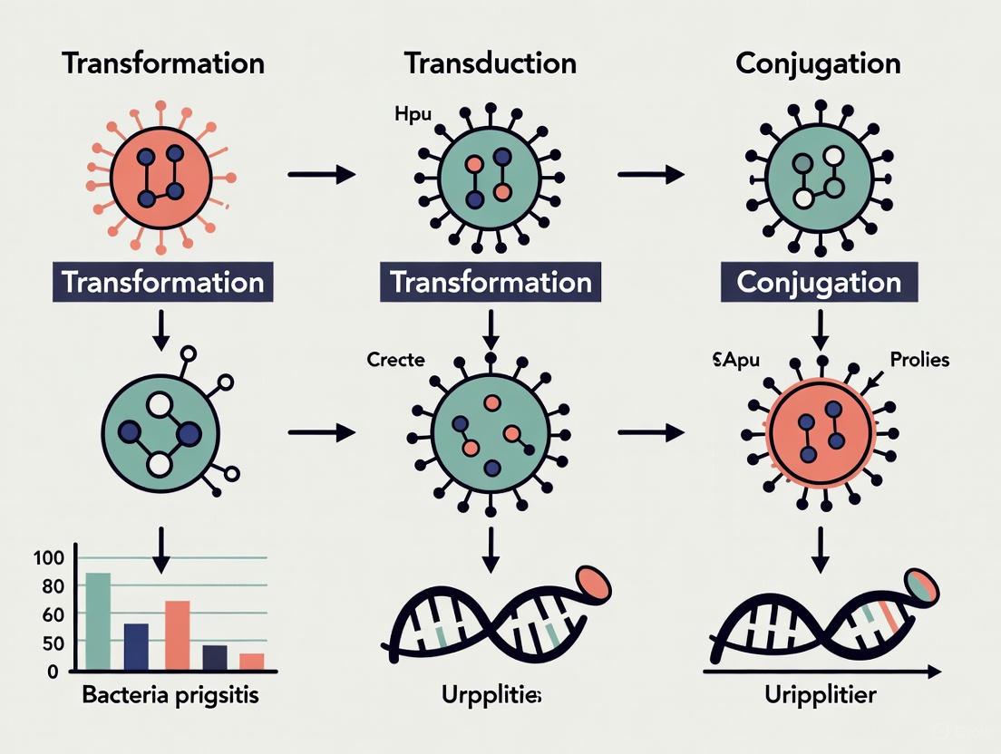
Visualization of HGT Mechanisms
Transformation Process
Transduction Process
Conjugation Process
Research Reagent Solutions for HGT Studies
Table 3: Essential Research Reagents for HGT Investigation
| Reagent/Category | Function/Application | Example Uses |
|---|---|---|
| Competent Cells | Chemical/electroporation-treated cells for transformation studies | Plasmid transformation efficiency assays [9] |
| Selection Antibiotics | Selective pressure for exconjugants/transformants | Isolation of successful HGT events [9] |
| Bacteriophages | Transduction studies and GTA characterization | Generalized/specialized transduction frequency analysis [7] |
| Plasmid Vectors | Conjugation and transformation studies | R-plasmid transfer tracking [10] [9] |
| Long-read Sequencing | Resolution of MGE architecture and context | Hybrid assembly for precise HGT visualization [10] |
| Bioinformatic Tools | HGT detection in genomic/metagenomic data | MetaCHIP, Daisy, LEMON for HGT identification [12] |
Experimental Protocols for HGT Detection
Conjugation Assay Protocol
Objective: To detect and quantify plasmid transfer between bacterial strains via conjugation.
Methodology:
- Grow donor and recipient strains to mid-logarithmic phase (OD600 ≈ 0.4-0.6) in appropriate selective media.
- Mix donor and recipient cells at approximately 1:1 ratio (typically 10^8 cells each) and concentrate on membrane filters (0.22 μm pore size).
- Incubate filters on non-selective agar plates for conjugation (typically 1-2 hours at optimal growth temperature).
- Resuspend cells from filters and plate serial dilutions on selective media containing antibiotics that distinguish exconjugants from donor and recipient cells.
- Include appropriate controls: donor alone, recipient alone, and mixed cells plated immediately without conjugation period.
- Calculate conjugation frequency as the number of exconjugants per recipient cell [9].
Antibiotic Selection Example: For conjugation between E. coli (donor) and P. aeruginosa (recipient) with an aminoglycoside resistance plasmid, use gentamicin (aminoglycoside) and nalidixic acid (quinolone). E. coli is naturally susceptible to nalidixic acid while P. aeruginosa is resistant, allowing selective isolation of exconjugants [9].
Genomic Detection of HGT Events
Objective: To identify horizontal gene transfer events in bacterial genomes using bioinformatic approaches.
Methodology:
- Sequence Acquisition and Assembly: Perform whole-genome sequencing using both short-read (Illumina) and long-read (PacBio, Nanopore) technologies. Generate hybrid assemblies for highly contiguous genomes [10].
- Comparative Genomics: Perform all-by-all alignment of genomes using tools such as nucmer. Filter results to retain alignments of at least 5 kb that share 100% identity between bacteria of different genera [10].
- Mobile Genetic Element Resolution: Precisely characterize MGE architecture and cargo using reference-based resolution of distinct plasmids and other mobile elements [10].
- Phylogenetic Analysis: Construct core gene-based phylogenies using tools such as the Genome Taxonomy Database Tool Kit (GTDBTk) to identify phylogenetic discrepancies indicative of HGT [10].
- Split-Site Analysis: For metagenomic data, use tools such as Daisy or LEMON to identify HGT breakpoints through split-read analysis, which identifies reads that map to different genomes [12].
Recent Applications: This approach has been successfully applied to identify shared sequences in 196 genomes belonging to 11 genera from healthcare-associated infections, grouped into 51 clusters of related sequences, with more than 80% of clusters encoding genes involved in DNA mobilization [10].
Implications for Antimicrobial Resistance and Drug Development
The role of HGT in disseminating antimicrobial resistance cannot be overstated. Horizontal gene transfer serves as the primary mechanism for the spread of antibiotic resistance in bacteria, with conjugative transfer of R-plasmids being especially problematic in clinical settings [7] [1]. Recent studies of healthcare-associated infections have demonstrated plasmid transfer independent from bacterial transmission, including instances of likely plasmid transfer within individual patients [10].
The magnitude of this problem is reflected in current global health statistics. Antimicrobial resistance is projected to cause 10 million deaths annually by 2050 if left unaddressed, with drug-resistant infections already contributing to more than 4.95 million deaths globally in 2019 [11]. Particularly alarming is the rise of resistance to last-resort antibiotics such as colistin and carbapenems in pathogens including Klebsiella pneumoniae and Acinetobacter baumannii, with treatment failure rates exceeding 50% in some regions [11].
Table 4: HGT-Mediated Antibiotic Resistance in Clinical Pathogens
| Pathogen | Resistance Mechanism | HGT Vehicle | Clinical Impact |
|---|---|---|---|
| MRSA | mecA gene encoding PBP2a | Staphylococcal cassette chromosome mec (SCCmec) | 10,000 annual deaths in US [11] |
| CRE | Carbapenemase genes (blaKPC, blaNDM) | Conjugative plasmids | High mortality in bloodstream infections [11] |
| ESBL-producing E. coli | Extended-spectrum β-lactamases | Plasmids, transposons | Limited therapeutic options [11] |
| VRE | vanA gene cluster | Conjugative transposons | Nosocomial infections with limited treatment [11] |
From a drug development perspective, understanding HGT mechanisms provides crucial insights for designing novel therapeutic strategies. Potential approaches include:
- Inhibition of Conjugation: Developing compounds that interfere with pilus formation or DNA transfer machinery.
- Phage Therapy: Utilizing engineered bacteriophages that target specific resistance plasmids.
- Anti-plasmid Compounds: Developing agents that specifically target plasmid maintenance or replication.
- CRISPR-based Approaches: Using sequence-specific nucleases to eliminate resistance genes from bacterial populations.
The continued evolution and dissemination of resistance mechanisms through HGT necessitates enhanced surveillance approaches. Advanced genomic tools now enable tracking of MGE movement in clinical settings, potentially identifying novel epidemiologic links not captured by traditional infection control methodologies [10]. This approach is particularly valuable for understanding the dynamics of MGE transfer in high-risk settings such as hospitals, where HGT has been shown to occur between distantly related bacteria, with isolates encoding shared sequence clusters more frequently cultured from patients with higher co-morbidity indices and solid organ transplantation [10].
Transformation is a fundamental mechanism of horizontal gene transfer (HGT) in which bacteria actively uptake and integrate extracellular environmental DNA (eDNA) into their own genomes [7]. This process enables bacteria to acquire new genetic traits from their environment rather than through vertical inheritance, serving as a powerful driver of bacterial evolution and adaptation [7]. The ability to undergo transformation, known as natural competence, is present in diverse bacterial species including notable examples such as Neisseria gonorrhoeae, Streptococcus pneumoniae, and Helicobacter pylori [7].
Environmental DNA comprises genetic material released into various ecosystems through multiple biological processes, including cell lysis, excretion of waste products, and active secretion from living organisms [13]. In aquatic environments, eDNA concentrations can reach up to 88 µg/L, while in terrestrial systems, soil eDNA content varies significantly from 0.03 to 200 µg/g depending on soil type, depth, and organic matter content [13]. The persistence and availability of this extracellular DNA create a widespread genetic reservoir that competent bacteria can exploit to rapidly adapt to selective pressures, including antibiotics and environmental stressors [7].
Molecular Mechanisms of Bacterial Transformation
The transformation process involves a sequence of highly coordinated molecular events, from DNA binding to genomic integration, facilitated by specialized bacterial machinery.
Stages of Natural Transformation
Transformation proceeds through four distinct stages: competence development, DNA binding and uptake, internalization, and genomic integration [7]. The following diagram illustrates this complete process:
DNA Uptake Mechanisms and Homologous Recombination
Naturally competent bacteria express specialized DNA-binding proteins on their cell surfaces that enable them to bind significantly more environmental DNA than noncompetent bacteria [7]. Once bound, DNA fragments approximately 10 genes in length are processed through one of two pathways: either both DNA strands penetrate the recipient cell, or a nuclease degrades one strand while the remaining single strand enters the recipient [7].
The internalized donor DNA fragment then undergoes homologous recombination with the recipient's genome, a process mediated by RecA proteins and other molecular facilitators [7]. This mechanism involves breakage and reunion of paired DNA segments with homologous regions sharing nearly identical nucleotide sequences, typically between similar bacterial strains or closely related species [7]. The successful integration of foreign DNA enables the acquisition of new functional genes, including virulence factors and antibiotic resistance determinants.
The availability and distribution of eDNA across different ecosystems significantly influences transformation frequency and evolutionary outcomes. The concentration and persistence of eDNA vary substantially across environmental matrices, as summarized in the table below.
Table 1: Environmental DNA Distribution Across Ecosystems
| Ecosystem | eDNA Concentration Range | Primary Sources | Persistence Factors |
|---|---|---|---|
| Freshwater | 2.5-88 µg/L [13] | Mucus, feces, skin cells, gametes [13] | Currents, temperature, pH, microbial activity [13] |
| Marine Sediments | 0.30-0.45 Gt total eDNA [13] | Degraded biological material | Particle adsorption protects from nucleases [13] |
| Soil | 0.03-200 µg/g [13] | Decomposition, root exudates, microbial activity | Soil composition, organic matter, depth [13] |
| River Sediments | 96.8 ± 19.8 µg/g [13] | Terrestrial runoff, in-situ production | Binding to mineral particles [13] |
Environmental DNA originates from various biological materials, including excretory products (urine, feces), sloughed epithelial cells, decomposing tissues, and active secretion mechanisms [13]. Release rates vary considerably among species and individuals, influenced by factors such as stress levels (which can increase shedding 100-fold), age, diet, water temperature, and biological community composition [13].
Beyond cellular lysis, eDNA can be actively introduced into environments through multiple mechanisms. Membrane vesicles (MVs) in Streptococcus mutans export eDNA that contributes to biofilm formation and maturation [13]. Neutrophil extracellular traps (NETs) represent another significant source, where complex biofilm structures composed of proteins and DNA are released in defense against pathogens [13]. Similar defense mechanisms occur in plant root tips, which release eDNA to combat pathogen invasions [13].
Experimental Methodologies for Studying Transformation
Research on bacterial transformation employs standardized molecular techniques to quantify DNA uptake, integration frequency, and functional outcomes. The following workflow illustrates a comprehensive experimental approach for investigating transformation:
Sample Collection and DNA Extraction
Environmental sampling protocols vary by ecosystem. Aquatic samples typically involve water collection followed by filtration through 0.22-1.2 μm filters to capture particulate matter and associated DNA [14]. Soil and sediment samples require core extraction with depth stratification, as eDNA concentration decreases significantly with increasing depth [13]. DNA extraction employs commercial kits optimized for different environmental matrices, with careful attention to inhibitor removal and DNA quality assessment.
Competence Induction and Transformation Assays
For transformation experiments, model bacteria such as Bacillus subtilis or Acinetobacter baylyi are commonly used due to their well-characterized natural competence systems [7]. Competence can be induced through specific growth conditions or chemical treatments. Standard transformation assays involve exposing competent cells to purified eDNA or environmental extracts, followed by incubation to allow DNA uptake and integration.
Selection markers, particularly antibiotic resistance genes, enable quantification of transformation frequency. The table below summarizes key reagents and methodologies used in transformation research:
Table 2: Research Reagent Solutions for Transformation Studies
| Reagent/Method | Function | Application Examples |
|---|---|---|
| RecA Proteins | Facilitates homologous recombination | Essential for DNA strand exchange and integration [7] |
| DNA Binding Proteins | Cell surface DNA recognition | Initial binding of extracellular DNA fragments [7] |
| Selective Media | Transformant selection | Antibiotic-containing media for resistance gene acquisition [7] |
| Metagenomic Sequencing | Comprehensive community analysis | Identifies potential donor sequences in eDNA [14] |
| MetaCHIP Software | HGT detection | Predicts horizontal transfer events from genomic data [3] |
Validation and Analysis Techniques
Transformation events are validated through multiple molecular approaches. PCR amplification with species-specific primers confirms the presence of acquired genes [14]. Quantitative PCR (qPCR) enables quantification of transformation frequency and gene copy number [14]. DNA sequencing provides comprehensive analysis of integrated fragments and any sequence modifications that occurred during recombination.
Bioinformatic tools like MetaCHIP enable community-level analysis of horizontal gene transfer by identifying candidate transfer events through nucleotide sequence similarity and reconciling gene and species trees using algorithms like Ranger-DTL [3]. This approach has revealed that Patescibacteria genomes contain approximately 1.0 horizontally transferred genes per genome, with up to 13% of their total genome length attributed to HGT [3].
Quantitative Analysis of Transformation and HGT
The contribution of transformation to bacterial genome evolution can be quantified through comparative genomic analyses. Studies of groundwater microbial communities, including streamlined Patescibacteria, reveal extensive HGT despite genome size constraints.
Table 3: Horizontal Gene Transfer Metrics in Bacterial Genomes
| Genome Category | HT Genes per Genome | HT Genes per Mbp | Sequence Divergence | Notable Findings |
|---|---|---|---|---|
| Patescibacteria | 1.0 ± 1.2 [3] | 1.1 ± 1.3 [3] | 23.7% ± 4.0 [3] | 54% of genomes show evidence of HT [3] |
| Other Community Members | 3.5 ± 4.8 [3] | 1.4 ± 2.1 [3] | 3.9-34.5% [3] | Comparable rate per Mbp despite larger genomes [3] |
Genomic analyses indicate that HGT events in natural environments often involve diverse taxonomic groups. In aquifer systems, transfer events occur not only between closely related bacteria but also between phylogenetically distinct lineages, such as exchanges of transcriptional regulator genes between Omnitrophota and Patescibacteria [3]. The acquired genes frequently encode metabolic functions, including transcription, translation, and DNA replication, recombination, and repair systems [3].
Transformation frequency correlates with several environmental factors. DNA concentration significantly influences transformation rates, with higher eDNA availability increasing potential recombination events [13]. Environmental conditions such as temperature, pH, and nutrient availability also impact both competence development and DNA persistence [13].
Research Implications and Future Directions
The study of transformation and eDNA uptake has profound implications for understanding bacterial evolution, antibiotic resistance spread, and microbial community dynamics. The integration of foreign DNA through transformation enables bacteria to rapidly acquire adaptive traits, including virulence factors and metabolic capabilities, without requiring mutational changes to existing genes [7]. This process is particularly significant in the context of pathogenicity islands—large, unstable genomic regions containing multiple virulence genes that can be transmitted to other bacteria through HGT [7].
Future research directions include developing more sensitive detection methods for rare transformation events, elucidating the regulatory networks controlling competence in diverse bacterial species, and understanding how environmental stressors modulate transformation frequency. The expanding application of eDNA monitoring technologies, including air sampling and sediment analysis, provides new opportunities to study transformation in natural environments [15] [13]. Additionally, metagenomic approaches coupled with single-cell analyses will enhance our understanding of transformation dynamics in complex microbial communities.
As transformation continues to be recognized as a major driver of bacterial adaptation and evolution, research in this field will remain crucial for addressing emerging challenges in antimicrobial resistance, environmental microbiology, and bacterial pathogenesis.
Horizontal Gene Transfer (HGT) represents a fundamental process enabling bacteria and archaea to acquire new genetic material without sexual reproduction, dramatically accelerating microbial evolution beyond the slow accumulation of mutational changes [16]. Among the three primary mechanisms of HGT—conjugation, transformation, and transduction—transduction stands out as the only one mediated by viral vectors. This process involves bacteriophages (viruses that infect bacteria) serving as accidental vehicles for transferring bacterial DNA between cells [17]. The abundance of tailed bacteriophages, estimated to outnumber bacteria by a factor of ten with approximately 10³¹ particles globally, makes them exceptionally influential genetic transfer agents in virtually every ecosystem, from the human gut to aquatic and soil environments [18].
Within the broader context of HGT research, transduction exemplifies how viral interactions can shape bacterial genomes, influencing everything from antibiotic resistance spread to pathogenicity evolution and ecological adaptations [19] [16]. The process exemplifies the complex co-evolutionary arms race between phages and their bacterial hosts, where bacteria develop sophisticated defense systems and phages counter with evasion strategies [20] [21]. Understanding transduction mechanisms is therefore critical not only for fundamental microbial ecology but also for addressing pressing public health challenges like antimicrobial resistance and developing novel therapeutic approaches.
Molecular Mechanisms of Transduction
Basic Phage Biology and Replication Cycles
Bacteriophages exhibit diverse life cycles that fundamentally influence their capacity to mediate gene transfer. Lytic phages hijack the host's cellular machinery immediately after infection, directing it to produce new phage particles that are eventually released through cell lysis [21]. In contrast, temperate phages can enter a lysogenic cycle where their genome integrates into the host chromosome as a prophage at a specific attachment site (att site) and replicates passively with the host cell until induced to enter the lytic cycle [17]. A third, less common chronic cycle involves continuous release of phage particles without immediate host cell lysis [21]. The life cycle determines the potential for gene transfer: temperate phages primarily facilitate specialized transduction, while lytic phages enable generalized transduction.
The phage replication cycle progresses through sequential stages: adsorption to specific bacterial surface receptors, invasion and DNA injection, uncoating, biosynthesis of viral components, assembly of progeny virions, and finally lysis and release [21]. Throughout this process, phages precisely package their genetic material into newly formed capsids using a terminase complex consisting of large (TerL) and small (TerS) subunits. TerL performs mechanical work including DNA cutting and translocation, while TerS provides packaging specificity through recognition of specific tag sequences in the phage genome [17]. This packaging precision is crucial but imperfect, creating opportunities for host DNA to be accidentally incorporated into viral particles.
Mechanisms of DNA Transfer
Table 1: Comparative Mechanisms of Phage-Mediated Gene Transfer
| Mechanism | Phage Type | Transferred DNA | Key Features | Frequency |
|---|---|---|---|---|
| Generalized Transduction | Lytic (primarily) | Any random fragment of host DNA | Results from packaging errors during phage assembly; non-specific DNA transfer | Varies; ~0.3-8×10⁻³ per PFU in freshwater environments [22] |
| Specialized Transduction | Temperate | Specific host genes adjacent to prophage integration site | Occurs through imprecise prophage excision; limited to flanking genes | Rare; ~1 transducing particle per 10⁴ virions in phage λ [17] |
| Lateral Transduction | Temperate | Extensive chromosomal regions | Initiated by prophage replication followed by packaging of adjacent host DNA | Highly efficient; can transfer hundreds of kilobases |
| Molecular Piracy | Various | Variable genetic elements | Phages capture and transfer mobile genetic elements like transposons | Dependent on element capture frequency |
Generalized Transduction
Generalized transduction occurs when bacteriophages accidentally package host bacterial DNA fragments instead of their own genome during the assembly stage of the lytic cycle [17]. This packaging error typically happens during the headful packaging mechanism employed by pac-type phages like Salmonella phage P22 and Escherichia coli phages P1 and T4 [17]. When the small terminase subunit recognizes the pac sequence, DNA packaging initiates continues until the phage head capacity is reached (typically 102-110% of genome size), with the cut determined by volume rather than specific sequence [17]. If bacterial DNA fragments containing pseudo-pac sites are recognized, they become packaged into phage capsids, creating transducing particles that contain no viral DNA but can inject bacterial DNA into subsequent hosts.
The injected donor DNA may then undergo homologous recombination with the recipient genome, permanently incorporating the transferred genes. Alternatively, if the DNA is from a plasmid or can replicate autonomously, it may persist without integration. The frequency of generalized transduction varies significantly across phage-host systems and environments, with studies in freshwater systems reporting frequencies of 0.3–8 × 10⁻³ per plaque-forming unit (PFU) [22].
Specialized Transduction
Specialized transduction is exclusively mediated by temperate phages and results from imprecise excision of the prophage from the host chromosome during induction from lysogenic to lytic cycle [17]. Unlike the precise excision that normally occurs, where the prophage cleanly detaches at the att sites, imprecise excision causes adjacent host genes to be cut out together with the phage genome [17]. These hybrid molecules containing both phage and bacterial DNA are then replicated and packaged into new virions.
Because specialized transduction depends on imprecise excision, it is typically restricted to bacterial genes immediately flanking the prophage integration site. In phage lambda, for instance, specialized transducing particles are produced at a rate of approximately 1 per 10⁴ virions, with successful transduction events occurring at frequencies around 1 in 10⁶ [17]. The limited gene range contrasts with generalized transduction but provides a targeted mechanism for specific genomic regions.
Visualization of Transduction Mechanisms
Quantitative Analysis of Transduction Frequency and Impact
Environmental Transduction Rates
Table 2: Experimentally Determined Transduction Frequencies Across Environments
| Environment/System | Phage Vector | Recipient Bacteria | Transduction Frequency | Detection Method |
|---|---|---|---|---|
| Freshwater systems | Phage P1, T4, EC10 | Plaque-forming Enterobacteriaceae | 0.3–8 × 10⁻³ per PFU | Culture-based methods [22] |
| Freshwater systems | Phage EC10 | Natural bacterial communities | Undetectable – 9 × 10⁻² per PFU | CPRINS-FISH [22] |
| Wastewater treatment systems | Indigenous phage consortia | Multidrug-resistant bacteria | Significant increase in ARG abundance in phage DNA | Metagenomic sequencing [19] |
| Phage λ system | Lambda | Escherichia coli | ~1 transducing particle per 10⁴ virions | Selective plating [17] |
Advanced detection methods like Cycling Primed In Situ Amplification-Fluorescent In Situ Hybridization (CPRINS-FISH) have revealed that more than 20% of cells carrying transferred genes retain viability in freshwater environments, indicating that transduction actively contributes to bacterial genome evolution in natural settings [22]. These findings demonstrate that gene exchange occurs frequently across a wide bacterial range, potentially promoting rapid prokaryotic genome evolution.
Genomic Impact of Phage-Mediated Transfer
The impact of transduction extends beyond immediate gene transfer to influence long-term bacterial genome architecture and evolution. Bacterial genomes contain numerous genomic islands—clusters of genes with foreign characteristics—many of which show evidence of phage-mediated integration [23]. In silico analyses provide strong statistical evidence for frequent lateral gene transfer (LGT) between virulent phages and prophages of their hosts, with bootstrap values of 91.3–100 and fit values of 91.433–100 in split decomposition analyses [23].
These phage-prophage interactions often entail genes encoding hypothetical proteins, but also affect functional genes including those for tail proteins, capsid proteins, holins, and transcriptional regulatory elements [23]. The discovered LGT events sometimes involve intergeneric recombination, particularly in E. coli and S. enterica virulent phages interacting with host prophages, demonstrating that transduction can transcend taxonomic boundaries [23].
Experimental Methods for Detecting and Measuring Transduction
Classical Microbial Techniques
Traditional transduction experiments rely on selective plating methods where donor and recipient strains with different genetic markers are co-cultured with phage vectors. Transductants are identified by their ability to grow on selective media that inhibit both the original donor and recipient strains [22]. For example, phage P1-mediated transduction in E. coli typically uses markers like antibiotic resistance or metabolic capabilities (e.g., ability to utilize specific carbon sources).
The efficiency of transduction is calculated as the number of transductants per plaque-forming unit (PFU) of the phage stock used. Controls must include recipient cells without phage (to verify selection stringency) and phage without donors (to confirm no pre-existing mutants). While straightforward, these culture-based methods have limitations, including inability to detect transductants that don't form colonies under laboratory conditions and potential underestimation of transduction rates for non-plaque-forming strains [22].
Molecular Detection Methods
Modern approaches employ molecular techniques that provide greater sensitivity and specificity:
CPRINS-FISH (Cycling Primed In Situ Amplification-Fluorescent In Situ Hybridization): This method combines in situ DNA amplification with fluorescence hybridization to detect transduction events at the single-cell level without cultivation bias [22]. The process involves sample fixation, permeabilization, in situ amplification with target-specific primers, and hybridization with fluorescent probes.
Metagenomic Analysis: High-throughput sequencing of phage and bacterial DNA from environmental samples allows detection of shared genetic elements and transduction events through comparative genomics [19] [23]. This approach identified increased antibiotic resistance gene abundance in phage fractions after co-cultivation in wastewater systems [19].
Recombination Detection Algorithms: Bioinformatics tools like SplitsTree and RDP4 implement multiple algorithms (RDP, GENECONV, BootScan, MaxChi, Chimaera, SiScan, 3Seq) to detect genetic recombination signals in genomic data [23]. These methods provide statistical evidence for LGT events through analysis of beginning and end breakpoints across homologous loci.
Visualization of Experimental Workflow for Transduction Detection
Research Reagent Solutions for Transduction Studies
Table 3: Essential Research Tools for Investigating Phage-Mediated Gene Transfer
| Reagent/Resource | Function/Application | Examples/Specifications |
|---|---|---|
| Model Phage Systems | Generalized and specialized transduction studies | Phage λ (specialized), Phage P1 (generalized), Phage T4, isolated environmental phages like EC10 [17] [22] |
| Bacterial Strains | Donor and recipient hosts for transduction assays | E. coli BW25113 (wild-type), LE392MP (amber-suppressor), isogenic strains with selectable markers [24] |
| Bioinformatics Tools | Detection of recombination events from genomic data | SplitsTree (split decomposition), RDP4 (multiple algorithms), PHASTER (prophage identification) [23] |
| Selection Markers | Detection and quantification of transductants | Antibiotic resistance genes (kanamycin, ampicillin), metabolic markers (lacZ, auxotrophic complements) |
| Molecular Detection Kits | Direct detection of transferred genes | CPRINS-FISH reagents, DNA extraction kits for metagenomics, sequencing library preparation kits [22] |
| Sequence Databases | Reference data for comparative genomics | NCBI RefSeq, PHROGs (phage protein families), ECOD (protein domains) [20] |
Current Research and Applications
Transduction in Antimicrobial Resistance Spread
Phage-mediated transduction significantly contributes to the dissemination of antibiotic resistance genes (ARGs) in diverse environments, particularly in clinical and wastewater settings [19] [18]. Cocultivation experiments with multidrug-resistant bacteria and bacteriophage consortia from wastewater treatment plants demonstrated significant increases in ARG abundance within phage DNA fractions, with 9 out of 11 identified ARGs showing substantial enrichment [19]. Notably, only 3.36% of detected plasmids were conjugative—significantly lower than the 25.2% found in broader plasmid databases—suggesting transduction may represent a more important ARG transfer mechanism than previously recognized in these environments [19].
The stability of ARGs carried by phages presents particular concern, as these genes remain functional through typical wastewater disinfection processes, allowing transducing particles to persist and locate infectable hosts [19]. This phenomenon creates environmental reservoirs of resistance determinants that can be accessed by pathogenic bacteria through phage infection.
Engineering Phages for Biotechnology Applications
Beyond their natural role in gene transfer, bacteriophages are being engineered as delivery vehicles for targeted bacterial genome editing. Recent work has modified phage λ to embed the DNA-editing all-in-one RNA-guided CRISPR-Cas transposase (DART) system, creating λ-DART phages that can infect E. coli and generate CRISPR RNA-guided transposition events in the host genome [24]. This system achieved editing efficiencies surpassing 50% of the targeted population in both monocultures and mixed bacterial communities, demonstrating the potential of engineered transduction for precise genetic manipulations [24].
Phage engineering employs sophisticated techniques like Cas13a-based counterselection paired with homologous recombination for precise phage modifications [24]. The λ-DART phages lack components essential for lysogeny, eliminating pathways for persistent phage maintenance while enabling efficient in situ gene integrations in bacteria [24]. This approach represents a convergence of natural transduction mechanisms with synthetic biology for microbiome engineering and therapeutic applications.
Ecological and Evolutionary Significance
The continuous evolutionary arms race between phages and their bacterial hosts drives diversification through mechanisms like domain shuffling in phage proteins [20]. Large-scale analyses of phage proteins reveal extensive domain mosaicism, where unrelated proteins from diverse functional classes frequently share homologous domains [20]. This phenomenon is particularly pronounced in receptor-binding proteins, endolysins, and DNA polymerases—proteins directly involved in host interactions—suggesting ongoing diversification via domain shuffling reflects co-evolutionary responses to bacterial defense mechanisms [20].
Metagenomic approaches are revolutionizing our understanding of these interactions by enabling comprehensive analysis of phage-bacteria dynamics without cultivation limitations [21]. These studies reveal that phage-mediated gene transfer shapes microbial community composition and function across diverse ecosystems, from the human gut to aquatic and terrestrial environments [21] [18]. The resulting genetic connectivity forms complex evolutionary networks rather than simple phylogenetic trees, with transduction providing key links in this web of microbial evolution [17].
Horizontal gene transfer (HGT) is a fundamental process driving bacterial and archaeal evolution, enabling rapid adaptation to environmental stresses such as antibiotics. Among HGT mechanisms, conjugation stands out as the primary vector for the dissemination of antibiotic resistance genes (ARGs) and virulence factors across microbial populations [25]. This process involves the direct cell-to-cell contact between a donor and a recipient bacterium, facilitating the unidirectional transfer of genetic material, most commonly plasmids and transposons [26]. Conjugation is universally conserved in bacteria and occurs in diverse environments, including soil, water, sewage, biofilms, and host-associated communities [26]. The transfer of plasmid-borne ARGs is particularly concerning from a clinical perspective, as it compromises the efficacy of widely used drugs, including last-resort antibiotics like carbapenems and colistin [25]. Understanding the molecular machinery, regulation, and impact of conjugation is therefore critical for public health and for framing a comprehensive thesis on HGT mechanisms. This review provides an in-depth technical guide to conjugation, detailing its mechanisms, regulation, and methodologies for study, specifically tailored for researchers, scientists, and drug development professionals.
Molecular Mechanisms of Conjugation
The Conjugative Apparatus: Pilus and Type IV Secretion System (T4SS)
The conjugation machinery is encoded by a set of transfer (tra) genes clustered on the mobile genetic element (MGE). In Gram-negative bacteria, this typically includes the genes for the elaboration of a conjugative pilus and a Type IV Secretion System (T4SS) [26]. The conjugative pilus, a multimeric assembly of the major pilin protein, is a key extracellular appendage that connects donor and recipient cells and serves as a conduit for DNA transfer [27] [26]. The T4SS is a membrane-spanning protein complex that enables the translocation of DNA across the cell envelope of the donor [26].
Historically, the F (Fertility) plasmid of Escherichia coli has served as the paradigm for conjugation studies. The tra operon of the F plasmid contains genes necessary for pilus biogenesis, mating pair formation, and DNA processing. Recent structural biology studies have revealed that conjugative pili are not limited to bacteria; archaea possess homologous systems. Cryo-EM structures of conjugative pili from the hyperthermophilic archaeon Aeropyrum pernix and the bacterium Agrobacterium tumefaciens show structural homology, suggesting a common evolutionary ancestor for these DNA transfer systems [27]. However, a key distinction is that in many hyperthermophilic archaea, the genes for the conjugation machinery (Ced system) are chromosomally encoded and "domesticated," meaning they are used to import cellular DNA rather than to spread proprietary MGEs [27].
The Relaxosome and DNA Processing
Prior to transfer, the plasmid must be processed into a transferable form. This is accomplished by the relaxosome, a nucleoprotein complex assembled at the origin of transfer (oriT) [26]. The core component of the relaxosome is the relaxase (e.g., TraI in the F plasmid), which nicks one strand of the double-stranded DNA at the oriT. The relaxase remains covalently bound to the 5' end of the nicked strand. Accessory proteins, such as TraY and TraM in the F plasmid, facilitate this process and regulate relaxase activity [26]. The nicked single-stranded DNA (ssDNA) is then unwound from its complementary strand and guided through the T4SS into the recipient cell. The coupling protein (T4CP, e.g., TraD), an AAA+ ATPase, is essential for connecting the relaxosome to the T4SS and powers the translocation of the nucleoprotein complex [26].
Plasmid Establishment in the Recipient Cell
Upon entry into the recipient cell (now termed a transconjugant), the ssDNA is converted into double-stranded DNA by host replication machinery. The plasmid must then overcome host defense systems, such as restriction-modification and CRISPR-Cas systems, to establish itself [26]. Successful establishment requires the early expression of "leading genes," which often include factors for plasmid replication, segregation, and anti-restriction functions. Once established, the plasmid can replicate autonomously and, if it carries the necessary tra genes, convert the new host into a donor cell, enabling further rounds of conjugation [26].
Regulation of Conjugative Transfer
The expression of tra genes is tightly regulated to balance the fitness cost of maintaining and expressing the conjugation machinery with the potential benefits of horizontal transfer. Regulation occurs at multiple levels and integrates both plasmid-encoded and host-encoded factors.
Table 1: Key Regulatory Systems in Plasmid Conjugation
| Regulatory System/ Factor | Plasmid/System | Mechanism of Action | Effect on Conjugation |
|---|---|---|---|
| FinOP Fertility Inhibition | F-like plasmids (e.g., R100, R1) | FinP (antisense RNA) & FinO (RNA chaperone) inhibit TraJ translation [26]. | Represses transfer; "superspreader" mutations in finO lead to constitutive transfer [26]. |
| Histone-like Nucleoid Structuring Protein (H-NS) | F plasmid, pSLT | Silences tra gene promoters (PY, PM); counteracted by TraJ and ArcA [26]. |
Growth-phase dependent transfer; maximum in exponential phase [26]. |
| Quorum Sensing (QS) | Ti plasmid (pTi) of Agrobacterium tumefaciens | TraR protein binds QS molecule OOHL, activating tra/trb operons [26]. |
Coordinates transfer with high cell density and host plant state [26]. |
| KorA/KorB Regulators | IncP plasmids | Binds operator DNA to repress tra gene transcription [28]. |
Modulates transfer rate; inhibited by ciprofloxacin, upregulated by indole [28]. |
Host-Derived Regulation and Environmental Cues
Chromosomally encoded host factors significantly influence conjugation efficiency. The histone-like nucleoid structuring protein (H-NS) acts as a global repressor by binding to and silencing AT-rich tra gene promoters, such as the PY promoter of the F plasmid [26]. This repression is growth-phase dependent, with conjugation rates peaking during the exponential phase when H-NS repression is counteracted by the plasmid-encoded activator TraJ and the host-encoded protein ArcA [26]. Furthermore, environmental signals can modulate transfer. For instance, the endogenous molecule indole was found to upregulate korA-korB expression, thereby inhibiting the transfer of broad-host-range IncP plasmids [28]. Conversely, sub-inhibitory concentrations of the antibiotic ciprofloxacin can stimulate plasmid transfer by repressing korA and korB [28].
Interplay Between Conjugation and Transposons
Conjugation and transposition are intimately linked processes that synergistically promote HGT. Plasmids act as effective vehicles for the horizontal transfer of transposons, which can carry ARGs, across taxonomic boundaries [25]. Once in a new host, transposons can jump between the newly acquired plasmid, the chromosome, or other resident plasmids.
A groundbreaking study revealed that the host nucleoid-associated protein H-NS serves as a transposon capture protein [29]. H-NS preferentially binds to horizontally acquired, AT-rich DNA regions, such as pathogenicity islands. Genome-wide mapping in Acinetobacter baumannii demonstrated that these H-NS-bound regions are "hotspots" for ISAba13 (an IS5 family transposon) insertion [29]. This targeting is mediated by the DNA-bridging activity of H-NS rather than the underlying DNA sequence alone. When H-NS is absent, transposition becomes more uniform across the genome, increasing the risk of disrupting essential genes. Therefore, H-NS directs transposition towards genetically "safe" regions, favoring evolutionary outcomes that are useful for the host cell, such as the creation of phenotypic diversity in capsule production, motility, and biofilm formation [29].
The following diagram illustrates this sophisticated mechanism of H-NS-mediated transposon targeting:
Quantitative Biology of Plasmids: Scaling Laws and Host Range
Understanding plasmid dynamics requires a quantitative analysis of their physical and genetic properties. A recent large-scale computational study analyzing 12,006 plasmids from 4,644 bacterial and archaeal genomes revealed three fundamental scaling laws that govern plasmid biology [30]:
- An inverse power-law correlation between plasmid copy number (PCN) and plasmid length. Smaller plasmids tend to have high copy numbers, while larger plasmids are maintained in low copy numbers.
- A positive linear correlation between the number of protein-coding genes and plasmid length.
- A positive correlation between the number of metabolic genes and plasmid length, particularly for large plasmids.
These scaling laws imply fundamental biophysical and evolutionary constraints. The inverse relationship between size and copy number suggests a cellular trade-off to manage the metabolic burden of plasmid replication and gene expression. Furthermore, as plasmids increase in length, they acquire more genes and converge toward chromosomal characteristics in both copy number and functional content [30].
Table 2: Plasmid Scaling Laws Derived from Genomic Analysis [30]
| Scaling Law | Mathematical Relationship | Functional Implication |
|---|---|---|
| Copy Number vs. Length | Inverse power-law | Cellular trade-off to manage metabolic burden; large plasmids are few, small plasmids are numerous. |
| Gene Number vs. Length | Positive linear correlation | Larger plasmids have a greater functional capacity and can carry more accessory genes. |
| Metabolic Genes vs. Length | Positive correlation (large plasmids) | Large plasmids contribute more significantly to the metabolic capabilities of the host cell. |
Another critical quantitative aspect is plasmid host range. A global analysis of over 10,000 plasmids led to the definition of Plasmid Taxonomic Units (PTUs), which are discrete genomic clusters of plasmids with high average nucleotide identity [31]. PTUs exhibit a characteristic host distribution, organized into a six-grade scale:
- Grade I: Restricted to a single host species.
- Grade VI: Able to colonize species from different phyla.
Notably, more than 60% of plasmids are in groups with host ranges beyond the species barrier, highlighting the extensive network for genetic exchanges in bacteria. Conjugative plasmids, which encode their own transfer machinery, are significantly more promiscuous and are overrepresented in PTUs with broad host ranges (Grades IV-VI) [31].
Experimental Methods and Research Tools
Estimating Plasmid Copy Number (PCN) with pseuPIRA
Accurate determination of Plasmid Copy Number (PCN) is crucial for understanding gene dosage effects and plasmid dynamics. A novel computational method, Pseudoalignment and Probabilistic Iterative Read Assignment (pseuPIRA), overcomes previous bottlenecks by enabling rapid, large-scale PCN estimation from short-read sequencing data [30].
Protocol: PCN Estimation with pseuPIRA [30]
- Input: A complete genome assembly (including chromosome and plasmid sequences) and linked short-read sequencing data.
- Pseudoalignment: Sequencing reads are rapidly mapped to all replicons (chromosomes and plasmids) using pseudoalignment, which quickly assigns reads to their potential origin without performing base-to-base alignment.
- Initial PCN Estimate: Unireads (reads that unambiguously map to a single replicon) are used to calculate an initial PCN estimate using the formula: PCN = (Reads mapped to plasmid / Plasmid length) / (Reads mapped to chromosome / Chromosome length).
- Multiread Handling: Multireads (reads that map to multiple replicons due to shared sequences like transposons) are re-aligned using traditional pairwise sequence alignment. Those that then map uniquely are added to the uniread count.
- Probabilistic Iterative Read Assignment (PIRA): The remaining multireads are probabilistically allocated to each replicon based on the current PCN estimates. The PCN estimates are iteratively updated based on this reallocation until convergence is reached.
- Output: A final, refined estimate of PCN for each plasmid in the genome.
pseuPIRA is more computationally efficient than alignment-based methods on large datasets and successfully handles the challenge of multireads, providing a robust and scalable solution for plasmid biology research [30].
Mapping Transposition Events with Native Tn-seq
To study transposition dynamics in a natural context, without artificial transposase induction, native Tn-seq was developed [29].
Protocol: Native Tn-seq for Genome-Wide Transposition Mapping [29]
- Sample Preparation: Genomic DNA is extracted from a wild-type bacterial culture where transposition is a rare, natural event.
- Library Preparation and Sequencing: DNA is prepared for sequencing with minimal amplification bias to avoid skewing the representation of natural transposon insertion sites.
- Bioinformatic Analysis: High-depth sequencing reads are aligned to a reference genome. The location and frequency of transposon insertions are mapped across the population.
- Identification of Hotspots: Insertion sites are correlated with genomic features (e.g., H-NS binding sites from ChIP-seq data) to identify transposition hotspots.
This method revealed that transposition is not random but is heavily biased towards H-NS-bound regions, a finding that would be obscured by conventional Tn-seq methods that use uniform, high-frequency insertion libraries [29].
The Scientist's Toolkit: Key Research Reagents and Methods
Table 3: Essential Reagents and Methods for Conjugation and Transposition Research
| Tool / Reagent / Method | Function / Description | Application in Research |
|---|---|---|
| pseuPIRA Algorithm [30] | Computational pipeline for Plasmid Copy Number (PCN) estimation from sequencing data. | Large-scale analysis of plasmid biology and dynamics across microbial genomes. |
| Native Tn-seq [29] | Maps natural transposon insertion sites genome-wide without artificial transposase induction. | Studying in vivo transposition patterns and identifying factors like H-NS that guide it. |
| ChIP-seq (Chromatin Immunoprecipitation followed by sequencing) [29] | Identifies genome-wide binding sites for DNA-associated proteins like H-NS. | Determining the genomic targets of regulatory proteins that influence gene transfer. |
| CRISPR-Cas12f / TnpB Systems [32] | RNA-guided endonucleases derived from bacterial immune systems and transposon-associated proteins. | Precision gene editing tools; TnpB is a particularly compact editor useful in biotechnological applications. |
| Conjugative Pilus Structural Models [27] | Atomic-resolution structures (from cryo-EM) of pili from bacteria and archaea. | Understanding the molecular basis of cell-cell contact and DNA transfer in conjugation. |
| Plasmid Taxonomic Units (PTUs) [31] | A natural classification scheme for plasmids based on genomic similarity and host range. | Ecological and evolutionary studies of plasmid spread and horizontal gene transfer networks. |
The following workflow diagram integrates these key methodologies to study conjugation and associated transposition:
Conjugation, as a cornerstone mechanism of horizontal gene transfer, plays an indispensable role in the evolution and adaptation of bacteria and archaea. Its sophisticated molecular machinery, involving the conjugative pilus, T4SS, and relaxosome, enables the efficient transfer of MGEs like plasmids and transposons. The process is under intricate multi-level regulation that integrates plasmid-encoded and host-encoded factors, fine-tuning transfer in response to physiological and environmental cues. The recently discovered scaling laws of plasmids and the organization of the plasmidome into PTUs with defined host ranges provide a quantitative framework for understanding the constraints and opportunities of plasmid-mediated gene flow. Furthermore, the interplay between conjugation and transposition, guided by host factors like H-NS, creates a powerful engine for generating genetic diversity and disseminating adaptive traits, including antibiotic resistance. For researchers and drug development professionals, a deep understanding of these mechanisms is paramount. The continued development of advanced tools—from computational methods like pseuPIRA and native Tn-seq to novel gene editors like TnpB—will be crucial in deciphering the complex dynamics of HGT and devising novel strategies to combat the spread of antimicrobial resistance.
Horizontal gene transfer (HGT) is a fundamental driver of evolution and adaptation in prokaryotes. While bacterial HGT mechanisms are well-characterized, archaea employ distinct, specialized systems that facilitate genetic exchange in extreme environments and contribute to their remarkable adaptability. This technical guide provides an in-depth analysis of three key archaeal-specific HGT mechanisms: the Crenarchaeal system for exchange of DNA (Ced), cytoplasmic bridges, and vesicle-mediated transfer. We synthesize current structural and functional data, present quantitative comparisons of DNA transfer capabilities, detail experimental methodologies for studying these systems, and visualize key mechanistic workflows. Understanding these archaeal-specific pathways provides crucial insights into microbial evolution and has implications for addressing antibiotic resistance and developing novel biotechnological applications.
Horizontal gene transfer represents a potent evolutionary force in archaea, enabling rapid adaptation to extreme environments including hyperthermal, acidic, and high-salinity conditions [16]. Unlike vertical gene transfer, HGT allows direct acquisition of genetic material from contemporary organisms, providing immediate access to beneficial traits. Archaea utilize both conserved and unique mechanisms for genetic exchange, with recent research revealing sophisticated, domain-specific adaptations [33] [34].
The Ced system represents a dedicated DNA import apparatus predominantly found in Crenarchaeota, while cytoplasmic bridges facilitate direct cellular connections for DNA exchange in Euryarchaeota such as Haloferax volcanii [35]. Additionally, membrane vesicle-mediated transfer provides a protected mechanism for intercellular genetic exchange across archaeal species [36] [37]. These systems operate alongside more universal mechanisms like transformation, transduction, and conjugation, but exhibit distinctive archaeal adaptations in their molecular machinery and regulation.
This review focuses on the molecular architecture, functional mechanisms, and experimental approaches for investigating these archaeal-specific HGT pathways, providing researchers with comprehensive methodological frameworks for advancing studies in archaeal genetics.
The Crenarchaeal System for Exchange of DNA (Ced)
Molecular Architecture and Components
The Ced system represents a specialized DNA import machinery identified in hyperthermophilic archaea, particularly within the Crenarchaeota phylum. This system was first characterized in Sulfolobus acidocaldarius, where it functions in chromosomal DNA exchange for DNA repair following UV damage [35]. Core components include four essential proteins with distinct structural and functional properties:
- CedA: A polytopic transmembrane protein predicted to contain 6-7 transmembrane domains that likely forms the central channel for DNA import across the membrane [35].
- CedB: A membrane-bound VirB4/HerA homolog AAA+ ATPase that provides the energy requirement for DNA translocation through hydrolysis of nucleoside triphosphates [35].
- CedA1 and CedA2: Small membrane proteins each containing two predicted transmembrane domains that form a complex with CedA and facilitate its function [35].
Recent structural studies have revealed that CedA1 homologs in Aeropyrum pernix form conjugative pili that are structurally homologous to bacterial mating pili, despite limited sequence similarity [34]. Cryo-EM analysis has determined the atomic structure of these archaeal conjugative pili at 3.3 Å resolution, demonstrating their functional equivalence to bacterial conjugation systems despite evolutionary divergence [34].
Functional Mechanism and Regulation
The Ced system operates in conjunction with the UV-inducible pili (Ups) system in a coordinated DNA repair response [35]. The functional sequence involves:
- UV Induction: DNA damage from UV irradiation triggers transcriptional upregulation of both Ups and Ced systems [35].
- Cellular Aggregation: Ups pili mediate species-specific cell aggregation through recognition of specific glycosylation patterns on pilin proteins and S-layers [35].
- DNA Import: The Ced machinery imports DNA fragments from adjacent cells within aggregates [35].
- Homologous Recombination: Imported DNA serves as a template for repair of double-strand breaks through homologous recombination [35].
Notably, the Ced system functions specifically in DNA import rather than export, distinguishing it from bacterial conjugation systems that typically export DNA [35]. This unidirectional import specialization optimizes the system for DNA repair functions. The system demonstrates species specificity, ensured by specific glycosylation patterns on the Ups pili and the protein S-layer that covers the cellular membrane [34].
Table 1: Core Components of the Archaeal Ced System
| Component | Key Features | Proposed Function | Homologs |
|---|---|---|---|
| CedA | 6-7 transmembrane domains, polytopic membrane protein | Forms transmembrane channel for DNA import | Limited homology to bacterial T4SS components |
| CedB | VirB4/HerA-like AAA+ ATPase, membrane-associated | Powers DNA translocation via NTP hydrolysis | VirB4/HerA ATPases |
| CedA1 | 2 transmembrane domains, forms pilus structures | Pilus formation for cell-cell contact | Bacterial major pilins |
| CedA2 | 2 transmembrane domains | Complex formation with CedA, regulation | No clear bacterial homologs |
Genomic Distribution and Evolutionary Context
The Ced system is widely distributed among Crenarchaeota, including orders Sulfolobales, Desulfurococcales, and Acidilobales [35]. Genomic analyses reveal conservation of the ced gene cluster organization across these lineages, with variations in additional domains present in CedA proteins of Desulfurococcales and Acidilobales [35].
Recent structural and genomic evidence indicates that the archaeal Ced system shares a common ancestor with bacterial type IV secretion systems (T4SS) [34]. However, a key evolutionary distinction is that the Ced system has been "domesticated" – its genes are encoded chromosomally rather than on mobile genetic elements, reflecting adaptation for cellular DNA repair rather than proprietary plasmid transfer [34].
Diagram 1: Ced System Functional Workflow. This diagram illustrates the UV-induced activation of the Ced DNA import system and its coordination with Ups pili for DNA repair.
Cytoplasmic Bridges and Cellular Fusion
Structural Characteristics and Formation
Cytoplasmic bridges represent a distinct HGT mechanism involving direct cytoplasmic connections between archaeal cells. This phenomenon was first described in the euryarchaeon Haloferax volcanii, where it facilitates exchange of large chromosomal DNA fragments [35]. These intercellular bridges differ from pilus-mediated connections by establishing full cytoplasmic continuity, allowing bidirectional transfer of cellular contents.
Bridge formation involves specialized membrane fusion proteins that create stable, pore-like connections between adjacent cells. These structures permit transfer of DNA fragments up to 500 kbp, significantly larger than typical fragments transferred through other HGT mechanisms [35]. The formation of these bridges in Haloferax species leads to the generation of diploid cells with mixed chromosomes, creating temporary heterozygotes that enhance genetic diversity and facilitate DNA repair [35].
Genetic Exchange and Functional Consequences
DNA transfer through cytoplasmic bridges exhibits several distinctive characteristics:
- Bidirectional transfer: Unlike the unidirectional import of the Ced system, bridge-mediated exchange allows DNA movement in both directions between connected cells [35].
- Large fragment size: The system accommodates extremely large DNA fragments (up to 500 kbp), enabling transfer of substantial genomic regions containing multiple operons or pathogenicity islands [35].
- Interspecies transfer: Bridges can form between different Haloferax species, promoting horizontal transfer across species boundaries [35].
This mechanism is particularly significant for DNA repair and adaptation in extreme environments, as it allows cells to access complete functional gene sets from neighbors, potentially conferring immediate adaptive advantages without requiring stepwise mutation accumulation.
Vesicle-Mediated Gene Transfer
Extracellular Vesicle Biogenesis and Composition
Extracellular vesicles (EVs) represent a ubiquitous mechanism for intercellular macromolecule transfer, including DNA, in both archaeal and bacterial domains. These spherical, membrane-bound nanostructures (typically 20-250 nm in diameter) are released through budding and pinching off of the membrane [37]. In archaea, EVs facilitate protected transport of genetic material between cells, particularly in extreme environments where naked DNA would be rapidly degraded.
Vesicle biogenesis involves multiple pathways:
- Outer membrane blebbing: Sections of outer membrane bleb away from the surface of intact cells [36].
- Membrane re-annealing following lysis: Small membrane fragments re-anneal after cell lysis events [36].
- Vesiculation-inducing signals: Molecules like the Pseudomonas quinolone signal (PQS) directly induce membrane curvature and vesiculation [37].
EVs contain diverse cargoes including proteins, metabolites, and nucleic acids. DNA is heterogeneously distributed within EV populations and may be incorporated through multiple mechanisms: formation of vesicles containing both inner and outer membranes, capture of DNA during re-annealing of lytic membrane fragments, or directed packaging systems [36].
DNA Packaging and Transfer Mechanisms
Marine studies comparing EVs and virus-like particles (VLPs) reveal distinct DNA carrying capacities between these nanoparticle types [36]. EVs contain DNA fragments ranging from hundreds of base pairs to over 180 kb, with a maximum observed length of 183 kb and N50 of approximately 3 kb [36]. This capacity sufficiently accommodates individual genes, complete operons, or mobile genetic elements.
Table 2: Quantitative Comparison of Nanoparticle DNA Transfer Capabilities
| Parameter | Extracellular Vesicles (EVs) | Virus-Like Particles (VLPs) |
|---|---|---|
| Size Range | 20-250 nm | 50-250 nm |
| DNA Fragment Length | 100s bp - 183 kb | Up to 233 kb |
| N50 Value | ~3 kb | ~37 kb |
| Maximum Capacity | 183 kb | 233 kb |
| DNA Packaging Mechanism | Passive enclosure, heterogeneous | Selective, active packaging |
| Enrichment in Mobile Elements | Yes | Yes |
The protected environment within EVs shields DNA from environmental nucleases during transit between cells, enhancing successful gene transfer compared to naked DNA transformation [37]. Sequencing analyses reveal that EV-associated DNA is enriched in mobile genetic elements (MGEs) including plasmids, transposons, integrative and conjugative elements (ICEs), and phage-inducible chromosomal islands (PICIs) compared to cellular chromosomal regions [36].
Vesicle-mediated transfer demonstrates broader host range compatibility compared to viral transduction, which is typically limited by receptor specificity [36]. This promiscuity makes EV-mediated HGT particularly significant for genetic exchange across diverse archaeal populations and potentially across domain boundaries.
Experimental Approaches and Methodologies
Genetic Analysis of Ced System Function
The functional characterization of the Ced system has employed targeted gene deletion complemented by DNA transfer assays [35]. The following protocol outlines key methodological approaches:
UV Induction and DNA Transfer Assay:
- Culture archaeal strains (e.g., Sulfolobus acidocaldarius) to mid-exponential phase in appropriate medium.
- Induce Ced system expression by UV irradiation (optimal dose determined empirically).
- Allow cellular aggregation for 4-6 hours post-induction.
- Separate aggregates from single cells by gentle sedimentation.
- Quantify DNA transfer using selectable markers or PCR-based detection of chromosomal markers.
- Compare transfer efficiency between wild-type and mutant strains (e.g., ΔcedA, ΔcedB).
Critical controls: Include DNase treatment to confirm cell-contact dependent transfer (Ced-mediated transfer should be DNase-resistant) [35].
Gene Deletion Construct Preparation:
- Amplify 5' and 3' flanking regions (approximately 500 bp each) of target ced genes.
- Clone flanking regions around a selectable marker (e.g., pyrE or lacS) in an E. coli-archaea shuttle vector.
- Transform deletion construct into recipient archaeal strain.
- Select for integrants using appropriate selective medium.
- Screen for double-crossover events by PCR and sequencing.
Vesicle Isolation and Characterization
Isolation and analysis of extracellular vesicles requires specialized approaches to separate vesicles from cells and free DNA:
Density Gradient Ultracentrifugation Protocol:
- Remove cells and debris from culture supernatant by sequential centrifugation at 4°C: 10,000 × g for 30 minutes, then 50,000 × g for 60 minutes.
- Concentrate vesicles by ultracentrifugation at 150,000 × g for 3 hours.
- Resuspend pellet in appropriate buffer (e.g., phosphate-buffered saline).
- Layer onto discontinuous iodixanol gradient (e.g., 10%-50%).
- Centrifuge at 100,000 × g for 16-18 hours.
- Collect vesicle-enriched fractions (typically 25%-35% iodixanol).
- Characterize by electron microscopy, nanoparticle tracking analysis, and protein quantification.
DNA Extraction and Analysis from Vesicles:
- Treat vesicle preparations with DNase I to remove externally adherent DNA.
- Verify DNase efficiency using control DNA samples.
- Lyse vesicles using detergent-based lysis buffer.
- Extract DNA using phenol-chloroform or commercial kits.
- Analyze DNA content by fluorometry, PCR, or sequencing approaches.
Diagram 2: Vesicle DNA Analysis Workflow. This experimental workflow outlines the key steps for isolating extracellular vesicles and analyzing their DNA content.
Research Reagent Solutions
Table 3: Essential Research Reagents for Studying Archaeal HGT Mechanisms
| Reagent/Category | Specific Examples | Function/Application |
|---|---|---|
| Selectable Markers | pyrE, lacS, hph (hygromycin resistance) | Selection of transformants and gene deletion mutants in archaeal systems |
| Archaeal Growth Media | DSMZ 197, ATCC 1303, appropriate extreme condition modifications | Support growth of specific archaeal species under optimal conditions |
| UV Crosslinkers | Spectrolinker XL-1000, CL-1000 | Controlled UV induction of Ced and Ups systems |
| Ultracentrifugation Equipment | Optima XPN, Type 70 Ti rotor | Vesicle isolation and purification via density gradient centrifugation |
| DNA Processing Enzymes | DNase I, Proteinase K, various restriction enzymes | DNA analysis, removal of external DNA contamination, molecular cloning |
| Structural Biology Tools | Cryo-electron microscopes (Titan Krios), image processing software | High-resolution structure determination of pili and membrane complexes |
| Sequence Analysis Platforms | BLASTP, HHpred, GTDB-Tk, antiSMASH | Genomic analysis, homology detection, taxonomy assignment, MGE identification |
Archaeal-specific HGT mechanisms represent sophisticated adaptations that facilitate genetic exchange and enhance survival in extreme environments. The Ced system exemplifies a domesticated DNA import apparatus optimized for DNA repair, while cytoplasmic bridges enable exchange of large genomic regions, and vesicle-mediated transfer provides protected intercellular DNA transport. These mechanisms collectively contribute to the remarkable adaptability and evolutionary success of archaea in diverse habitats.
Future research directions should focus on structural characterization of full Ced membrane complexes, regulatory networks controlling these HGT systems, and exploration of potential intersections between different transfer mechanisms. Additionally, understanding how archaeal HGT contributes to antibiotic resistance spread and metabolic adaptation will provide valuable insights for therapeutic development and biotechnology applications. The continued development of genetic tools and high-resolution analytical approaches will be essential for advancing our understanding of these fascinating molecular systems.
The Role of Horizontal Transposon Transfer (HTT) in Genome Evolution Across Domains
Horizontal Transposon Transfer (HTT) represents a powerful evolutionary force, defined as the non-genealogical transmission of transposable elements (TEs) between genomes, distinct from vertical parent-to-offspring inheritance [1]. While horizontal gene transfer (HGT) has long been recognized as a cornerstone of prokaryotic evolution, recent research has established that HTT is a common and widespread phenomenon in eukaryotes as well [1]. This process enables the rapid mobilization of genetic elements across species boundaries, fundamentally reshaping genomes and accelerating evolutionary innovation beyond the constraints of vertical descent.
The study of HTT has gained renewed importance with growing recognition of its impact on genome architecture and function. HTT provides a mechanistic explanation for the patchy distribution of certain TEs across divergent taxa and the sudden appearance of new transposable elements in lineages without evolutionary precursors [38] [1]. For researchers investigating microbial evolution and drug development, understanding HTT is crucial as it facilitates the spread of antibiotic resistance genes among pathogenic bacteria and can introduce new genetic variation that complicates therapeutic targeting [1]. This whitepaper examines the mechanisms, distribution, and evolutionary consequences of HTT within the broader framework of horizontal gene transfer mechanisms in bacteria and archaea.
Mechanisms of Horizontal Transposon Transfer
Horizontal Transposon Transfer occurs through diverse molecular pathways that enable TEs to cross between organisms. The mechanisms differ significantly between prokaryotic and eukaryotic systems, though some principles remain conserved across domains of life.
General HTT Mechanisms
The passage of mobile DNA segments between genomes can occur through multiple vectors. In prokaryotes, HTT is intimately linked with established HGT mechanisms—transformation, transduction, and conjugation—which can facilitate the transfer of transposons along with other genetic material [1]. Bacteriophages (viruses that infect bacteria) serve as particularly efficient vectors for HTT through transduction, packaging bacterial DNA including transposons and delivering it to new host cells [1].
In eukaryotes, proposed vectors for HTT include arthropods, viruses, endosymbiotic bacteria, and intracellular parasitic bacteria [1]. The actual transportation mechanism of TEs from donor to host cells remains incompletely characterized, though circulating naked DNA and RNA in bodily fluid has been proposed as a potential medium for transfer [1]. Recent evidence suggests that giant viruses may contribute to introner transfer across divergent eukaryotic lineages [38].
Transposon-Specific Transfer Considerations
The structural characteristics of different transposon classes significantly impact their propensity for horizontal transfer. DNA transposons and LTR retroelements are more likely to undergo HTT compared to non-LTR retroelements because both have a stable, double-stranded DNA intermediate that is sturdier than the single-stranded RNA intermediate of non-LTR retroelements, which can be highly degradable [1].
Autonomous elements, which encode the proteins required for their own mobilization, may be more likely to transfer horizontally compared to non-autonomous elements that rely on trans-acting factors for movement [1]. The structure of these non-autonomous elements generally consists of an intronless gene encoding a transposase protein, and may or may not have a promoter sequence [1]. Those lacking promoter sequences rely on adjacent host promoters for expression after successful horizontal transfer [1].
Table 1: Transposable Element Propensity for Horizontal Transfer
| TE Category | Molecular Intermediate | HTT Likelihood | Key Factors |
|---|---|---|---|
| DNA Transposons | Double-stranded DNA | High | Stable DNA intermediate, encodes transposase |
| LTR Retrotransposons | Double-stranded DNA | High | Stable DNA intermediate, reverse transcription step |
| Non-LTR Retrotransposons | Single-stranded RNA | Lower | Degradable RNA intermediate |
| Autonomous Elements | DNA or RNA | Variable | Encodes necessary proteins for mobilization |
| Non-autonomous Elements | DNA or RNA | Variable | Requires trans-acting factors for mobility |
Distribution and Diversity of HTT Across Organisms
Recent genomic analyses have revealed that HTT occurs across an exceptionally broad taxonomic range, from prokaryotes to multicellular eukaryotes, challenging earlier assumptions about the restricted distribution of this phenomenon.
HTT in Prokaryotic Systems
Horizontal gene transfer, including HTT, is common among bacteria and occurs even between very distantly related species, as well as between bacteria and archaea [1]. This process is a significant cause of increased drug resistance when one bacterial cell acquires resistance genes and transfers them to other species [1]. In archaea, research into HGT has historically lagged behind bacterial studies, though some mechanisms of gene exchange—such as plasmids that transmit via membrane vesicles and cytoplasmic bridges that allow transfer of both chromosomal and plasmid DNA—may be archaea-specific [39].
HTT in Eukaryotic Lineages
A systematic search of 8,716 annotated eukaryotic genome assemblies revealed diverse species whose genomes contain introns derived from recent transposition, with HTT occurring in an exceptionally broad range of eukaryotic species [38]. HTT is particularly abundant in aquatic organisms, unicellular species, and fungi (>98% of introner-containing species fall into these categories) [38]. Recent studies have also identified HTT in expanded taxonomic ranges including land plants (e.g., Panicum virgatum and Salvia splendens) and an echinoderm (Strongylocentrotus purpuratus) [38].
Massive HTT-mediated intron gain has been documented in certain species, such as the parasitic dinoflagellate Amoebophrya sp. A120, which harbors introners attributed to non-LTR retrotransposons, LTR retrotransposons, and diverse TIR DNA transposons [38]. In some cases, these diverse introners generate tens of introns in a single gene, demonstrating the profound impact HTT can have on genome architecture [38].
Table 2: Documented HTT Events Across Eukaryotic Taxa
| Taxonomic Group | Example Species | TE Types Transferred | Genomic Impact |
|---|---|---|---|
| Dinoflagellates | Amoebophrya sp. A120 | Non-LTR retrotransposons, LTR retrotransposons, TIR DNA transposons | Tens of introns per gene in some cases |
| Dinoflagellates | Polarella glacialis | Multiple diverse introners | Significant ongoing intron gain |
| Grasses | Panicum virgatum | Copia LTR elements | Intron gain via autonomous and non-autonomous elements |
| Basidiomycete Fungi | Suillus subalutaceus | Helitrons | Intron generation with characteristic TT/TC insertion sites |
| Marine Diatom | Parmales sp. scaly parma | Unknown mechanism | >91% of recognizable intron gains from one introner family |
Methodologies for HTT Detection and Analysis
Accurately identifying HTT events requires complementary bioinformatic and experimental approaches that can distinguish horizontal transfer from vertical inheritance.
Bioinformatics Detection Methods
HTT is typically inferred using bioinformatics methods, which generally fall into two categories: parametric methods that identify atypical sequence signatures, and phylogenetic methods that identify strong discrepancies between the evolutionary history of particular sequences compared to that of their hosts [1]. The transferred gene (xenolog) found in the receiving species is more closely related to the genes of the donor species than would be expected under vertical inheritance [1].
One demonstrated method to find HGT events is through Shotgun Metagenomics, which involves breaking down then sequencing DNA in a sample by its contiguous regions and looking for phylogenetic mismatches that can be inferred as instances of Horizontal Gene Transfer [1]. For HTT specifically, detection is complicated by the fact that it is an ongoing phenomenon constantly changing in frequency and TE composition within host genomes [1].
Experimental Validation Protocols
To confirm bioinformatically predicted HTT events, several experimental approaches can be employed:
1. Horizontal Transfer Assay Protocol
- Purpose: To experimentally demonstrate the transfer of transposable elements between bacterial strains or species.
- Methodology:
- Donor and recipient strains are cultivated in mixed cultures, with selection markers distinguishing the strains.
- Selectable markers (e.g., antibiotic resistance) are used to identify recipients that have acquired the TE.
- PCR and Southern blotting confirm the presence and genomic integration of the transferred TE.
- Controls include monocultures of both donor and recipient to detect spontaneous mutation.
2. Transposition Activity Assay
- Purpose: To verify that horizontally transferred TEs remain functionally active in the recipient genome.
- Methodology:
- The candidate TE is cloned into a plasmid vector.
- The construct is introduced into recipient cells via transformation.
- Transposition events are detected using genetic screens or reporters that activate upon integration.
- DNA sequencing validates the precise integration and target site duplications characteristic of the TE family.
Diagram 1: HTT detection workflow
Research Reagent Solutions for HTT Studies
Investigating Horizontal Transposon Transfer requires specialized reagents and tools. The following table outlines essential materials for experimental HTT research.
Table 3: Research Reagent Solutions for HTT Investigation
| Reagent/Tool | Function in HTT Research | Application Examples |
|---|---|---|
| Selectable Marker Genes | Tracking transferred elements | Antibiotic resistance genes for selection of recipient cells |
| Plasmid Vectors | Cloning and transferring TEs | Shuttle vectors for inter-species TE transfer experiments |
| Metagenomic Sequencing Kits | Comprehensive DNA profiling | Shotgun metagenomics for HTT detection in microbial communities |
| PCR Primers for TE Signatures | Amplifying transposon sequences | Detection of specific TEs in potential recipient species |
| Southern Blot Hybridization Probes | Verifying genomic integration | Confirming TE presence and copy number in recipient genomes |
| Phage & Virus Collection | HTT vector studies | Investigating transduction-mediated TE transfer |
| Bacterial Conjugation Systems | Studying conjugation-mediated transfer | Mobilizable plasmids for testing inter-species TE transfer |
Evolutionary Consequences and Genomic Impact
HTT has profound effects on genome evolution, influencing both genome architecture and functional capacity across diverse organisms.
Intron Gain and Genome Restructuring
Introners, which are specialized transposable elements that generate introns upon insertion, represent a significant consequence of HTT in eukaryotic genomes [38]. These elements can create thousands of introns within a single genome and are derived from functionally diverse TEs including terminal-inverted-repeat DNA TEs, retrotransposons, cryptons, and helitrons [38]. Introners represent the only mechanism that could explain the "bursts" of intron gains observed across diverse eukaryotic lineages [38].
Recent research has revealed that intron-generating TEs span exceptional mechanistic diversity, arising from TEs spanning approximately 80% of orders and at least 50% of superfamilies of known mobile genetic elements [38]. These include diverse terminal inverted repeat (TIR) DNA transposons, long terminal repeat (LTR) retrotransposons, non-LTR retrotransposons, rolling circle helitrons and tyrosine recombinase (Crypton) elements [38]. The ancient origins of these diverse TEs suggests that introners have likely been generating introns throughout eukaryotic evolution [38].
Adaptive Evolution and Host Fitness
The arrival of a new TE in a host genome can have detrimental consequences because TE mobility may induce deleterious mutations [1]. However, HTT can also be beneficial by introducing new genetic material into a genome and promoting the shuffling of genes and TE domains among hosts, which can be co-opted by the host genome to perform new functions [1]. Furthermore, transposition activity increases the TE copy number and generates chromosomal rearrangement hotspots, potentially creating novel regulatory networks and genetic innovations [1].
In prokaryotes, HTT plays a crucial role in adaptive evolution, facilitating the rapid acquisition of antibiotic resistance and metabolic capabilities that enable colonization of new ecological niches [40] [1]. HGT accelerates evolutionary rates, facilitates adaptive innovations, and shapes microbial pangenomes, with HTT serving as a specialized mechanism for distributing mobile genetic elements that can carry adaptive traits [40].
Future Research Directions
Despite significant advances, key aspects of HTT remain poorly understood and represent fertile ground for future investigation. The molecular mechanisms driving introner proliferation remain almost entirely unexplored, and it is unclear whether introners arise from diverse transposable elements or are restricted to a specific mechanism of mobilization [38]. The precise vectors facilitating HTT between eukaryotic species require further characterization, though current evidence suggests arthropods, viruses, freshwater snails, endosymbiotic bacteria, and intracellular parasitic bacteria may play roles [1].
A new wave of research seeks to predict how HGT shapes microbial evolution within natural communities, especially during rapid ecological shifts [40] [8]. Quantifying the dynamics of HGT is critical for understanding microbial adaptation in natural and engineered environments, with HTT representing a specialized but important component of this genetic exchange [8]. Future studies should aim to quantify HTT rates within diverse ecological contexts and determine the fitness effects of horizontally transferred transposable elements across different host backgrounds.
Diagram 2: HTT research framework
Horizontal Transposon Transfer represents a significant evolutionary mechanism that operates across all domains of life, facilitating the rapid exchange of mobile genetic elements between divergent lineages. The integration of HTT research within the broader framework of horizontal gene transfer enriches our understanding of microbial evolution and provides critical insights into the dynamics of genome innovation. For researchers and drug development professionals, appreciating the mechanisms and consequences of HTT is essential for understanding the spread of antibiotic resistance, the emergence of new pathogenic traits, and the fundamental processes that shape genomic diversity. As detection methods improve and more genomes are sequenced, the full extent of HTT's role in evolution will continue to be revealed, potentially offering new strategies for managing genetic diseases and controlling the spread of undesirable genetic elements in pathogenic organisms.
Detecting and Harnessing HGT: Bioinformatic Tools and Applications in Research and Therapy
Horizontal Gene Transfer (HGT), also known as Lateral Gene Transfer (LGT), represents a fundamental mechanism in microbial evolution, enabling the direct transmission of genetic material between disparate organisms outside of vertical inheritance. This process serves as a critical driver of adaptive evolution, facilitating the rapid acquisition of novel traits such as antibiotic resistance, pathogenicity determinants, and metabolic capabilities that allow bacteria and archaea to colonize new ecological niches, including extreme environments [16] [2]. The detection and analysis of HGT events are therefore paramount to understanding microbial evolution, pathogenesis, and environmental adaptation.
Bioinformatic approaches for HGT detection have evolved into two principal methodological frameworks: parametric methods and phylogenetic methods. Parametric methods identify horizontally acquired genes by detecting significant deviations in sequence composition from the host genomic average, while phylogenetic methods identify genes whose evolutionary history conflicts with the accepted species phylogeny [2]. These approaches operate on complementary principles and exhibit different strengths, limitations, and time sensitivities, making them suitable for different research scenarios and evolutionary timescales. This technical guide provides an in-depth examination of these core methodologies, their implementation, and their integration in contemporary bacterial and archaeal research.
Parametric Detection Methods
Parametric methods, often termed "sequence composition-based methods," operate on the principle that each genome possesses a unique genomic signature—a characteristic pattern of sequence composition that remains relatively consistent across native genes but becomes disrupted in recently acquired foreign genes. These methods function without requiring comparative data from multiple genomes, relying instead on intrinsic sequence properties of the genome under investigation [2].
Core Principles and Genomic Signatures
The fundamental assumption of parametric methods is that horizontally transferred DNA segments initially retain the distinct compositional features of their donor genome. These signatures include nucleotide composition, oligonucleotide frequencies, codon usage bias, and structural DNA features. Over time, a process called amelioration occurs, where the transferred sequences gradually adopt the genomic signature of the recipient genome through mutational processes, making ancient HGT events increasingly difficult to detect [2] [41].
Table 1: Primary Genomic Signatures Used in Parametric HGT Detection
| Signature Type | Description | Detection Capability | Key Limitations |
|---|---|---|---|
| Nucleotide Composition (GC Content) | Measures Guanine-Cytosine (GC) percentage in genomic segments. Simple to compute. | Effective for detecting recent HGT from donors with different GC% [2]. | High intragenomic variability in native genes can cause false positives. Ameliorates quickly. |
| Oligonucleotide Spectrum (k-mer frequencies) | Frequency analysis of all possible nucleotide sequences of length k (e.g., tetranucleotides) [2]. | Highly discriminative due to large number of possible patterns; captures species-specific signals [2]. | Requires optimization of sliding window size; large windows reduce sensitivity to small HGT regions [2]. |
| Codon Usage Bias | Measures preference for specific synonymous codons within coding sequences [2]. | One of the first methodical assessment approaches; can identify HGT where bias differs significantly [2]. | Bias influenced by gene expression levels; highly expressed native genes may show atypical patterns [2]. |
| Structural Features | Encodes structural DNA properties (e.g., interaction energies, twist angles, deformability) into numerical sequences [2]. | Can provide supporting evidence through periodicity spectrum analysis; validated in massive HGT cases [2]. | Complex to compute and interpret; requires specialized analytical approaches [2]. |
Methodological Considerations and Limitations
The effective implementation of parametric methods requires careful consideration of several analytical challenges. A critical parameter is the sliding window size used for scanning genomes. Larger windows better account for natural intragenomic variability but reduce sensitivity for detecting small horizontally transferred regions, with a reported compromise of 5 kb windows with 0.5 kb steps for tetranucleotide analysis [2]. Furthermore, parametric methods are inherently limited to detecting recent HGT events before amelioration is complete, typically within the last 100 million years for bacterial genomes [2]. They also struggle with identifying transfers between organisms with similar genomic signatures, particularly among closely related species.
Phylogenetic Detection Methods
Phylogenetic methods represent a more direct approach for identifying HGT events by comparing the evolutionary history of individual genes with a trusted species phylogeny. These methods leverage the fundamental principle that in the absence of HGT, all genes should produce phylogenetic trees that are congruent with the species tree. Significant incongruence between gene trees and the species tree provides evidence for horizontal transfer [2] [42].
Methodological Frameworks and Applications
Phylogenetic approaches explicitly reconstruct evolutionary relationships for individual genes and compare them to a reference species tree. These methods can be further divided into those that perform full phylogenetic tree reconstruction and those that use surrogate measures, such as sequence similarity distributions, in place of complete trees [2]. The availability of numerous sequenced genomes has made phylogenetic methods increasingly powerful, as they can integrate information from multiple taxa using evolutionary models [2].
Table 2: Phylogenetic Approaches for HGT Detection
| Method Category | Description | Advantages | Tools/Implementations |
|---|---|---|---|
| Explicit Tree Reconciliation | Reconstructs phylogenetic trees for individual genes and compares them to a reference species tree. | Can characterize HGT events (donor, timing); uses evolutionary models; suitable for ancient transfers [2]. | HGTphyloDetect [42], AvP [42]. |
| Similarity Distribution Analysis | Analyzes distribution of BLAST hits across taxonomic groups to identify atypical patterns. | High-throughput capability; uses comprehensive database searches; suitable for genome-wide scans [42] [41]. | DarkHorse [43], HGTector [41]. |
| Alien Index (AI) Scoring | Calculates a score based on best BLAST hit comparisons between ingroup and outgroup lineages. | Effective for detecting inter-kingdom transfers; simple statistical threshold (AI ≥45 indicates foreign origin) [42]. | HGTphyloDetect [42]. |
Advanced Phylogenetic Framework: HGTphyloDetect
HGTphyloDetect exemplifies modern phylogenetic approaches that combine high-throughput analysis with rigorous phylogenetic inference. This computational toolbox implements dual workflows for detecting HGT from both evolutionarily distant and closely related organisms, addressing a critical gap in earlier methodologies [42].
For distantly acquired genes, HGTphyloDetect calculates an Alien Index (AI) score using the formula:
AI = log((best ingroup E-value + e-200) / (best outgroup E-value + e-200))
where the ingroup lineage includes species inside the same kingdom but outside the subphylum, and the outgroup includes all species outside the kingdom. Genes with AI ≥ 45 and an outgroup percentage (out_pct) ≥ 90% are considered strong HGT candidates [42].
For detecting HGT from closely related organisms, the software employs a Comparative Similarity Index calculated as the bitscore of the best hit in a potential donor divided by the bitscore of the best hit in the recipient. Genes with an index ≥ 50% and where ≥ 80% of hits come from potential donors are identified as HGT candidates [42].
The workflow incorporates a comprehensive phylogenetic analysis pipeline that selects top homologs, performs multiple sequence alignment with MAFFT, refines alignments with trimAl, constructs phylogenetic trees with IQ-TREE using ultrafast bootstrapping, and generates visualizations for biological interpretation [42].
Diagram 1: HGT detection workflow in HGTphyloDetect, showing dual pathways for identifying transfers from evolutionarily distant and closely related organisms.
Phylogenetic Incongruence from Biological Processes
Phylogenetic incongruence, the phenomenon where different genomic regions tell conflicting evolutionary stories, arises from multiple biological processes beyond HGT. Incomplete Lineage Sorting (ILS) occurs when ancestral polymorphisms persist through successive speciation events, leading to gene trees that reflect the timing of allele divergence rather than species divergence [44] [45]. Hybridization and introgression involve the exchange of genetic material between partially reproductively isolated lineages, creating phylogenetic patterns that can be difficult to distinguish from HGT [44] [45]. Additionally, gene duplication and loss events can create apparent incongruence when paralogous genes are mistakenly analyzed as orthologs [2].
Discriminating between these processes requires sophisticated analytical approaches. Studies on Allium plants have demonstrated how coalescent simulation, Quartet Sampling (QS), and MSCquartets can be employed to systematically evaluate phylogenetic discordance and decipher its underlying drivers, revealing that significant incongruences often stem from combined effects of ILS and reticulate evolution [45]. Similarly, research on Anastrepha fruit flies has highlighted how introgression and ancestral polymorphism complicate phylogenetic inference, particularly in recent radiations [44].
Integrated Approaches and Experimental Protocols
Protocol for Genome-Wide HGT Screening
A robust protocol for genome-wide HGT detection integrates both parametric and phylogenetic approaches to leverage their complementary strengths:
Data Acquisition and Preparation: Obtain complete genome sequences of target organisms from public databases (NCBI, ENA). Annotate protein-coding genes using standardized pipelines.
Parametric Screening:
- Calculate genomic signatures (GC content, k-mer frequencies) across the genome using sliding windows.
- Identify regions with significant deviations from genomic averages (Z-score > 3).
- Filter false positives by excluding regions with inherent atypical signatures (e.g., ribosomal gene clusters, highly expressed genes).
Phylogenetic Screening with HGTector:
- Perform all-against-all BLASTP searches against the non-redundant protein database.
- Define taxonomic categories: self (recipient genome), close (vertical inheritance expected), and distal (phylogenetically distant).
- Calculate normalized BLAST bit scores and weight distributions for each category.
- Apply statistical cutoffs to identify genes with atypical distribution patterns [41].
Phylogenetic Validation with HGTphyloDetect:
- For candidate genes, retrieve top homologs with diverse taxonomic representation.
- Perform multiple sequence alignment using MAFFT with default parameters.
- Refine alignments with trimAl using the '-automated1' option to remove ambiguous regions.
- Construct phylogenetic trees using IQ-TREE with 1000 ultrafast bootstrap replicates.
- Root trees at midpoint and visualize conflicting topologies [42].
Comparative Analysis:
- Compare candidates identified by different methods.
- Prioritize genes with support from multiple approaches.
- Perform functional annotation of high-confidence HGT candidates.
Table 3: Essential Computational Tools and Databases for HGT Research
| Tool/Resource | Function | Application in HGT Detection |
|---|---|---|
| HGTphyloDetect | Phylogeny-based HGT identification | Detects HGT from both distant and closely related species; combines AI scoring with phylogenetic validation [42]. |
| HGTector | Genome-wide HGT discovery | Uses BLAST hit distribution patterns in predefined taxonomic categories to identify putative horizontal transfers [41]. |
| DarkHorse | Phylogenetically atypical protein identification | Calculates Lineage Probability Index (LPI) to rank proteins based on phylogenetic distance to database matches [43]. |
| IQ-TREE | Phylogenetic tree inference | Implements maximum likelihood tree building with ultrafast bootstrap approximation for phylogenetic validation [46] [42]. |
| MAFFT | Multiple sequence alignment | Creates accurate alignments for phylogenetic analysis; essential for tree-based HGT detection methods [46] [42]. |
| NCBI nr Database | Comprehensive protein sequence database | Reference database for homology searches and taxonomic distribution analysis [42] [41]. |
| CIPRES Science Gateway | High-performance computing portal | Provides computational resources for computationally intensive phylogenetic analyses [46]. |
Discussion and Future Perspectives
The integrated application of parametric and phylogenetic methods provides a powerful framework for unraveling the complex evolutionary history of bacterial and archaeal genomes. While parametric methods offer rapid screening for recent HGT events, phylogenetic approaches enable deeper evolutionary investigation and can detect both recent and ancient transfers. The persistent challenge of distinguishing HGT from other sources of phylogenetic incongruence, particularly in rapidly evolving microbial genomes, necessitates continued methodological refinement.
Future directions in HGT detection will likely focus on improved model integration that simultaneously accounts for HGT, ILS, and gene duplication/loss events, as well as machine learning approaches that combine multiple genomic features to enhance prediction accuracy. The growing availability of metagenomic datasets from diverse environments also presents opportunities for discovering novel HGT events in uncultured microbial diversity, further expanding our understanding of this fundamental evolutionary process.
For researchers investigating HGT mechanisms in bacteria and archaea, a tiered approach that combines rapid screening with rigorous phylogenetic validation provides the most robust strategy. The continuing development of user-friendly computational tools that implement these integrated approaches will make comprehensive HGT analysis increasingly accessible to the broader research community, accelerating discoveries in microbial evolution, ecology, and pathogenesis.
Shotgun Metagenomics for Identifying HGT Events in Complex Communities
Horizontal Gene Transfer (HGT) represents a fundamental mechanism driving bacterial and archaeal evolution, enabling microorganisms to rapidly acquire adaptive traits beyond vertical inheritance. This process facilitates the spread of antibiotic resistance genes, virulence factors, and metabolic capabilities across microbial communities. Shotgun metagenomics has emerged as a powerful, culture-independent approach for identifying HGT events directly within complex microbial ecosystems, providing insights into the dynamics and functional consequences of genetic exchange in environments ranging from the human gut to extreme habitats. This technical guide examines current methodologies, bioinformatic tools, and experimental frameworks for detecting and characterizing HGT using metagenomic data, with particular relevance for research on microbial adaptation and drug development challenges.
Core Concepts and Biological Significance
HGT Mechanisms and Genetic Elements
Horizontal gene transfer occurs through three primary mechanisms: transformation (uptake of free environmental DNA), transduction (phage-mediated gene transfer), and conjugation (plasmid transfer via direct cell contact) [12]. These processes involve various mobile genetic elements (MGEs), including plasmids, transposons, integrons, and bacteriophages, which facilitate the movement of genetic material between organisms. Genomic islands (GIs), defined as large horizontally acquired genomic segments (>10 kb), often contain genes that provide selective advantages under specific conditions, such as antibiotic resistance or specialized metabolic capabilities [12].
Functional and Ecological Impacts
HGT serves as a rapid evolutionary mechanism for microbial adaptation, significantly impacting human health and ecosystem functioning. In the human gut microbiome, HGT enables bacteria to acquire new functionalities that enhance fitness in response to dietary changes, pharmaceutical exposures, and host physiological factors [47]. Recent longitudinal studies demonstrate that proton pump inhibitor usage correlates with increased transfer of multidrug transporter genes, illustrating how host medications directly influence HGT dynamics [47]. Beyond clinical settings, HGT facilitates adaptation to extreme environments, including high temperatures, acidity, and antibiotic pressure, contributing to the spread of resistance genes across natural and human-made ecosystems [16].
Bioinformatic Tools for HGT Detection
Shotgun metagenomics data analysis employs specialized computational tools to identify HGT events, each utilizing distinct strategies and offering unique advantages. The table below summarizes key bioinformatic tools for HGT detection from metagenomic data.
Table 1: Bioinformatic Tools for Detecting Horizontal Gene Transfer in Metagenomic Data
| Tool | Detection Strategy | Data Input | Key Features | Considerations |
|---|---|---|---|---|
| MetaCHIP [12] | Phylogenetic tree comparison + comparative genomics | Assembled contigs or MAGs | Detects HGT in metagenome-assembled genomes; estimates timing of transfer events | Requires adequate assembly quality; may miss recent HGT |
| Daisy [12] | Split-site identification | Raw reads + reference genomes | Identifies HGT boundaries using split reads; uses coverage for validation | Dependent on complete reference genomes |
| LEMON [12] | Split-site clustering | Raw reads | Clusters split reads using DBSCAN algorithm; no prior genome knowledge required | Computationally intensive for large datasets |
| LocalHGT [12] | K-mer based fuzzy matching | Raw reads | Fast breakpoint discovery; 82% reduced CPU usage vs. LEMON | Newer method with less extensive validation |
| RANGER-DTL [12] | Gene tree/species tree reconciliation | Whole genomes | Infers HGT in both closely and distantly related organisms | Best with cultured isolate genomes |
| PopCOGenT [12] | Genetic variation analysis | Whole genomes | Detects HGT in closely related species by identifying regions with low genetic variation | Limited to phylogenetically close bacteria |
| Meteor2 [48] | MSP-based profiling + SNV tracking | Raw reads | Provides taxonomic, functional, and strain-level profiling; tracks single nucleotide variants | Environment-specific catalogues may limit applicability |
These tools employ three primary detection strategies: sequence homology-based approaches identifying highly similar regions between divergent organisms; split-site methods detecting junctions between transferred and native DNA; and phylogenetic reconciliation comparing gene trees against species trees to identify discordances suggesting HGT [12]. The choice of tool depends on available data quality, reference genomes, computational resources, and research objectives.
Experimental Design and Workflow
Sample Collection and DNA Extraction
Proper experimental design begins with appropriate sample collection and preservation. For human gut microbiome studies, fecal samples should be immediately frozen at -80°C or preserved in specialized solutions like RNAlater to maintain DNA integrity [49]. Environmental samples (soil, water, sediment) require standardized collection protocols to ensure representative microbial community sampling. DNA extraction should use kits designed for metagenomic studies, such as the QIAamp Fast DNA Stool Mini Kit for fecal samples or PowerSoil DNA Isolation Kit for environmental samples with high inhibitor content [49]. Extraction efficiency and DNA quality should be quantified using fluorometric methods (e.g., Qubit Fluorometer) and assessed for fragment size distribution via agarose gel electrophoresis [49].
Sequencing Strategies and Considerations
Shotgun metagenomic sequencing comprehensively samples all genes of all organisms in a complex sample, enabling simultaneous taxonomic, functional, and HGT analysis [50]. The Illumina platform remains dominant due to high accuracy (error rate: 0.1-1%) and substantial output (up to 6Tb per run on NovaSeq 6000) [51]. Sequencing depth critically impacts HGT detection sensitivity; while shallow sequencing may suffice for community profiling, deeper sequencing (≥10-20 million reads per sample) enhances detection of low-abundance taxa and rare HGT events [50] [52]. Alternative technologies like Oxford Nanopore and Pacific Biosciences offer long-read capabilities that can span entire HGT regions but have higher error rates (≥2.5%) [51]. For large-scale studies, shallow shotgun sequencing provides a cost-effective alternative to deep sequencing while maintaining higher discriminatory power than 16S rRNA sequencing [50].
Metagenomic Assembly and Binning
The bioinformatic workflow for HGT detection typically involves quality control of raw reads using tools like FASTQC or Trimmomatic to remove adapters and low-quality sequences [51]. For HGT analysis, metagenomic assembly reconstructs longer contiguous sequences (contigs) from short reads using assemblers such as MEGAHIT or metaSPAdes. Subsequent binning groups contigs into Metagenome-Assembled Genomes (MAGs) based on sequence composition and abundance profiles across samples [53]. High-quality MAGs facilitate more accurate HGT detection by providing genomic context for transferred elements. Recent studies have successfully recovered thousands of MAGs from complex environments, including 3,978 MAGs from wastewater systems, enabling identification of antimicrobial resistance gene carriers [53].
Table 2: Comparative Analysis of Metagenomic Approaches for HGT Studies
| Parameter | Whole Genome Shotgun Metagenomics | 16S rRNA Amplicon Sequencing |
|---|---|---|
| HGT Detection | Direct detection possible from assembled contigs/MAGs | Limited to inference from taxonomic anomalies |
| Taxonomic Resolution | Species and strain level [51] | Genus level (limited species/strain) [52] |
| Functional Profiling | Comprehensive gene content and metabolic potential [51] | Limited to prediction from taxonomy |
| Reference Dependence | Can detect novel elements via de novo assembly | Requires primer matching to known taxa |
| DNA Input Requirements | Higher quantity/quality needed | Effective with lower biomass |
| Cost per Sample | Higher | Lower |
| Detection Sensitivity | Higher for low-abundance taxa with sufficient sequencing depth [52] | Limited for rare taxa |
HGT Detection Workflow
The following diagram illustrates the comprehensive bioinformatic workflow for detecting horizontal gene transfer from shotgun metagenomic data:
Diagram 1: Bioinformatic Workflow for HGT Detection in Metagenomic Data. The workflow begins with raw read processing, proceeds through assembly and binning, then applies multiple HGT detection strategies before functional annotation and biological interpretation.
Research Reagent Solutions
The table below outlines essential research reagents and computational resources for conducting HGT studies using shotgun metagenomics:
Table 3: Essential Research Reagents and Resources for Metagenomic HGT Studies
| Category | Specific Product/Resource | Application/Function | Considerations |
|---|---|---|---|
| DNA Extraction Kits | QIAamp Fast DNA Stool Mini Kit [49] | Fecal DNA extraction | Optimized for difficult stool samples with inhibitors |
| PowerSoil DNA Isolation Kit [49] | Environmental DNA extraction | Effective for soil/sediment with high humic acids | |
| Library Preparation | Illumina Nextera XT DNA Library Prep Kit [49] | Metagenomic library construction | Suitable for low-input DNA (1ng) |
| Sequencing Platforms | Illumina MiSeq/NovaSeq [51] | High-throughput sequencing | Balance of output, read length, and cost |
| Oxford Nanopore MinION [51] | Long-read sequencing | Useful for spanning complete MGEs | |
| Reference Databases | CARD Database [54] | Antibiotic resistance gene annotation | Focus on clinically relevant ARGs |
| KEGG Orthology [48] | Functional annotation of genes | Metabolic pathway analysis | |
| GTDB (r220) [48] | Taxonomic classification | Updated microbial taxonomy | |
| ResFinder [48] | ARG identification from isolates | Curated database of resistance genes | |
| Analysis Tools | Meteor2 [48] | Taxonomic/functional/strain profiling | Integrated TFSP using microbial gene catalogues |
| MetaPhlAn4 [52] | Taxonomic profiling | Marker gene-based community analysis | |
| bowtie2 [48] | Read alignment to reference | Fast and memory-efficient mapping | |
| Trimmomatic [51] | Read quality control | Adapter removal and quality filtering |
Applications and Case Studies
Antimicrobial Resistance Dissemination
Shotgun metagenomics has revealed critical insights into the role of HGT in disseminating antimicrobial resistance (AMR) across diverse environments. A comprehensive wastewater study recovered 3,978 metagenome-assembled genomes, finding that 13.6% carried antimicrobial resistance genes, with tetracycline and oxacillin resistance being most prevalent [53]. The research identified "microbial dark matter" - yet-uncultivated microorganisms - as reservoirs for clinically relevant ARGs, highlighting the advantage of culture-independent approaches [53]. Similarly, analysis of urban settlements in Kathmandu, Nepal detected 53 ARG subtypes across human, animal, and environmental samples, with poultry samples showing the highest ARG diversity, suggesting intensive antibiotic use in agriculture drives resistance dissemination [49].
Gut Microbiome Dynamics
Longitudinal studies tracking gut microbiota over time have transformed our understanding of HGT dynamics within human populations. Research analyzing 676 fecal samples from 338 individuals collected four years apart identified 5,644 high-confidence HGT events occurring within approximately the past 10,000 years across 116 gut bacterial species [47]. This study revealed that species pairs with HGT relationships were more likely to maintain stable co-abundance relationships, suggesting gene exchange contributes to community stability [47]. Additionally, an individual's mobile gene pool remains highly personalized and stable over time, indicating host lifestyle factors drive specific gene transfer patterns [47].
Environmental Adaptation
HGT plays a crucial role in microbial adaptation to extreme environments, with metagenomic studies identifying numerous horizontally acquired genes encoding stress resistance functions. Recent research demonstrates that thermophiles, psychrophiles, acidophiles, and other extremophiles have extensively utilized HGT to acquire adaptations to their respective niches [16]. Comparison of fungal-dominated versus bacterial-rich fermentation environments revealed distinct resistome profiles, with bacterial-rich samples exhibiting higher ARG prevalence and diversity, suggesting ecological factors significantly influence HGT dynamics [54].
Technical Challenges and Methodological Considerations
Limitations and Validation
Despite advances in metagenomic approaches, HGT detection faces several methodological challenges. False positive identifications may arise from conserved vertical genes or contamination, requiring careful validation through multiple detection methods [12]. Fragmented assemblies can break HGT regions across multiple contigs, obscuring complete context of transferred elements [51]. Strain heterogeneity within samples complicates precise assignment of transferred regions to specific genomic locations [47]. Recommended validation approaches include PCR amplification across predicted HGT junctions, targeted sequencing of candidate regions, and independent verification using multiple bioinformatic tools with different detection principles [12].
Quantitative and Temporal Analysis
Advanced metagenomic approaches now enable not only detection but also quantification of HGT rates and dynamics. Longitudinal sampling designs allow researchers to track transfer events over time, revealing that recent HGTs (0-10,000 years) are enriched for defense mechanisms, intracellular trafficking, and secretion functions, while ancient transfers primarily involve metabolic genes [12]. Tools like MetaCHIP can infer the timing of transfer events based on sequence similarity of homologous genes, providing evolutionary context for HGT events [12]. For drug development applications, understanding the timescales of resistance gene transfer is particularly valuable for predicting the trajectory of AMR dissemination.
Shotgun metagenomics provides powerful, culture-independent approaches for identifying and characterizing horizontal gene transfer in complex microbial communities. Integration of sophisticated bioinformatic tools, appropriate experimental design, and functional validation enables comprehensive profiling of HGT events and their contributions to microbial adaptation, antibiotic resistance dissemination, and ecosystem functioning. As sequencing technologies advance and analytical methods refine, metagenomic approaches will continue to illuminate the dynamics, mechanisms, and functional consequences of gene transfer, with significant implications for understanding bacterial evolution and addressing public health challenges, particularly in antimicrobial resistance. For drug development professionals, these methods offer crucial insights into the dissemination mechanisms of resistance genes and potential strategies for interrupting this process.
Analyzing HGT's Role in Adaptive Evolution and Niche Specialization
Horizontal gene transfer (HGT), the non-vertical transmission of genetic material between organisms, represents a fundamental evolutionary force that profoundly shapes microbial adaptation and diversification [8]. In contrast to gradual accumulation of mutations, HGT enables the rapid acquisition of novel genetic traits, serving as a cornerstone for microbial evolution by facilitating adaptive innovations and shaping pangenomes [8]. This process is particularly consequential for bacterial and archaeal lineages, which demonstrate extensive and ongoing gene transfer and loss, resulting in substantial genome content differences even among closely related isolates [55]. The functional consequences of HGT are far-reaching, enabling pathogens to acquire antibiotic resistance, species to adapt to extreme environments, and organisms to colonize new ecological niches [56] [16]. Understanding the mechanisms, dynamics, and impacts of HGT is therefore crucial for researchers investigating microbial evolution, ecology, and pathogenesis, as well as for drug development professionals combatting the spread of antimicrobial resistance.
Within the broader context of horizontal gene transfer mechanisms in bacteria and archaea research, this technical guide synthesizes current understanding of how HGT potentiates adaptation and drives niche specialization. We explore the eco-evolutionary models that explain HGT dynamics, present key experimental evidence demonstrating its adaptive significance, and detail methodologies for detecting and analyzing transfer events. By integrating recent advances from comparative genomics, experimental evolution, and machine learning approaches, this whitepaper provides researchers with both theoretical frameworks and practical tools for investigating HGT's role in microbial evolution.
Theoretical Frameworks: Ecological and Evolutionary Dynamics of HGT
The Ecological Structuring of Gene Transfer
Horizontal gene transfer is not a random process but is strongly constrained by environmental and ecological factors. Recent research demonstrates that habitat and niche play pivotal roles in structuring HGT networks, leading to a model of ecological speciation via gradual genetic isolation triggered by differential habitat association of nascent populations [55]. This ecological structuring helps explain how bacteria and archaea form populations that display both ecological cohesion and high genomic diversity despite rapid gene turnover.
The concept of genotypic clusters is central to understanding this apparent paradox. Microbes organize into genotypic clusters evident from comparison of multiple genes representing the core genome, though these clusters can vary considerably in their delineation and sequence diversity [55]. The formation of these clusters occurs through a process in which an ancestral, ecologically uniform population differentiates into novel, ecologically distinct populations that gradually develop into genotypic clusters [55]. This process begins with evolution of an adaptive allele or gene via mutation or recombination, followed by differential habitat association that creates a genetic barrier to homologous recombination, ultimately leading to ecological specialization and independent evolutionary trajectories [55].
Table 1: Key Genomic Elements in Bacterial Adaptation via HGT
| Genomic Category | Definition | Role in Adaptation |
|---|---|---|
| Core Genome | Genes shared by all isolates of a taxonomic group | Encodes basic functions necessary for common niche |
| Flexible Genome | Genes variably present among isolates | Confers specialized properties and niche-specific adaptations |
| Pan Genome | Total set of genes found in a sample | Represents genetic repertoire for colonizing totality of habitats |
Migration as a Catalyst for Horizontal Sweeps
Traditional models suggested that asexual reproduction would overpower horizontal transfer, greatly limiting its effects due to competition between strains [57]. However, incorporating migration completely changes these predictions. With migration, the rates and impacts of horizontal transfer are greatly increased, and transfer becomes most frequent for loci under positive natural selection [57]. This explains how ecologically important loci can sweep through competing strains and species.
This migration-driven model reveals that microbial genomes can evolve to become ecologically diverse where different genomic regions encode for partially overlapping, but distinct, ecologies [57]. Under these conditions, ecological species do not exist in the traditional sense because genes, not species, inhabit niches. This framework fundamentally reshapes our understanding of microbial evolution and ecology, highlighting the necessity of considering both gene flow and organismal migration in studies of adaptation.
Quantitative Models: Predicting HGT Networks and Dynamics
Machine Learning Approaches to HGT Prediction
Recent advances in machine learning have enabled remarkably accurate prediction of HGT networks based on functional gene content. One comprehensive study applied multiple algorithms to a curated set of diverse bacterial genomes and found that functional content accurately predicts the HGT network with an area under the receiver operating characteristic curve (AUROC) of 0.983 [56]. Performance improved further (AUROC = 0.990) for transfers involving antibiotic resistance genes, highlighting the particular importance of HGT machinery, niche-specific, and metabolic functions in predicting transfer events [56].
These models demonstrated that functional similarity outperforms both phylogenetic distance and ecological co-occurrence as a predictor of HGT, though all factors contribute to transfer likelihood. The research identified specific functional categories that are particularly predictive of transfer events, including transfer machinery itself, niche-specific functions, and metabolic genes [56]. This predictive capability enables researchers to identify high-probability, not-yet-detected antibiotic resistance gene transfer events, which appear to be almost exclusive to human-associated bacteria based on current data [56].
Table 2: Performance of HGT Prediction Models by Feature Type
| Predictor Features | Model Type | Performance (AUROC) | Key Insights |
|---|---|---|---|
| 16S rRNA Distance | Logistic Regression | 0.848 | Phylogeny alone provides decent prediction |
| Functional Content | Logistic Regression | 0.917 | Outperforms phylogenetic distance |
| Functional Content | Random Forest | 0.983 | Captures non-linear relationships |
| Functional Content + Network Topography | Graphical Convolutional Neural Network | 0.990 | Leverages transfer network structure |
Evolutionary Dynamics of HGT in Experimental Populations
Experimental evolution studies have provided crucial insights into the population dynamics of horizontally transferred genes. Research using Helicobacter pylori demonstrated that HGT alters evolutionary dynamics so that deleterious genetic variants, including antibiotic resistance genes, can establish in populations without selection [58]. This occurs because HGT increases the range of selective conditions under which genes can spread through a population, allowing deleterious and neutral genetic variants to become established and potentially contribute to adaptation after environmental change [58].
In these experiments, HGT treatment populations evolved higher fitness than non-HGT controls even in antibiotic-free environments, with most horizontally transferred genetic variants establishing at low frequencies (approximately 1%) in the population [58]. When challenged with antibiotics, this low-level variation potentiated adaptation, with HGT populations flourishing in conditions where non-potentiated populations went extinct [58]. This demonstrates how HGT can act as an evolutionary force that facilitates the spread of non-selected genetic variation and expands the adaptive potential of microbial populations.
Diagram 1: HGT Potentiation of Adaptation. Horizontal gene transfer creates a low-frequency variant pool that enables rapid adaptation when environmental conditions change.
Methodologies: Experimental Protocols and Detection Frameworks
Experimental Evolution with H. pylori
Protocol Overview: This methodology tracks the evolutionary dynamics of horizontally acquired and de novo genetic variants using whole-genome metagenomic sequencing in naturally competent Helicobacter pylori [58].
Key Steps:
- Strain Preparation: Utilize antibiotic-sensitive H. pylori P12 as recipient strain and an antibiotic-resistant isolate as donor strain (differing by 34 fixed genetic variations including mutations in rdxA and frxA genes conferring metronidazole resistance)
- Population Propagation: Propagate replicate populations in metronidazole-free growth media divided into HGT and non-HGT treatment groups
- HGT Induction: Regularly add purified donor DNA to HGT treatment cultures at intervals of approximately 23 generations
- Long-term Evolution: Culture populations for approximately 161 generations (7 weeks) in antibiotic-free conditions
- Selection Challenge: Transfer evolved populations to growth media supplemented with metronidazole
- Variant Tracking: Sequence populations to minimum 500-fold coverage at multiple time points to track frequencies of de novo and HGT-originating mutations
- Fitness Assays: Perform competitive fitness assays on all replicate populations after evolution
Technical Considerations: The natural competence of H. pylori enables DNA uptake without experimental manipulation, making it an ideal model for HGT studies. The protocol allows quantification of both establishment of transferred variants and their contribution to adaptation under changing selective conditions.
Bioinformatics Detection of HGT Events
Computational Pipeline: The nf-core/hgtseq pipeline provides a standardized, automated workflow for detecting HGT from sequencing data [59].
Workflow Steps:
- Quality Control: Assess read quality using FastQC and trim adapters with Trim Galore!
- Host Sequence Removal: Map reads to host reference genome and extract unmapped reads
- Taxonomic Assignment: Classify unmapped reads using Kraken2 against comprehensive microbial database
- Contaminant Filtering: Remove reads associated with known contaminants from extraction kits or laboratory environments
- Assembly: De novo assembly of microbial reads using metaSPAdes
- HGT Identification: Identify putative HGT events through integration site analysis and phylogenetic methods
- Visualization: Generate interactive reports for exploratory analysis
Implementation: The pipeline is implemented in Nextflow, ensuring portability across computing environments, and utilizes containerization (Docker/Singularity) for reproducibility [59]. It accepts both whole-genome and whole-exome sequencing data, enabling reanalysis of existing datasets.
Table 3: Research Reagent Solutions for HGT Studies
| Reagent/Resource | Function/Application | Example Use Case |
|---|---|---|
| Naturally Competent Bacterial Strains | DNA uptake without manipulation | H. pylori experimental evolution |
| Donor Genomic DNA | Source of transferable genetic variants | Antibiotic resistance gene transfer studies |
| Antibiotic Supplements | Selective pressure for HGT-acquired traits | Fitness cost-benefit analysis |
| nf-core/hgtseq Pipeline | Automated HGT detection | Identification of transfer events in sequencing data |
| KEGG Database | Functional annotation of genes | Determining functional predictors of HGT |
| Earth Microbiome Project Data | Ecological distribution reference | Correlating HGT with environmental co-occurrence |
Adaptive Consequences: HGT in Niche Specialization and Extreme Environments
Genomic Signatures of Host Adaptation
Comparative genomic analyses reveal distinct strategies employed by bacterial pathogens during adaptation to different hosts. Studies of 4,366 high-quality bacterial genomes isolated from various hosts and environments demonstrate significant variability in bacterial adaptive strategies [60]. Human-associated bacteria, particularly from the phylum Pseudomonadota, exhibit higher detection rates of carbohydrate-active enzyme genes and virulence factors related to immune modulation and adhesion, indicating co-evolution with the human host [60]. In contrast, bacteria from environmental sources show greater enrichment in genes related to metabolism and transcriptional regulation, highlighting their high adaptability to diverse environments [60].
These analyses reveal that different bacterial phyla employ distinct adaptive strategies: Pseudomonadota utilize gene acquisition through HGT, while Actinomycetota and certain Bacillota employ genome reduction as an adaptive mechanism [60]. This demonstrates how HGT provides a versatile mechanism for niche specialization, with different lineages evolving different strategic approaches to leveraging gene transfer for adaptation.
Adaptation to Extreme Environments
HGT represents a faster way to adapt to new or extreme habitats than accumulation of de novo mutations, and evidence demonstrates the importance of gene exchange in organismal adaptations to extreme environments [16]. Acquisition of a gene already beneficial in extreme environments is fast and less costly than evolving such capabilities through mutation, making HGT an advantageous strategy for adjusting to survival and growth in challenging conditions [16].
Documented examples include thermophiles living at high temperatures, psychrophiles found at low temperatures, acidophiles inhabiting high acidity environments, halophiles in high salt environments, and organisms that withstand high levels of ionizing radiation [16]. In each case, horizontally acquired genes provide immediate functionality that would be unlikely to emerge through gradual mutation, enabling relatively rapid colonization of extreme niches.
Horizontal gene transfer represents a fundamental evolutionary mechanism that profoundly shapes adaptive evolution and niche specialization in bacteria and archaea. Through ecological structuring of transfer networks, migration-facilitated horizontal sweeps, and functional selection of transferred genes, HGT enables microbial populations to rapidly acquire adaptive traits and diversify into new ecological niches. The experimental and computational methodologies outlined in this whitepaper provide researchers with powerful tools for investigating HGT dynamics, while the theoretical frameworks offer context for interpreting findings.
Future research directions include expanding predictive models of HGT networks, elucidating the molecular mechanisms that facilitate or constrain gene transfer, and exploring the therapeutic implications of HGT in clinical settings, particularly regarding antibiotic resistance spread. As detection methods improve and datasets grow, our understanding of HGT's role in microbial evolution will continue to refine, offering new insights into one of biology's most dynamic evolutionary processes.
Horizontal gene transfer (HGT), also known as lateral gene transfer, represents a fundamental process in prokaryotic evolution whereby organisms acquire genetic material from unrelated individuals, bypassing traditional vertical inheritance. This mechanism stands in stark contrast to vertical gene transfer, where genes are passed from parent to offspring. In the context of pathogenic bacteria and archaea, HGT serves as a powerful engine for rapid adaptation, enabling the acquisition of novel traits such as antibiotic resistance, enhanced virulence, and metabolic versatility without the slow accumulation of beneficial mutations. The evolutionary impact of HGT is profound; it allows for large genetic changes that can immediately confer new functionalities, facilitating adaptation to changing environments, including those posed by host immune responses and clinical interventions [61].
The transfer of antibiotic resistance genes (ARGs) and virulence determinants through HGT has emerged as a critical global health challenge. Evidence indicates that HGT is a potent evolutionary force in prokaryotes, with studies showing that between 1.5% to 14.5% of genes in completely sequenced genomes have been acquired through horizontal transfer [61]. The World Health Organization has recognized antimicrobial resistance as a growing crisis, with HGT playing a central role in the development and dissemination of multidrug resistance (MDR). The emergence of "superbugs" that carry multiple HGT-transferred ARGs on mobile genetic elements and tolerate almost all antibiotics underscores the critical importance of understanding these processes [62]. Global estimates suggest that MDR could account for 10 million deaths annually by 2050 if current trends continue, exceeding mortality from cancer and highlighting the urgent need for innovative strategies to curtail the spread of resistance genes [62].
Mechanisms of Horizontal Gene Transfer
Bacteria employ three primary mechanisms for horizontal gene transfer, each with distinct molecular processes and biological requirements. These mechanisms facilitate the movement of genetic material within and between species, dramatically accelerating microbial evolution.
Conjugation
Conjugation represents the most efficient and widely distributed mechanism for HGT, involving direct cell-to-cell contact and the transfer of mobile genetic elements. This process requires specialized apparatus and can occur between diverse bacterial species:
Process Overview: Conjugation involves the direct physical transfer of plasmid DNA or integrative and conjugative elements (ICEs) from a donor bacterium to a recipient through a specialized conjugative pilus or pore structure [63]. The establishment of direct cell-to-cell contact initiates the formation of a mating pair, followed by the directional transfer of single-stranded DNA from donor to recipient cell.
Molecular Machinery: The process is mediated by self-transmissible plasmids or ICEs that encode all necessary components for transfer, including the origin of transfer (oriT), relaxase enzymes, and type IV secretion systems (T4SS) that form the channel for DNA passage [62].
Clinical Relevance: Conjugation is particularly significant in the spread of multidrug resistance. Plasmids carrying carbapenemase resistance genes (blaKPC, blaNDM, blaOXA-48) in Gram-negative bacteria can be rapidly transmitted to susceptible strains within host environments like the gastrointestinal tract [63]. Similarly, ICE-mediated resistance transmission has been documented in Gram-positive pathogens, including Streptococcus species [63].
Transformation
Transformation involves the uptake and incorporation of extracellular DNA from the environment, providing a pathway for genetic exchange without direct cell-to-cell contact:
Natural Competence: Transformation requires that recipient bacteria enter a physiological state of "competence," during which they express the molecular machinery necessary for DNA binding, uptake, and integration [63]. Competence development is often regulated by environmental conditions and quorum-sensing mechanisms.
DNA Uptake and Integration: Competent cells bind extracellular DNA fragments, which may be released from lysed donor bacteria, and transport them across the cell membrane. Once internalized, homologous recombination facilitates the integration of the DNA into the recipient's chromosome if sufficient sequence similarity exists.
Pathogen Examples: Several clinically important pathogens utilize natural transformation for genetic exchange, including Neisseria gonorrhoeae, Vibrio cholerae, and Streptococcus pneumoniae [63]. Research indicates that even Escherichia coli can absorb DNA under natural conditions within the gut environment, suggesting transformation may contribute to ARG dissemination in mammalian hosts [63].
Transduction
Transduction utilizes bacteriophages (bacterial viruses) as vectors to transfer genetic material between bacterial cells, representing a virus-mediated gene transfer mechanism:
Generalized vs. Specialized Transduction: In generalized transduction, any bacterial DNA fragment can be mistakenly packaged into phage particles during the lytic cycle, while specialized transduction involves the transfer of specific genes adjacent to prophage integration sites during excision [63].
Phage Vectors: Temperate bacteriophages, which can integrate into the bacterial chromosome as prophages, serve as efficient vectors for gene transfer. During the lytic cycle, bacterial DNA may be packaged into phage capsids instead of viral DNA, creating transducing particles that inject bacterial DNA into new hosts.
Clinical Impact: Transduction plays a significant role in the spread of virulence factors and antibiotic resistance, particularly in pathogens like Staphylococcus aureus. Methicillin-resistant Staphylococcus aureus (MRSA) acquires the mecA gene through phage-mediated transduction, and bacteriophage φ80α has been shown to mediate transfer of penicillin and tetracycline resistance genes to multidrug-resistant S. aureus strains [63].
Table 1: Comparative Analysis of Primary HGT Mechanisms
| Feature | Conjugation | Transformation | Transduction |
|---|---|---|---|
| Vector Required | Self-transmissible plasmids, ICEs | None (naked DNA) | Bacteriophages |
| Cell-Cell Contact | Required | Not required | Not required |
| DNA Type Transferred | Plasmid DNA, ICEs | Chromosomal fragments, plasmids | Chromosomal fragments, plasmids |
| Host Range | Broad, often cross-species | Usually within same species | Specific to phage host range |
| Significance in ARG Spread | High (primary mechanism) | Moderate | Variable (significant in Staphylococci) |
| Key Pathogen Examples | Enterobacteriaceae, Streptococcus | Neisseria, Streptococcus, Vibrio | Staphylococcus aureus |
Emerging and Alternative HGT Mechanisms
Beyond the three classical mechanisms, recent research has identified additional pathways that contribute to gene transfer:
Membrane Vesicles (MVs): Gram-negative bacteria secrete outer membrane vesicles (20-400 nm in size) that can package and deliver DNA, including antibiotic resistance genes, to recipient cells [63]. Studies demonstrate that Acinetobacter baumannii and Escherichia coli can transfer β-lactamase genes via MVs, providing a previously underestimated route for HGT [63].
Gene Transfer Agents (GTAs): These are defective bacteriophage-like particles produced by some bacteria that randomly package and transfer host DNA, representing a dedicated mechanism for genetic exchange.
Conjugative Transposons: These mobile genetic elements can excise themselves from the chromosome, form circular intermediates, and mediate their own transfer through conjugation mechanisms, then integrate into the recipient genome.
Quantitative Genomic Evidence of HGT in Pathogens
Genomic analyses of completely sequenced bacterial and archaeal genomes provide compelling evidence for the widespread occurrence and impact of HGT in microbial evolution, particularly in pathogens.
Variation in HGT Rates Across Microbial Lineages
Comparative genomic studies reveal significant variation in the extent of horizontally acquired genes among different microbial lineages, with important implications for pathogen evolution:
Archaea and Non-pathogenic Bacteria: Exhibit generally higher percentages of horizontally transferred genes, with some archaeal species showing HGT rates exceeding 14% [61]. This pattern may reflect the greater opportunity for genetic exchange in diverse environmental communities.
Pathogenic Bacteria: Show generally lower percentages of HGT, with notable exceptions such as Mycoplasma genitalium (14.47%) and Bacillus subtilis (14.47%) [61]. The reduced HGT rates in some pathogens may reflect their restricted ecological niches or specialized lifestyles.
Functional Categorization: Informational genes (those involved in replication, transcription, and translation) are less frequently transferred than operational genes (involved in metabolism, stress response, and pathogenicity) [61]. This "complexity hypothesis" suggests that informational genes function within highly integrated networks that are less tolerant of foreign components.
Table 2: Documented HGT Percentages in Selected Bacterial and Archaeal Genomes
| Organism | Pathogenicity | Genome Size (bp) | ORFs | HGT Genes | Percentage HGT |
|---|---|---|---|---|---|
| Bacillus subtilis | Non-pathogenic | 4,214,814 | 4100 | 537 | 14.47% |
| Mycoplasma genitalium | Urethritis | 580,074 | 480 | 67 | 14.47% |
| Aeropyrum pernix | Archaeon | 1,669,695 | 2694 | 370 | 14.01% |
| Thermotoga maritima | Hyperthermophile | 1,860,725 | 1846 | 198 | 11.63% |
| Escherichia coli | Variable | 4,639,221 | 4289 | 381 | 9.62% |
| Treponema pallidum | Syphilis | 1,138,011 | 1031 | 77 | 8.32% |
| Helicobacter pylori 26695 | Ulcer | 1,667,867 | 1553 | 89 | 6.41% |
| Haemophilus influenzae | Pneumonia | 1,830,138 | 1709 | 96 | 6.19% |
| Chlamydia pneumoniae | Pneumonia, bronchitis | 1,230,230 | 1052 | 55 | 5.70% |
| Mycobacterium tuberculosis | Tuberculosis | 4,411,529 | 3918 | 187 | 5.01% |
| Borrelia burgdorferi | Lyme disease | 910,724 | 850 | 12 | 1.56% |
Case Study: HGT in Vibrio harveyi Pathogen Evolution
The genomic analysis of Vibrio harveyi strain 345 provides a compelling case study of how HGT contributes to the evolution of virulence and antibiotic resistance in pathogens:
Genomic Features: The complete genome of V. harveyi 345 consists of two circular chromosomes (3,713,225 bp and 2,220,396 bp) and two megaplasmids (185,327 bp and 66,874 bp), encoding 5678 predicted genes [64]. This genomic complexity itself reflects the cumulative impact of historical gene transfer events.
Virulence and Resistance Genes: The genome encodes 487 virulence genes contributing to pathogenesis and 25 antibiotic-resistance genes (ARGs) conferring multidrug resistance [64]. Specific ARGs identified include tetm, tetb, qnrs, dfra17, and sul2 located on the pAQU-type plasmid p345-185, providing direct evidence for HGT-mediated resistance dissemination.
Mobile Genetic Elements: The genome contains 71 genomic islands encoding virulence factors, including type III and type VI secretion system proteins, along with prophage sequences that serve as HGT vehicles [64]. These elements facilitate the mobilization and transfer of pathogenicity determinants between strains.
Comparative Genomics: Analysis of 31 V. harveyi strains identified 217 genes and 7 gene families specific to strain 345, including a class C beta-lactamase gene, a virulence-associated protein D gene, and an OmpA family protein gene, all likely acquired through HGT from other bacteria [64].
Mobile Genetic Elements as HGT Vehicles
Mobile genetic elements (MGEs) serve as the primary vehicles for horizontal gene transfer, facilitating the movement of antibiotic resistance and virulence genes within and between bacterial populations. These elements range from small transposable elements to large conjugative plasmids and integrative elements.
Plasmids
Plasmids are extrachromosomal DNA molecules that replicate independently of the bacterial chromosome and represent the most significant vectors for antibiotic resistance gene dissemination:
Structural Diversity: Plasmids vary considerably in size (from a few kilobases to several hundred kilobases), replication mechanisms, and host range. Broad-host-range plasmids can shuttle ARGs between different genera, orders, and even phyla, dramatically expanding the potential for resistance dissemination [63].
Conjugative Plasmids: These self-transmissible plasmids encode all the genetic information required for conjugation, including the mating pair formation (MPF) system and DNA processing machinery. They exhibit a wide host range and can efficiently transfer ARGs among diverse bacterial populations in various environments [63].
Clinical Significance: Plasmids play a crucial role in the simultaneous transfer of multiple ARGs, creating multidrug-resistant "superbugs" in a single transfer event. Surveillance studies have identified the global emergence of "superbugs" carrying MDR plasmids (e.g., NDM-1 and MCR-1) in various environmental niches, including patients, animals, and soil [62].
Evolution and Adaptation: Plasmids demonstrate remarkable evolutionary plasticity, with documented cases of plasmid fusion through illegitimate recombination producing novel plasmids with combined ARG repertoires from ancestral plasmids [62]. The diversification and evolution of specific plasmid types (e.g., IncHI5) through recombination and mutation events further contributes to their success as gene transfer vehicles.
Transposons and Insertion Sequences
Transposable elements facilitate gene mobility within genomes and can catalyze the rearrangement and dissemination of resistance genes:
Composite Transposons: These consist of antibiotic resistance genes flanked by insertion sequences (IS elements) that provide the transposition machinery. The characterization of novel mobile transposons like Tn6242, which contains ARGs flanked by IS26 in multidrug-resistant uropathogenic Escherichia coli ST405, demonstrates their clinical significance [62].
Complex Transposons: These elements carry additional genes beyond those required for transposition, including multiple antibiotic resistance determinants. They often transpose through replicative mechanisms, generating duplicate copies during the transfer process.
Regulatory Impact: Beyond moving ARGs, transposons can exacerbate antibiotic resistance through insertional activation of gene expression. Insertion of transposons into promoter regions can activate transcription of genes associated with conjugation, significantly improving conjugation frequency [62]. Similarly, insertion of the ISCR1 (Insertion Sequence Common Region) element can enhance expression of downstream ARGs, explaining the frequent association of ISCR1 with clinical resistance [62].
Integrative and Conjugative Elements (ICEs)
ICEs, also known as conjugative transposons, are chromosomal elements that can excise themselves, transfer via conjugation, and integrate into the recipient genome:
Hybrid Nature: ICEs combine features of plasmids (conjugative transfer) and phages (chromosomal integration), typically persisting as integrated elements in the host chromosome while retaining the ability to excise and transfer.
Gene Carrying Capacity: These elements frequently carry antibiotic resistance genes and virulence determinants, contributing to the emergence of multidrug-resistant pathogens. ARGs have been identified in ICEs on bacterial chromosomes in various clinical isolates [62].
Regulation: ICE transfer is typically regulated by complex genetic switches that respond to environmental signals, potentially linking stress conditions to increased gene transfer rates.
Genomic Islands and Bacteriophages
Genomic Islands: These large chromosomal regions, often flanked by direct repeats and associated with tRNA genes, are frequently acquired through HGT and encode accessory functions including virulence factors (pathogenicity islands) and antibiotic resistance determinants.
Bacteriophages: Temperate phages can integrate into bacterial chromosomes as prophages and serve as vectors for specialized transduction, transferring specific bacterial genes adjacent to their integration sites. Prophages also frequently carry virulence genes, contributing to the evolution of pathogenic bacteria.
Experimental Methodologies for Tracking HGT
Investigating horizontal gene transfer requires sophisticated experimental approaches that combine genomic, phenotypic, and computational methods to detect, quantify, and track gene transfer events in various environments.
Genomic Detection Methods
Computational analysis of genomic sequences provides powerful tools for identifying putative horizontally acquired genes:
Sequence Composition Analysis: This approach identifies genes with anomalous sequence characteristics compared to the recipient genome, including:
- G+C Content Deviation: Horizontally transferred genes often exhibit G+C content significantly different from the genomic average [61].
- Codon Usage Bias: Foreign genes may display distinct patterns of synonymous codon usage that differ from the recipient genome's characteristic pattern [61].
- Dinucleotide Frequency: The relative abundance of dinucleotide sequences in putative foreign genes may deviate from genomic norms.
Phylogenomic Methods: These comparative approaches detect HGT by identifying phylogenetic inconsistencies, where the evolutionary history of a gene conflicts with the species phylogeny:
- Unusual Distribution Patterns: Genes present in distantly related taxa but absent in close relatives suggest horizontal transfer.
- Discordant Tree Topologies: Significant differences between individual gene trees and the species tree indicate potential transfer events.
Mobile Genetic Element Association: The physical linkage of genes to known mobile genetic elements (plasmids, transposons, phages) provides direct evidence of transfer potential.
Diagram 1: HGT Detection Methodologies
In Vitro and In Vivo Experimental Models
Controlled laboratory experiments provide direct evidence for HGT and enable quantification of transfer frequencies under defined conditions:
In Vitro Conjugation Assays: These experiments quantify plasmid transfer between donor and recipient strains under laboratory conditions:
- Filter Mating: Donor and recipient cells are mixed and collected on filters, then incubated on solid media to allow conjugation [63].
- Liquid Mating: Cells are mixed in liquid medium, allowing natural cell-to-cell contact and plasmid transfer.
- Transfer Frequency Calculation: The conjugation frequency is typically expressed as the number of transconjugants per donor or recipient cell.
Transformation Protocols: Natural transformation assays evaluate the uptake of extracellular DNA by competent bacteria:
- DNA Preparation: Purified DNA containing selectable markers (e.g., antibiotic resistance genes) is added to competent cells.
- Competence Development: Conditions are optimized to induce the competent state in recipient bacteria.
- Transformation Efficiency: Calculated as the number of transformants per microgram of DNA or per recipient cell.
In Vivo Models: These systems study HGT in environments that more closely mimic natural conditions:
- Animal Models: Mouse and other animal models allow investigation of gene transfer in host environments, particularly the gastrointestinal tract [63].
- Human Microbiome Studies: Analysis of clinical isolates and human gut microbiota provides direct evidence for HGT in natural settings [63].
- Environmental Models: Systems simulating soil, water, and other natural environments enable study of HGT in ecologically relevant contexts [62].
Table 3: Key Experimental Approaches for Studying HGT
| Method Type | Specific Techniques | Key Measured Parameters | Applications |
|---|---|---|---|
| In Vitro Conjugation | Filter mating, Liquid mating | Transfer frequency, Host range | Plasmid transfer efficiency, Donor-recipient specificity |
| Transformation Assays | Natural transformation, Artificial competence | Transformation efficiency, DNA uptake specificity | Competence regulation, DNA integration mechanisms |
| Transduction Experiments | Phage propagation, Transduction assays | Transduction frequency, Packaging specificity | Phage-mediated gene transfer, Host-phage interactions |
| In Vivo Models | Animal models, Human microbiome studies | In vivo transfer rates, Population dynamics | HGT in natural environments, Therapeutic interventions |
| Genomic Analysis | Whole-genome sequencing, Comparative genomics | HGT percentage, Foreign gene identification | Evolutionary history, Pathogen tracking |
The Researcher's Toolkit: Essential Reagents and Materials
Investigating horizontal gene transfer requires specialized reagents and experimental tools designed to detect, quantify, and characterize gene transfer events:
Selectable Markers: Antibiotic resistance genes (e.g., ampicillin, kanamycin, chloramphenicol resistance) serve as crucial selection tools for identifying transconjugants, transformants, and transductants in experimental systems [63]. These markers enable counter-selection against donor and recipient strains while specifically identifying successful gene transfer events.
Fluorescent Reporter Systems: Genes encoding fluorescent proteins (GFP, RFP, etc.) permit visual tracking and quantification of gene transfer through fluorescence microscopy and flow cytometry without requiring selection [62]. These systems enable real-time monitoring of transfer dynamics and spatial organization of HGT events.
Mobile Genetic Elements: Well-characterized plasmids, bacteriophages, and transposons serve as reference standards for method development and comparative studies [62]. The broad-host-range plasmid RP4, marked with gfp, has been particularly valuable for monitoring plasmid transfer in complex environments like soil [62].
DNA Uptake Systems: For transformation studies, specialized systems facilitate DNA entry into cells, including chemical competence treatments (CaCl₂), electroporation apparatus, and natural competence-inducing growth conditions [63].
Bioinformatic Tools: Computational resources for HGT detection include programs for G+C content analysis (GCProfile), codon usage bias (CodonW), phylogenetic analysis (PhyloPhlAn), and genomic island prediction (IslandViewer) [61].
Environmental and Clinical Significance of HGT
The transfer of antibiotic resistance and virulence genes extends beyond laboratory settings into diverse natural and clinical environments, with profound implications for public health and ecosystem functioning.
HGT in Natural Environments
Horizontal gene transfer occurs in virtually all environments where bacteria exist, with certain settings serving as particularly active hotspots for genetic exchange:
Soil Ecosystems: Soil represents a complex matrix where diverse bacterial communities interact, facilitating extensive genetic exchange. Research has demonstrated that broad host range plasmids like RP4 can transfer into soil bacteria spanning 15 different phyla within 75 days, highlighting the extensive connectivity of soil bacterial gene pools [62].
Aquatic Environments: Freshwater and marine systems support active HGT, with studies of river sediments revealing that plasmid-mediated adhesion to particles contributes to selective enrichment of mobile genetic elements, creating reservoirs of ARGs in aquatic ecosystems [62].
Extreme Environments: Recent evidence indicates that HGT plays a crucial role in adaptations to extreme environments, including high temperatures, acidity, salinity, and pressure [16]. Gene exchange occurs in every extreme habitat examined, including human-made extremes, affecting all extremophiles including eukaryotes [16].
HGT in Clinical and Host-Associated Environments
Within host environments, HGT contributes significantly to the evolution and adaptation of pathogens, with important consequences for disease treatment and control:
Human Gut Microbiome: The human intestinal tract represents a key environment for HGT, characterized by high bacterial densities and diverse microbial populations that facilitate genetic exchange [63]. The gut microbiota serves as an important reservoir for ARGs, with opportunistic pathogens frequently acquiring resistance genes through HGT within this environment [63].
Infection Sites: Localized infections can create microenvironments conducive to HGT, with high bacterial densities, stress conditions, and antibiotic exposure potentially stimulating transfer events. Studies have documented plasmid transfer encoding carbapenem resistance (OXA-48) between Enterobacteriaceae family members in the gastrointestinal tract [63].
Hospital Environments: Healthcare settings represent hotspots for HGT due to the concentration of antibiotics, disinfectants, and diverse bacterial pathogens in close proximity. The selection pressure exerted by antibiotic use in these environments favors the expansion of resistant clones acquired through HGT.
Implications for Antibiotic Resistance Management
Understanding the role of HGT in antibiotic resistance dissemination has crucial implications for clinical practice and public health policy:
Transmission Prevention: Infection control measures must account for the potential for HGT within patients and healthcare environments, not merely the transmission of resistant strains between individuals.
Antibiotic Stewardship: Prudent antibiotic use is essential to reduce selective pressure that favors the expansion and transfer of resistance determinants.
Environmental Monitoring: Comprehensive resistance management requires surveillance of HGT in environmental settings, including wastewater treatment plants, agricultural systems, and natural waterways where resistance genes can persist and disseminate.
Novel Therapeutic Approaches: Developing interventions that specifically target HGT mechanisms, such as conjugation inhibitors or natural competence blockers, represents a promising avenue for limiting the spread of antibiotic resistance.
Horizontal gene transfer represents a fundamental biological process with profound implications for bacterial evolution, pathogen emergence, and the global spread of antibiotic resistance. Through conjugation, transformation, transduction, and emerging mechanisms like membrane vesicle transfer, pathogens continuously exchange genetic material, rapidly acquiring new virulence factors and resistance determinants. Genomic analyses reveal that HGT contributes substantially to microbial genomes, with transferred genes encoding critical adaptive functions that enable pathogens to overcome host defenses and therapeutic interventions.
The investigation of HGT requires sophisticated methodological approaches spanning genomic analysis, experimental models, and environmental surveillance. As research in this field advances, integrating multidisciplinary perspectives from genomics, microbiology, ecology, and clinical medicine will be essential for developing innovative strategies to mitigate the spread of antibiotic resistance and virulence genes. In an era of increasing antimicrobial resistance, understanding and addressing the mechanisms of horizontal gene transfer represents an urgent priority for global health security.
Horizontal Gene Transfer (HGT) represents a fundamental evolutionary mechanism enabling bacteria to rapidly acquire adaptive traits, including antimicrobial resistance. For Staphylococcus aureus, a major human pathogen, HGT is the primary catalyst for the emergence of highly resistant strains such as Methicillin-Resistant S. aureus (MRSA) and Vancomycin-Resistant S. aureus (VRSA). Clinical surveillance that incorporates knowledge of HGT mechanisms moves beyond passive monitoring and into the realm of predictive genomics. Understanding that resistance genes are not static but are dynamically exchanged via mobile genetic elements (MGEs) such as plasmids, bacteriophages, and transposons allows for the development of sophisticated tracking systems. These systems can identify not only existing resistant clones but also the genetic potential for future resistance emergence. This technical guide details the methodologies and frameworks for integrating HGT knowledge into active, genomic-based surveillance programs for MRSA and VRSA, providing researchers and public health officials with the tools to anticipate and counter the evolving threat of resistant staphylococci.
Genetic Mechanisms of HGT-Mediated Resistance
Key Mobile Genetic Elements and Resistance Transfer
The S. aureus genome is remarkably fluid, with approximately 15–20% of its content comprised of MGEs that can be acquired or lost through HGT [65]. These elements are the primary vehicles for the dissemination of antibiotic resistance genes. The table below summarizes the major MGEs involved in the transfer of methicillin and vancomycin resistance.
Table 1: Key Mobile Genetic Elements Involved in MRSA and VRSA Development
| Mobile Genetic Element | Primary Resistance Conferred | Key Genetic Determinants | Transfer Mechanism |
|---|---|---|---|
| SCCmec (Staphylococcal Cassette Chromosome mec) | Methicillin (β-lactams) | mecA or mecC gene complex | Integration into the chromosome via site-specific recombination [66] |
| Plasmids | Vancomycin, Tetracycline, Aminoglycosides | vanA operon, tet(K), dfrG, ant(6)-Ia | Conjugation; Plasmid fusion and transfer between species (e.g., from VRE to MRSA) [66] [65] |
| Bacteriophages (φ3 family) | Not direct resistance, but host adaptation | Immune evasion genes (sak, chp, scn) | Transduction (viral-mediated DNA transfer) [65] |
| Transposons (e.g., Tn1546, Tn916) | Vancomycin, Tetracycline | vanRSHAXYZ gene cluster, tet(M) | "Cut-and-paste" or "copy-and-paste" transposition; can jump between chromosomes and plasmids [66] |
The Vancomycin Resistance Paradigm: A Case Study in Inter-Generic HGT
The emergence of VRSA is a canonical example of HGT overcoming taxonomic barriers. High-level vancomycin resistance (MIC > 16 µg/mL) is predominantly mediated by the vanA gene cluster, which is carried on transposon Tn1546 [66]. This transposon is often located on conjugative plasmids in Enterococcus faecium or Enterococcus faecalis (Vancomycin-Resistant Enterococci, VRE).
The transfer event leading to VRSA involves two critical steps, as elucidated by genomic analysis of clinical isolates from a patient in a long-term-care facility [66]:
- Plasmid Transfer: A multidrug-resistance plasmid harboring the vanA cluster is transferred from a coinfecting VRE strain to a compatible MRSA strain.
- Chromosomal Integration: The plasmid can subsequently integrate into the S. aureus chromosome via homologous recombination. This was observed to occur between regions derived from transposon remnants (e.g., Tn5405), which carry antibiotic resistance genes like ant(6)-sat4-aph(3") [66]. This integration can enhance the stability of the vanA operon, potentially allowing for the persistence of vancomycin resistance even in the absence of antibiotic selective pressure.
Figure 1: HGT Pathway from VRE to MRSA Leading to VRSA. The pathway involves initial co-infection, followed by plasmid transfer and potential chromosomal integration of the vanA cluster.
Genomic Surveillance Methodologies for Detecting HGT
Effective surveillance requires a multi-faceted approach that combines classical microbiology with advanced whole-genome sequencing (WGS) and bioinformatic analysis.
Laboratory Protocols for Isolate Characterization
Protocol 1: Whole-Genome Sequencing for HGT Detection
- Objective: To obtain closed genome assemblies for identifying single-nucleotide polymorphisms (SNPs), MGEs, and precise HGT integration sites.
- Method:
- DNA Extraction: Use a kit-based method (e.g., DNeasy Blood & Tissue Kit) to extract high-molecular-weight genomic DNA. For nanopore sequencing, DNA integrity is critical; avoid excessive shearing.
- Sequencing Technology: Employ a combination of long-read (e.g., Oxford Nanopore Technologies, PacBio) and short-read (e.g., Illumina) sequencing platforms [66]. Long-reads are essential for resolving repetitive regions and assembling complete plasmids and MGEs, while short-reads provide high base-pair accuracy for variant calling.
- Library Preparation & Sequencing: Follow manufacturer protocols for both long- and short-read library prep. Sequence to a minimum coverage of 50x for short-reads and 100x for long-reads.
- Bioinformatic Analysis:
- Assembly: Perform hybrid assembly using tools like Unicycler to generate high-quality, complete genome sequences.
- Annotation: Use automated annotation pipelines (e.g., Prokka, NCBI PGAP) to identify genes, including resistance determinants and virulence factors.
- MGE Identification: Manually curate assemblies by BLASTing against databases of known MGEs (e.g., ACLAME, ICEberg) and visualizing with tools like Artemis or Geneious to identify plasmid boundaries, prophage regions, and SCCmec structures.
- Variant Calling: Map short-reads to a completed reference genome to identify SNPs using tools like BWA and GATK.
Protocol 2: Tracking HGT Dynamics In Vivo Using Experimental Evolution
- Objective: To observe real-time HGT and adaptation during co-colonization with multiple strains [65].
- Method:
- Strain Selection: Select isogenic or closely related strains (e.g., human-associated and livestock-associated CC398 MRSA) carrying different, selectable markers (e.g., antibiotic resistance or colony color phenotypes).
- Inoculation: Introduce a defined mixture of strains into a relevant animal model (e.g., gnotobiotic piglets) [65]. A negative control using in vitro co-culture is essential.
- Sampling: Collect bacterial samples from multiple body sites (nares, skin) over a time course (e.g., 4 hours to 16 days).
- Population Analysis: Plate serial dilutions on selective and non-selective media to quantify the total population and sub-populations. Pick hundreds of colonies for PCR-screening of MGE acquisition or for WGS to track genomic changes at high resolution.
- Key Insight: This protocol has demonstrated that HGT of bacteriophages and plasmids occurs at a significantly higher frequency in vivo than in vitro, and that co-colonization leads to diversified populations of bacterial variants rather than a single dominant clone [65].
The Scientist's Toolkit: Essential Research Reagents
Table 2: Key Reagents and Tools for HGT and Resistance Surveillance Research
| Reagent / Tool | Function / Application | Example Use Case |
|---|---|---|
| Gnotobiotic Piglet Model | Provides a sterile, controlled host environment to study in vivo bacterial adaptation and HGT dynamics. | Tracking the transfer of φ3 bacteriophages and plasmids between human and livestock-associated CC398 strains during co-colonization [65]. |
| Long-Read Sequencing (Nanopore/PacBio) | Generates long DNA sequence reads (>>10 kb) essential for assembling complete plasmids, phages, and complex repeat regions. | Resolving the complete structure of a mosaic multidrug resistance plasmid and its site of chromosomal integration in a VRSA isolate [66]. |
| Hybrid Assembly Software (Unicycler) | Combines the high accuracy of short-read data with the contiguity of long-read data to produce optimal genome assemblies. | Creating a closed reference genome for a VRSA isolate to serve as a basis for SNP analysis and comparative genomics. |
| Selective Culture Media | Allows for the isolation and quantification of specific bacterial sub-populations based on antibiotic resistance. | Differentiating between MRSA and VRSA colonies from a polymicrobial patient sample, or tracking the recipient and donor strains in an experimental evolution study [65]. |
| DarkHorse Algorithm | A probability-based, lineage-weighted bioinformatic method for identifying potential horizontally transferred genes. | Detecting HGT events between distantly related organisms, such as archaea and bacteria, by analyzing shared genes [67]. |
Data Integration and Analytical Frameworks
Quantifying HGT's Impact on Resistance Prevalence
Surveillance data must be contextualized within local and global frameworks. The following table synthesizes key findings from recent studies to illustrate the variability in MRSA and VRSA prevalence, which is influenced by local antibiotic practices, infection control, and HGT flux.
Table 3: Comparative Prevalence of MRSA and VRSA from Select Studies
| Study Location / Context | MRSA Prevalence | VRSA Prevalence | Key Associated Factors |
|---|---|---|---|
| Hawassa, Ethiopia (2019-2023) [68] | 17.9% (27/151 isolates) | 0% (0/151 isolates) | Admission to surgical ward, female gender. All isolates were vancomycin-susceptible (MIC ≤ 2 µg/mL). |
| Global Estimate (WHO) [68] | >20% (exceeds in all regions) | ~1.5% (global estimate) | Not specified in the search results. |
| Single Patient, USA (2004) [66] | Multiple MRSA and VRE isolates co-colonizing | 11 VRSA isolates evolved | Prolonged antibiotic exposure (vancomycin, levofloxacin, etc.) and presence of a medical device (nephrostomy tube). |
Predictive Modeling and Pathway Visualization
Integrating HGT data into predictive models requires mapping the logical relationships between patient factors, microbial ecology, and genetic outcomes. The following diagram outlines a surveillance and response workflow that incorporates HGT risk assessment.
Figure 2: HGT-Informed Clinical Surveillance and Response Workflow. This logic flow integrates patient risk factors with active genomic screening to preemptively flag high-risk scenarios for VRSA emergence.
Discussion and Future Directions
The integration of HGT knowledge into clinical surveillance represents a paradigm shift from reactive to proactive public health action. The case study of the New York patient [66] provides a powerful template: routine surveillance that had included WGS of co-colonizing VRE and MRSA could have flagged the presence of a transferable vanA plasmid before the emergence of a full-blown VRSA infection. This would create a window for intervention, such as intensified decolonization or isolation protocols.
Future advancements in this field will rely on several key developments:
- Real-Time Metagenomic Sequencing: Moving beyond isolate-based sequencing to direct sequencing of clinical samples (metagenomics) will allow for the detection of low-frequency HGT events and resistance genes within a complex microbial community, providing an even earlier warning system.
- Standardized HGT Reporting: Surveillance programs must develop standardized metrics and nomenclature for reporting the mobility of resistance genes, such as the plasmid incompatibility groups and transposon types that are circulating.
- Integration of Eco-Evolutionary Theory: As highlighted by models, HGT can itself stabilize microbial diversity by dynamically altering the fitness of competitors [69]. This means that intervention strategies must consider the ecological context, as eliminating one resistant strain may simply create a niche for another via HGT.
In conclusion, the fight against MRSA and VRSA is a race against microbial evolution. By leveraging deep knowledge of Horizontal Gene Transfer mechanisms, clinical surveillance can evolve in tandem with the pathogens it seeks to control, transforming from a historical record-keeper into a forecasting system capable of informing pre-emptive strategies to curb the spread of antibiotic resistance.
HGT as a Tool for Genetic Engineering and Synthetic Biology
Horizontal Gene Transfer (HGT), the non-inherited acquisition of genetic material, has revolutionized genetic engineering and synthetic biology by enabling precise, programmable genetic exchange between microorganisms [70] [71]. While vertical gene transfer limits genetic inheritance to parent-offspring relationships, HGT mechanisms—including conjugation, transformation, and transduction—facilitate rapid dissemination of functional traits across microbial populations [71]. This technical guide examines how HGT transforms microbial community engineering through stabilized gene abundance, enhanced adaptive potential, and sophisticated intercellular communication systems. By leveraging natural HGT mechanisms, researchers can now design synthetic microbial consortia with predictable, robust functionalities for therapeutic, industrial, and environmental applications.
Theoretical Framework: HGT-Mediated Genetic Stability
Dynamic Stabilization Mechanism
HGT enables remarkable functional stability in microbial communities despite compositional fluctuations. Theoretical models demonstrate that HGT implements dynamic functional redundancy, where gene flow across species buffers against population variations that would otherwise compromise community function [72].
The stability of gene abundance (ϕ) against compositional fluctuations increases with HGT rate according to the relationship:
ϕ = 1/σ(Xi)
Where Xi represents the normalized relative abundance of a target gene across multiple parallel communities with different species compositions, and σ(Xi) quantifies the variation across these communities [72]. As transfer rates increase, the response curve of gene abundance to species ratio flattens, rendering community function less sensitive to population composition changes [72].
Quantitative Modeling Approaches
Computational models of HGT dynamics in two-species systems reveal that increased conjugation rates directly correlate with enhanced functional stability. These models account for species-specific transfer rates, plasmid loss due to segregation error, and differential growth dynamics [72]. The table below summarizes key parameters from HGT stability models:
Table 1: Key Parameters in HGT-Mediated Stability Models
| Parameter | Description | Impact on Stability |
|---|---|---|
| Conjugation Rate (γ) | Rate of plasmid transfer between cells | Primary determinant; higher rates increase stability |
| Segregation Error Rate (ε) | Probability of plasmid loss during cell division | Inverse relationship with stability |
| Species Ratio (S₁:S₂) | Relative abundance of community members | Stability less sensitive to changes at high HGT rates |
| Plasmid Burden (β) | Fitness cost to host cell | Can limit stability if cost exceeds benefit |
| Dilution Rate (δ) | Community turnover rate in continuous culture | Affects steady-state dynamics and stability |
Experimental Validation of HGT Applications
Stabilizing Gene Abundance in Engineered Consortia
Experimental validation using engineered E. coli consortia (MG1655 and Top10 strains) transferring conjugative plasmid R388 demonstrates HGT-mediated functional stabilization. Researchers modulated community composition using streptomycin selection and controlled conjugation rates with linoleic acid, a known conjugation inhibitor [72].
Table 2: Experimental Parameters for HGT Stability Validation
| Experimental Component | Specification | Purpose |
|---|---|---|
| Bacterial Strains | E. coli MG1655 (Strp-sensitive) and Top10 (Strp-resistant) | Enable compositional modulation via antibiotic selection |
| Conjugative Plasmid | R388 (trimethoprim resistance) | Track gene transfer and abundance |
| Composition Modulation | Streptomycin (0-40 μg/mL) | Selectively pressure strain ratios |
| HGT Inhibition | Linoleic acid (0-8 mM) | Control conjugation rates |
| Culture Regimen | Daily dilution (10⁴ or 10⁵ ratio) over 15 days | Simulate dynamic community conditions |
| Monitoring | plating on selective media every 5 days | Quantify composition and plasmid abundance |
Results demonstrated that despite drastic composition changes, plasmid abundance remained stable when HGT was uninhibited. With linoleic acid treatment reducing conjugation, plasmid stability decreased significantly, confirming HGT's role in functional buffering [72].
Protocol: Validating HGT-Mediated Stability
Materials:
- E. coli donor and recipient strains with differential antibiotic resistance
- Conjugative plasmid with selectable marker (e.g., R388 with trimethoprim resistance)
- LB media and appropriate antibiotics for selection
- Conjugation inhibitor (e.g., linoleic acid) for control conditions
Methodology:
- Prepare separate overnight cultures of donor and recipient strains
- Mix cultures at appropriate ratios (e.g., 1:10 donor:recipient)
- Allow conjugation to proceed on solid media via filter mating (2-4 hours)
- Resuspend cells and plate on selective media to quantify transconjugants
- For community stability assays, establish chemostat or serial dilution regimes
- Modulate composition using species-specific antibiotics
- Monitor plasmid abundance and composition regularly via selective plating
- Calculate stability metric ϕ = 1/σ(Xi) across replicate communities
HGT-Potentiated Adaptation
HGT facilitates adaptation by maintaining deleterious genetic variants at low frequencies, creating "genetic reservoirs" that potentiate rapid response to environmental change. Experimental evolution of Helicobacter pylori with HGT from antibiotic-resistant donors demonstrated that resistance alleles established at ~1-5% frequency even without antibiotic selection [58]. When challenged with metronidazole, these HGT-potentiated populations flourished while controls went extinct [58].
Advanced Synthetic Biology Applications
Integrase-Based Intercellular Logic Systems
Advanced synthetic biology applications employ HGT for sophisticated genetic programming. Integrase-mediated systems enable intercellular Boolean logic via bacterial conjugation, creating programmable cellular communication networks [71].
Table 3: Components for Integrase-Based DNA Messaging Systems
| Component | Function | Examples |
|---|---|---|
| Orthogonal Integrases | Catalyze site-specific DNA recombination | TP901-1, Bxb1, phiC31 |
| Attachment Sites | Target sequences for recombination | attP (phage), attB (bacterial) |
| Conjugative Plasmids | DNA message transfer between cells | RP4-based vectors with oriT |
| Layered Strain Architecture | Hierarchical signal processing | Donor, router, processor, actuator layers |
| Genetic Logic Gates | Implement Boolean operations in cells | AND, OR, NOT gates via integrase regulation |
These systems implement multi-layer signal processing through engineered E. coli strains organized in hierarchical frameworks: donor cells initiate DNA messages, router cells propagate signals, processor cells execute genetic logic operations, and actuator cells produce functional outputs [71].
Protocol: Implementing Intercellular Logic Gates
Materials:
- Engineered E. coli S17-1 λpir donor strains with RP4 conjugation system
- Recipient strains with genetic circuits containing integrase attachment sites
- Orthogonal integrase systems (TP901-1, Bxb1, phiC31)
- Selective media for transconjugant isolation
- Induction agents for circuit activation (aTc, IPTG, etc.)
Methodology:
- Design genetic circuits with integrase recognition sites (attP/attB) flanking regulatory elements
- Clone circuits into conjugative vectors containing RP4 oriT
- Transform donor strains and verify circuit function
- Mix donor and recipient strains at optimal ratios (typically 1:1 to 1:10)
- Allow conjugation (4-24 hours) on solid media or in liquid culture
- Plate on selective media to isolate transconjugants
- Characterize logic function by measuring output (e.g., fluorescence) after induction
- Implement multi-layer systems through sequential conjugation steps
Visualization of HGT Engineering Frameworks
HGT-Mediated Gene Stability Mechanism
Integrase-Based Intercellular Logic System
Research Reagent Solutions
Table 4: Essential Research Reagents for HGT Engineering
| Reagent/Category | Specific Examples | Function/Application |
|---|---|---|
| Conjugative Plasmids | R388 (Tm⁶), RP4-based vectors | Enable DNA transfer between bacterial cells |
| Model Bacterial Strains | E. coli MG1655, Top10, S17-1 λpir | Engineered hosts with defined HGT capabilities |
| Orthogonal Integrase Systems | TP901-1, Bxb1, phiC31 | Enable site-specific recombination for genetic logic |
| Selection Antibiotics | Trimethoprim, Streptomycin, Ampicillin | Selective pressure for plasmids and strain composition |
| Conjugation Modulators | Linoleic acid (inhibitor), Synthetic inducters | Control HGT rates experimentally |
| Genetic Circuit Parts | attB/attP sites, promoters, reporters | Build synthetic systems for HGT programming |
HGT has evolved from a natural evolutionary mechanism to a programmable tool for advanced genetic engineering. By leveraging HGT for gene stability, adaptive potential, and intercellular communication, synthetic biologists can design microbial consortia with robust, predictable functions. Integrase-based systems and conjugation networks represent the cutting edge of this field, enabling sophisticated programming of multicellular behaviors. As HGT engineering continues to advance, it promises to transform therapeutic development, bioproduction, and environmental applications through precisely controlled genetic exchange systems.
Barriers and Biases in HGT: Understanding the Restrictions on Genetic Exchange
Physical and Ecological Barriers to Gene Flow
Horizontal gene transfer (HGT) is a fundamental driver of evolution in bacteria and archaea, enabling rapid acquisition of novel traits such as antibiotic resistance, virulence factors, and metabolic adaptations to extreme environments [16] [73]. Unlike vertical inheritance, HGT involves the lateral movement of genetic material between organisms, potentially across distant taxonomic boundaries. However, this gene flow is not unrestricted; it encounters significant physical and ecological barriers that determine the success and permanence of transferred genetic material. Understanding these barriers is crucial for comprehending microbial evolution, niche adaptation, and the emergence of pathogenic traits.
The mechanisms of HGT—transformation, conjugation, and transduction—facilitate genetic exchange, but multiple factors determine whether acquired genes become functional and persist in recipient genomes. This review synthesizes current knowledge on the complex interplay of genetic incompatibilities, ecological specialization, and molecular constraints that collectively shape gene flow patterns in microbial populations, with implications for antibiotic development and public health interventions.
Genetic and Molecular Barriers
Sequence Divergence and Homologous Recombination
Genetic exchange in bacteria occurs primarily through homologous recombination, which requires sufficient sequence similarity between donor and recipient DNA. Research across >2,600 bacterial species demonstrates that interruption of gene flow typically occurs at genomic identity thresholds ranging from 90% to 98%, with the conventional 95% species boundary representing an approximate rather than absolute barrier [74]. This dependency on sequence similarity creates a fundamental genetic barrier to HGT, as illustrated in Figure 1.
Figure 1: Relationship between genomic divergence and gene flow in bacteria
Restriction-Modification Systems
Restriction-modification (R-M) systems constitute a primary defense mechanism against foreign DNA, functioning as a potent physical barrier to gene flow. These systems employ methyltransferases to modify specific sequence motifs in endogenous DNA, while cognate restriction endonucleases cleave unmethylated foreign DNA containing the same motifs [75]. In Serratia marcescens, incompatible R-M systems significantly reduce successful genetic exchange between clusters, maintaining genetic separation even among closely related populations [75].
Gene-Specific Functional Constraints
Experimental studies transferring 44 orthologous genes from Salmonella enterica serovar Typhimurium to Escherichia coli have quantified fitness effects and identified specific molecular properties that create barriers to successful HGT [76]. Table 1 summarizes the quantitative impact of these gene-specific barriers.
Table 1: Gene-specific factors affecting horizontal gene transfer success
| Factor | Impact on Fitness Effect | Experimental Evidence | Proposed Mechanism |
|---|---|---|---|
| Gene Length | Significant negative correlation (p<0.05) [76] | Longer genes more deleterious | Increased metabolic cost & integration complexity |
| Dosage Sensitivity | Significant effect [76] | Genes sensitive to expression levels disrupt cellular homeostasis | Imbalance in stoichiometric relationships |
| Intrinsic Protein Disorder | Significant effect [76] | Proteins with higher disorder more deleterious | Increased spurious protein-protein interactions |
| Functional Category | Not significant [76] | No difference between informational/operational genes | - |
| Protein-Protein Interactions | Not significant [76] | Number of interactions does not predict fitness cost | - |
Contrary to the "complexity hypothesis," the number of protein-protein interactions does not reliably predict HGT success [76]. Similarly, the traditional distinction between informational genes (involved transcription, translation) and operational genes (involved in metabolism) shows no significant correlation with fitness effects in experimental transfers [76].
Ecological and Population Barriers
Habitat Specialization and Niche Adaptation
Ecological separation creates effective barriers to gene flow by limiting physical contact between potential donors and recipients. In Serratia marcescens, genetic clusters show distinct habitat associations, with Cluster 1 significantly enriched in clinical settings and negatively associated with environmental sources, while Clusters 3 and 5 show enrichment in environmental and animal sources [75]. This ecological divergence reduces opportunities for genetic exchange between populations occupying different niches.
HGT serves as a rapid adaptation mechanism, particularly in extreme environments where acquired genes can immediately enhance survival capabilities [16]. Thermophiles, psychrophiles, acidophiles, and other extremophiles frequently acquire niche-specific adaptations through HGT, including specialized metabolic pathways, stress response proteins, and membrane modifications [16].
Physical Separation and Community Structure
The structure of microbial communities significantly influences HGT dynamics. In aquifer systems, Patescibacteria (Candidate Phyla Radiation) engage in extensive gene transfer despite genome streamlining constraints, with HGT accounting for up to 13% of genome content [3]. Gene flow occurs predominantly between co-existing community members, demonstrating that physical proximity and stable community structure facilitate genetic exchange, while spatial separation acts as a barrier [3].
Methodologies for Studying Gene Flow Barriers
Experimental Approaches
Controlled experimental systems allow precise measurement of HGT fitness effects. The methodology illustrated in Figure 2 employs fluorescently labeled competitors to quantify selection coefficients with high precision (Δs ≈ 0.005) [76].
Figure 2: Experimental workflow for determining HGT fitness effects
Computational and Genomic Approaches
Bioinformatic methods for detecting HGT events leverage two complementary approaches: phylogenetic methods that identify discordance between gene and species trees, and parametric methods that detect compositional anomalies (e.g., atypical GC content, codon usage) in acquired genes [73]. Network-based analyses reconstruct gene flow patterns across entire microbial communities, revealing directional transfer between taxa [73].
Advanced tools like MetaCHIP enable community-level HGT detection from metagenome-assembled genomes (MAGs), identifying donor-recipient pairs and transfer directionality with >80% accuracy [3]. The Jenson-Shannon Codon Bias (JS-CB) method groups genes with similar codon usage patterns, enabling identification of foreign genes and their potential donor sources through compositional similarity [73].
Table 2: Bioinformatic methods for detecting horizontal gene transfer
| Method Type | Approach | Strengths | Limitations |
|---|---|---|---|
| Phylogenetic | Incongruence between gene trees and species trees | Detects ancient transfer events; provides evolutionary context | Computationally intensive; requires multiple genomes |
| Parametric/Composition | Atypical sequence features (GC content, codon usage) | Detects recent transfers; identifies "orphan" genes without homologs | Fails to detect ameliorated ancient transfers |
| Phyletic Pattern | Patchy distribution among close relatives | Does not require sequence composition analysis | Misses transfers from closely related taxa |
| Network Analysis | Gene clustering and similarity networks | Identifies directional transfer; reveals community-level patterns | Complex interpretation; requires large datasets |
Table 3: Key research reagents and computational tools for HGT studies
| Reagent/Tool | Function/Application | Specific Example/Use Case |
|---|---|---|
| Fluorescent Reporter Systems | Precise fitness measurement in competition assays | CFP/YFP labeling for frequency monitoring [76] |
| Controlled Expression Vectors | Standardized gene expression across transferred genes | Identical promoter systems for fitness effect isolation [76] |
| MetaCHIP | Community-level HGT detection from MAGs | Identifying donor-recipient pairs in aquifer communities [3] |
| Jenson-Shannon Codon Bias (JS-CB) | Composition-based foreign gene detection | Identifying horizontally acquired genes based on codon usage patterns [73] |
| Average Nucleotide Identity (ANI) | Species delineation and recombination boundary mapping | Determining 90-98% identity thresholds for gene flow interruption [74] |
| Ranger-DTL | Gene/species tree reconciliation | Validating HGT predictions and determining transfer directionality [3] |
Physical and ecological barriers to gene flow in bacteria and archaea operate across multiple levels, from sequence-specific molecular constraints to population-level ecological separation. Genetic barriers include sequence divergence thresholds that limit homologous recombination, restriction-modification systems that defend against foreign DNA, and gene-specific factors like length and dosage sensitivity that determine fitness effects. Ecological barriers arise from habitat specialization and physical separation that limit contact between potential donors and recipients.
The interplay of these barriers creates a complex landscape for gene flow, where successful HGT events represent exceptions that overcome multiple constraints. Future research should focus on integrating experimental and computational approaches to better predict HGT outcomes across different environmental and genetic contexts, with significant implications for understanding antibiotic resistance spread, bacterial pathogenesis, and microbial ecosystem dynamics.
Within the relentless evolutionary arms race of the microbial world, prokaryotes are under constant threat from mobile genetic elements (MGEs) like phages and plasmids, which drive horizontal gene transfer (HGT). To counter this, bacteria and archaea have evolved sophisticated, multi-layered defense systems. This whitepaper provides an in-depth technical analysis of three principal cellular defense mechanisms—Restriction-Modification (R-M), CRISPR-Cas, and Toxin-Antitoxin (TA) systems—framed within the context of their role in regulating HGT. Understanding these systems is paramount for research in microbiology, evolutionary biology, and for developing novel antimicrobial and biotechnological tools.
Restriction-Modification Systems
Restriction-Modification systems are the prokaryotic equivalent of an innate immune system. They provide a rapid, first line of defense against invading DNA that lacks the host's specific methylation pattern.
Core Mechanism and Types
R-M systems consist of two opposing enzyme activities: a restriction endonuclease (REase) that cleaves DNA at specific short palindromic sequences, and a methyltransferase (MTase) that modifies the same sequence, protecting it from cleavage. The primary types are summarized below.
Table 1: Major Types of Restriction-Modification Systems
| Type | Subunit Composition | Cofactor Requirement | Recognition Sequence | Cleavage Characteristics |
|---|---|---|---|---|
| I | Multisubunit (HsdR, HsdM, HsdS) | ATP, Mg²⁺, AdoMet | Bipartite and asymmetric (e.g., E. coli K12: AAC(N)₆GTGC) | Cleavage occurs at variable distances (up to 1000 bp) from recognition site. |
| II | Separate REase and MTase | Mg²⁺ | Short, palindromic (e.g., EcoRI: GAATTC) | Cleavage occurs within or at a defined position adjacent to the recognition site. |
| IIB | Single polypeptide | Mg²⁺ | Specific sequence | Cleaves both strands on both sides of the recognition site, excising it. |
| III | Multisubunit (Mod, Res) | ATP, Mg²⁺ | Short, asymmetric (e.g., EcoP15I: CAGCAG) | Cleavage requires two inversely oriented recognition sites; occurs 25-27 bp downstream. |
| IV | (e.g., McrBC) | GTP, Mg²⁺ | Methylated motifs (e.g., RᵐC) | Cleaves DNA containing modified cytosines (hydroxymethylated or methylated). |
Experimental Protocol: Assessing Restriction Activity In Vivo
Objective: To determine the restriction efficiency of a bacterial strain against a bacteriophage or plasmid.
Methodology:
- Phage Titering (Efficiency of Plating - EOP):
- Prepare a high-titer lysate of the phage of interest.
- Mix a known quantity of phage particles (e.g., 10⁵ PFU) with a soft-agar overlay containing ~10⁸ cells of the test bacterial strain (containing the R-M system) and a control restriction-deficient strain (e.g., an hsdR mutant).
- Pour the overlay onto an LB-agar plate and incubate overnight at 37°C.
- Count the resulting plaques. The EOP is calculated as (PFU on test strain) / (PFU on control strain). A low EOP (e.g., 10⁻⁴ to 10⁻⁶) indicates strong restriction activity.
- Plasmid Transformation Efficiency:
- Isolate an unmethylated plasmid (e.g., by propagating it in a dam-/dem- E. coli strain).
- Perform a transformation assay, introducing equal amounts of unmethylated and in vivo methylated (prepared from the host strain) plasmid DNA into competent cells of the test strain.
- Plate transformations on selective media and count colonies after incubation.
- Calculate the transformation efficiency as (number of colonies / μg of DNA). The ratio of efficiency for unmethylated vs. methylated plasmid quantifies restriction barrier strength. A high ratio indicates effective restriction.
Diagram 1: R-M System Logic
CRISPR-Cas Systems
CRISPR-Cas is an adaptive immune system that provides sequence-specific protection by incorporating snippets of foreign DNA into the host genome, which are then used to target and destroy subsequent invasions.
Classification and Key Features
CRISPR-Cas systems are divided into two classes, six types, and numerous subtypes based on their cas gene complement and effector module structure.
Table 2: Classification and Quantitative Features of Major CRISPR-Cas Systems
| Class | Type | Key Effector Protein(s) | crRNA Biogenesis | Target Interference | PAM Requirement | Prevalence* (%) |
|---|---|---|---|---|---|---|
| 1 | I | Cascade + Cas3 | Cas6 | Multi-subunit complex | Yes (variable) | ~50 |
| III | Cas10-Csm/Cmr complex | Cas6 | Multi-subunit complex | No | ~10 | |
| IV | Csf1, Cas5, Cas7, Cas8 | Unknown | Multi-subunit complex | Unknown | ~5 | |
| 2 | II | Cas9 | RNase III | Single effector | Yes (e.g., NGG for SpCas9) | ~35 |
| V | Cas12 (e.g., Cpf1) | Cas12 itself | Single effector | Yes (e.g., TTTN for FnCas12a) | ~15 | |
| VI | Cas13 | Cas13 itself | Single effector | No (targets RNA) | ~2 |
*Approximate prevalence in bacterial genomes; many systems are untypeable or hybrid.
Experimental Protocol: CRISPR Interference Assay
Objective: To validate the functionality of a CRISPR-Cas system by demonstrating sequence-specific cleavage of a target plasmid.
Methodology:
- Spacer Identification: Sequence the CRISPR array locus of the strain of interest to identify existing spacers.
- Target Plasmid Construction: Clone a protospacer sequence (matching a identified spacer) into a suitable plasmid vector. Ensure the protospacer is flanked by the correct Protospacer Adjacent Motif (PAM) for the specific Cas enzyme (e.g., 5'-NGG-3' for Type II-A).
- Control Plasmid Construction: Create a control plasmid with a mutated protospacer or PAM sequence.
- Transformation: Introduce both the target and control plasmids into electrocompetent cells of the CRISPR-containing strain. Include a transformation into a crispr-negative (Δcrispr) mutant as a baseline control.
- Analysis:
- Efficiency of Transformation (EOT): Calculate the transformation efficiency (CFU/μg DNA) for each plasmid/strain combination. A significant reduction in EOT for the target plasmid only in the CRISPR-positive strain indicates functional interference.
- Diagnostic Restriction Digestion: Recover plasmids from a few transformants and perform diagnostic digestion or sequencing to check for mutations or lack of recovery, confirming cleavage.
Diagram 2: CRISPR-Cas Adaptive Immunity
Toxin-Antitoxin Systems
TA systems are genetic modules typically consisting of a stable toxin protein that disrupts essential cellular processes and a labile antitoxin (a protein or RNA) that neutralizes the toxin. They are often linked to plasmid maintenance and stress response, potentially acting as anti-phage defense by inducing abortive infection.
Classification and Mechanisms
TA systems are classified into eight types (I-VIII) based on the nature and mode of action of the antitoxin.
Table 3: Characteristics of Major Toxin-Antitoxin System Types
| Type | Antitoxin Nature | Neutralization Mechanism | Example Toxin | Toxin Activity |
|---|---|---|---|---|
| I | antisense RNA | Binds toxin mRNA, inhibits translation | Hok (E. coli) | Membrane depolarization |
| II | Protein | Protein-protein interaction, blocks toxin active site | MazF (E. coli) | mRNA cleavage (ribonuclease) |
| III | RNA | RNA-protein interaction, directly sequesters toxin | ToxN (P. atrosepticum) | RNA cleavage (ribonuclease) |
| IV | Protein | Protects target protein instead of toxin | CbtA-CbeA (E. coli) | Inhibits cytoskeleton proteins |
| V | Protein | Cleaves toxin mRNA (endoribonuclease) | GhoT (E. coli) | Damages cell membrane |
| VI | Protein | Promotes toxin degradation via protease | SocB (E. coli) | Inhibits DNA replication |
Experimental Protocol: Validating a Type II TA System
Objective: To confirm the identity of a putative Type II TA pair and demonstrate toxin-mediated growth inhibition.
Methodology:
- Cloning and Expression:
- Clone the putative toxin gene alone into an inducible expression vector (e.g., pBAD/arabINOSE or pET/IPTG).
- Clone the full TA operon (toxin and antitoxin) into the same vector.
- Growth Curves:
- Transform the two constructs and an empty vector control into a suitable expression strain (e.g., E. coli BL21(DE3)).
- Inoculate cultures and grow to mid-log phase.
- Induce expression with the appropriate inducer (e.g., 0.2% arabINOSE for pBAD).
- Monitor optical density (OD₆₀₀) every 30-60 minutes for 6-8 hours post-induction.
- Plating Assay:
- Perform serial dilutions of the induced cultures.
- Spot dilutions onto LB-agar plates containing the inducer and plates without inducer.
- Incubate overnight and compare growth.
- Expected Results: Expression of the toxin gene alone should result in severe growth inhibition or cell death, observable as a flat growth curve and absence of colonies on induced plates. Expression of the full TA operon should show normal growth, similar to the empty vector control, demonstrating antitoxin-mediated neutralization.
Diagram 3: TA System Activation
The Scientist's Toolkit: Research Reagent Solutions
Table 4: Essential Reagents for Studying Prokaryotic Defense Systems
| Reagent / Material | Function / Application | Example Product / Strain |
|---|---|---|
| dam-/dem- E. coli strains | Propagate unmethylated plasmid DNA for R-M restriction assays. | E. coli ER1821, GM2163 |
| Type II Restriction Enzymes | In vitro digestion of DNA; used as benchmarks and tools. | EcoRI, HindIII, BamHI (NEB) |
| Phage Lambda Vir | A standard virulent phage for in vivo restriction (EOP) assays. | ATCC 23724-B2 |
| pCRISPR Plasmids | Customizable CRISPR system delivery for interference assays. | pCRISPR (Addgene #42875) |
| Cas9 Nickase | Catalytically "dead" Cas9 (dCas9) for gene repression without cleavage. | dCas9 (Addgene #47106) |
| Anti-CRISPR Proteins | Inhibitors of specific Cas proteins; used as control tools. | AcrIIA4 (e.g., Sigma-Aldrich) |
| T7 Expression System | High-level, inducible protein expression for TA toxin studies. | pET vectors / BL21(DE3) |
| ArabINOSE-Inducible System | Tightly regulated protein expression for toxic genes. | pBAD vectors / Top10 |
| Protease Inhibitor Cocktails | Prevent antitoxin degradation during TA complex purification. | e.g., Roche cOmplete |
| Bacterial Two-Hybrid System | Study protein-protein interactions (e.g., Toxin-Antitoxin binding). | Euromedex BACTH System |
Restriction-Modification, CRISPR-Cas, and Toxin-Antitoxin systems represent a spectrum of defense strategies—innate, adaptive, and altruistic/abortive, respectively—that prokaryotes deploy to manage the constant influx of genetic material via HGT. Their study not only elucidates fundamental microbial ecology and evolution but also provides the foundational tools (restriction enzymes, CRISPR gene editing) that have revolutionized molecular biology and therapeutic development. Continued research into their intricate mechanisms and interplay is crucial for addressing the growing challenge of antibiotic resistance and for pioneering next-generation genetic technologies.
Horizontal gene transfer (HGT) serves as a powerful engine of evolutionary innovation in prokaryotes, yet its efficacy is constrained by sequence-specific barriers that govern genetic exchange. This review delves into the molecular mechanisms of two critical constraints: specific DNA uptake sequences that facilitate the initial internalization of extracellular DNA, and homology requirements that dictate the success of recombination events. We explore how these barriers collectively shape the genetic landscape of bacteria and archaea, ensuring that HGT is both a targeted and efficient process. Framed within the context of a broader thesis on HGT mechanisms, this analysis synthesizes recent findings on the conserved nucleases that process environmental DNA, the minimal homology lengths required for successful recombination, and the phylogenetic boundaries that limit plasmid propagation. For researchers and drug development professionals, understanding these fundamental processes provides crucial insights into the spread of antibiotic resistance and offers potential targets for novel therapeutic strategies aimed at controlling pathogenic evolution.
Horizontal gene transfer (HGT) represents a potent evolutionary force in prokaryotes, contributing significantly to genomic plasticity and adaptation. Genomic analyses reveal that horizontally acquired genes can constitute from 1.5% to 14.5% of a prokaryotic genome, with archaea and nonpathogenic bacteria often displaying the highest percentages [61]. This genetic exchange occurs through three primary mechanisms: transformation (uptake of environmental DNA), conjugation (plasmid-mediated transfer), and transduction (virus-mediated transfer). However, these processes are not indiscriminate; they operate within constraints imposed by sequence-specific barriers that ensure the selective incorporation of genetic material.
The efficacy of HGT is fundamentally governed by two sequential checkpoints: (1) the initial recognition and uptake of extracellular DNA, often mediated by specific DNA uptake sequences, and (2) the homology-dependent integration of this DNA into the recipient genome through recombination. These barriers exist on a spectrum, with some bacteria exhibiting high stringency for specific uptake sequences, while others employ more generalized mechanisms with stricter homology requirements downstream. This review examines the molecular machinery underpinning these barriers, their evolutionary implications, and experimental approaches for their quantification.
Understanding these mechanisms provides critical insights into microbial evolution, pathogen emergence, and the dissemination of antibiotic resistance genes. For instance, the discovery of conserved nucleases facilitating DNA uptake across Gram-positive pathogens highlights potential targets for therapeutic intervention [77]. Similarly, delineating minimal homology requirements informs genetic engineering approaches and synthetic biology applications in both academic and industrial settings.
DNA Uptake Sequences and Environmental DNA Processing
The initial step in bacterial transformation involves the recognition and internalization of extracellular DNA from the environment. While some bacteria exhibit sequence-specificity during this uptake, others employ more general mechanisms, relying instead on downstream homology requirements. Recent research has identified key molecular players in this process, particularly in model organisms like Bacillus subtilis.
A Conserved Nuclease in DNA Uptake
In Bacillus subtilis, the protein YhaM has been identified as a conserved 3'-deoxyribonuclease essential for processing single-stranded DNA (ssDNA) during natural transformation [77]. YhaM assembles into hexamers in the presence of divalent cations, which enhances its substrate binding capacity through a conserved oligonucleotide-binding domain. This assembly is crucial for its function, as cells lacking YhaM show a severe defect in the uptake of both plasmid and genomic DNA, while transduction of double-stranded DNA by bacteriophages remains unaffected. This highlights YhaM's specific role in the maturation of ssDNA during natural transformation, a function conserved across various Gram-positive human pathogens, including Staphylococcus aureus [77].
The mechanistic role of YhaM appears to be in processing internalized DNA fragments to create substrates suitable for homologous recombination. This processing step represents a critical barrier—without proper maturation, DNA fragments cannot proceed to the homology search and strand invasion stages of integration. The conservation of this mechanism across pathogens suggests it could contribute significantly to the spread of antibiotic resistance genes, presenting a potential target for therapeutic intervention.
Sequence-Specificity in Uptake Versus Processing
The molecular inventory mediating DNA uptake extends beyond nucleases to include pilus structures and membrane complexes. In B. subtilis, DNA uptake occurs all over the cell surface through a dynamic pilus structure [77], suggesting a less sequence-specific uptake mechanism compared to some other bacteria. Instead, the primary sequence-specific barrier appears to operate at the level of DNA processing and recombination, rather than initial internalization.
This stands in contrast to organisms like Neisseria gonorrhoeae, which utilize specific DNA uptake sequences (DUS) that are recognized by outer membrane receptors. The variation in uptake mechanisms across bacterial species underscores the diversity of evolutionary solutions to balancing genetic openness with maintenance of genomic integrity.
Homology Requirements in Recombination
Once DNA is successfully internalized and processed, the next critical barrier is homology-dependent recombination. The efficiency of this process depends on both the length and quality of homologous sequences, with distinct requirements for different recombination pathways.
Minimal Efficient Processing Segment
Research in Saccharomyces cerevisiae has provided precise quantification of homology requirements for double-strand break repair via homologous recombination. When a double-strand break occurs with one end perfectly homologous to a donor sequence and the other end requiring processing of a non-homologous tail, the efficiency of repair is highly dependent on the length of homologous sequences [78] [79].
Key findings include:
- When homology at the matched end is ≤150 bp, efficient repair depends on the Recombination Enhancer (RE), which tethers the donor template near the break site [78] [79].
- This 150 bp threshold represents an apparent "minimum efficient processing segment"—homology shorter than this can be rescued by physical proximity through tethering mechanisms.
- When homology at the second end is ≤150 bp, second-end capture becomes inefficient and repair shifts from gene conversion to break-induced replication (BIR) [78] [79].
These findings demonstrate that homology requirements are not absolute but are influenced by cellular context, including the spatial organization of genetic material within the nucleus.
Transformation-Associated Recombination (TAR) Cloning
The minimal homology requirement for specific gene isolation has been systematically investigated using Transformation-Associated Recombination (TAR) cloning technology. This approach uses S. cerevisiae to isolate specific genes from complex genomes through homologous recombination between genomic DNA and a vector containing targeting sequences ("hooks") [80].
A critical experiment using the Tg.AC mouse transgene as a target revealed a sharp cutoff in cloning efficiency based on hook length [80]:
- With hooks ≥60 bp, approximately 2% of transformants contained the target transgene.
- Efficiency dramatically decreased with hooks of 40 bp or less.
- No transgene-positive YAC clones were detected when a yeast origin of replication (ARS element) was incorporated into the vector.
These results establish ~60 bp as the minimal length of unique sequence required for efficient gene isolation by TAR cloning, providing a quantitative benchmark for homology requirements in eukaryotic systems.
Table 1: Minimal Homology Requirements in Different Systems
| System | Minimal Homology | Context | Key Findings |
|---|---|---|---|
| Yeast DSB Repair [78] [79] | ≤150 bp | Double-strand break repair | Shorter homology can be rescued by donor tethering; affects choice between gene conversion and BIR |
| TAR Cloning [80] | ~60 bp | Gene isolation from complex genomes | Sharp cutoff in efficiency; decreased dramatically below 60 bp |
| Bacterial Transformation [77] | Not precisely quantified | Natural transformation | Efficiency depends on multiple factors including DNA processing and RecA-mediated strand invasion |
Homology and Pathway Choice
The length of available homology not only affects recombination efficiency but also determines the specific repair pathway employed. When both ends of a double-strand break share sufficient homology with a donor template, repair occurs primarily through gene conversion (GC), which involves synthesis of a short patch of new DNA [78]. However, when homology is reduced at the second end (≤150 bp), repair shifts toward break-induced replication (BIR), a more error-prone process that can lead to non-reciprocal translocations [78] [79].
This pathway competition is influenced by helicases including:
- Sgs1 (RecQ family): Promotes synthesis-dependent strand annealing with short second-end homology.
- Mph1 (FANCM-related): Promotes BIR when second-end homology is limited.
The balance between these pathways represents an additional layer of control in HGT, ensuring that genetic exchanges occur only when sufficient homology exists to maintain genomic stability.
Experimental Approaches and Methodologies
Assessing Homology Requirements
The precise quantification of homology requirements has been enabled by several sophisticated experimental systems:
Yeast Mating-Type Switching System [78] [79]:
- Utilizes synchronous induction of site-specific DSBs at the MAT locus by HO endonuclease.
- Measures repair efficiency using engineered donors with varying lengths of homology.
- Distinguishes between GC and BIR outcomes through genetic and physical assays.
- Employs RE deletion mutants to assess the role of donor tethering in compensating for short homology.
Transformation-Associated Recombination (TAR) Cloning [80]:
- Constructs vectors with specific "hooks" of varying lengths (20-800 bp).
- Transforms yeast spheroplasts with linearized vectors and genomic DNA.
- Quantifies successful recombination by detecting target gene in transformants.
- Controls for ARS activity to ensure selective pressure for genomic acquisition.
Directed Evolution of Recombinases [81]:
- Performs deep scanning mutagenesis and DNA shuffling to enhance recombinase specificity.
- Uses intra-plasmid recombination reporters with attH1 and attP sites for selection.
- Combines additive mutational combinations to optimize both efficiency and specificity.
- Employs dCas9 fusions for simultaneous target and donor recruitment.
Research Reagent Solutions
Table 2: Essential Research Reagents for Studying Homology and DNA Uptake
| Reagent / Tool | Function | Example Application |
|---|---|---|
| TAR Cloning Vectors [80] | Specific gene isolation | Isolation of chromosomal regions and genes from complex genomes |
| HO Endonuclease System [78] | Site-specific DSB induction | Study of DSB repair mechanisms and homology requirements in yeast |
| YhaM Nuclease [77] | ssDNA processing | Investigation of DNA maturation during natural transformation |
| Engineered LSR Variants [81] | Site-specific genome insertion | Large DNA sequence integration for research and therapeutic applications |
| Plasmid Taxonomic Unit (PTU) Classification [31] | Plasmid host range analysis | Global mapping of horizontal gene transfer boundaries |
Visualization of Key Mechanisms
Homology-Dependent Repair Pathways
Figure 1: Decision pathways for double-strand break repair based on homology availability. When homology is sufficient at both ends, repair proceeds through efficient gene conversion. With homology at only one end, repair shifts to break-induced replication. Limited homology (≤150 bp) can be rescued by tethering mechanisms like the Recombination Enhancer (RE) [78] [79].
DNA Uptake and Processing in Natural Transformation
Figure 2: Sequential barriers in natural transformation. Environmental DNA must first pass through potential sequence-specific uptake mechanisms, then undergo processing by nucleases like YhaM, before facing the critical homology requirement barrier that determines successful integration [77].
Implications for Horizontal Gene Transfer and Evolutionary Dynamics
The sequence-specific barriers governing DNA uptake and homology requirements have profound implications for the patterns and outcomes of HGT in natural environments. These barriers create a complex fitness landscape where genetic exchange is balanced against genomic stability.
Phylogenetic Barriers to Plasmid Propagation
Global analysis of plasmid genomes reveals that HGT is constrained by phylogenetic distance between potential hosts. Plasmids organize into discrete genomic clusters called Plasmid Taxonomic Units (PTUs) with characteristic host distributions [31]. Analysis of over 10,000 reference plasmids shows:
Table 3: Plasmid Host Range Distribution [31]
| Grade | Host Range | Percentage of Plasmids |
|---|---|---|
| I | Restricted to single species | <40% |
| II | Beyond species, within genus | Not specified |
| III | Beyond genus, within family | Not specified |
| IV | Beyond family, within order | Not specified |
| V | Beyond order, within class | Not specified |
| VI | Beyond class, different phyla | <10% |
More than 60% of plasmids can transfer beyond the species barrier (Grades II-VI), with less than 10% capable of colonizing species from different phyla (Grade VI) [31]. This demonstrates that while phylogenetic distance constrains plasmid propagation, a significant proportion of plasmids possess broad host ranges that facilitate genetic exchanges across considerable taxonomic distances.
Evolutionary Implications
The sequence-specific barriers described in this review create evolutionary trade-offs. Strict uptake sequences may increase the specificity of acquired DNA but limit the pool of potential genetic material. Conversely, generalized uptake with stricter homology requirements allows broader sampling of environmental DNA while maintaining integration specificity.
These dynamics influence:
- Antibiotic resistance spread: Barriers affect how quickly resistance genes move between species in clinical environments [77].
- Genomic innovation: The balance between openness to new genes and maintenance of genomic coherence shapes adaptive trajectories.
- Speciation: Differential barriers to HGT contribute to reproductive isolation and the formation of distinct evolutionary lineages.
Sequence-specific barriers governing DNA uptake and homology requirements represent fundamental constraints on horizontal gene transfer in prokaryotes. From the conserved nuclease YhaM that processes environmental DNA in Gram-positive bacteria, to the ~60 bp minimal homology required for TAR cloning and the 150 bp threshold distinguishing gene conversion from break-induced replication, these mechanisms collectively ensure that genetic exchange is both targeted and efficient.
For researchers and drug development professionals, understanding these barriers provides crucial insights into the spread of antibiotic resistance and the evolutionary dynamics of pathogens. The experimental approaches outlined—from directed evolution of recombinases to global plasmidomics—offer powerful tools for investigating and potentially intervening in these processes. As our understanding of these fundamental mechanisms deepens, so too does our capacity to harness them for biomedical advancement and to combat the growing threat of antimicrobial resistance.
Challenges in Accurately Detecting and Validating HGT Events in Eukaryotes and Complex Genomes
Horizontal Gene Transfer (HGT), also referred to as Lateral Gene Transfer (LGT), represents the non-inherited movement of genetic material between organisms, operating as a crucial evolutionary mechanism distinct from vertical descent [82]. Initially documented in prokaryotes, HGT is now recognized to impact all domains of life, including eukaryotes [16] [82]. While the process facilitates rapid adaptation to environmental pressures—such as antibiotic resistance in bacteria or survival in extreme environments—accurately detecting and validating these transfer events presents substantial methodological challenges [16] [82]. These challenges are particularly pronounced in eukaryotic and complex genomes, where genomic architecture, reduced HGT frequency, and analytical limitations complicate identification. This technical guide examines these core challenges within the broader context of bacterial and archaeal HGT research, providing researchers with current methodologies and computational frameworks for advancing studies in this evolving field.
Core Computational and Methodological Challenges
Phylogenetic Incongruence and Detection Limitations
A primary method for HGT detection involves comparing gene trees with reference species trees to identify topological conflicts that suggest horizontal transfer. This phylogenetic approach, while powerful, faces significant limitations. The method depends heavily on the accuracy of both gene and reference tree reconstruction, where incomplete lineage sorting, gene duplication, and loss can create incongruences mistakenly attributed to HGT [83]. Research on prokaryotic genomes reveals that even frequently transferred genes, such as those encoding the 30S ribosomal subunit protein S21 (HGT-index: 0.80) or aminoglycoside resistance kinase, are not "universally" exchanged, highlighting the quantitative challenges in distinguishing true HGT events from other evolutionary phenomena [83].
Complex Genomic Architecture in Eukaryotes
Eukaryotic genomes present unique complications for HGT detection due to their structural complexity. The presence of introns, repetitive elements, and complex regulatory regions complicates the alignment and assembly of horizontally acquired sequences [84]. Furthermore, the integration of viral sequences—such as Human Papillomavirus (HPV) in cervical cancer or Hepatitis B Virus (HBV) in liver cancer—demonstrates how HGT can drive oncogenesis, yet these events are difficult to detect in genomes like breast cancer where no viral integrations were found, illustrating the context-dependent nature of these events [84]. The HGT-ID workflow was specifically designed to address such challenges by leveraging both discordant reads and soft-clipped sequencing reads to pinpoint integration sites within complex human genomes [84].
Signal-to-Noise Ratio and Background Contamination
Distinguishing bona fide HGT events from background noise remains a fundamental challenge. False positives frequently arise from contamination during sequencing library preparation, undetected paralogy, or taxonomic misclassification [84] [83]. The HGT-ID algorithm addresses this through a scoring function that prioritizes integration events supported by multiple independent reads, effectively differentiating true biological integrations from random chimeric artifacts [84]. This approach is particularly crucial for eukaryotic systems where HGT events may be rare yet biologically significant.
Table 1: Key Challenges in HGT Detection for Complex Genomes
| Challenge Category | Specific Technical Issues | Impact on Detection Accuracy |
|---|---|---|
| Phylogenetic Resolution | Incongruence from gene duplication/loss vs. true HGT | False positives/negatives in tree reconciliation |
| Genomic Complexity | Introns, repetitive elements, regulatory regions | Obscured integration site identification |
| Sequence Composition | GC content, codon usage bias masking foreign origin | Reduced sensitivity for ancient transfer events |
| Technical Artifacts | Sequencing chimeras, library contamination | Inflation of false positive calls |
| Evolutionary Scope | Limited taxonomic sampling in reference databases | Incomplete detection of donor-recipient relationships |
Methodological Gaps in Current Approaches
Current HGT detection methodologies exhibit significant gaps, particularly for ancient transfer events where sequence composition has ameliorated to resemble the host genome. Composition-based methods (e.g., GC content, codon usage) lose effectiveness over evolutionary time, while phylogenetic methods struggle with deep evolutionary relationships where taxonomic sampling is sparse [83]. The development of tools like HGT-ID represents progress in addressing these gaps through multi-pronged approaches combining sequence alignment, linguistic complexity filtering, and statistical validation [84].
Computational Frameworks and Workflows
The HGT-ID Workflow: A Specialized Approach
The HGT-ID workflow exemplifies a modern computational framework designed specifically to address HGT detection challenges in complex eukaryotic systems, particularly for viral integration sites in human genomes. This workflow employs a structured four-step process that includes pre-processing of unaligned reads, viral detection via subtraction approach, integration site identification using discordant and soft-clipped reads, and candidate prioritization through a specialized scoring function [84]. The method demonstrates improved sensitivity and specificity compared to earlier tools like VirusFinder2 and BATVI, especially in well-characterized systems such as HPV-associated cervical cancers [84].
Diagram 1: HGT-ID computational workflow for detecting viral integrations in human genomes
Detection in Streamlined and Complex Genomes
Studies of Patescibacteria (Candidate Phyla Radiation) reveal HGT dynamics in genomically streamlined organisms, where despite strong reductive evolutionary pressures, up to 13% of genome content can be attributed to horizontally acquired elements [3]. MetaCHIP, a community-level HGT detection tool, identified 1,487 transfer events among 396 groundwater metagenome-assembled genomes (MAGs), with 124 genes horizontally acquired by 68 Patescibacteria organisms [3]. This demonstrates that HGT persists even in severely reduced genomes, though detection requires specialized approaches that account for their unique genomic constraints.
Table 2: Computational Tools for HGT Detection and Their Applications
| Tool Name | Methodology | Target Data | Strengths | Limitations |
|---|---|---|---|---|
| HGT-ID | Subtraction approach + scoring function | WGS/RNA-Seq (eukaryotic) | High sensitivity/specificity, primer design | Optimized for human-viral integration |
| MetaCHIP | Phylogenetic tree reconciliation | Metagenomic assemblies | Community-level analysis, direction prediction | Computationally intensive |
| HGTree Database | DTL reconciliation with 16S rRNA reference | Prokaryotic genomes | Large pre-calculated dataset, evolutionary focus | Limited to complete genomes only |
| VirusFinder2 | VERSE reassembly algorithm | RNA-Seq data | Comprehensive viral detection | Stringent thresholds reduce sensitivity |
| BATVI | k-mer alignment based | WGS data | Fast processing | Lower sensitivity for complex integrations |
Functional and Evolutionary Analysis of Transferred Genes
Beyond identification, understanding the functional impact of HGT requires specialized analytical approaches. Genomic context analysis examines whether genes originate from typical horizontal transfer vectors like plasmids, phages, or genomic islands [82] [3]. Functional annotation of transferred genes reveals that HGT affects diverse biological processes including metabolism (3,582 genes), transport (3,565 genes), transcription (2,766 genes), and stress response (285 genes) in prokaryotes [83]. In ultra-small Patescibacteria, HGT has introduced critical metabolic functions including transcription, translation, and DNA repair mechanisms despite genomic streamlining [3].
Experimental Validation Frameworks
Multi-Method Verification Approaches
Computational predictions of HGT require rigorous experimental validation to confirm biological reality. A multi-pronged verification approach is essential, combining:
- Functional Analysis: Experimental demonstration of acquired traits through gene knockout/complementation studies [82]
- PCR Amplification: Primer-driven amplification of predicted integration junctions [84]
- Sanger Sequencing: Direct confirmation of breakpoint sequences and chimeric junctions [84]
- Mobile Element Detection: Identification of plasmids, integrative conjugative elements (ICEs), transposons, or phage sequences flanking putative HGT regions [82]
The HGT-ID workflow incorporates built-in primer design functionality specifically for experimental validation of predicted integration sites, bridging computational prediction and laboratory confirmation [84].
Reagent and Resource Requirements
Table 3: Essential Research Reagents for HGT Validation
| Reagent/Resource | Function in HGT Research | Application Context |
|---|---|---|
| RefSeq Viral Database | Reference for viral sequence alignment | Viral integration detection in host genomes |
| BWA-MEM Aligner | Sequence alignment to host and reference genomes | Pre-processing and viral detection steps |
| High-Quality MAGs | (>70% complete, <5% contaminated) | Community-level HGT detection in metagenomics |
| Species-Specific Primers | Amplification of integration junctions | Experimental validation of predicted HGT events |
| Phylogenetic Tools | (Ranger-DTL) Tree reconciliation | Directionality and evolutionary analysis |
| Linguistic Complexity Filter | Removal of low-complexity sequences | Reduction of false positives in alignment |
Future Directions and Concluding Remarks
The field of HGT detection continues to evolve with several promising directions. Improved algorithms that combine multiple detection signals—phylogenetic, compositional, and structural—show potential for enhanced accuracy [84] [83]. Long-read sequencing technologies may better resolve complex integration architectures, while single-cell approaches could reveal HGT dynamics within heterogeneous populations [3]. For clinical applications, refined detection of viral integrations in oncogenesis represents a critical frontier with direct diagnostic and therapeutic implications [84].
As evidence mounts for the biological significance of HGT across the tree of life—from adaptation in extreme environments to cancer development—addressing these detection challenges becomes increasingly urgent [16] [84]. The development of specialized workflows like HGT-ID for eukaryotic systems and MetaCHIP for community-level analysis represents significant progress, yet substantial hurdles remain in distinguishing true transfer events from analytical artifacts, particularly for ancient transfers and in complex genomic contexts [84] [3] [83]. Continued refinement of computational and experimental frameworks will be essential to fully elucidate the scope and evolutionary impact of horizontal genetic exchange.
Horizontal Gene Transfer (HGT) represents a fundamental evolutionary process whereby organisms exchange genetic material through mechanisms decoupled from vertical inheritance [2]. This phenomenon serves as a primary driver of bacterial adaptation, facilitating the rapid spread of antibiotic resistance genes, pathogenicity determinants, and metabolic innovations [2] [1]. The accurate detection of HGT events is therefore paramount for research spanning microbial evolution, infectious disease management, and drug development. However, the presence of repeat-rich genomic regions—including transposable elements, duplicated sequences, and structural repeats—introduces significant biases that complicate detection efforts and can lead to both false positives and false negatives [3].
Repeat-rich regions pose particular challenges for HGT inference because they violate core assumptions of standard detection methods. Parametric approaches, which identify foreign DNA through deviations in genomic signatures like GC content or codon usage, struggle to distinguish recently acquired sequences from native repeat-rich regions that may exhibit similar compositional biases [2]. Similarly, phylogenetic methods, which identify HGT through conflicts between gene trees and species trees, can be misled by unrecognized paralogy resulting from gene duplication events followed by differential loss [2]. As research extends beyond prokaryotes to encompass eukaryotes with repeat-dense genomes, these challenges become increasingly pronounced [1].
This technical guide examines the specific biases introduced by repeat-rich regions in HGT detection and provides a comprehensive framework of methodological strategies to overcome them. By addressing these challenges, researchers can achieve more accurate characterizations of HGT dynamics, ultimately advancing our understanding of microbial evolution and adaptation mechanisms with significant implications for antimicrobial development and public health initiatives.
Methodological Approaches to HGT Detection and Their Limitations
Contemporary HGT detection methodologies fall into two primary categories: parametric methods and phylogenetic methods. Each approach possesses distinct strengths and vulnerabilities when applied to repeat-rich genomic contexts.
Parametric Methods
Parametric methods operate by identifying genomic regions with signatures that significantly deviate from the host genomic average [2]. These approaches rely on the premise that horizontally acquired DNA retains compositional properties (e.g., nucleotide bias, codon usage, oligonucleotide frequencies) characteristic of its donor organism, creating detectable anomalies within the recipient genome.
Table 1: Parametric Methods for HGT Detection
| Method Type | Detection Basis | Key Strengths | Limitations with Repeat-Rich Regions |
|---|---|---|---|
| Nucleotide Composition | GC content deviation from genomic average | Simple, computationally efficient; effective for recent transfers with distinct signatures [2] | Repeat-rich regions often have atypical composition; fails with ameliorated sequences [2] |
| Oligonucleotide Spectrum | k-mer frequency deviations | Captures higher-order sequence patterns; can identify transfers from unknown donors [2] | Repeats create false anomalies; requires uniform host signature [2] |
| Codon Usage Bias | Deviation from synonymous codon preferences | Sensitive to coding sequence adaptations | Confounded by highly expressed native genes with atypical codon usage [2] |
| Structural Features | DNA structural properties (e.g., twist, energy) | Can validate transfers identified by other methods [2] | Limited application to non-coding repeats; specialized computational requirements |
A critical limitation of parametric methods is "amelioration"—the process whereby transferred sequences gradually adopt the host's genomic signature through mutational processes over evolutionary time [2]. This process causes the erasure of the very signals that parametric methods depend upon, rendering ancient HGT events undetectable. Furthermore, repeat-rich regions frequently exhibit intrinsic compositional biases that mimic foreign signatures, leading to overprediction of HGT events when these regions are incorrectly flagged as horizontal acquisitions [2].
Phylogenetic Methods
Phylogenetic methods identify HGT by detecting conflicts between the evolutionary history of specific genes and the established species phylogeny [2]. These approaches leverage the growing availability of genomic data to reconstruct detailed genealogical relationships.
Table 2: Phylogenetic Methods for HGT Detection
| Method Type | Detection Basis | Key Strengths | Limitations with Repeat-Rich Regions |
|---|---|---|---|
| Tree Reconciliation | Discordance between gene trees and species trees | Can identify donor and recipient lineages; provides evolutionary context [2] | Misinterprets paralogy (e.g., from gene duplication) as HGT [2] |
| Surrogate Measures | Sequence similarity metrics without full tree reconstruction | Computationally efficient for large datasets [85] | Similarity can result from conservation rather than transfer [86] |
| Database-Dependent Methods | Best match identification against reference databases | High-throughput capability [86] | Highly sensitive to database composition and completeness [86] |
The application of phylogenetic methods to repeat-rich genomes introduces specific challenges. Gene duplication events followed by differential loss can create topological discrepancies in gene trees that mimic patterns expected from HGT, leading to false positives [2]. Additionally, the presence of repetitive elements can complicate multiple sequence alignment and phylogenetic reconstruction, potentially introducing artifacts that obscure true evolutionary relationships.
Specific Biases in HGT Detection
Database Representation Bias
The composition of reference databases significantly impacts HGT detection accuracy, particularly for methods that rely on similarity searches against existing genomic data. Systematic imbalances in database representation—such as the current overrepresentation of bacterial sequences compared to eukaryotic sequences—can create artificial patterns that mimic horizontal transfer [86].
Recent research demonstrates this effect unequivocally. When detecting interdomain HGT in Pezizomycotina fungi, the number of identified HGT candidates decreased dramatically as database eukaryotic representation increased—from 79 events with a database containing 25% eukaryotic sequences down to 0 events with a fully representative eukaryotic database [86]. This pattern held consistently across all proteomes tested, revealing that database imbalance leads to systematic overestimation of HGT frequency [86].
Database Bias Impact on HGT Detection
This bias is particularly problematic for detecting HGT in repeat-rich regions, as these areas are often incompletely represented in databases due to assembly challenges, creating a cyclical effect where methodological limitations reinforce database gaps.
Paralogy and Repeat-Induced Phylogenetic Confusion
Repeat-rich genomes frequently contain numerous paralogous genes—homologous sequences related through duplication events rather than speciation. The presence of uncharacterized paralogs represents a significant source of error in phylogenetic HGT detection, as differential paralog loss across lineages can create patterns indistinguishable from true horizontal transfer events [2].
This problem is exacerbated in genomes with high transposable element activity, where repeated duplication and insertion events create complex gene families with evolutionary histories that diverge from the species tree. Without careful characterization of orthology/paralogy relationships, researchers risk misattuting these complex evolutionary patterns to HGT.
Advanced Strategies for Bias Mitigation
Integrated Multi-Method Approaches
Given the complementary strengths and limitations of different HGT detection methods, combining predictions from multiple approaches represents the most robust strategy for mitigating biases introduced by repeat-rich regions. Integrated frameworks that leverage both parametric and phylogenetic signals have demonstrated improved accuracy compared to single-method applications [2].
The implementation of consensus approaches—where HGT events are only accepted when supported by multiple detection methods—can significantly reduce false positives arising from repeat-induced anomalies. For instance, a region flagged by parametric methods due to compositional bias but lacking phylogenetic evidence of transfer may represent a native repeat-rich region rather than a true horizontal acquisition.
Machine Learning and Deep Learning Applications
Emerging deep learning approaches offer promising alternatives to conventional HGT detection methods by directly learning sequence features associated with insertion sites, potentially bypassing biases introduced by repeat-rich regions.
The DeepHGT model exemplifies this innovation—a deep residual network trained on approximately 1.55 million sequence segments that achieves an area under curve (AUC) value of 0.8782 in recognizing HGT insertion sites based solely on sequence patterns [85]. This approach successfully identified biologically relevant features, including palindromic subsequences (P-value = 0.0182) characteristic of mobile genetic element boundaries, without explicit compositional assumptions [85].
Deep Learning HGT Detection Workflow
By learning relevant features directly from data rather than relying on predefined genomic signatures, deep learning models can potentially discriminate between true HGT events and native repeat-rich regions based on subtle patterns imperceptible to traditional methods.
Experimental Validation Framework
Computational HGT predictions require rigorous validation, particularly when working with repeat-rich genomes where false positives are prevalent. The following experimental protocol provides a systematic approach for validating putative HGT events:
Protocol 1: HGT Prediction Validation in Repeat-Rich Genomes
Contig Validation: Screen contigs containing HGT candidates for potential bacterial contamination using BLASTn against bacterial genome databases. Eliminate contigs showing exclusively bacterial hits without host genomic context [86].
Orthology Assessment: Conduct comprehensive orthology analysis using tools such as OrthoFinder to distinguish true orthologs from paralogs. This step is crucial for avoiding misinterpretation of gene duplication events as HGT [2].
Phylogenetic Confirmation: Reconstruct detailed phylogenetic trees for candidate genes and closely related sequences. Apply statistical tests (e.g., AU test) to evaluate whether the candidate gene tree significantly conflicts with the accepted species tree [86].
Compositional Analysis: Compare compositional features (GC content, codon usage, dinucleotide frequency) of candidate regions with both the host genome and putative donor groups. Exercise caution with repeat-rich regions that may exhibit intrinsic biases [2].
Genomic Context Inspection: Examine flanking sequences for features associated with mobile genetic elements (integrases, transposases, repeat sequences) that support horizontal transfer [2].
Functional Correlation: Assess whether acquired genes provide plausible selective advantages in the recipient's ecological context, which supports the biological plausibility of the transfer event [16].
Research Reagent Solutions
Table 3: Essential Research Reagents and Computational Tools for HGT Studies
| Resource Name | Type | Primary Function | Application Context |
|---|---|---|---|
| Darkhorse v2.0 [86] | Algorithm | Lineage-based anomaly detection | Identifies HGT candidates based on taxonomic discordance |
| MetaCHIP [3] | Computational Tool | Community-level HGT detection | Infers gene flow direction via gene/species tree reconciliation |
| DeepHGT [85] | Deep Learning Model | HGT insertion site recognition | Identifies insertion sites using sequence patterns (AUC = 0.878) |
| LEMON [85] | Detection Tool | HGT detection via split read realignment | Identifies and labels HGT insertion sites for training data generation |
| WAAFLE [87] | Bioinformatics Pipeline | HGT identification from metagenomes | Detects potential HGT events in metagenomic contigs |
| PyFeat [85] | Feature Extraction | Generates sequence features for ML | Provides comparative baseline for deep learning approaches |
The accurate detection of horizontal gene transfer in repeat-rich genomic regions remains a formidable challenge in microbial genomics, with significant implications for understanding bacterial evolution, antibiotic resistance spread, and adaptive responses to environmental pressures. The biases introduced by repetitive elements—through database imbalances, phylogenetic artifacts, and compositional anomalies—necessitate sophisticated methodological approaches that extend beyond conventional single-method applications.
Future advancements in HGT research will likely emerge from three intersecting frontiers: first, the refinement of integrated detection frameworks that leverage both parametric and phylogenetic signals within a consensus-based architecture; second, the continued development of machine learning approaches capable of discerning subtle patterns that distinguish true HGT events from native repeat-rich regions; and third, the expansion of curated genomic resources that provide balanced taxonomic representation to minimize database-driven artifacts.
As these methodological innovations converge with increasingly powerful sequencing technologies, researchers will gain unprecedented capacity to resolve the complex dynamics of horizontal gene transfer across the tree of life, ultimately illuminating fundamental evolutionary processes and informing critical public health interventions in an era of escalating antimicrobial resistance.
Comparative Analysis of HGT: Contrasting Evolutionary Impact in Bacteria versus Archaea
Horizontal gene transfer (HGT) is a fundamental evolutionary force that profoundly shapes the architecture and function of prokaryotic genomes. Unlike vertical gene transfer, where genetic material is passed from parent to offspring, HGT enables the direct exchange of genetic material between distantly related organisms, including different bacterial and archaeal species [88]. This process accelerates microbial evolution, facilitates the rapid acquisition of adaptive traits and drives genomic innovation across the tree of life [8]. Understanding the prevalence and quantifying the scale of HGT is therefore critical for deciphering microbial evolution, tracking the spread of antibiotic resistance, and engineering microbial communities for biotechnological applications. This review synthesizes current methodologies for detecting HGT, presents quantitative data on its occurrence across diverse prokaryotic lineages, details experimental protocols for its study, and discusses the evolutionary impact of gene transfer on bacterial and archaeal biology.
The Quantitative Landscape of HGT Across Prokaryotes
Genomic analyses have revealed that HGT is not a rare occurrence but a pervasive force affecting a significant proportion of prokaryotic genes. The extent of HGT, however, varies substantially across different phylogenetic lineages and ecological niches.
Genomic Variation in HGT Prevalence
Early comparative genomic studies employing statistical analyses of G+C content, codon usage, amino acid usage, and gene position predicted that the percentage of horizontally transferred genes varies from 1.5% to 14.5% across complete bacterial and archaeal genomes [61]. As illustrated in Table 1, non-pathogenic bacteria and archaea generally exhibit higher percentages of HGT, while pathogenic bacteria (with the exception of Mycoplasma genitalium) typically show lower percentages [61].
Table 1: Horizontal Gene Transfer Prevalence Across Prokaryotic Genomes
| Species | Classification | Genome Size (bp) | Number of Open Reading Frames | HGT Genes | Percentage HGT |
|---|---|---|---|---|---|
| Bacillus subtilis | Gram-positive bacteria | 4,214,814 | 4100 | 537 | 14.47% |
| Mycoplasma genitalium | Pathogenic bacteria | 580,074 | 480 | 67 | 14.47% |
| Thermotoga maritima | Bacteria | 1,860,725 | 1846 | 198 | 11.63% |
| Aeropyrum pernix | Archaea | 1,669,695 | 2694 | 370 | 14.01% |
| Methanobacterium thermoautotrophicum | Archaea | 1,751,377 | 1869 | 179 | 10.73% |
| Escherichia coli | Proteobacteria | 4,639,221 | 4289 | 381 | 9.62% |
| Treponema pallidum | Pathogenic bacteria | 1,138,011 | 1031 | 77 | 8.32% |
| Helicobacter pylori 26695 | Pathogenic bacteria | 1,667,867 | 1553 | 89 | 6.41% |
| Mycobacterium tuberculosis | Pathogenic bacteria | 4,411,529 | 3918 | 187 | 5.01% |
| Deinococcus radiodurans | Bacteria | 2,648,638 | 2580 | 95 | 3.92% |
| Rickettsia prowazekii | Pathogenic bacteria | 1,111,523 | 834 | 28 | 3.62% |
| Borrelia burgdorferi | Pathogenic bacteria | 910,724 | 850 | 12 | 1.56% |
Ecological and Functional Patterns in HGT
More recent studies have confirmed that HGT frequencies are strongly influenced by ecological relationships. Mesophilic anaerobic organisms exhibit particularly high frequencies of genetic exchange, engaging in HGT approximately twice as frequently as their aerobic counterparts [89]. Networks of genetic exchange preferentially form between organisms with overlapping ecological niches, with inter-phylum HGT affecting up to approximately 16% of the total genes and ~35% of the metabolic genes in some genomes [89].
Functional categorization of horizontally transferred genes reveals distinct patterns. Informational genes (those involved in information storage and processing) are transferred less frequently than operational genes (involved in housekeeping functions) [61]. This functional bias is consistent with the complexity hypothesis, which posits that genes involved in complex, multi-subunit systems are less likely to be successfully transferred because their products must interact with numerous cellular components [89].
Methodologies for Detecting and Quantifying HGT
Researchers employ diverse methodological approaches to detect and quantify HGT events, ranging from computational genomic analyses to experimental assays.
Computational Detection Methods
Bioinformatic approaches for HGT detection typically rely on identifying sequence signatures that deviate from genomic norms:
- Sequence Composition Analysis: Detection of anomalies in G+C content, codon usage bias, and amino acid usage patterns compared to the rest of the genome [61]
- Phylogenetic Incongruence: Identification of genes whose phylogenetic relationships conflict with the species tree [88]
- Topological Data Analysis (TDA): Application of persistent homology to detect non-tree-like evolutionary patterns in resistomes, which indicate HGT events [90]
These computational methods have been instrumental in revealing the extensive history of HGT in prokaryotic evolution, though they primarily detect older, stabilized transfer events that have been retained in genomes.
Experimental Approaches for Monitoring HGT
Experimental methods enable researchers to study HGT as it occurs, providing insights into transfer mechanisms and frequencies:
- Traditional Culture Methods: Mating assays in flasks or well plates where donor and recipient cells are mixed and cultured, with transconjugants identified on selective media [91]
- Microfluidics: Platforms that simulate natural microbial habitats and enable high-throughput monitoring of HGT events [91]
- Pock Assays: Specific to Streptomyces, these assays visualize conjugal transfer through the appearance of circular areas where receptor mycelium development is delayed [92]
These experimental approaches have demonstrated that HGT frequencies vary dramatically depending on the mechanism, with conjugation generally representing the most efficient pathway [91].
Experimental Protocols for HGT Research
This section provides detailed methodologies for key experiments in HGT research, enabling researchers to implement these approaches in their own laboratories.
Conjugation Assay Protocol
Principle: This method detects plasmid-mediated transfer through direct cell-to-cell contact [91].
Procedure:
- Grow donor and recipient strains separately to mid-exponential phase in appropriate selective media.
- Mix donor and recipient cells at approximately 1:10 ratio (donor:recipient) and concentrate by centrifugation.
- Resuspend cell mixture in small volume and spot onto non-selective solid medium.
- Incubate for mating (typically 2-18 hours, depending on bacterial growth rates).
- Resuspend mating mixture and plate serial dilutions onto selective media that:
- Count donor cells (select against recipient)
- Count recipient cells (select against donor)
- Count transconjugants (select for recipient with plasmid marker)
- Calculate conjugation frequency as: Number of transconjugants / Number of donor cells.
Applications: Tracking plasmid-borne antibiotic resistance gene dissemination; studying conjugation dynamics in different environments.
Monitoring HGT through Population Dynamics Modeling
Principle: This approach combines experimental data with mathematical modeling to predict HGT detection probabilities in structured populations [93].
Procedure:
- Establish bacterial populations exposed to exogenous DNA under defined conditions.
- Sample populations at multiple time points and screen for HGT events using selective plating or PCR-based methods.
- Model population growth dynamics using the equation for selected variant frequency:
[ \frac{p}{1-p} = \frac{p0}{1-p0} e^{m t_g} ]
Where (p) is current frequency, (p0) is initial frequency, (m) is Malthusian relative fitness per generation, and (tg) is number of generations [93].
- Account for stochastic timing of rare HGT events by integrating over all possible event timings.
- Compute probability of detection given that HGT actually occurred, incorporating sampling design parameters.
Applications: Estimating realistic time frames for HGT detection in natural environments; guiding sampling design for monitoring programs; risk assessment of antibiotic resistance gene dissemination.
Experimental Evolution with Selection for Gene Duplications
Principle: This protocol demonstrates how antibiotic selection drives HGT through duplication of resistance genes [94].
Procedure:
- Engineer Escherichia coli strains containing a minimal transposon with an antibiotic resistance gene (e.g., tetA) flanked by terminal repeats.
- Include an external transposase source (e.g., Tn5 transposase in chromosome) to enable transposition.
- Subject strains to antibiotic selection (e.g., 50 μg/mL tetracycline) for defined periods (1-9 days).
- Propagate parallel control populations without antibiotic selection.
- Sequence entire populations or isolated clones using long-read technologies to resolve duplicated regions.
- Identify transposition events and gene duplications through comparative genomic analysis.
Applications: Studying how selection drives gene duplications via mobile genetic elements; understanding adaptive evolution of antibiotic resistance; investigating copy number variation under selection.
Figure 1: Experimental Workflow for HGT Detection and Quantification. ARG: Antibiotic Resistance Gene.
Table 2: Key Research Reagent Solutions for HGT Studies
| Reagent/Resource | Function | Application Examples |
|---|---|---|
| Selective Media | Selective growth of donors, recipients, and transconjugants | Antibiotic-containing agar plates for conjugation assays [91] |
| Broad-Host-Range Plasmids | Vehicle for inter-species gene transfer | RP4 plasmid for studying conjugation in diverse bacterial phyla [62] |
| Minimal Transposons | Engineered mobile genetic elements with selectable markers | tetA-Tn5 transposon for studying selection-driven duplications [94] |
| Long-read Sequencing | Resolution of repetitive regions and mobile genetic elements | Oxford Nanopore or PacBio for identifying duplicated ARGs [94] |
| Computational Pipelines | Bioinformatic detection of HGT events | ICEScreen for ICE/AICE identification; TDA for resistome analysis [90] [92] |
| Microfluidic Devices | Simulation of natural habitats for HGT studies | High-throughput monitoring of conjugation in structured environments [91] |
Horizontal gene transfer represents a powerful evolutionary mechanism that continually reshapes prokaryotic genomes. Quantitative assessments reveal that HGT affects a substantial proportion of bacterial and archaeal genes, with prevalence rates varying from 1.5% to over 14% across different lineages [61]. The development of sophisticated computational methods and experimental protocols has enabled researchers to not only detect historical transfer events but also monitor HGT as it occurs in real-time. The ongoing refinement of long-read sequencing technologies, combined with advanced mathematical modeling and experimental evolution approaches, continues to enhance our understanding of the scale and evolutionary impact of HGT. As these methodologies become increasingly accessible, they will undoubtedly yield new insights into the dynamic nature of prokaryotic genomes and the role of gene transfer in microbial adaptation and diversification.
Horizontal Gene Transfer (HGT), the non-sexual movement of genetic information between distinct genomes, represents a potent evolutionary force with profound implications for microbial speciation and diversity. Long acknowledged as a fundamental mechanism in prokaryotic evolution, HGT enables the rapid acquisition of novel traits, allowing microorganisms to adapt to new ecological niches and changing environmental conditions at a pace that far exceeds the adaptive capacity of vertical gene transfer alone [39] [8]. In prokaryotic genomes, the percentage of horizontally transferred genes varies significantly, from 1.5% to 14.5% across different species, with archaea and nonpathogenic bacteria generally exhibiting the highest percentages [61]. This gene flow directly challenges classic evolutionary models by enabling dynamic changes in microbial fitness on ecological timescales, fundamentally reshaping our understanding of microbial evolution and ecosystem dynamics [69] [8]. This review synthesizes current understanding of how HGT drives speciation and maintains diversity in bacterial and archaeal populations, providing a technical foundation for researchers investigating microbial evolution, ecology, and drug development.
Molecular Mechanisms of Horizontal Gene Transfer
Archaea and bacteria employ diverse mechanisms for horizontal gene acquisition, each with distinct molecular pathways and genetic outcomes. In archaea, certain mechanisms may be lineage-specific, including plasmid transmission via membrane vesicles and the formation of cytoplasmic bridges that facilitate transfer of both chromosomal and plasmid DNA [39]. These processes enable the acquisition of genomic islands (GIs)—clusters of genes acquired via HGT that integrate into host chromosomes through site-specific recombination [95].
Genomic islands typically exhibit distinctive features that differentiate them from the core genome, including: sporadic distribution, instability, spontaneous excision capability, sequence composition bias, atypical codon usage, large size, proximity to tRNA genes, and flanking direct repeats (DRs) [95]. These features serve as biomarkers for identifying historical HGT events in genomic sequences. Functional analysis of GIs in acidophilic archaea reveals they frequently carry genes related to genetic information processing, metabolism, and specialized adaptive functions such as iron oxidation, mercury reduction, and toxin-antitoxin systems, thereby enhancing environmental adaptability [95].
The following diagram illustrates the primary mechanisms of Horizontal Gene Transfer in bacteria and archaea, and their contributions to microbial evolution:
Table 1: Quantitative Genomic Evidence of Horizontal Gene Transfer Across Microbial Lineages
| Organism Type | Representative Species | Genome Size (Mbp) | Horizontally Transferred Genes (%) | Common Features of HGT Regions |
|---|---|---|---|---|
| Archaea | Aeropyrum pernix | 1.67 | 14.01% | Clustered integration, often near tRNA genes |
| Non-pathogenic Bacteria | Bacillus subtilis | 4.21 | 14.47% | Alien genomic strips with divergent GC content |
| Pathogenic Bacteria | Escherichia coli | 4.64 | 9.62% | Pathogenicity islands, virulence factors |
| Extremophiles | Thermotoga maritima | 1.86 | 11.63% | Metabolic adaptation genes, catabolic routes |
| Minimal Genomes | Mycoplasma genitalium | 0.58 | 14.47% | Limited biosynthetic capabilities |
HGT-Driven Speciation in Bacteria and Archaea
Accelerated Adaptation and Niche Specialization
Horizontal gene transfer acts as a potent speciation mechanism by enabling rapid acquisition of complex adaptive traits that would require considerable time to evolve through point mutations alone. This accelerated adaptation is particularly evident in extreme environments, where HGT provides pre-adapted genetic modules that confer immediate fitness advantages [16]. Analysis of acidophilic archaea reveals that genomic islands frequently carry genes involved in stress response, heavy metal resistance, and specialized metabolic pathways, enabling colonization of acidic, metal-rich environments like acid mine drainage systems [95]. Through this process, recipient organisms can rapidly transition to new ecological niches, establishing reproductively isolated populations that may ultimately diverge into distinct species.
The tempo of HGT-mediated adaptation can be remarkably rapid, as demonstrated by experimental evolution studies. When Bacillus subtilis was subjected to high-salinity stress over 504 generations, populations acquired extensive foreign DNA from close phylogenetic donors, with some cells incorporating dozens of fragments simultaneously in "burst" events [96]. In one notable instance, nearly 2% of the recipient genome was replaced by horizontally acquired DNA in a single transfer event. These horizontally acquired segments tended to cluster in integration hotspots within the genome and were accompanied by spontaneous point mutations, collectively accelerating adaptation to osmotic stress [96].
Phylogenetic Patterns and Genetic Barriers
The efficiency of HGT as a speciation mechanism is constrained by phylogenetic distance, with genetic compatibility acting as a barrier to interspecies gene exchange. Experimental evidence demonstrates that HGT occurs most frequently between closely related taxa, with efficiency declining as phylogenetic distance increases [96]. In the B. subtilis evolution experiment, foreign DNA integration was extensive from close Bacillus donors but minimal from more phylogenetically distant species, despite exposure to DNA from diverse pre-adapted or naturally salt-tolerant donors [96].
Functional analyses reveal that genes involved in informational processes (transcription, translation, replication) are less frequently transferred than operational genes (metabolism, stress response) across microbial lineages [61]. This "complexity hypothesis" suggests that informational genes typically function within large, interconnected networks and are therefore less likely to integrate successfully into divergent genetic backgrounds. This transfer bias contributes to the mosaic nature of microbial genomes, where core informational genes reflect vertical inheritance while peripheral metabolic and adaptive genes show evidence of horizontal acquisition.
HGT as a Mechanism for Maintaining Microbial Diversity
Overcoming Biodiversity Limits
Classic ecological theory predicts that homogeneous environments with limited resources should support only a small number of competing species due to competitive exclusion principles. However, natural microbial ecosystems routinely defy this expectation, maintaining substantial diversity even in seemingly uniform habitats [69]. Horizontal gene transfer resolves this paradox by enabling dynamic neutralization of fitness differences among competing species through continuous genetic exchange.
Theoretical modeling demonstrates that HGT can dramatically increase coexistence feasibility—the fraction of parameter space allowing stable multispecies coexistence—by enabling dynamic changes in species growth rates mediated by mobile genetic elements [69]. This effect strengthens with increasing transfer rates, regardless of competition strength, and is robust across different distribution models of microbial fitness. In communities with multiple competing species, HGT expands the range of conditions permitting biodiversity by enabling dynamic neutrality, where fitness differentials fluctuate as genes move between populations, preventing competitive exclusion by any single superior competitor [69].
Table 2: Impact of Horizontal Gene Transfer on Microbial Community Properties
| Community Property | Effect of HGT | Underlying Mechanism | Experimental Evidence |
|---|---|---|---|
| Species Coexistence | Increased feasibility of stable coexistence | Dynamic neutralization of fitness differences | Modeling shows expanded parameter space for coexistence [69] |
| Metabolic Interactions | Enhanced network connectivity | Acquisition of novel catabolic routes | Genomic analysis of 835 species shows 69% of gained genes are catabolic [97] |
| Stress Resistance | Improved community stability | Spread of mobile resistance genes | Soil microcosms with conjugative plasmids show increased stability [98] |
| Functional Redundancy | Maintenance of ecosystem functions | Distributed traits across taxa | Coupled gene gains and losses in metabolic networks [97] |
Eco-evolutionary Dynamics and Community Stability
Horizontal gene transfer creates intimate connections between evolutionary processes and ecological dynamics in microbial communities, generating feedback loops that maintain diversity. The acquisition of mobile genetic elements carrying resistance genes to stressors like antibiotics or environmental pollutants significantly enhances community stability when faced with perturbations [98]. Modeling demonstrates that adding resistance genes to a microbiome typically increases its overall stability, particularly when these genes are encoded on mobile elements with high transfer rates that efficiently spread resistance throughout the community [98].
The stability benefits conferred by HGT are not uniformly distributed across community members but vary based on ecological interactions and gene mobility. Mobile resistance genes generally increase the stability of previously susceptible recipient taxa but may decrease the stability of the originally resistant donor taxon in competitive environments [98]. This differential impact creates dynamic stability patterns that maintain diversity by preventing competitive dominance. Experimental validation using soil microcosms confirms that mobile resistance genes encoded on conjugative plasmids increase community stability to heavy metal perturbation, whereas the same genes encoded chromosomally (non-mobile) decrease stability under identical conditions [98].
Experimental Approaches and Methodologies
Evolution Experiments with Controlled Gene Flow
Experimental evolution under controlled laboratory conditions provides powerful insights into HGT dynamics and its evolutionary consequences. A representative protocol involves serial dilution evolution of naturally competent bacteria like Bacillus subtilis under specific selective pressures, with controlled introduction of foreign DNA from phylogenetically diverse donors [96].
Detailed Methodology:
- Strain Preparation: Utilize a genetically tractable strain such as B. subtilis 168 with engineered competence systems (e.g., comK under inducible promoter) to regulate DNA uptake capability [96].
- Selection Pressure: Apply consistent environmental stress, such as high salinity (0.8M NaCl in LB medium), creating strong selective pressure for adaptive mutations and gene acquisitions [96].
- DNA Donor Selection: Include diverse donor organisms spanning phylogenetic distances to assess barriers to gene transfer, from closely related Bacillus species to distant extremophiles [96].
- Evolution Protocol: Subject populations to serial dilution (e.g., 1:100 daily transfer) for extended periods (≥500 generations), maintaining parallel lineages with and without foreign DNA supplementation [96].
- Genomic Monitoring: Sequence populations at intermediate time points and endpoint isolates to identify HGT events, point mutations, and their population dynamics [96].
This approach revealed that HGT occurs in bursts, with single cells occasionally acquiring dozens of foreign fragments simultaneously, and that transferred segments tend to cluster in genomic hotspots, often near tRNA genes [96].
The following diagram illustrates a generalized experimental workflow for investigating HGT in microbial evolution studies:
Computational and Genomic Approaches
Bioinformatic methods for detecting historical HGT events leverage distinctive sequence features that differentiate horizontally acquired genes from vertically inherited genomic background. The following computational pipeline represents current best practices:
Genomic Island Prediction Pipeline:
- Sequence Composition Analysis: Identify regions with anomalous nucleotide composition (GC content, codon usage) compared to genomic averages using tools like IslandPath-DIMOB [95] [61].
- Comparative Genomics: Apply phylogenetic profiling to detect genes with patchy distributions using IslandPick, which identifies regions present in the target genome but absent from closely related species [95].
- Integration Site Analysis: Scan for tRNA and tmRNA genes that frequently serve as integration sites for genomic islands using Islander [95].
- Functional Annotation: Classify predicted horizontally acquired genes into functional categories using systems like COG (Clusters of Orthologous Groups) or EggNOG to identify enrichment for specific adaptive functions [95] [61].
- Phylogenetic Validation: Reconstruct gene trees for putative horizontally transferred genes and compare to species trees to identify topological conflicts that indicate HGT [8].
This integrated approach successfully identified 176 genomic islands across 25 acidophilic archaeal genomes, revealing their role in adaptation to extreme environments through acquisition of genes involved in iron oxidation, mercury reduction, and toxin-antitoxin systems [95].
Table 3: Research Reagent Solutions for HGT Studies
| Reagent Category | Specific Examples | Function/Application | Technical Notes |
|---|---|---|---|
| Competent Model Organisms | Bacillus subtilis 168 (comK-inducible) | Experimental evolution studies | Xylose-inducible competence enables controlled DNA uptake [96] |
| DNA Donor Strains | Phylogenetically diverse Bacillus species, extremophiles | Source of adaptive genes for HGT | Vary phylogenetic distance to assess transfer barriers [96] |
| Selection Media | LB with 0.8M NaCl, antibiotics, heavy metals | Applying selective pressure for HGT events | Concentration must balance selection strength with cell viability [96] |
| Bioinformatics Tools | IslandViewer4, tRNAscan-SE2.0, EggNOG5.0 | Predicting and annotating genomic islands | Integrates multiple prediction algorithms for improved accuracy [95] |
| Plasmid Vectors | Conjugative plasmids with marker genes | Studying mobility of resistance genes | Vary transfer rates to assess mobility effects on stability [98] |
| Metabolic Databases | KEGG REACTION, COG/arCOG | Functional annotation of gained/lost genes | Enables reconstruction of metabolic network evolution [97] |
Horizontal gene transfer stands as a fundamental process driving both speciation and diversity maintenance in bacterial and archaeal populations. Through diverse molecular mechanisms including transformation, conjugation, and archaea-specific vesicle transfer, HGT enables rapid niche adaptation and functional diversification that would be inaccessible through point mutation alone. By dynamically altering microbial fitness on ecological timescales, genetic exchange overcome classical biodiversity limits, explaining the paradoxical coexistence of numerous competing species in homogeneous environments. The experimental and computational methodologies reviewed here provide powerful approaches for investigating HGT dynamics in natural and engineered systems, with significant implications for understanding antibiotic resistance spread, designing microbial consortia for biotechnology, and deciphering fundamental evolutionary processes across the tree of life. As research in this field accelerates, particularly with advances in predicting rates and fitness effects of horizontally transferred genes, our understanding of how gene flow shapes microbial evolution during rapid ecological shifts continues to deepen [8].
Methicillin-resistant Staphylococcus aureus (MRSA) represents a major global public health threat, responsible for significant morbidity and mortality in both healthcare and community settings. The evolution of MRSA is a powerful example of bacterial adaptation, largely driven not by spontaneous mutation but by horizontal gene transfer (HGT). Unlike vertical gene transfer, HGT enables the direct acquisition of genetic material from other bacteria, including distantly related species, allowing for rapid phenotypic shifts. In MRSA, the defining characteristic – resistance to all β-lactam antibiotics – is conferred by the mecA gene (or its homolog mecC), which is carried on a distinctive mobile genetic element called the staphylococcal cassette chromosome mec (SCCmec). The dissemination of SCCmec and other mobile elements encoding virulence and additional resistance genes across S. aureus populations is primarily facilitated by HGT mechanisms. This case study examines the specific HGT mechanisms driving MRSA evolution, the experimental methodologies used to study them, and the broader implications for antimicrobial resistance research and drug development.
HGT Mechanisms inS. aureusand MRSA Evolution
Staphylococcus aureus utilizes three principal mechanisms for HGT: transduction, conjugation, and transformation. The relative contribution and efficiency of each pathway vary significantly, shaping the evolutionary trajectory of MRSA clones.
Transduction: The Predominant Pathway
Generalized transduction, mediated by bacteriophages, is considered the most efficient and prevalent HGT mechanism in S. aureus [99] [100]. In this process, bacterial DNA, including plasmid or chromosomal fragments, is mistakenly packaged into a bacteriophage capsid during the lytic cycle. When this phage particle infects a new bacterial host, it injects the bacterial DNA, which can then recombine into the recipient's genome.
- Key Evidence: Transduction is strongly implicated in the transfer of the SCCmec element, which carries the mecA gene encoding the alternative penicillin-binding protein PBP2a that confers β-lactam resistance [101]. Environmental studies have detected the mecA gene in viral fractions, suggesting that bacteriophages act as a reservoir and vector for antibiotic resistance genes in nature [101].
- Lineage Association: The carriage and transfer of bacteriophages are not random; they are strongly associated with specific S. aureus lineages. This is partly due to barriers like Restriction-Modification (RM) systems [100]. Each lineage possesses a unique Type I RM system that degrades foreign DNA, effectively creating a barrier to HGT between unrelated strains and guiding evolution along lineage-specific paths [99] [100].
Conjugation: Plasmid-Mediated Transfer
Conjugation involves the direct, contact-dependent transfer of mobile genetic elements, most commonly plasmids, from a donor to a recipient cell. This mechanism is particularly important for the spread of multidrug resistance.
- Protocol: The filter-mating method is a standard laboratory protocol to study conjugation [102]. Donor and recipient strains are mixed, concentrated on a filter membrane, and incubated on enriched medium to allow cell-to-cell contact. The cells are then resuspended and plated on selective media to isolate and quantify transconjugants that have acquired both the plasmid-borne resistance and a chromosomal marker from the recipient [102].
- Clinical Relevance: Conjugative plasmids, such as pGO1, can carry multiple antibiotic resistance genes simultaneously (e.g., for aminoglycosides, macrolides, and disinfectants), facilitating the emergence of multidrug-resistant (MDR) MRSA clones in a single transfer event [102] [99].
Natural Transformation: A Recently Discovered Pathway
Natural transformation is the uptake and integration of free environmental DNA by a physiologically competent cell. While historically not considered a major HGT pathway in S. aureus, recent evidence confirms its capability.
- Competence Development: The expression of the SigH factor, a cryptic secondary transcription sigma factor, is critical for enabling S. aureus to enter a competent state and take up extracellular DNA [102].
- In Vivo Significance: HGT events detected in vivo, such as in the airways of cystic fibrosis patients, occur at a significantly higher frequency than can be replicated in vitro, underscoring the importance of the host environment in driving genetic exchange [65] [103]. In these chronic infections, mixed populations of different S. aureus lineages engage in extensive HGT, acquiring novel mobile genetic elements that likely aid in long-term adaptation and persistence [103].
Table 1: Key Horizontal Gene Transfer Mechanisms in S. aureus
| Mechanism | Transferred Material | Key Elements | Primary Role in MRSA Evolution |
|---|---|---|---|
| Transduction | Any DNA fragment (plasmids, chromosomal DNA) | Bacteriophages, SCCmec | Transfer of mecA and virulence genes (e.g., PVL, immune evasion cluster) |
| Conjugation | Plasmids, Transposons | Conjugative machinery (e.g., Tra operon in pGO1) | Spread of multidrug resistance plasmids |
| Transformation | Free extracellular DNA | Competence factors (e.g., SigH) | Uptake of resistance/virulence genes in specific conditions |
Quantitative Data and Experimental Evidence
Empirical data from both in vitro and in vivo studies provide compelling evidence for the scale and impact of HGT in MRSA populations.
High-Frequency Transfer In Vivo
A seminal experimental evolution study in gnotobiotic piglets demonstrated the astonishing speed and extent of HGT. During co-colonization of human- and pig-associated S. aureus strains, the transfer of mobile genetic elements (MGEs)—including bacteriophages and plasmids—was detected within just 4 hours of inoculation [65]. Over 16 days, this resulted in a population with a wide diversity of mobilomes, with no detectable changes in the core genome, highlighting MGEs as the primary drivers of rapid adaptation [65].
Quantitative Transfer Frequencies
Experimental protocols allow for the quantification of HGT efficiency. The table below summarizes typical frequencies and outcomes from controlled studies.
Table 2: Quantitative Data from HGT Experiments in S. aureus
| Experiment Type | Donor → Recipient | Transferred Gene | Transfer Frequency | Key Finding / Outcome |
|---|---|---|---|---|
| Conjugation [102] | N315-45 → COL/Mu50 | cfr (PhLOPSA resistance) | Transconjugants selected on 32 mg/L chloramphenicol + 8 mg/L tetracycline | Successful transfer confirmed by colony PCR and antibiogram |
| Phage Transduction [102] | N315-45 → N315, COL, Mu50 | cfr | Transductants selected on 32 mg/L chloramphenicol | Transfer efficiency depends on phage titer and calcium concentration |
| In Vivo Co-colonization [65] | Pig-associated → Human-associated CC398 | Multiple MGEs (bacteriophages, plasmids) | Detected within 4 hours; extensive and repeated transfer | HGT frequency in vivo was significantly higher than detectable in vitro |
Essential Experimental Protocols for Studying HGT
To investigate HGT in MRSA, standardized and reliable laboratory protocols are essential. The following are detailed methodologies for the three main HGT mechanisms.
Principle: To facilitate direct cell-to-cell contact and allow for the conjugative transfer of plasmids.
- Culture Preparation: Grow overnight cultures of donor (e.g., N315-45, CmR) and recipient (e.g., COL, TetR) strains in Tryptic Soy Broth (TSB) with and without selective antibiotics, respectively.
- Cell Mixing: Adjust the OD600 of cultures to 1.0. Mix 0.5 mL of donor with 0.5 mL of recipient culture. Add 1 mL of phosphate-buffered saline (PBS) and vacuum-filter the mixture onto a 0.45 µm filter membrane.
- Incubation: Place the filter membrane on a sheep blood agar plate and incubate at 37°C for 24 hours to allow conjugation.
- Cell Harvesting: Transfer the filter to a tube with 10 mL of PBS and vortex thoroughly to resuspend the bacteria.
- Selection and Analysis: Perform serial dilutions and plate onto TSA plates containing both chloramphenicol (32 mg/L) and tetracycline (8 mg/L) to select for transconjugants. Plate on tetracycline alone to count the total recipient population. Confirm transconjugants via colony PCR for the transferred gene and antibiogram analysis.
Principle: To use bacteriophages as vectors to transfer bacterial DNA from a donor to a recipient strain.
A. Phage Amplification on the Donor Strain:
- Grow the donor strain (e.g., N315-45) in Nutrient Broth with 3.6 mM CaCl₂ (NBCaCl₂).
- Prepare a subculture and infect with serial dilutions of a transducing phage (e.g., MR83a).
- Incubate overnight with gentle shaking. Select a cleared lysate.
- Add chloroform to the lysate, centrifuge, and collect the supernatant containing the phage particles.
B. Measuring Phage Titer (Plaque Assay):
- Prepare warm NBCaCl₂ agar and pour into Petri dishes.
- Mix an overnight culture of a susceptible strain with the diluted phage preparation and add to molten top agar.
- Pour over the base agar, let solidify, and incub overnight. Count plaques to determine the plaque-forming units (PFU) per mL.
C. Transduction:
- Grow the recipient strain in NBCaCl₂ to mid-exponential phase.
- Infect with the phage lysate at a suitable multiplicity of infection (MOI).
- Incubate to allow infection and gene expression.
- Plate on selective media (e.g., 32 mg/L chloramphenicol) to select for transductants. Confirm by PCR and susceptibility testing.
Principle: To induce a state of genetic competence for the uptake and integration of free DNA.
- Strain Selection: Use a recipient strain with a constitutively expressed SigH factor to induce competence.
- DNA Preparation: Isolate genomic DNA from a donor strain carrying a selectable marker (e.g., cfr gene for chloramphenicol resistance).
- Induction of Competence: Grow the recipient strain in an appropriate competence medium to the optimal growth phase (varies by strain).
- Transformation: Add the donor DNA to the competent cells and incubate to allow DNA uptake and recombination.
- Selection and Analysis: Plate the culture on selective media. Confirm transformants via colony PCR and antibiogram to ensure the recipient's susceptibility profile is maintained except for the acquired resistance.
Visualization of HGT Mechanisms and Workflows
The following diagrams illustrate the core HGT mechanisms and a computational workflow for detecting HGT events from sequencing data.
Diagram 1: Three primary HGT mechanisms in S. aureus. Transduction involves bacteriophages as vectors. Conjugation requires direct cell contact for plasmid transfer. Transformation is the uptake of free environmental DNA by a competent cell.
Diagram 2: HGT-ID computational workflow for detecting viral integration in host genomes. This bioinformatics pipeline analyzes next-generation sequencing data to identify and prioritize high-confidence horizontal gene transfer events, such as the integration of oncogenic viruses [104].
The Scientist's Toolkit: Key Research Reagents and Materials
Research into HGT relies on a specific set of reagents, bacterial strains, and methodologies. The following table details essential components for designing and executing HGT experiments.
Table 3: Essential Research Reagents and Materials for HGT Studies in S. aureus
| Category | Item / Reagent | Specification / Example Strain | Function in HGT Research |
|---|---|---|---|
| Bacterial Strains | Donor Strains | N315-45 (carrying cfr gene) [102], Clinical MRSA isolates [103] | Source of mobile genetic elements (MGEs) or antibiotic resistance genes for transfer. |
| Recipient Strains | COL (TetR) [102], Mu50 [102], Competent B. subtilis 168 [96] | Recipient of genetic material; often possesses counter-selectable markers for isolation. | |
| Growth Media & Supplements | Tryptic Soy Broth (TSB) / Agar | Standard formulation [102] | General growth medium for cultivation of S. aureus. |
| Nutrient Broth with CaCl₂ | Supplemented with 3.6 mM Ca²⁺ [102] | Essential for phage infection and transduction experiments. | |
| Selective Antibiotics | Chloramphenicol (32 mg/L), Tetracycline (8 mg/L) [102] | For selection of transconjugants, transductants, or transformants. | |
| Molecular Biology Tools | Bacteriophages | Generalized transducing phage MR83a [102], φ80α [103] | Vector for generalized transduction of host DNA. |
| DNA Extraction Kits | Commercial genomic DNA isolation kits | To prepare donor DNA for transformation experiments or whole-genome sequencing. | |
| Bioinformatics Tools | HGT-ID Workflow | [104] | Computational pipeline to detect HGT/viral integration from NGS data (WGS/RNA-Seq). |
| BWA-mem | Alignment tool [104] | Used within HGT-ID for precise read mapping to host and viral reference genomes. | |
| RAST / CLC Genomics Workbench | [103] | Platforms for genome annotation, assembly, and comparative genomic analysis. |
The evolution of MRSA is a paradigm of rapid microbial adaptation driven predominantly by horizontal gene transfer. The acquisition of SCCmec via transduction, the spread of multidrug resistance plasmids through conjugation, and the potential for gene uptake via transformation collectively equip S. aureus with a powerful and versatile genetic toolkit to overcome antimicrobial pressure and host defenses. The experimental methodologies outlined here provide a framework for dissecting these complex processes.
Understanding the precise molecular mechanisms and ecological drivers of HGT is not merely an academic exercise; it is critical for public health. Insights gained from studying HGT can inform novel therapeutic strategies aimed at blocking the transfer of resistance genes, a concept known as "curing" resistance. Furthermore, genomic surveillance powered by advanced bioinformatics tools like HGT-ID can track the real-time evolution and spread of high-risk MRSA clones, enabling more targeted and proactive interventions. As the battle against antimicrobial resistance intensifies, disrupting the very processes that fuel the evolution of superbugs like MRSA represents a promising frontier for future drug development.
Horizontal Gene Transfer (HGT) represents a fundamental mechanism of evolutionary innovation in prokaryotes, enabling the acquisition of novel traits beyond the constraints of vertical inheritance. While the prevalence and impact of HGT in bacteria have been extensively documented, its role in shaping archaeal evolution has emerged as a critical area of research with profound implications for understanding the origins of major archaeal clades. This case study examines the pivotal debate surrounding gene acquisitions from bacteria into archaeal lineages, analyzing the methodologies used to detect these transfer events and their proposed role in major evolutionary transitions. The discourse encompasses both the initial claims of massive, clade-defining gene transfers and subsequent research that challenges the scale and interpretation of these acquisitions, providing a comprehensive technical analysis of this evolving field.
The investigation of gene flow across domain boundaries has transformed our understanding of microbial evolution. Early genomic analyses suggested that horizontal gene transfer played a significant role in archaeal evolution, with some studies reporting that bacterial genes constituted a substantial portion of certain archaeal genomes [61]. One seminal study by Nelson-Sathi et al. proposed that the origins of major archaeal lineages corresponded to massive, group-specific gene acquisitions from bacteria [105]. However, this interpretation has been challenged by subsequent reanalyses suggesting these transfer events were vastly overestimated due to methodological limitations [105]. This scientific dialogue highlights the complexities of reconstructing ancient evolutionary events and the importance of methodological considerations in phylogenomic analyses.
Quantifying Gene Transfer: Analytical Frameworks and Debates
Methodological Approaches for Detecting Horizontal Gene Transfer
The accurate detection of horizontally acquired genes requires robust phylogenetic methods and appropriate evolutionary models. Early approaches often relied on sequence composition bias such as G+C content deviations and codon usage patterns to identify putative foreign genes [61]. These methods operate on the principle that acquired genes may retain the distinctive sequence signatures of their donor organisms until mutational processes gradually ameliorate these patterns over evolutionary time. While useful for detecting recent transfers, these approaches have limitations for identifying ancient transfer events where sequence signatures have been largely erased.
More sophisticated phylogenetic methods compare gene trees against established species trees to identify discordances that suggest HGT. These methods typically involve:
- Gene tree-species tree reconciliation: Using probabilistic models to infer transfer events based on topological discordance
- Ancestral state reconstruction: Inferring the presence or absence of genes at ancestral nodes
- Phylogenetic profile analysis: Examining the distribution of genes across extant genomes to infer gain and loss events
A significant methodological advancement came with the development of models that account for the dynamics of gene gain and loss across lineages, providing more realistic estimates of transfer events [105]. These models recognize that the absence of a gene in descendants does not necessarily indicate its absence in ancestors, addressing a key limitation of parsimony-based approaches.
The Debate on Scale and Timing of Gene Acquisitions
The scale and evolutionary significance of bacterial gene acquisitions in archaea remain contentious. Initial studies proposed that 14-24% of genes in certain archaeal genomes were acquired from bacteria [61], with thermophilic archaea like Thermotoga maritima showing particularly high levels (up to 24%) of bacterial genes [61]. A provocative study by Nelson-Sathi et al. suggested that major archaeal clades originated through massive, concentrated acquisition of bacterial genes, implying that these transfer events were foundational to archaeal diversification [105].
However, a critical reexamination by Groussin et al. challenged these findings, arguing that the methodology used systematically inflated the number of genes acquired at the root of each major archaeal lineage [105]. The reanalysis suggested these acquisitions occurred more continuously over time rather than in concentrated bursts associated with clade origins. This alternative interpretation emphasizes continuous acquisition of genes over long evolutionary periods rather than discrete, cladogenesis-prompting events [105].
Table 1: Reported Percentages of Horizontally Transferred Genes in Archaeal Genomes
| Archaeal Species | Reported HGT Percentage | Primary Donor | Confidence Assessment |
|---|---|---|---|
| Aeropyrum pernix | 14.01% | Bacteria | Methodologically contested |
| Archaeoglobus fulgidus | 8.44% | Bacteria | Methodologically contested |
| Methanobacterium thermoautotrophicum | 10.73% | Bacteria | Methodologically contested |
| Methanococcus jannaschii | 5.00% | Bacteria | Methodologically contested |
| Thermotoga maritima | 11.63% | Bacteria | Early estimate, requires reassessment |
Mechanisms of Gene Flow in Archaea
Molecular Mechanisms of DNA Transfer
Archaea employ diverse mechanisms for horizontal genetic exchange that parallel yet differ from bacterial systems. The primary documented mechanisms include:
Natural competence: Several archaeal species can actively take up environmental DNA through specialized membrane complexes, though the regulation of this process differs from bacterial transformation systems.
Virus-mediated transduction: Archaeal viruses serve as vectors for DNA transfer between cells, with recent studies revealing an integrative viral genome in Heimdallarchaeota that bidirectionally replicates in circular form [106]. This mechanism may facilitate the transfer of genetic material between archaea and across domain boundaries.
Plasmid conjugation: While less characterized than in bacteria, conjugative plasmids have been identified in some archaeal lineages and may facilitate intercellular DNA transfer.
Membrane vesicle transfer: Some archaea produce extracellular vesicles that may contain DNA and facilitate genetic exchange between cells.
The frequency and impact of these mechanisms vary across archaeal lineages and environments, with certain hyperthermophilic archaea showing particularly high rates of genetic exchange [107].
Barriers and Facilitators of Cross-Domain Gene Transfer
Successful integration of bacterial genes into archaeal genomes depends on overcoming multiple barriers:
- DNA entry barriers: Differences in membrane composition and cell walls between bacteria and archaea may limit direct DNA uptake
- Restriction-modification systems: Archaeal defense systems can degrade foreign DNA
- Sequence divergence: Significant sequence divergence reduces homologous recombination efficiency
- Functional integration: Acquired genes must be compatible with archaeal transcriptional and translational machinery
Despite these barriers, certain factors facilitate cross-domain gene transfer:
- Shared thermophily: Thermophilic bacteria and archaea inhabiting similar environments may have more compatible cellular systems
- Syntrophic relationships: Close physical associations between bacteria and archaea in symbiotic relationships increase transfer opportunities
- Mobile genetic elements: Specialized elements like the "aloposons" found in Heimdallarchaeota—transposons encoding giant proteins—can acquire and shuttle genes between domains [106]
Table 2: Molecular Tools and Reagents for Studying HGT in Archaea
| Tool/Reagent Category | Specific Examples | Function in HGT Research |
|---|---|---|
| Phylogenomic Analysis Tools | ClonalFrame, ClonalOrigin, PopCOGenT | Reconstruct evolutionary relationships; detect recombination events; define populations based on recent gene flow [108] [109] |
| Sequence Analysis Metrics | Average Nucleotide Identity (ANI), G+C content deviation, codon usage bias | Identify putative horizontally acquired genes based on sequence composition anomalies [61] |
| Cultural Model Systems | Sulfolobus islandicus, Heimdallarchaeum species | Experimental investigation of gene flow patterns and mechanisms in defined systems [108] [106] |
| Mobile Element Identification | Transposase mutants, integrative viral vectors | Study the role of specific mobile elements in facilitating cross-domain gene transfer [106] |
Experimental Approaches and Technical Protocols
Population Genomic Analysis of Gene Flow
The development of PopCOGenT (Populations as Clusters Of Gene Transfer) represents a significant methodological advancement for analyzing recent gene flow in microbial populations [109]. This approach defines populations based on recent gene transfer rather than overall genetic similarity, enabling identification of ecologically meaningful units. The protocol involves:
Genome assembly and alignment: High-quality genome sequences are obtained from environmental isolates or metagenome-assembled genomes (MAGs) and aligned to identify shared regions
Identity block detection: The algorithm identifies long stretches of identical DNA sequences between genomes that indicate recent gene flow
Network construction: Genomes are connected in a network where edge weights represent the amount of recent gene transfer
Community detection: Distinct populations are identified as clusters in the gene flow network with higher connectivity within than between clusters
This method successfully differentiated ecologically distinct populations of Vibrio bacteria and identified Ruminococcus gnavus populations associated with Crohn's disease versus healthy individuals [109], demonstrating its utility for connecting genetic exchange patterns with ecological specialization.
Reverse Ecology for Linking Gene Flow to Adaptation
The reverse ecology approach identifies selective sweeps within defined populations to reveal adaptations driving ecological differentiation [109]. This method involves:
- Population definition: First defining populations using gene flow metrics like PopCOGenT
- Selective sweep detection: Identifying genomic regions with reduced DNA variation within populations, indicating recent strong selection
- Function mapping: Annotating genes in swept regions to infer the selected traits
- Environmental correlation: Mapping population distributions to environments or hosts to validate adaptive hypotheses
This approach successfully identified specific genes under selection in different R. gnavus populations, connecting genetic adaptation to disease association without prior environmental knowledge [109].
Ecological and Evolutionary Implications
Ecological Speciation in Archaea
Research on Sulfolobus islandicus populations from the Mutnovsky Volcano in Kamchatka provides compelling evidence for ecological speciation in archaea [108]. Genomic analysis of 12 strains from a single hot spring revealed:
- Differential gene flow: Higher recombination rates within versus between two coexisting groups, fitting the biological species concept [108]
- Incipient speciation: Decreasing gene flow between groups over time, indicating ongoing evolutionary divergence
- Ecological differentiation: Each group possessed unique genetic islands encoding distinct physiological functions and growth phenotypes
- Genomic continent divergence: Genetic differentiation occurred in large genomic regions rather than being uniformly distributed
This study demonstrated that archaeal species can be maintained by ecological differentiation rather than physical barriers to gene flow [108], challenging previous assumptions about microbial speciation.
Pervasive Gene Flow and Species Boundaries
Large-scale analyses across >2600 bacterial species provide context for understanding gene flow in archaea [74]. Key findings include:
- Rare clonality: Only 2.6% of bacterial species appear truly clonal, suggesting predominantly sexual evolution across prokaryotes [74]
- Porous species barriers: Gene flow frequently occurs between distinct species, analogous to introgression in sexual organisms
- Universal isolation mechanism: Decreasing frequency of identical DNA segments required for homologous recombination appears to be the primary determinant of species barriers
- Functional division: Different scaling laws for archaea- versus bacteria-related genes in Asgard archaea suggest continuous bacterial gene import throughout evolution [106]
These findings support models where prokaryotic evolution is shaped by similar forces driving the evolution of sexual organisms, with interrupted gene flow establishing permanent species boundaries [74].
Case Study: Gene Flow in Asgard Archaea and Eukaryogenesis
Genomic Features of Heimdallarchaeota
The recent recovery of circularized genomes of Heimdallarchaeum species provides unprecedented insight into gene flow at the prokaryote-eukaryote boundary [106]. Analysis of Candidatus Heimdallarchaeum endolithica PR6 and Candidatus Heimdallarchaeum aukensis PM71 revealed:
- Large genome size: 3.32 and 3.08 million base pairs respectively, substantially larger than many other archaea
- Diverse mobile elements: Unique transposons called "aloposons" encoding giant proteins (Otus and Ephialtes) of ~5,000 amino acids
- Bacterial gene acquisitions: Mobile elements have captured various genes from bacteria and bacteriophages
- Scaling laws: Archaea- and bacteria-related genes follow different genome size-dependent scaling laws, with bacterial gene import representing a continuous process
These findings support the Heimdall nucleation–decentralized innovation–hierarchical import model of eukaryogenesis, which accounts for the emergence of eukaryotic complexity through progressive gene acquisitions [106].
Implications for the Origin of Eukaryotes
The gene flow patterns observed in Asgard archaea have profound implications for understanding eukaryotic origins:
- Continuous import: Bacterial gene acquisition appears to have been a continuous process rather than a single massive transfer event
- Pre-eukaryotic innovations: Many eukaryotic signature proteins (ESPs) were already present in the Asgard archaeal ancestor
- Metabolic integration: Metabolic genes acquired from bacteria complemented archaeal informational systems
- Scalable gene pool: The expansion of bacterial genes in archaeal genomes followed predictable scaling laws, suggesting a systematic rather than random process
This model reconciles the chimeric nature of eukaryotic genomes—combining archaeal and bacterial features—through continuous gene flow rather than singular fusion events [106].
Table 3: Evolutionary Significance of Documented Gene Transfer Events
| Evolutionary Process | Proposed Role of HGT | Evidence Level | Alternative Interpretation |
|---|---|---|---|
| Origin of Major Archaeal Clades | Massive bacterial gene acquisitions triggered cladogenesis [105] | Contested | Continuous acquisition over long periods; methodological inflation of root transfers [105] |
| Ecological Speciation | Differential gene flow maintains ecologically distinct populations [108] | Strong (empirical) | Supported by population genomic studies of Sulfolobus islandicus [108] |
| Eukaryotic Origins | Continuous bacterial gene import into archaeal ancestors [106] | Emerging | Scalable import following genome size-dependent laws in Asgard archaea [106] |
| Maintenance of Species Boundaries | Porous barriers allow introgression while maintaining cohesion [74] | Strong (large-scale analysis) | <10% of bacterial species truly clonal; porous species boundaries [74] |
The investigation of gene acquisitions from bacteria into archaeal lineages has evolved from initial discoveries of individual transfer events to sophisticated models of continuous gene flow and its evolutionary consequences. While early studies suggested massive, clade-defining gene acquisitions, more recent analyses indicate a more complex picture of continuous gene flow shaped by ecological factors and scalable according to genome size-dependent laws.
Future research directions should include:
- Experimental validation: Developing genetic systems to test the functional impact of acquired genes
- Time-resolved analyses: Improved dating of transfer events to distinguish ancient from recent acquisitions
- Environmental context: Linking gene flow patterns to specific ecological conditions and microbial interactions
- Eukaryotic implications: Further elucidating how continuous gene flow contributed to eukaryotic origins
The study of gene flow across domain boundaries continues to reshape our understanding of microbial evolution, suggesting that archaeal evolution—like that of bacteria and eukaryotes—is predominantly shaped by recombination and gene exchange rather than purely clonal diversification.
Functional Analysis of Horizontally Acquired Genes in Metabolism and Stress Response
Horizontal gene transfer (HGT) serves as a fundamental mechanism for the rapid acquisition of novel traits in prokaryotes, significantly influencing their evolutionary trajectories and adaptive capabilities. This whitepaper provides an in-depth technical examination of the functional roles played by horizontally acquired genes, with a specialized focus on metabolic versatility and stress response adaptation. By synthesizing recent findings from diverse ecological niches—including soil, marine environments, and animal/human microbiomes—we delineate standardized methodologies for the detection, validation, and functional characterization of HGT events. The presented data and protocols offer a comprehensive resource for researchers investigating microbial evolution, adaptation mechanisms, and the dissemination of functions critical for survival in challenging environments.
Horizontal gene transfer represents a pivotal evolutionary process enabling the direct exchange of genetic material between organisms outside of traditional vertical inheritance. In prokaryotes, HGT acts as a primary driver of genome innovation, facilitating the rapid spread of traits that confer selective advantages in specific ecological contexts [110] [111]. The functional analysis of horizontally acquired genes reveals significant enrichment in categories related to metabolic capabilities and stress response systems, underscoring their importance in microbial adaptation [67] [112]. Within human-associated microbiota, HGT activity is markedly elevated compared to environmental microorganisms, with over half of all genes in these genomes having been transferred horizontally at some evolutionary point [113]. This whitepaper consolidates current methodologies and findings regarding the functional roles of these acquired genes, providing a technical framework for researchers engaged in dissecting the mechanisms and consequences of HGT in bacterial and archaeal systems. The focus on metabolism and stress response is particularly relevant for understanding how microorganisms rapidly adapt to anthropogenic changes, antibiotic exposure, and other environmental pressures.
Methodologies for HGT Detection and Functional Analysis
Bioinformatics Pipelines for HGT Identification
Accurate detection of HGT events requires sophisticated computational approaches that can distinguish horizontally transferred genes from those inherited vertically. Current methodologies fall into three primary categories, each with distinct strengths and applications:
Phylogenomic Reconstruction and Reconciliation: This approach involves constructing gene trees for putative orthologous sequences and reconciling them with a trusted species tree to identify topological conflicts indicative of HGT events. Implementations like the HGTree pipeline employ parsimony-based reconciliation to identify the most probable evolutionary scenario, assigning costs to different events (speciation, duplication, transfer, loss) to find the most parsimonious explanation for observed tree discrepancies [113]. This method is particularly valuable for detecting both ancient and recent transfer events, though it requires reliable multiple sequence alignments and an accurate reference species tree.
Similarity-Based and Synteny Approaches: These methods identify nearly identical genes shared between distantly related taxa as evidence of recent HGT. The pipeline described by [110] identifies genes with ≥99% nucleotide identity over ≥99% global alignment length across different genera. Complementary to this, synteny-based approaches like the Synteny Index (SI) method detect HGT through disruptions in conserved gene order across genomes, using probabilistic frameworks to distinguish true transfer events from vertical inheritance [114]. These approaches are particularly effective for detecting recent HGT among closely related strains and species.
Metagenomic HGT Detection: For complex microbial communities without isolated representatives, tools like WAAFLE identify HGT directly from metagenome-assembled contigs by aligning them against reference databases and identifying regions with conflicting phylogenetic signals [115]. Recent advancements such as the HDMI workflow enable longitudinal tracking of HGT events within human gut microbiomes, allowing researchers to monitor gene flux over time and correlate transfer events with host factors [47].
Experimental Validation of HGT Events
Computational predictions of HGT require experimental validation to confirm transfer and assess functional integration:
Quantitative HGT Rate Measurement: Controlled laboratory studies enable precise quantification of transfer rates for specific HGT mechanisms (conjugation, transformation, transduction). These approaches typically employ selectable markers (e.g., antibiotic resistance genes) to track gene acquisition in recipient populations under defined conditions [111]. Conjugation efficiency assays on solid surfaces or in liquid media provide standardized metrics for plasmid transfer rates, while transformation assays quantify uptake of exogenous DNA under varying competency-inducing conditions.
Functional Integration Assessment: Following predicted HGT events, researchers can examine whether acquired genes are fully integrated into host regulatory networks through transcriptomic analyses (RNA-seq) and proteomic verification. Additional validation includes demonstrating that the acquired function confers a measurable phenotypic advantage under specific selective conditions relevant to the gene's predicted function [112].
Table 1: Bioinformatics Tools for HGT Detection
| Tool Name | Methodology | Primary Application | Key Features |
|---|---|---|---|
| HGTree | Phylogenomic reconciliation | Genome-wide HGT detection in sequenced isolates | Parsimony-based; detects ancient and recent transfers |
| WAAFLE | Metagenomic contig alignment | HGT detection in complex communities without isolation | Works directly with metagenome-assembled contigs |
| Similarity Pipeline | Nearly identical gene identification | Recent HGT detection in cultured isolates | Identifies genes with ≥99% identity across genera |
| MetaCHIP | Phylogenetic + best-match | Metagenomic datasets | Combines phylogenetic and similarity approaches |
| Synteny Index (SI) | Gene order conservation | Closely related strains/species | Detects HGT through loss of synteny |
Figure 1: Comprehensive workflow for identification and functional analysis of horizontally acquired genes, integrating computational prediction with experimental validation.
Functional Analysis of Horizontally Acquired Genes
Metabolic Capabilities Acquired via HGT
Horizontally transferred genes significantly expand the metabolic repertoire of recipient organisms, enabling utilization of novel nutrient sources and adaptation to specific ecological niches. Functional enrichment analyses reveal consistent patterns of metabolic gene transfer across diverse environments:
Carbon and Nitrogen Metabolism: In nitrogen-amended soil systems, HGT events preferentially involve genes related to energy metabolism, particularly under continuous nitrogen enrichment conditions [115]. Between archaea and bacteria, HGT disproportionately involves genes involved in inorganic ion transport and metabolism, amino acid metabolism, and energy conversion systems [67]. These transfers are particularly enriched between co-habiting taxa in anaerobic, high-temperature environments such as hot springs, marine sediments, and oil wells, suggesting environment-specific selective pressures driving metabolic adaptation.
Specialized Metabolic Pathways: In intertidal red algae (Pyropia haitanensis), HGT has delivered critical metabolic enzymes from bacterial donors, including lipoxygenase genes that enable complex chemical defenses and carbonic anhydrase genes that enhance carbon utilization during the sporophyte stage [112]. Similarly, human gut microbiota exhibit HGT-mediated acquisition of carbohydrate-active enzymes that enhance dietary fiber breakdown, demonstrating how host diet can select for specific metabolic acquisitions [47].
Table 2: Metabolic Genes Frequently Acquired via HGT
| Gene Category | Specific Gene Examples | Donor Organisms | Functional Consequence |
|---|---|---|---|
| Energy Metabolism | Sirohydrochlorin ferrochelatase, Energy conservation systems | Bacteria → Algae [112], Archaea → Bacteria [67] | Enhanced ATP production, adaptation to energy limitations |
| Carbohydrate Metabolism | Glycosyl hydrolases, Carbohydrate-active enzymes | Marine bacteria → Human gut bacteria [47], Various prokaryotes [116] | Access to novel carbohydrate sources, dietary adaptation |
| Amino Acid & Inorganic Ion Transport | Amino acid permeases, Ion transporters | Archaea → Bacteria [67] | Enhanced nutrient acquisition in nutrient-poor environments |
| Xenobiotic Degradation | Detoxification enzymes, Reductases | Soil bacteria [115] | Resistance to environmental toxins, antibiotic resistance |
| Cofactor Biosynthesis | Vitamin B12-associated genes | Soil bacteria [115] | Auxotrophy complementation, metabolic independence |
Stress Response Genes Acquired via HGT
HGT serves as a rapid adaptation mechanism for coping with diverse environmental stressors, with clear enrichment of stress-related functions among horizontally transferred genes:
Oxidative and Heat Stress Resistance: In intertidal algae, HGT-derived genes include those encoding heat shock proteins (HSP20), superoxide dismutase (SOD), and peptide-methionine (R)-S-oxide reductase, all contributing to protection against heat and oxidative damage [112]. Thermotolerant cyanobacteria and archaea have extensively shared genes involved in redox homeostasis and protein stabilization under high-temperature conditions [67].
Antibiotic Resistance and Detoxification: The dissemination of antibiotic resistance genes represents the most clinically significant form of HGT, with transfer events occurring at high frequencies within human gut microbiomes and other host-associated communities [110] [47]. Beyond clinical settings, HGT enables the spread of heavy metal resistance determinants and xenobiotic degradation pathways in contaminated environments, facilitating microbial community adaptation to anthropogenic pollution [115].
pH and Osmotic Stress Adaptation: In soil systems subjected to nitrogen fertilization, transferred genes include those involved in maintaining pH homeostasis and resisting metal ion toxicity resulting from soil acidification [115]. Similarly, halophilic microorganisms share genes encoding compatible solute biosynthesis and ion transport systems that enable survival in high-salinity environments [67].
Figure 2: Stress response pathways enhanced through horizontal gene transfer, demonstrating how HGT provides rapid adaptive solutions to environmental challenges.
Environmental and Ecological Drivers of HGT
The frequency and functional direction of HGT are strongly influenced by environmental factors that create selective pressures favoring specific genetic acquisitions:
Nutrient Availability and Stress Conditions: In grassland soil ecosystems, nitrogen addition significantly increases HGT frequency, with both continuous application and ceased application after historical exposure resulting in elevated transfer rates compared to untreated controls [115]. This suggests that both ongoing nutrient enrichment and legacy effects of past perturbation can stimulate genetic exchange. The specific functions transferred under these conditions reflect adaptation to the associated environmental changes, including soil acidification and metal ion mobilization resulting from nitrogen amendment.
Physical Proximity and Phylogenetic Relatedness: HGT occurrence demonstrates a strong distance-decay relationship, with higher transfer frequencies between closely related organisms and between taxa inhabiting the same physical niche [113]. Within the human body, microbial populations residing in different sites exhibit distinct HGT networks, though significant "crosstalk" occurs between body sites, suggesting transfer mechanisms that span different anatomical regions [113]. This phylogenetic effect highlights the importance of both genetic compatibility (affecting integration success) and ecological association (affecting transfer opportunity) in shaping HGT dynamics.
Host Lifestyle and Anthropogenic Factors: Longitudinal studies of human gut microbiomes reveal that an individual's mobile gene pool remains highly personalized and stable over time, indicating that host-specific factors including diet, medication use, and lifestyle drive specific gene transfer events [47]. For example, proton pump inhibitor usage correlates with increased transfer of multidrug transporter genes, demonstrating how pharmaceutical exposure can directly shape HGT networks [47]. Similarly, infant delivery mode (vaginal vs. cesarean section) influences the initial HGT profile of developing gut microbiota, with differential enrichment of genes involved in carbohydrate metabolism and immune modulation [116].
Table 3: Environmental Factors Influencing HGT Frequency and Direction
| Environmental Factor | Effect on HGT Frequency | Examples of Transferred Functions |
|---|---|---|
| Nitrogen Enrichment | Significant increase in HGT events | Energy metabolism, xenobiotic degradation, membrane transport [115] |
| Anaerobic Conditions | Increased archaea-bacteria transfer | Energy conversion, inorganic ion metabolism [67] |
| High Temperature | Enhanced transfer between thermophiles | Heat shock proteins, DNA repair systems, redox homeostasis [67] [112] |
| Host Association | Elevated vs. environmental microbes | Antibiotic resistance, nutrient metabolism, host interaction factors [113] |
| Antibiotic Exposure | Selection for resistance gene transfer | β-lactamases, efflux pumps, drug modification enzymes [110] [47] |
| Physical Proximity | Increased transfer between co-localized taxa | Niche-specific adaptive genes [113] |
The Scientist's Toolkit: Essential Research Reagents and Materials
Successful functional analysis of horizontally acquired genes requires specialized reagents and computational resources:
Table 4: Essential Research Reagents and Materials for HGT Functional Analysis
| Reagent/Resource | Specifications | Application in HGT Research |
|---|---|---|
| High-Purity DNA Extraction Kits | Minimum 50kb fragment size for long-read sequencing | Obtaining high-molecular-weight DNA for metagenomic assembly and HGT detection [110] |
| Long-Read Sequencing Platforms | PacBio HiFi-CCS, Oxford Nanopore | Complete genome assembly, resolution of repetitive regions flanking HGT events [112] |
| Metagenomic Assembly Software | MEGAHIT, metaSPAdes | Reconstructing genomes from complex communities without cultivation [47] [116] |
| HGT Detection Algorithms | WAAFLE, MetaCHIP, HGTree | Identifying putative HGT events from genomic and metagenomic data [115] [110] [113] |
| Functional Annotation Databases | KEGG, COG, eggNOG | Categorizing predicted functions of horizontally acquired genes [115] [110] |
| Selective Culture Media | Antibiotic-supplemented, defined carbon sources | Validating functional capabilities conferred by acquired genes [112] |
| Axenic Culture Systems | Gnotobiotic setups, antibiotic cocktails | Establishing symbiont-free hosts for functional validation [112] |
| Plasmid Capture Systems | Conjugation assays, transformation protocols | Experimental measurement of HGT rates and mechanisms [111] |
Functional analysis of horizontally acquired genes in metabolism and stress response reveals HGT as a fundamental evolutionary mechanism driving microbial adaptation across diverse environments. The standardized methodologies and datasets presented in this technical guide provide researchers with a comprehensive framework for investigating HGT in various biological contexts. Future research directions should prioritize the development of more sensitive computational tools capable of detecting HGT across greater phylogenetic distances, expanded experimental systems for validating the functional consequences of gene acquisitions, and longitudinal studies tracking HGT dynamics in complex natural communities over time. Understanding the functional implications of horizontally acquired genes remains crucial for addressing pressing challenges in antimicrobial resistance, climate change adaptation, and microbiome engineering.
Horizontal Gene Transfer (HGT), the non-inherited movement of genetic material between organisms, is a fundamental evolutionary force driving genomic innovation across the tree of life. While extensively studied in bacteria, research now reveals that significant genetic exchange occurs across the most fundamental biological divisions—Archaea, Bacteria, and Eukarya. This inter-domain HGT challenges traditional, tree-like conceptualizations of evolution and has profound implications for understanding how complex traits emerge in diverse lineages. Within the context of bacterial and archaeal research mechanisms, this process is not merely a curiosity but a fundamental process that shapes functional capabilities, ecological adaptations, and metabolic networks. For drug development professionals, understanding inter-domain HGT is increasingly crucial, as it explains the origin of virulence factors, antibiotic resistance mechanisms, and novel metabolic pathways that can be exploited for therapeutic benefit. This whitepaper synthesizes current evidence, methodologies, and implications of inter-domain genetic exchange, providing a technical guide for researchers navigating this complex landscape.
Mechanisms of Inter-Domain Horizontal Gene Transfer
Genetic material traverses the boundaries between domains through several mechanistic pathways, some shared with intra-domain transfer and others potentially unique to inter-domain exchange.
Transduction: Virus-mediated gene transfer is a key mechanism. Archaea are infected by diverse, often uniquely shaped double-stranded DNA viruses (e.g., bottle, hooked rod forms) that can package host DNA and transfer it to new archaeal or potentially even bacterial hosts during subsequent infections [39] [117]. This process parallels bacteriophage transduction in bacterial systems.
Transformation: The uptake of free environmental DNA is a well-characterized pathway in bacteria. While less common in eukaryotes, it remains a feasible route for inter-domain transfer, particularly in microbial eukaryotes with competent stages in their life cycles [1].
Conjugation-like Processes: Direct cell-to-cell contact can facilitate DNA transfer. Evidence suggests that archaea engage in a form of conjugation, possibly involving cytoplasmic bridges that allow the transfer of both chromosomal and plasmid DNA [39]. Some archaea-specific mechanisms, such as plasmid transmission via membrane vesicles, have been proposed [39].
Gene Transfer Agents (GTAs) and Transposons: GTAs, virus-like elements encoded by host genomes, can facilitate transfer in some bacterial lineages [1]. Furthermore, Horizontal Transposon Transfer (HTT) enables mobile DNA segments to "jump" between domains. The stable double-stranded DNA intermediates of DNA transposons and LTR retroelements make them particularly prone to HTT, with proposed vectors including arthropods, viruses, and endosymbiotic bacteria [1].
Table 1: Core Mechanisms of Inter-Domain Horizontal Gene Transfer
| Mechanism | Description | Potential Domains Involved | Key Features |
|---|---|---|---|
| Transduction | Virus-mediated transfer of host DNA. | Archaea→Archaea, Archaea→Bacteria | Involves unique archaeal viruses; parallels bacterial phage transduction. |
| Transformation | Uptake and incorporation of environmental DNA. | Bacteria→Eukarya, Archaea→Eukarya | More common in prokaryotes; possible in unicellular eukaryotes. |
| Conjugation | Direct cell-to-cell transfer via a pilus or other bridge. | Archaea→Archaea, Archaea→Bacteria | Poorly understood in archaea; may involve unique cytoplasmic bridges. |
| Horizontal Transposon Transfer (HTT) | Movement of transposable elements. | All Domains | DNA transposons are sturdiER; vectors can include parasites and endosymbionts. |
Detection and Methodologies for Investigating Inter-Domain HGT
Rigorous identification of inter-domain HGT events requires sophisticated bioinformatic and experimental protocols to distinguish true transfer from other evolutionary phenomena.
Bioinformatics and Genomic Analysis
Computational detection primarily relies on identifying phylogenetic incongruities—where the evolutionary history of a gene does not match that of its host organism.
Best-Match and Triplet Analysis: To overcome database biases (e.g., over-represented taxa), a robust bioinformatic pipeline uses genome triplet analysis. Each triplet includes a reference genome, a genome from the same phylum ("insider"), and a genome from a different phylum ("outsider") [118]. All genes from the reference are searched against a database of proteins from the insider and outsider. A statistically significant excess of best-matches to the outsider genome indicates potential inter-phylum HGT. This method controls for the effect of genetic divergence, which can cause false positives [118].
Phylogenetic Reconstruction: Building detailed gene trees and comparing them to the accepted species tree is a gold standard. A gene of bacterial origin nested within a clade of archaeal or eukaryotic sequences provides strong evidence for HGT [119]. This method was used to demonstrate the transfer of a bacterial GH25 muramidase gene to archaea, plants, and aphids [119].
Pan-Genome Analysis: Studying the full complement of genes in a species can reveal "unique genes" that are candidates for recent HGT. By performing BLAST searches with these unique genes against diverse taxonomic groups, researchers can infer inter-phylum transfer events and even deduce the directionality of transfer [120].
The following diagram illustrates a generalized workflow for detecting HGT using genomic data:
Experimental Validation and Functional Characterization
Bioinformatic predictions require functional validation to confirm HGT and elucidate its biological impact.
PCR Verification from Environmental Samples: To confirm that a putative HGT event is not a sequencing artifact, researchers can design primers for the candidate gene and test for its presence in natural populations. For example, the bacterial GH25 muramidase gene was verified in field isolates of the archaeon Aciduliprofundum boonei from deep-sea vents worldwide [119].
Gene Expression and Phenotypic Assays: Demonstrating that a transferred gene is functional in its new host is critical. This involves:
- Transcriptional Analysis: Quantifying gene expression (e.g., via RT-qPCR) under relevant conditions. The transcription of the archaeal GH25 muramidase was shown to be upregulated during coculture with bacterial competitors [119].
- Biochemical Characterization: Purifying the recombinant protein and testing its activity. The recombinant lysozyme from A. boonei exhibited broad-spectrum, dose-dependent antibacterial activity [119].
- Heterologous Complementation: Introducing the candidate gene into a model organism (e.g., E. coli) and assessing its ability to confer a new function or complement a mutant. This approach helped confirm the functional nature of the transferred lysozyme [119].
Key Case Studies and Quantitative Data
The GH25 Muramidase: A Niche-Transcending Gene
One of the best-documented cases of cross-domain HGT involves a bacterial glycosyl hydrolase 25 (GH25) muramidase, a lysozyme that breaks down bacterial cell walls.
Distribution: This bacterial gene has been independently transferred to all domains of life, including the plant Selaginella moellendorffii, the deep-sea vent archaeon Aciduliprofundum boonei, the pea aphid Acyrthosiphon pisum, and numerous fungi [119]. This represents a niche-transcending adaptation, providing a potent advantage—antibacterial activity—in vastly different physiological contexts.
Function: In the archaeon A. boonei, the lysozyme is not a remnant but a functional, expressed antibiotic weapon. When the archaeon is co-cultured with a bacterial competitor, transcription of the muramidase gene is upregulated. The recombinant enzyme kills a broad range of bacteria in a dose-dependent manner, representing the first characterized antibacterial gene in archaea [119]. This transfer effectively arms the recipient against competitors, a powerful selective advantage that explains its successful establishment across domains.
Quantitative Scope of Inter-Phylum HGT
Genome-wide analyses reveal that inter-phylum HGT is not a rare occurrence but a major evolutionary force, particularly in shaping metabolic capabilities.
Table 2: Quantitative Impact of Horizontal Gene Transfer on Genomes
| Metric | Value | Context / Notes | Source |
|---|---|---|---|
| Genomes with HGT Signal | 811 of 847 | Number of evaluated bacterial genomes showing significant inter-phylum HGT. | [118] |
| Max. Total Genes from HGT | ~16% | Conservative estimate of the proportion of total genes in some genomes. | [118] |
| Max. Metabolic Genes from HGT | ~35% | Proportion of metabolic genes in some genomes affected by HGT. | [118] |
| HGT Frequency: Anaerobic vs. Aerobic | 2x higher | Mesophilic anaerobic organisms engage in HGT twice as frequently as aerobes. | [118] |
| Ribosomal Gene HGT Frequency | ≥150x less | Universal genes are transferred far less often than metabolic genes. | [118] |
| Unique GH25 Sequences | 75 | Non-redundant sequences found across Archaea, Bacteria, Eukarya, and viruses. | [119] |
The data demonstrate that HGT is highly biased toward certain gene functions. Metabolic genes, such as those encoding various dehydrogenases and ABC transport systems, are among the most promiscuous, while universal, informational genes like ribosomal proteins are rarely transferred [118]. This functional bias is key to understanding how HGT contributes to functional redundancy and metabolic diversity in microbial communities.
Furthermore, ecology is a major driver. Organisms with overlapping ecological niches, such as mesophilic anaerobes, form networks of genetic exchange where HGT is significantly more frequent [118]. This suggests that physical proximity and shared environmental pressures are more important than phylogenetic relatedness in facilitating genetic exchange.
The Scientist's Toolkit: Research Reagent Solutions
Research into inter-domain HGT relies on a suite of biological and computational reagents.
Table 3: Essential Research Reagents and Resources for HGT Studies
| Reagent / Resource | Function in HGT Research | Example Application |
|---|---|---|
| Curated Genomic Databases | Provide the raw sequence data for comparative genomics and best-match analysis. | NCBI RefSeq, UniProt, specialized databases for Archaea. |
| Bioinformatics Pipelines | Automate homology searches, phylogenetic reconstruction, and statistical tests for HGT. | Custom scripts for triplet analysis; tools like OrthoFinder, FastTree. |
| Environmental DNA Samples | Act as a source for verifying putative HGT genes in natural populations via PCR. | DNA extracted from hydrothermal vent samples to verify archaeal lysozyme [119]. |
| Heterologous Expression Systems | Enable production and purification of proteins from candidate HGT genes for functional assays. | Using E. coli to express and purify an archaeal lysozyme [119]. |
| Specialized Growth Chambers | Allow for co-culture experiments under controlled, often extreme, conditions to study HGT in ecologically relevant contexts. | Bioreactors for co-culturing vent archaea with bacterial competitors [119]. |
Implications for Drug Discovery and Future Research
The discovery of functional inter-domain HGT, particularly of antibacterial genes, opens new frontiers for drug development. The case of the GH25 muramidase in archaea suggests that this underexplored domain of life could be a rich repository for novel antibiotics [119]. Screening archaeal genomes for other bacterial-derived antibacterial genes may yield new therapeutic leads with unique mechanisms of action.
Future research must focus on elucidating the molecular mechanisms of DNA transfer between domains, particularly the archaea-specific processes like plasmid transfer via membrane vesicles [39]. Furthermore, expanding functional studies beyond single case histories will be essential to understand the full scope of how HGT rewires metabolic and functional networks across the tree of life. For drug development professionals, integrating HGT analysis into the study of pathogen evolution will be critical for anticipating the spread of antibiotic resistance and virulence factors.
Conclusion
Horizontal gene transfer is a fundamental, ongoing evolutionary force that profoundly shapes the genomes of bacteria and archaea, driving adaptation and diversification. Understanding the distinct and shared mechanisms, from bacterial conjugation to archaeal-specific systems like Ced, provides critical insights into microbial evolution, particularly the rapid spread of antibiotic resistance. For biomedical research, this knowledge is paramount for developing next-generation surveillance tools and therapeutic strategies that target the mobile genetic elements responsible for virulence and drug resistance. Future research must focus on elucidating the molecular details of understudied archaeal mechanisms, quantifying the real-time dynamics of HGT within host-associated microbiomes, and leveraging this understanding to design innovative interventions that stay ahead of pathogen evolution.
References
- 1. Horizontal gene transfer [https://en.wikipedia.org/wiki/Horizontal_gene_transfer]
- 2. Inferring Horizontal Gene Transfer - PMC [https://pmc.ncbi.nlm.nih.gov/articles/PMC4462595/]
- 3. Genetic exchange shapes ultra-small Patescibacteria ... [https://pmc.ncbi.nlm.nih.gov/articles/PMC12455944/]
- 4. About the Guest Editors | Horizontal gene transfer [https://www.nature.com/collections/jegbdfbggi/guest-editors]
- 5. HGTree v2.0: a comprehensive database update for ... [https://pmc.ncbi.nlm.nih.gov/articles/PMC9825516/]
- 6. A comparative study indicates vertical inheritance and ... [https://www.sciencedirect.com/science/article/abs/pii/S1055790322000926]
- 7. 3.1: Horizontal Gene Transfer in Bacteria [https://bio.libretexts.org/Bookshelves/Microbiology/Microbiology_(Kaiser)/Unit_2%3A_Bacterial_Genetics_and_the_Chemical_Control_of_Bacteria/3%3A_Bacterial_Genetics/3.1%3A_Horizontal_Gene_Transfer_in_Bacteria]
- 8. Unraveling the tempo and mode of horizontal gene transfer ... [https://www.sciencedirect.com/science/article/pii/S0966842X25001003]
- 9. Bacterial Gene Manipulation by Horizontal Gene Transfer ... [https://www.goldbio.com/blogs/articles/bacteria-gene-manipulation?srsltid=AfmBOooyAHV5GfPrKtVhu9CmE4ILHRoB6TsIQQSxkcb4cc-UzJKj45TT]
- 10. Systematic detection of horizontal gene transfer across ... [https://pmc.ncbi.nlm.nih.gov/articles/PMC7156236/]
- 11. The Global Challenge of Antimicrobial Resistance [https://pmc.ncbi.nlm.nih.gov/articles/PMC12284435/]
- 12. Unveiling horizontal gene transfer in the gut microbiome [https://pmc.ncbi.nlm.nih.gov/articles/PMC12118462/]
- 13. Environmental DNA (eDNA): A review of ecosystem ... [https://link.springer.com/article/10.1007/s10531-025-03112-y]
- 14. Application of Environmental DNA in Aquatic Ecosystem ... [https://www.mdpi.com/2073-4441/17/5/661]
- 15. Environmental DNA: Using eDNA to track world's biodiversity [https://www.npr.org/2025/05/22/g-s1-67081/environmental-dna-edna-air-track-biodiversity]
- 16. Impact of Horizontal Gene Transfer on Adaptations to ... [https://www.sciencedirect.com/science/article/pii/S0022283625004693]
- 17. Phage-mediated horizontal gene transfer and its ... [https://pmc.ncbi.nlm.nih.gov/articles/PMC9006064/]
- 18. Bacteriophages: Potential Candidates for the ... [https://www.mdpi.com/2813-3137/3/3/25]
- 19. The role of bacteriophages in facilitating the horizontal ... [https://www.sciencedirect.com/science/article/abs/pii/S0043135424016750]
- 20. Ongoing shuffling of protein fragments diversifies core viral ... [https://pmc.ncbi.nlm.nih.gov/articles/PMC10684548/]
- 21. The arms race in bacteria-phage interaction [https://www.frontiersin.org/journals/microbiology/articles/10.3389/fmicb.2025.1687307/full]
- 22. High-frequency phage-mediated gene transfer in ... [https://www.nature.com/articles/ismej2009145]
- 23. Genetic recombination-mediated evolutionary interactions ... [https://bmcmicrobiol.biomedcentral.com/articles/10.1186/s12866-024-03312-6]
- 24. Phage-based delivery of CRISPR-associated transposases ... [https://pmc.ncbi.nlm.nih.gov/articles/PMC12318184/]
- 25. Plasmids, a Molecular Cornerstone of Antimicrobial ... [https://pmc.ncbi.nlm.nih.gov/articles/PMC12440250/]
- 26. Plasmid Transfer by Conjugation in Gram-Negative Bacteria [https://pmc.ncbi.nlm.nih.gov/articles/PMC7690428/]
- 27. Archaeal DNA-import apparatus is homologous to bacterial ... [https://pmc.ncbi.nlm.nih.gov/articles/PMC9905601/]
- 28. Editorial: Plasmid transfer-mechanisms, ecology, evolution ... [https://www.frontiersin.org/journals/microbiology/articles/10.3389/fmicb.2022.993628/full]
- 29. H-NS is a bacterial transposon capture protein [https://www.nature.com/articles/s41467-024-51407-5]
- 30. Scaling laws of bacterial and archaeal plasmids [https://www.nature.com/articles/s41467-025-61205-2]
- 31. Pathways for horizontal gene transfer in bacteria revealed ... [https://www.nature.com/articles/s41467-020-17278-2]
- 32. Application of the transposon-associated TnpB system ... [https://www.frontiersin.org/journals/microbiology/articles/10.3389/fmicb.2025.1604032/full]
- 33. Horizontal Gene Transfer: From Evolutionary Flexibility to ... [https://pmc.ncbi.nlm.nih.gov/articles/PMC7248198/]
- 34. Archaeal DNA-import apparatus is homologous to bacterial ... [https://www.nature.com/articles/s41467-023-36349-8]
- 35. The archaeal Ced system imports DNA - PMC [https://pmc.ncbi.nlm.nih.gov/articles/PMC4780597/]
- 36. Distinct horizontal gene transfer potential of extracellular ... [https://www.nature.com/articles/s41467-025-57276-w]
- 37. Gene Transfer Potential of Outer Membrane Vesicles ... [https://pmc.ncbi.nlm.nih.gov/articles/PMC8198371/]
- 38. Horizontal transmission of functionally diverse transposons ... [https://pmc.ncbi.nlm.nih.gov/articles/PMC12130899/]
- 39. Horizontal Gene Transfer in Archaea-From Mechanisms to ... [https://pubmed.ncbi.nlm.nih.gov/35667126/]
- 40. Unraveling the tempo and mode of horizontal gene transfer in ... [https://pubmed.ncbi.nlm.nih.gov/40274494/]
- 41. HGTector: an automated method facilitating genome-wide ... [https://bmcgenomics.biomedcentral.com/articles/10.1186/1471-2164-15-717]
- 42. HGTphyloDetect: facilitating the identification and ... [https://pmc.ncbi.nlm.nih.gov/articles/PMC10025432/]
- 43. DarkHorse HGT Candidate Resource [https://darkhorse.ucsd.edu/]
- 44. Identifying sets of phylogenetically informative markers for ... [https://www.nature.com/articles/s41598-025-16399-2]
- 45. Phylogenomic Analyses Reveal Species Relationships ... [https://www.mdpi.com/2223-7747/14/13/2083]
- 46. Annotated Bioinformatic Pipelines for Phylogenomic ... [https://pmc.ncbi.nlm.nih.gov/articles/PMC11896780/]
- 47. Longitudinal gut microbiota tracking reveals the dynamics ... [https://www.nature.com/articles/s41467-025-66612-z]
- 48. Accurate profiling of microbial communities for shotgun ... [https://microbiomejournal.biomedcentral.com/articles/10.1186/s40168-025-02249-w]
- 49. Metagenomic analysis of human, animal, and ... [https://www.nature.com/articles/s41598-025-90777-8]
- 50. Shotgun Metagenomic Sequencing | Study unculturable ... [https://www.illumina.com/areas-of-interest/microbiology/microbial-sequencing-methods/shotgun-metagenomic-sequencing.html]
- 51. Metagenomic approaches in microbial ecology: an update ... [https://pmc.ncbi.nlm.nih.gov/articles/PMC7641418/]
- 52. Comparison between 16S rRNA and shotgun sequencing ... [https://www.nature.com/articles/s41598-021-82726-y]
- 53. Genome-resolved metagenomics uncovers antimicrobial ... [https://www.sciencedirect.com/science/article/pii/S0048969725022478]
- 54. Shotgun Metagenomics Reveals Antibiotic Resistome ... [https://www.frontiersin.org/journals/microbiology/articles/10.3389/fmicb.2025.1626799/full]
- 55. Horizontal Gene Transfer and the Evolution of Bacterial ... [https://pmc.ncbi.nlm.nih.gov/articles/PMC3760709/]
- 56. Functions predict horizontal gene transfer and the emergence ... [https://pmc.ncbi.nlm.nih.gov/articles/PMC8535800/]
- 57. Migration and horizontal gene transfer divide microbial ... [https://www.nature.com/articles/ncomms9924]
- 58. Horizontal gene transfer potentiates adaptation by reducing ... [https://pmc.ncbi.nlm.nih.gov/articles/PMC7604491/]
- 59. hgtseq: A Standard Pipeline to Study Horizontal Gene ... [https://www.mdpi.com/1422-0067/23/23/14512]
- 60. Comparative genomics reveals key adaptive mechanisms ... [https://www.frontiersin.org/journals/microbiology/articles/10.3389/fmicb.2025.1543610/full]
- 61. Horizontal Gene Transfer in Bacterial and Archaeal ... [https://pmc.ncbi.nlm.nih.gov/articles/PMC310969/]
- 62. Horizontal Gene Transfer Mediated Bacterial Antibiotic ... [https://www.frontiersin.org/journals/microbiology/articles/10.3389/fmicb.2019.01933/full]
- 63. The Spread of Antibiotic Resistance Genes In Vivo Model [https://pmc.ncbi.nlm.nih.gov/articles/PMC9314185/]
- 64. Horizontal gene transfer contributes to virulence and ... [https://pmc.ncbi.nlm.nih.gov/articles/PMC6805501/]
- 65. Extensive Horizontal Gene Transfer during Staphylococcus ... [https://pmc.ncbi.nlm.nih.gov/articles/PMC4224341/]
- 66. Genomic Analysis of Vancomycin-Resistant Staphylococcus ... [https://pmc.ncbi.nlm.nih.gov/articles/PMC10100801/]
- 67. Effect of the environment on horizontal gene transfer ... [https://pmc.ncbi.nlm.nih.gov/articles/PMC5624296/]
- 68. Prevalence and associated factors of methicillin-resistant and ... [https://pmc.ncbi.nlm.nih.gov/articles/PMC12542227/]
- 69. Horizontal gene transfer is predicted to overcome the ... [https://www.nature.com/articles/s41467-024-45154-w]
- 70. Horizontal gene transfer from genetically modified plants [https://www.frontiersin.org/journals/bioengineering-and-biotechnology/articles/10.3389/fbioe.2022.971402/full]
- 71. Integrase enables synthetic intercellular logic via bacterial ... [https://www.sciencedirect.com/science/article/abs/pii/S2405471225001012]
- 72. Horizontal gene transfer enables programmable ... [https://pmc.ncbi.nlm.nih.gov/articles/PMC10018779/]
- 73. Reconstructing horizontal gene flow network to understand ... [https://pmc.ncbi.nlm.nih.gov/articles/PMC9708380/]
- 74. Gene flow and introgression are pervasive forces shaping the ... [https://genomebiology.biomedcentral.com/articles/10.1186/s13059-022-02809-5]
- 75. Genetic barriers more than environmental associations ... [https://www.nature.com/articles/s42003-024-06069-w]
- 76. Experimental determination of evolutionary barriers to ... [https://bmcmicrobiol.biomedcentral.com/articles/10.1186/s12866-020-01983-5]
- 77. A conserved nuclease facilitates environmental DNA uptake [https://pubmed.ncbi.nlm.nih.gov/40421801/]
- 78. Homology requirements and competition between gene ... [https://pmc.ncbi.nlm.nih.gov/articles/PMC5291802/]
- 79. Homology Requirements and Competition between Gene ... [https://www.sciencedirect.com/science/article/pii/S1097276516308164]
- 80. Defining the minimal length of sequence homology ... [https://pmc.ncbi.nlm.nih.gov/articles/PMC29761/]
- 81. Site-specific DNA insertion into the human genome with ... [https://www.nature.com/articles/s41587-025-02895-3]
- 82. – Meaning, Methods and Horizontal Gene Transfer Detection [https://www.technologynetworks.com/immunology/articles/horizontal-gene-transfer-meaning-methods-detection-and-outcomes-397911]
- 83. Frontiers | A Preliminary List of Horizontally Transferred in... Genes [https://www.frontiersin.org/journals/genetics/articles/10.3389/fgene.2017.00112/full]
- 84. -ID: an efficient and sensitive workflow to HGT human-viral... detect [https://bmcbioinformatics.biomedcentral.com/articles/10.1186/s12859-018-2260-9]
- 85. Deep learning for HGT insertion sites recognition [https://bmcgenomics.biomedcentral.com/articles/10.1186/s12864-020-07296-1]
- 86. Database Bias in the Detection of Interdomain Horizontal ... [https://www.mdpi.com/2079-7737/13/7/469]
- 87. Gene horizontal transfers and functional diversity ... [https://www.nature.com/articles/s41522-024-00588-4]
- 88. Mechanisms, detection, and impact of horizontal gene transfer ... [https://pmc.ncbi.nlm.nih.gov/articles/PMC12448838/]
- 89. Inter-phylum HGT has shaped the metabolism of many ... [https://www.nature.com/articles/ismej2014193]
- 90. Topological data analysis captures horizontal gene transfer ... [https://www.frontiersin.org/journals/microbiology/articles/10.3389/fmicb.2025.1461293/full]
- 91. Current examining methods and mathematical models of ... [https://pmc.ncbi.nlm.nih.gov/articles/PMC11025395/]
- 92. Prevalence and mobility of integrative and conjugative ... [https://www.frontiersin.org/journals/microbiology/articles/10.3389/fmicb.2022.970179/full]
- 93. Assessing the Probability of Detection of Horizontal Gene ... [https://pmc.ncbi.nlm.nih.gov/articles/PMC3282476/]
- 94. Duplicated antibiotic resistance genes reveal ongoing ... [https://www.nature.com/articles/s41467-024-45638-9]
- 95. promotion of horizontal gene transfer by genomic islands [https://pmc.ncbi.nlm.nih.gov/articles/PMC12302457/]
- 96. Experimental Evolution of Bacillus subtilis Reveals the ... [https://pmc.ncbi.nlm.nih.gov/articles/PMC7536860/]
- 97. Horizontal gene transfer drives the evolution of ... [https://pmc.ncbi.nlm.nih.gov/articles/PMC9108730/]
- 98. Horizontal gene transfer and ecological interactions jointly ... [https://pmc.ncbi.nlm.nih.gov/articles/PMC9678337/]
- 99. Staphylococcus aureus genomics and the impact of ... [https://www.sciencedirect.com/science/article/abs/pii/S1438422113001835]
- 100. Carriage and Horizontal Gene Transfer is Lineage ... [https://www.frontiersin.org/journals/cellular-and-infection-microbiology/articles/10.3389/fcimb.2012.00006/full]
- 101. "Transduction as the method of horizontal gene transfer of the ... [https://commons.lib.jmu.edu/honors201019/76/]
- 102. Methodology for the Study of Horizontal Gene Transfer in ... [https://pmc.ncbi.nlm.nih.gov/articles/PMC5409224/]
- 103. In vivo competition and horizontal gene transfer among ... [https://bmcmicrobiol.biomedcentral.com/articles/10.1186/s12866-018-1308-3]
- 104. HGT-ID: an efficient and sensitive workflow to detect ... [https://pmc.ncbi.nlm.nih.gov/articles/PMC6050683/]
- 105. Gene Acquisitions from Bacteria at the Origins of Major ... [https://pubmed.ncbi.nlm.nih.gov/26541173/]
- 106. Unique mobile elements and scalable gene flow at the ... [https://www.nature.com/articles/s41564-021-01039-y]
- 107. Mechanisms of gene flow in archaea [https://pubmed.ncbi.nlm.nih.gov/28502981/]
- 108. Patterns of Gene Flow Define Species of Thermophilic ... [https://pmc.ncbi.nlm.nih.gov/articles/PMC3283564/]
- 109. Using recent gene flow to define microbe populations [https://news.mit.edu/2019/define-microbes-gene-flow-0808]
- 110. Detecting horizontal gene transfer among microbiota [https://pmc.ncbi.nlm.nih.gov/articles/PMC10782964/]
- 111. Quantitative analysis of horizontal gene transfer in complex ... [https://www.sciencedirect.com/science/article/abs/pii/S1369527421000552]
- 112. Horizontal gene transfer and symbiotic microorganisms ... [https://www.nature.com/articles/s42003-024-06663-y]
- 113. Horizontal gene transfer in human-associated ... [https://www.nature.com/articles/s41598-019-42227-5]
- 114. Detecting horizontal gene transfer: a probabilistic approach [https://bmcgenomics.biomedcentral.com/articles/10.1186/s12864-019-6395-5]
- 115. Gene horizontal transfers and functional diversity negatively ... [https://pmc.ncbi.nlm.nih.gov/articles/PMC11569254/]
- 116. Horizontal gene transfers differentially shape the functional ... [https://www.sciencedirect.com/science/article/abs/pii/S0024320525006423]
- 117. 7.11F: Gene Transfer in Archaea [https://bio.libretexts.org/Bookshelves/Microbiology/Microbiology_(Boundless)/07%3A_Microbial_Genetics/7.11%3A_Genetic_Transfer_in_Prokaryotes/7.11F%3A_Gene_Transfer_in_Archaea]
- 118. Inter-phylum HGT has shaped the metabolism of many ... [https://pmc.ncbi.nlm.nih.gov/articles/PMC4817696/]
- 119. Antibacterial gene transfer across the tree of life [https://elifesciences.org/articles/04266]
- 120. Inferring inter-phylum gene transfer events from unique ... [https://www.sciencedirect.com/science/article/pii/S1055790325000466]