Harnessing Pan-Genome Analysis for Specific PCR Primer Development: A Guide for Biomedical Researchers
This article provides a comprehensive guide on leveraging pan-genome analysis to develop highly specific PCR primers for detecting pathogens and other microorganisms.
Harnessing Pan-Genome Analysis for Specific PCR Primer Development: A Guide for Biomedical Researchers
Abstract
This article provides a comprehensive guide on leveraging pan-genome analysis to develop highly specific PCR primers for detecting pathogens and other microorganisms. It covers the foundational concepts of core and accessory genomes, details step-by-step methodologies using modern bioinformatics tools like Roary and BPGA, and addresses common troubleshooting and optimization challenges. Through case studies and validation strategies from recent research, we demonstrate how this comparative genomics approach significantly enhances detection accuracy, reduces false positives, and advances diagnostics in biomedical and clinical research.
Understanding Pan-Genomics: The Foundation for Precision Primer Design
The concept of the pan-genome represents a fundamental shift in genomics, moving beyond the limitations of a single reference genome to encompass the entire set of genes found across all strains within a clade [1]. Originally developed for bacterial genomics, this approach has revolutionized our understanding of genetic diversity, evolution, and adaptation in microbial populations [2]. The pan-genome is partitioned into three distinct components: the core genome containing genes present in all strains, the accessory genome (sometimes called dispensable genome) comprising genes present in a subset of strains, and strain-specific genes found only in single strains [1] [2].
This framework has profound implications for understanding bacterial evolution and pathogenesis. The core genome typically houses essential housekeeping genes responsible for basic cellular functions, while the accessory and strain-specific genomes often contain genes related to niche adaptation, virulence, antibiotic resistance, and other specialized functions [1] [2]. The pan-genome concept has proven particularly valuable for developing precise molecular diagnostics, as it enables identification of genetic markers specific to pathogenic strains that would be impossible to detect using single reference genomes [3] [4].
Computational Pan-Genome Analysis
Essential Bioinformatics Tools
Multiple software tools have been developed for pan-genome analysis, each with distinct strengths, limitations, and optimal use cases. The table below summarizes key tools used in contemporary pan-genome research:
Table 1: Bioinformatics Tools for Pan-Genome Analysis
| Tool | Key Features | Advantages | Limitations | Reference |
|---|---|---|---|---|
| PGAP2 | Fine-grained feature analysis; ortholog identification; quality control | High precision and scalability; quantitative outputs | Requires computational expertise | [5] |
| Roary | Rapid pan-genome analysis; pre-clustering approach | Fast processing; visualization capabilities | Lower sensitivity with highly divergent genomes | [3] |
| BPGA | Functional annotation; orthologous group clustering | User-friendly; provides functional insights | Limited scalability; requires high-quality assemblies | [3] |
| panX | Interactive visualization; phylogenetic integration | Combines evolutionary context with genomic data | Limited scalability for very large datasets | [3] |
| EDGAR | Web-based platform; comparative genomics | Intuitive interface; comprehensive visualization | Limited to smaller genome sets | [3] |
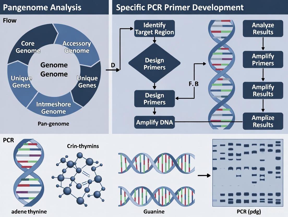
Pan-Genome Analysis Workflow
The typical workflow for computational pan-genome analysis involves multiple stages, from data preparation to biological interpretation. The following diagram illustrates this process with specific emphasis on applications for PCR primer development:
Diagram 1: Pan-genome analysis workflow for PCR primer development
Experimental Protocols for Diagnostic Marker Development
Protocol 1: Identification of Species-Specific Chromosomal Markers
This protocol outlines the procedure for identifying chromosomal markers specific to a target pathogen, based on the methodology successfully applied to Bacillus anthracis [4].
Materials and Reagents
Table 2: Essential Research Reagents for Pan-Genome Analysis
| Reagent/Resource | Specification | Function/Purpose |
|---|---|---|
| Bacterial Genomes | Complete genome sequences from public databases (NCBI) | Provides input data for comparative analysis |
| Prokka | Version 1.11 or higher | Rapid prokaryotic genome annotation |
| Roary | Version 3.13.0 or higher | Pan-genome analysis and gene clustering |
| BLAST+ | Version 2.12.0 or higher | Specificity validation of candidate markers |
| Perl/Python Scripts | Custom scripts for data filtering | Identification of strain-specific genes |
Step-by-Step Procedure
Genome Dataset Curation
- Download complete genomes of target species and closely related non-target species from NCBI
- For B. anthracis detection, include 50 genomes each of B. anthracis, B. cereus, and B. thuringiensis, plus one B. weihenstephanensis as outgroup [4]
- Ensure balanced representation across taxonomic groups
Genome Annotation
- Perform de novo annotation using Prokka with default parameters
- Command:
prokka --outdir [output_directory] --prefix [strain_name] [input_genome.fna] - Generate GFF3 files for all genomes for input to Roary
Pan-Genome Construction
- Execute Roary with Prokka annotations as input
- Command:
roary -e --mafft -p 8 -i 90 -cd 99 *.gff - Parameters: -i 90 (minimum percentage identity for BLASTP), -cd 99 (core definition threshold)
Identification of Species-Specific Genes
- Use custom Perl/Python scripts to extract genes present in all target species genomes but absent in non-target species
- Generate gene presence/absence matrix for manual verification
Specificity Validation
- Perform nucleotide BLAST (BLASTn) search of candidate genes against non-target genomes in NCBI
- Confirm absence of significant hits (e-value < 1e-10, identity >90%) in non-target species
- Validate presence across all available target species genomes using local BLAST alignment
Protocol 2: Development and Validation of Multiplex PCR Assays
This protocol describes the process for translating identified genetic markers into functional multiplex PCR assays for pathogen detection.
Materials and Reagents
- DNA Extraction Kits (commercial kits for bacterial genomic DNA isolation)
- PCR Reagents: Taq DNA polymerase, dNTPs, buffer solutions, MgCl₂
- Primer Synthesis: Custom primers designed from identified marker genes
- Agarose Gel Electrophoresis equipment or real-time PCR system
- Reference Strains: Target and non-target species for specificity testing
Step-by-Step Procedure
Primer Design
- Select 2-3 specific marker genes identified in Protocol 1
- Design primers with melting temperatures of 58-62°C, length 18-22 bp, and amplicon size 100-300 bp
- Ensure primer specificity by in silico PCR against non-target genomes
Multiplex PCR Optimization
- Test individual primer pairs separately before multiplexing
- Optimize primer concentrations (typically 0.1-0.5 µM each)
- Standard PCR conditions: initial denaturation 95°C for 2 min; 35 cycles of 95°C for 30s, 55-60°C for 30s, 72°C for 30s; final extension 72°C for 5 min
- Adjust annealing temperature and MgCl₂ concentration for optimal specificity
Analytical Specificity Testing
- Test multiplex PCR against DNA from target species (n=15-20 strains) and non-target species (n=40-50 strains)
- Include closely related species to verify absence of cross-reactivity
- For B. anthracis, include B. cereus and B. thuringiensis strains [4]
Sensitivity Determination
- Perform limit of detection (LOD) testing with serial dilutions of target DNA
- Determine minimum detectable DNA concentration (e.g., 10-100 fg/µL)
Application Testing
- Validate assay performance with spiked clinical or food samples
- Compare with conventional culture methods or established molecular tests
Data Analysis and Interpretation
Quantitative Pan-Genome Metrics
The following table summarizes key quantitative parameters for interpreting pan-genome analysis results, particularly in the context of primer development:
Table 3: Quantitative Parameters for Pan-Genome Analysis
| Parameter | Calculation/Definition | Interpretation | Application to Primer Development |
|---|---|---|---|
| Core Genome Size | Number of genes shared by 100% of genomes | Indicates genetic stability and essential functions | Avoid for species-specific detection; useful for broad-range assays |
| Pan-Genome Size | Total non-redundant genes across all genomes | Measures total gene repertoire | Larger pan-genomes offer more candidate markers |
| Heap's Law α-value | Power law parameter: n = kN^(-α) | α > 1 = closed pan-genome; α ≤ 1 = open pan-genome | Open pan-genomes may require ongoing marker validation as new strains are sequenced |
| Gene Frequency Distribution | Percentage of core, shell, and cloud genes | Reflects population diversity | Strain-specific (cloud) genes ideal for specific detection |
| Unique Genes per Genome | Average strain-specific genes | Measures individual strain uniqueness | Source of highly specific markers |
Case Study:Bacillus anthracisDetection
A recent study demonstrated the power of this approach by identifying 30 chromosome-encoded genes exclusive to B. anthracis through pan-genome analysis of 151 genomes [4]. Among these, 20 were located in known lambda prophage regions, while 10 represented newly discovered markers. The study established three distinct multiplex PCRs using genes BA1698, BA5354, and BA5361, which successfully detected diverse B. anthracis strains from Zambia and Mongolia while showing no cross-reactivity with closely related B. cereus and B. thuringiensis strains [4].
Pan-genome analysis provides a powerful framework for identifying genetic markers that enable specific detection of bacterial pathogens. The structured approach outlined in these application notes—from computational identification of strain-specific genes to experimental validation of multiplex PCR assays—offers researchers a validated pathway for developing robust diagnostic tools. This methodology is particularly valuable for distinguishing closely related species where conventional targets like 16S rRNA lack sufficient discriminatory power [3]. As sequencing technologies continue to advance and more genomes become available, pan-genome-driven approaches will play an increasingly important role in molecular diagnostics, vaccine development, and public health surveillance.
The use of a single, linear reference genome has long been the standard for genomic studies, including the critical task of PCR primer design. However, population-scale studies increasingly demonstrate that this approach creates systematic blind spots by collapsing natural genetic diversity into a single representative sequence [6]. A single reference genome inevitably omits alleles and sequence paths found in other individuals, leading to reference bias where reads from non-reference alleles map poorly or not at all [6]. This bias produces false negatives, skewed allele frequencies, and missed genotype-phenotype associations that undermine the reliability of molecular assays.
In primer design specifically, this limitation manifests as primers that fail to bind to target sequences in certain individuals or populations, exhibit reduced amplification efficiency, or produce non-specific binding to off-target regions [3] [4]. The fundamental problem is that designing primers against a single reference fails to account for the natural genetic variation present in real-world populations, resulting in assays with inconsistent performance across diverse samples.
The Fundamental Shortcomings of Single-Reference Primer Design
Conceptual Limitations and Technical Consequences
The traditional single-reference approach suffers from several interconnected limitations that directly impact primer efficacy:
Systematic Blind Spots: Single references necessarily collapse population-specific insertions, divergent haplotypes, and repetitive elements into one sequence [6]. This creates systematic blind spots, particularly in regions with high divergence or complex structure, leading to primers that cannot recognize missing variants.
Reference Bias: During alignment, sequences absent from the reference genome map poorly or not at all, producing false negatives and skewed allele frequencies [6]. This bias means that primers designed to variable regions may work optimally only for individuals closely matching the reference sequence.
Incomplete Variant Representation: Single references under-detect presence-absence variation (PAV) that removes entire genes in some individuals while introducing novel genes in others [6]. Similarly, copy-number variation (CNV) is misestimated when the reference lacks or misrepresents duplicated segments.
Practical Impacts on PCR Assay Development
These limitations translate directly to practical problems in molecular assay development:
Reduced Assay Robustness: Primers designed against a single reference may exhibit unpredictable performance across diverse samples, requiring extensive empirical optimization and potentially failing with specific variants [3].
False Results in Diagnostic Applications: In clinical diagnostics, single-reference designed primers can yield false negatives when target sequences contain polymorphisms at primer binding sites, or false positives through non-specific amplification of similar sequences [3] [4].
Inefficient Resource Utilization: The need for repeated optimization and validation of primers across different sample types increases time and resource expenditures in research and diagnostic development.
Pan-Genome Analysis: A Solution for Comprehensive Primer Design
Conceptual Framework of Pan-Genome Analysis
Pan-genome analysis addresses the limitations of single-reference approaches by capturing the full repertoire of sequences and variants across multiple individuals, separating genomic content into core elements (shared by almost all individuals) and accessory elements (variable between populations or strains) [6]. This comprehensive perspective enables researchers to distinguish between truly conserved genomic regions ideal for universal primer binding and variable regions that may require specialized primer sets for different variants.
A pan-genome can be represented as a graph-based reference that replaces the single linear sequence with a network of paths representing alternate alleles, insertions, deletions, and complex structural variants in a unified coordinate system [6]. This approach fundamentally transforms primer design by providing a complete map of genetic variation within a target species or population.
Quantitative Advantages of Pan-Genome Approaches
Table 1: Comparative Performance of Single Reference vs. Pan-Genome Approaches
| Parameter | Single Reference Genome | Pan-Genome Approach | Improvement |
|---|---|---|---|
| Sequence Coverage | Limited to reference sequence and closely related variants | Expands to include population-specific sequences and structural variants | Adds 119 million base pairs of euchromatic polymorphic sequences and 1,115 gene duplications in human pangenome [7] |
| Variant Detection | Under-represents structural variants and presence-absence variations | Comprehensive variant catalog including PAV, CNV, and complex rearrangements | 104% increase in structural variants detected per haplotype compared to GRCh38 [7] |
| CpG Site Identification | Limited to reference-compatible sites | Expanded detection across diverse haplotypes | 7.4% more CpGs called genome-wide using T2T-CHM13 vs. GRCh38 [8] |
| Primer Specificity | Specificity checked against limited reference context | Specificity validated across full spectrum of known variation | Enables development of primers with 100% specificity for target serotypes [3] |
| Cross-Population Applicability | Biased toward reference population | Balanced representation across diverse haplotypes | Identifies cross-population and population-specific unambiguous probes [8] |
Table 2: Pan-Genome Analysis Tools for Primer Design
| Tool | Primary Function | Advantages | Limitations |
|---|---|---|---|
| Roary | Pan-genome visualization for prokaryotes | Fast and efficient; visualization of output data | Limited to bacterial genomes; lower sensitivity in highly divergent genomes [3] |
| BPGA (Bacterial Pan Genome Analysis pipeline) | Functional annotation and orthologous group clustering | Identification of functional insight; ease of use | Limited scalability; demands high-quality genome assemblies [3] |
| Panaroo | Pan-genome construction with error correction | Effective error correction mechanisms; retains sequence continuity | Limited to prokaryotic genomes [9] |
| PGAP-X | Whole-genome alignments and genetic variation analysis | High scalability; suitable for large datasets and customization | High computational demand; requires advanced bioinformatics skills [3] |
| varVAMP | Primer design from multiple sequence alignments | Designed specifically for pan-specific primer design; handles high diversity | Primarily focused on viral pathogens [10] |
Experimental Protocols for Pan-Genome Informed Primer Design
Protocol 1: Identification of Species-Specific Markers Through Pan-Genome Analysis
This protocol outlines the process for identifying species-specific chromosomal markers for highly specific PCR detection, based on the approach successfully used for Bacillus anthracis detection [4].
Materials and Reagents
- High-quality genome assemblies for target and related species
- Computing infrastructure with sufficient memory and storage
- Prokka annotation software (v1.11 or higher)
- Roary pan-genome analysis tool (v3.13.0 or higher)
- BLAST+ suite for sequence similarity search
- Perl or Python environment for custom script execution
Methodology
- Dataset Curation: Collect complete genomes from NCBI for the target species and closely related species. Include an outgroup species for comparison. For the B. anthracis study, 151 complete genomes were used (50 each of B. anthracis, B. cereus, and B. thuringiensis, plus one B. weihenstephanensis as an outgroup) [4].
Genome Annotation: Perform de novo annotation of all genomes using Prokka with default parameters. This ensures consistent annotation across all sequences regardless of their original annotation status.
Pan-Genome Construction: Execute Roary using the Prokka annotations as input with standard parameters. Roary will generate a gene presence-absence spreadsheet that forms the basis for identifying unique genes.
Identification of Exclusive Genes: Use custom Perl or Python scripts to parse the Roary output and identify genes present in all target species strains but absent in related species genomes.
Specificity Validation: Submit each candidate gene to a nucleotide BLAST (BLASTn) search against the entire NCBI database, excluding the target species, to verify absence from non-target organisms.
Consistency Verification: Perform local BLAST alignment against a comprehensive set of target species genomes to confirm consistent presence across diverse strains.
Primer Design: Select validated unique genes as targets and design primers using standard tools such as Primer-BLAST, with verification of specificity against the pan-genome data.
Expected Results and Interpretation This protocol successfully identified thirty chromosome-encoded genes specific to B. anthracis [4]. Twenty were located in known lambda prophage regions, while ten were in previously undefined chromosomal regions. Three of these genes (BA1698, BA5354, and BA5361) were used to establish multiplex PCR assays that accurately distinguished B. anthracis from closely related species.
Protocol 2: Development of Pan-Specific Primers for Diverse Viral Pathogens
This protocol describes an approach for designing pan-specific primers capable of detecting diverse viral genotypes, based on methods developed for poliovirus and other highly variable viruses [10].
Materials and Reagents
- Representative viral sequences covering known genetic diversity
- Multiple sequence alignment tool (MAFFT v7.526 or higher)
- varVAMP primer design tool
- EMBOSS tool suite for sequence manipulation
- Standard PCR reagents for experimental validation
Methodology
- Sequence Collection: Compile a comprehensive set of viral genome sequences representing the full genetic diversity of the target virus. For poliovirus, this included representatives of all three serotypes with approximately 70% pairwise sequence identity [10].
Sequence Degapping (if necessary): If starting with pre-aligned sequences, use the EMBOSS
degapseqtool to remove alignment gaps and recover original sequences.Multiple Sequence Alignment: Perform multiple sequence alignment using MAFFT with the FFT-NS-2 (fast, progressive method) algorithm. This balances speed and accuracy for large viral datasets.
Pan-Specific Primer Design: Execute varVAMP using the multiple sequence alignment as input. Set parameters according to experimental needs:
- For qPCR: Design two primers and one probe
- For tiled-amplicon sequencing: Define amplicon size based on sequencing technology
- Set conservation thresholds based on required breadth of detection
Specificity Verification: Validate candidate primers in silico against comprehensive sequence databases and check for off-target binding potential.
Experimental Validation: Test primer performance against representative viral strains spanning the genetic diversity, quantifying sensitivity and specificity empirically.
Expected Results and Interpretation This approach enables development of primer sets capable of amplifying highly diverse viral sequences. For poliovirus, which shows approximately 70% sequence identity across serotypes, this method successfully identified conserved regions suitable for pan-specific detection [10]. The resulting primers provide broader detection capability compared to those designed using single reference sequences.
Workflow Visualization: Pan-Genome Informed Primer Design
Case Studies: Successful Applications of Pan-Genome Informed Primer Design
Bacterial Pathogen Detection and Differentiation
Table 3: Case Studies in Pan-Genome Informed Primer Design for Bacterial Detection
| Species | Pan-Genome Tool | Target Genes | Specificity Achieved | Application Validation |
|---|---|---|---|---|
| Salmonella Montevideo | panX | Species-specific genes | High sensitivity and selectivity for target serovar | Food samples (raw chicken, peppers) [3] |
| Salmonella E serogroup | Roary (v3.11.2) | Serogroup-specific markers | Specific detection of E serogroup | Artificially contaminated foods [3] |
| Salmonella Infantis | BPGA (v1.3) | SIN_02055 | 100% accuracy for target serovar | 60 Salmonella serovars profiled [3] |
| Bacillus anthracis | Roary | BA1698, BA5354, BA5361 | Specific distinction from B. cereus and B. thuringiensis | 62 bacterial strains tested [4] |
| Acinetobacter baumannii | Panaroo + Ptolemy | Beta-lactam resistance genes | Identification of novel plasmid structures | 70 clinical isolates [9] |
The application of pan-genome analysis for Salmonella detection demonstrates the flexibility of this approach for targeting different taxonomic levels. Researchers identified gene targets for Salmonella enterica serovar Montevideo through pan-genome analysis of 706 S. enterica strains, including 23 strains of S. Montevideo [3]. The resulting primer-probe sets showed significantly improved detection capability in challenging food matrices like red pepper and black pepper compared to conventional culture methods.
Similarly, for Bacillus anthracis, pan-genome analysis of 151 genomes identified thirty chromosome-encoded genes specific to this pathogen, enabling the development of multiplex PCR assays that accurately distinguish it from closely related B. cereus and B. thuringiensis strains [4]. This addresses a critical diagnostic challenge where plasmid-based detection methods fail with plasmid-deficient strains, and previously described chromosomal markers have shown cross-reactivity with other species.
Beneficial Microorganisms and Agricultural Applications
Pan-genome approaches have also proven valuable for detecting beneficial microorganisms such as Lactobacillus species used in food fermentation and probiotics [3]. Additionally, in agricultural contexts, pan-genome analysis of Malus species (apple) enabled the development of molecular markers for disease resistance traits, leveraging the graph-based pan-genome to capture shared and species-specific structural variations [11].
These applications demonstrate how pan-genome informed primer design supports not only pathogen detection but also the identification and characterization of beneficial microorganisms in food products and agricultural settings.
Essential Research Reagents and Computational Tools
Table 4: Research Reagent Solutions for Pan-Genome Informed Primer Design
| Reagent/Tool | Function | Application Notes |
|---|---|---|
| Roary | Rapid pan-genome visualization for prokaryotes | Ideal for bacterial species; uses pre-clustering approach for efficiency [3] [9] |
| BPGA Pipeline | Functional annotation and orthologous group clustering | Incorporates functional insights; user-friendly interface [3] |
| Primer-BLAST | Primer design with specificity checking | Integrates with NCBI databases; combines Primer3 with BLAST [12] [13] |
| varVAMP | Pan-specific primer design from MSAs | Specialized for highly diverse viral pathogens [10] |
| MAFFT | Multiple sequence alignment | Creates alignments for diverse sequences; essential for varVAMP input [10] |
| Prokka | Rapid prokaryotic genome annotation | Provides consistent annotations for pan-genome analysis [4] |
| Panaroo | Pan-genome construction with error correction | Effective annotation error correction; maintains sequence continuity [9] |
The limitations of traditional single-genome references for primer design are both conceptual and practical, resulting in assays with inherent biases and inconsistent performance across diverse populations. Pan-genome analysis addresses these limitations by providing a comprehensive map of genetic variation within target species, enabling the design of primers with enhanced specificity, sensitivity, and cross-population applicability.
The case studies presented demonstrate that pan-genome informed primer design successfully supports a wide range of applications, from foodborne pathogen detection to clinical diagnostics and agricultural improvement. As sequencing technologies continue to advance and computational tools become more accessible, pan-genome approaches will likely become standard practice for molecular assay development.
Future developments in pan-genome methodologies, including improved graph reference formats, more efficient computational algorithms, and enhanced integration with primer design tools, will further streamline the process of developing robust, population-aware PCR assays. This evolution represents a necessary paradigm shift from a one-size-fits-all approach to precision primer design that accounts for the rich tapestry of natural genetic diversity.
The Critical Shift from 16S rRNA to Pan-Genome-Derived Markers
For decades, the 16S ribosomal RNA (rRNA) gene has served as the gold standard for bacterial identification and taxonomic classification [14]. This conserved gene region has enabled researchers to profile complex microbial communities and establish phylogenetic relationships across bacterial species. However, the advent of high-throughput sequencing and comparative genomics has revealed significant limitations in 16S rRNA-based approaches. Studies have demonstrated that the 16S rRNA gene often lacks sufficient resolution to distinguish between closely related bacterial species and strains, leading to false-positive identifications in diagnostic applications [3] [15]. The gene's conserved nature, while useful for broad phylogenetic analysis, prevents discrimination of recently diverged lineages that may possess dramatically different pathogenic potentials or metabolic capabilities.
The fundamental problem stems from genetic similarity among organisms that differ markedly in phenotype. As noted in studies of foodborne pathogens, "primers targeting the 16S rRNA region have been conventionally employed in PCR analyses [but] several studies have highlighted limitations and false-positive results" [3]. This resolution problem is particularly acute in clinical and diagnostic settings where accurate identification to the strain level can directly impact patient outcomes and public health responses. Furthermore, research has shown that single-nucleotide substitutions exist between intragenomic copies of the 16S gene within the same organism, creating additional challenges for accurate strain-level discrimination [15]. These limitations have prompted a paradigm shift toward pan-genome-derived markers that offer superior specificity and resolution for bacterial detection and characterization.
Pan-Genome Analysis: A New Paradigm for Marker Discovery
Conceptual Framework and Definitions
The pan-genome represents the full complement of genes found across all individuals within a defined taxonomic group, encompassing both shared and variable genomic content [16]. This concept, first introduced by Tettelin et al. in 2005, recognizes that a single reference genome cannot capture the complete genetic diversity of a species [14] [16]. The pan-genome is typically divided into three core components: (1) the core genome - genes present in all individuals; (2) the shell genome - genes found in most but not all individuals; and (3) the cloud genome - genes present in only a few individuals [17]. This classification system provides a powerful framework for understanding bacterial evolution, niche adaptation, and functional diversity.
From a practical standpoint, the pan-genome concept enables researchers to identify genetic elements unique to specific pathogens, lineages, or phenotypic traits. By comparing entire genomic repertoires rather than single genes, pan-genome analysis facilitates the discovery of highly specific markers that can distinguish even closely related bacterial strains. This approach has proven particularly valuable for distinguishing pathogenic from non-pathogenic variants within the same species complex, as demonstrated in studies of Bacillus cereus group organisms where traditional markers failed to provide sufficient discrimination [4].
Comparative Analysis: 16S rRNA vs. Pan-Genome-Derived Markers
Table 1: Quantitative comparison between 16S rRNA and pan-genome-derived markers
| Characteristic | 16S rRNA Markers | Pan-Genome-Derived Markers |
|---|---|---|
| Taxonomic Resolution | Limited to genus/species level [15] | Species/strain level [3] [4] |
| Discriminatory Power | 56% of V4 amplicons fail species-level classification [15] | 100% specificity demonstrated for multiple pathogens [3] [4] |
| Genetic Basis | Single gene with variable regions | Multiple unique genes/genomic regions |
| Detection Accuracy | Prone to false positives due to conservation [3] | High specificity; minimal cross-reactivity |
| Application Flexibility | Limited to broad classification | Customizable for specific serotypes/virulence strains [3] |
| Representation of Diversity | Partial (~1500 bp) | Comprehensive (entire gene repertoire) |
The limitations of 16S rRNA become particularly evident when examining its performance across different bacterial taxa. Research has demonstrated that "the V4 region performed worst, with 56% of in-silico amplicons failing to confidently match their sequence of origin" at the species level [15]. Different variable regions also exhibit taxonomic biases, with certain regions performing poorly for specific bacterial groups. For instance, the V1-V2 region shows limited resolution for Proteobacteria, while V3-V5 struggles with Actinobacteria classification [15].
In contrast, pan-genome-derived markers leverage the full genomic diversity of bacterial species, enabling the development of highly specific detection assays. For example, in a study targeting Salmonella enterica serovar Montevideo, pan-genome analysis of 706 S. enterica strains enabled the development of primer-probe sets that demonstrated high sensitivity and selectivity in complex food matrices [3]. Similarly, research on Bacillus anthracis identified 30 chromosome-encoded genes exclusively present in this pathogen, enabling specific detection that distinguishes it from closely related B. cereus and B. thuringiensis strains [4].
Bioinformatics Workflow for Pan-Genome-Based Marker Discovery
Computational Tools and Pipelines
The identification of specific markers through pan-genome analysis relies on a suite of bioinformatics tools that facilitate genome comparison, ortholog identification, and unique gene discovery. Multiple software options exist with complementary strengths and applications. Roary represents a widely-used tool for rapid pan-genome analysis, particularly suitable for prokaryotic genomes, though it may exhibit reduced sensitivity with highly divergent sequences [3]. The Bacterial Pan Genome Analysis (BPGA) pipeline incorporates functional annotation and orthologous group clustering, providing valuable insights for marker selection [3]. More recently developed tools like PGAP2 offer enhanced accuracy through fine-grained feature analysis and constrained regional strategies, improving ortholog identification across diverse datasets [5].
Table 2: Bioinformatics tools for pan-genome analysis and their applications
| Tool | Primary Function | Advantages | Limitations |
|---|---|---|---|
| Roary | Pan-genome visualization | Fast, efficient for prokaryotes | Lower sensitivity in highly divergent genomes [3] |
| BPGA | Functional annotation & ortholog clustering | Ease of use, functional insights | Limited scalability [3] |
| PGAP2 | Ortholog identification via fine-grained feature analysis | High precision, robust with large datasets | High computational demand [5] |
| EDGAR | Comparative genomics & visualization | Intuitive web interface | Limited to small genome sets [3] |
| panX | Phylogenetic & genomic integration | Interactive visualization, evolutionary context | Limited scalability [3] |
The selection of appropriate tools depends on the specific research objectives, dataset size, and desired level of analysis. For large-scale studies involving thousands of genomes, PGAP2 offers superior performance in ortholog identification, while smaller datasets may be effectively analyzed using BPGA or Roary depending on the need for functional annotation or visualization capabilities [3] [5].
Experimental Protocol: From Genomes to Specific Markers
Protocol: Pan-genome analysis for specific marker discovery
Step 1: Data acquisition and quality control
- Obtain complete genome sequences for target and reference strains from public databases (NCBI, ENA)
- Perform quality assessment using FastQC or similar tools
- For PGAP2: Designate representative genome based on gene similarity; classify outliers using Average Nucleotide Identity (ANI < 95%) or unique gene counts [5]
Step 2: Genome annotation and ortholog identification
- Annotate genomes using Prokka [4] or similar annotation pipelines
- Identify orthologous groups using OrthoFinder [16] or pan-genome analysis tools
- For BPGA: Utilize built-in orthologous clustering algorithms [3]
Step 3: Pan-genome profiling and unique gene identification
- Generate presence/absence matrix of gene families across all strains
- Calculate frequency of each orthogroup across samples:
frequency = sum(presence)/number_of_genomes[16] - Classify genes into categories: Core (frequency = 1), Softcore (frequency ≥ 0.9), Dispensable (1 < frequency < 0.9), Private (frequency = 1/numberofgenomes) [16]
- Identify target-specific genes using custom Perl or Python scripts to extract genes present in all target strains but absent from non-target strains [4]
Step 4: Specificity validation and marker selection
- Verify specificity of candidate markers using BLASTN against non-target genomes [4]
- Select multiple markers (3-5 candidates) for experimental validation
- Consider genomic context, avoiding mobile genetic elements when possible
Step 5: Primer design and in silico validation
- Design primers using standard tools (Primer3, BLAST)
- Validate specificity in silico against comprehensive database
- Optimize primer parameters for compatibility with intended detection platform (qPCR, LAMP, etc.)
Application Notes: Case Studies in Pathogen Detection
Specific Detection of Bacillus anthracis
The challenge of distinguishing Bacillus anthracis from closely related B. cereus and B. thuringiensis represents a compelling case study in the application of pan-genome-derived markers. Traditional methods relying on plasmid-encoded virulence factors proved inadequate due to potential plasmid loss or transfer between species [4]. Similarly, previously described chromosomal markers such as BA813 were subsequently found in B. cereus strains, resulting in false positives [4].
In this study, researchers analyzed 151 complete genomes (50 each of B. anthracis, B. cereus, and B. thuringiensis, plus one B. weihenstephanensis as an outgroup) using a comprehensive pan-genome approach [4]. Genomes were annotated with Prokka, and pan-genome analysis was performed with Roary to generate a gene presence/absence matrix. Through comparative analysis, thirty chromosome-encoded genes exclusively present in B. anthracis were identified. Of these, twenty were located in known lambda prophage regions, while ten represented novel discoveries from previously undefined chromosomal regions [4].
Three genes (BA1698, BA5354, and BA5361) were selected for multiplex PCR development, resulting in three distinct assays that accurately identified all B. anthracis strains while showing no cross-reactivity with other Bacillus species [4]. This approach demonstrated 100% specificity across 62 bacterial strains, including geographically and temporally diverse B. anthracis isolates from Zambia and Mongolia [4].
Targeted Detection of Salmonella Serovars
Salmonella enterica comprises over 2600 serovars with varying host specificities, pathogenic potential, and phenotypic characteristics. Pan-genome analysis has enabled the development of detection methods targeting specific serovars of public health concern. In one study, researchers utilized the panX tool to analyze 706 S. enterica strains, including 23 strains of S. Montevideo [3]. This analysis identified unique gene targets that enabled the development of primer-probe sets for specific detection of this serovar.
The resulting real-time PCR assays demonstrated superior performance compared to conventional culture methods using XLD media, particularly in challenging food matrices such as raw chicken meat, red pepper, and black pepper [3]. In a separate study, BPGA-based analysis of 60 Salmonella serovars identified the SIN_02055 gene as a specific marker for S. Infantis, enabling detection with 100% accuracy [3]. These examples highlight the flexibility of pan-genome analysis in developing detection methods targeting either multiple serovars or individual high-risk strains.
Table 3: Research reagent solutions for pan-genome-based marker development
| Resource Category | Specific Tools/Reagents | Application & Function |
|---|---|---|
| Bioinformatics Software | Roary, BPGA, PGAP2, OrthoFinder | Pan-genome construction, ortholog identification, phylogenetic analysis |
| Genome Annotation | Prokka, PGAP | Automated annotation of bacterial genomes |
| Primer Design & Validation | Primer3, BLAST, varVAMP | In silico design and specificity testing of PCR primers |
| Reference Databases | NCBI GenBank, ENA, Species-specific databases | Source of genomic data for comparative analysis |
| Laboratory Validation | qPCR reagents, Multiplex PCR kits, DNA extraction kits | Experimental confirmation of marker specificity |
| Programming Environments | R, Python with BioPython, Perl | Custom scripts for data analysis and visualization |
The transition from 16S rRNA to pan-genome-derived markers represents a fundamental advancement in microbial detection and characterization. This paradigm shift addresses the critical need for specific identification of pathogens at the strain level, enabling more accurate diagnostics, improved outbreak investigations, and enhanced surveillance capabilities. The case studies presented demonstrate the practical application of this approach across diverse bacterial pathogens, with consistent improvements in specificity and reliability compared to traditional methods.
Future developments in pan-genome analysis will likely focus on several key areas. First, the increasing availability of high-quality genome assemblies will enhance the resolution of pan-genome maps, particularly for underrepresented taxonomic groups. Second, improvements in computational efficiency will enable real-time pan-genome analysis for rapid response during outbreak situations. Finally, integration of machine learning approaches may facilitate the automated identification of optimal marker sets for specific detection scenarios. As these technical advances mature, pan-genome-derived markers are poised to become the new gold standard for microbial detection across clinical, food safety, and public health applications.
Open vs. Closed Pan-Genomes and Their Impact on Assay Design
In the fields of molecular biology and genetics, a pan-genome represents the entire set of genes from all strains within a clade, serving as the union of all genomes for a given taxonomic group [1]. This concept has fundamentally shifted genomic analysis from a single linear reference to a comprehensive framework that captures the full genetic repertoire of a species [18]. The pan-genome is typically partitioned into three components: the core genome (genes present in all individuals), the shell genome (genes present in two or more but not all strains), and the cloud genome (genes unique to single strains, also known as the accessory or dispensable genome) [1]. This classification provides critical insights for designing molecular assays, particularly for pathogen detection and typing.
The distinction between open and closed pan-genomes represents a fundamental principle with direct implications for assay design [1]. Species with a closed pan-genome reach a point where sequencing additional genomes adds few or no new genes, making the total gene repertoire predictable. In contrast, species with an open pan-genome continue to accumulate new genes with each additional sequenced genome, presenting ongoing challenges for comprehensive assay development [18]. This classification is mathematically determined using Heaps' law ((N=kn^{-α})), where (α > 1) indicates a closed pan-genome and (α ≤ 1) indicates an open pan-genome [1].
Pan-Genome Openness and Its Experimental Implications
Characteristics of Open and Closed Pan-Genomes
Table 1: Characteristics of Open vs. Closed Pan-Genomes
| Feature | Open Pan-Genome | Closed Pan-Genome |
|---|---|---|
| Gene Discovery Rate | New genes continue to be added with each sequenced genome | Gene number stabilizes; few new genes added after sufficient sampling |
| Mathematical Parameter (α) | α ≤ 1 | α > 1 |
| Typical Ecological Niche | Diverse environments, sympatric lifestyle | Restricted niche, host-restricted or specialist |
| Horizontal Gene Transfer | Frequent | Limited |
| Examples | Escherichia coli (89,000 gene families), Alcaligenes sp., Serratia sp. [1] | Streptococcus pneumoniae, Staphylococcus lugdunensis [1] |
| Impact on Assay Design | Requires broader target selection; ongoing surveillance needed | More stable target selection; comprehensive coverage achievable |
Practical Implications for PCR-Based Detection
The openness or closure of a pathogen's pan-genome directly influences the strategy for developing molecular detection assays. For species with closed pan-genomes, researchers can design PCR assays with greater confidence that the targets will remain relevant across most strains. After analyzing a sufficient number of genomes (which varies by species), the core genome stabilizes, allowing for the selection of conserved targets that will likely detect future isolates [1]. For example, Streptococcus pneumoniae exhibits a closed pan-genome where the predicted number of new genes drops to zero after sequencing approximately 50 genomes [1].
In contrast, for species with open pan-genomes like Escherichia coli, the continuous discovery of new genes with each sequenced genome complicates assay design [1]. These species typically undergo frequent horizontal gene transfer, leading to substantial variation in gene content. Detection assays for such pathogens must either target multiple conserved regions or focus on highly stable core genes that remain despite the ongoing genomic flux. This necessitates ongoing surveillance and potential updates to detection protocols as new strains emerge.
Pan-Genome Analysis Tools for Assay Development
Table 2: Bioinformatics Tools for Pan-Genome Analysis in Assay Development
| Tool | Primary Function | Advantages | Limitations | Applicability to Assay Design |
|---|---|---|---|---|
| Roary | Rapid pan-genome analysis pipeline | Fast, efficient visualization | Lower sensitivity with highly divergent genomes; limited to bacteria [3] | Quick identification of core genes for broad-specificity assays [3] |
| BPGA (Bacterial Pan Genome Analysis Pipeline) | Functional annotation and orthologous group clustering | Ease of use; functional insights | Limited scalability; requires high-quality assemblies [3] | Linking target genes to functional traits for diagnostic development [3] |
| PGAP2 | Pan-genome analysis based on fine-grained feature networks | High precision; robust with large datasets; quantitative outputs | Requires computational expertise [5] | Large-scale target identification across thousands of genomes [5] |
| panX | Phylogenetic and genomic visualization | Interactive visualization; evolutionary context | Limited scalability [3] | Serotype-specific target identification [3] |
| Panaroo | Pan-genome analysis with error correction | Handles assembly errors; graph-based output | Moderate computational demand [9] | Accurate identification of core genes from diverse datasets [9] |
| EasyPrimer | Pan-PCR/HRM primer design | User-friendly; identifies conserved regions flanking variable segments | Web-based with dependency on interface [19] | Direct primer design for strain discrimination [19] |
Workflow for Pan-Genome Informed Assay Design
The following diagram illustrates the comprehensive workflow for designing pan-genome-informed detection assays:
Experimental Protocols for Pan-Genome Informed Primer Design
Protocol 1: Core Gene Identification for Broad-Specificity Detection
Objective: Identify conserved core genes suitable for PCR-based detection of a target species across diverse strains.
Materials:
- Genomic sequences of multiple strains (minimum 10-15 for preliminary analysis)
- Bioinformatics tools: Roary, PGAP2, or Panaroo
- Computing resources (Linux workstation or cluster)
Procedure:
- Data Collection and Curation: Collect complete or draft genome sequences for representative strains of the target species from public databases (NCBI, PATRIC). Ensure data quality by filtering for contamination and completeness [5].
Pan-Genome Calculation:
- Input genome annotations (GFF3 format) and sequences (FASTA format) into the selected pan-genome analysis tool.
- For Roary: Execute with default parameters initially (
roary -f output_dir -e -n *.gff). - For PGAP2: Use the integrated quality control to identify outliers before core gene analysis [5].
Core Gene Identification:
- Extract the list of core genes (present in ≥95% or 99% of strains) from the tool output.
- Sort core genes by sequence conservation (% identity across strains).
- Prioritize genes with functional annotations relevant to detection goals (e.g., essential metabolic genes).
Target Validation:
- Perform multiple sequence alignment of candidate core genes across strains.
- Identify conserved regions suitable for primer binding (typically ≥18-25 bp with 100% conservation).
- Verify specificity by BLAST analysis against non-target genomes.
Expected Output: A ranked list of core genes with conserved regions suitable for broad-specificity detection assay design.
Protocol 2: Accessory Gene Profiling for Strain Discrimination
Objective: Identify accessory genes that differentiate strains within a species for typing applications.
Materials:
- Genomic sequences of target strains
- Pan-genome analysis tool with accessory gene output (Roary, PIRATE, Panaroo)
- Primer design software (Primer3, EasyPrimer)
Procedure:
- Pan-Genome Profiling:
- Compute pan-genome using selected tool with focus on accessory gene identification.
- Use clustering algorithms (e.g., CD-HIT as implemented in pan-PCR) to group accessory genes [20].
Discriminatory Gene Selection:
- Apply greedy approximation algorithm (as in pan-PCR) to select minimal gene set that maximizes strain discrimination [20].
- For 10 strains, approximately 3-4 targets are theoretically sufficient (log₂N principle).
- Filter out mobile genetic elements (phage, transposons) unless specifically targeted.
Multiplex Primer Design:
- Design primers for each target gene with different product sizes for multiplex PCR.
- Use tools like EasyPrimer to identify conserved regions flanking variable segments in alignments [19].
- For HRM applications, target regions with single-nucleotide polymorphisms that alter melting temperature.
In Silico Validation:
- Verify primer specificity against the input genomes.
- Check for potential primer-dimer formations in multiplex configurations.
- Predict amplicon sizes and melting temperatures for HRM applications.
Expected Output: A multiplex PCR or HRM assay targeting accessory genes that can differentiate strains within the target species.
Case Studies in Pathogen Detection
Salmonella Serotyping Using Pan-Genome Analysis
Salmonella enterica represents a species with significant diversity, requiring sophisticated detection approaches. Researchers have successfully applied pan-genome analysis to develop precise detection assays for various Salmonella serovars [3]:
Methodology:
- For Salmonella Montevideo: Used panX tool to analyze 706 S. enterica strains (including 23 S. Montevideo). Identified serovar-specific gene targets and developed primer-probe sets for real-time qPCR [3].
- For Salmonella E serogroup: Applied Roary to identify genetic targets specific to serogroup E (Weltevreden, London, Meleagridis, Senftenberg). Validated primers in artificially contaminated food samples using conventional PCR [3].
- For 60 Salmonella serovars: Employed BPGA to design unique gene markers for 60 common serovars. Verified detection accuracy through real-time PCR with 100% specificity for targeted serovars [3].
Results: All studies demonstrated that pan-genome informed primer design provided superior specificity compared to conventional 16S rRNA-based approaches. The S. Montevideo assay successfully detected targets in challenging food matrices like black pepper and red pepper, where conventional culture methods face limitations [3].
Klebsiella pneumoniae Typing via HRM
Klebsiella pneumoniae represents a pathogen with significant strain diversity requiring discrimination below the species level. Researchers developed an HRM typing scheme using the hypervariable wzi gene [19]:
Methodology:
- Gene Selection: Selected wzi gene for its high variability and phylogenetic relevance.
- Primer Design: Used EasyPrimer to identify conserved regions flanking variable segments in a wzi gene alignment.
- Assay Development: Designed two primer pairs targeting different variable regions of wzi.
- Validation: Tested against 17 K. pneumoniae strains from different sequence types (STs) and compared to MLST-based HRM.
Results: The wzi-based HRM scheme demonstrated comparable discriminatory power to an 8-primer MLST HRM scheme while requiring only two primer pairs. The assay successfully reconstructed a nosocomial outbreak, correctly clustering outbreak strains and distinguishing non-outbreak strains. This approach reduced typing time from days (for traditional MLST) to under five hours [19].
The Scientist's Toolkit: Essential Research Reagents and Materials
Table 3: Essential Research Reagents for Pan-Genome Informed Assay Development
| Category | Specific Items | Function/Application | Examples/Specifications |
|---|---|---|---|
| Bioinformatics Tools | Roary, PGAP2, Panaroo, BPGA | Pan-genome calculation and visualization | Roary for rapid prokaryotic pan-genome analysis; PGAP2 for large-scale datasets [3] [5] |
| Primer Design Software | Primer3, varVAMP, EasyPrimer | Primer and probe selection | EasyPrimer for identifying conserved regions in alignments [19]; varVAMP for viral primer schemes [10] |
| Sequence Alignment | MAFFT, MUSCLE | Multiple sequence alignment | MAFFT with FFT-NS-2 algorithm for progressive alignment [10] |
| Specificity Verification | BLAST, VSEARCH | In silico validation of primer specificity | BLAST against NT database for off-target binding check [20] |
| Laboratory Reagents | DNA Polymerase, dNTPs, Buffer Systems | PCR amplification | Polymerase with high fidelity for accurate amplification |
| Detection Chemistry | SYBR Green, TaqMan Probes, HRM Dyes | Signal detection in real-time PCR | Intercalating dyes for HRM; hydrolysis probes for specific detection [19] |
| Positive Controls | Genomic DNA from reference strains | Assay validation and quality control | Well-characterized strains representing target diversity |
The classification of pan-genomes as open or closed provides a critical framework for designing molecular detection assays. For species with closed pan-genomes, stable and comprehensive assays can be developed with relative confidence, while open pan-genome species require more flexible approaches that accommodate ongoing genetic diversity. The integration of pan-genome analysis into assay development workflows enables researchers to make informed decisions about target selection, ultimately leading to more robust and reliable detection methods. As pan-genome analysis tools continue to evolve, particularly with advancements in graph-based representations and long-read sequencing, the precision and efficiency of molecular assay development will continue to improve, supporting enhanced pathogen detection, typing, and surveillance capabilities across diverse research and clinical applications.
Pan-genome analysis represents a paradigm shift in genomic studies, moving beyond the limitations of single reference genomes to encompass the complete gene repertoire of a species. The pan-genome is categorized into three components: the core genome, consisting of genes shared by all strains; the accessory genome, containing genes present in two or more but not all strains; and the unique genome, comprising strain-specific genes [21]. This comprehensive approach is particularly powerful for understanding genetic diversity, evolutionary dynamics, and specialized adaptations in bacterial populations [3]. In recent years, pan-genome analysis has found valuable applications in molecular diagnostics and detection assay development, enabling researchers to identify unique genetic targets for highly specific PCR primer design [3] [22]. This methodology offers significant advantages over traditional approaches that target conserved regions like 16S rRNA, which have been associated with false-positive and false-negative results due to insufficient discriminatory power [3] [22].
The development of specialized bioinformatics tools has been instrumental in facilitating robust pan-genome analyses. Among the numerous available platforms, Roary, BPGA, PGAP-X, and panX have emerged as prominent solutions, each with distinct algorithmic approaches and functional capabilities. These tools enable researchers to process multiple genome sequences, identify core and accessory genetic elements, and extract targets for diagnostic applications. This article provides a comprehensive technical overview of these four essential tools, focusing on their application within the context of developing specific PCR primers for detecting microorganisms in research and diagnostic settings.
Tool Specifications and Comparative Analysis
Table 1: Technical Specifications and Primary Applications of Pan-Genome Analysis Tools
| Tool | Primary Function | Core Algorithm | Input Formats | Execution Speed | Key Outputs |
|---|---|---|---|---|---|
| Roary | Pan-genome visualization & core genome analysis | Pre-clustering approach (fast) | GFF3 files | Fast, efficient for prokaryotes [3] | Core/accessory gene sets, phylogenetic trees [3] |
| BPGA | Comprehensive pan-genome analysis with functional annotation | USEARCH (default), CD-HIT, OrthoMCL [23] | GenBank, FASTA, binary matrix [23] | Ultra-fast pipeline [23] | Pan/core genome plots, COG/KEGG mappings, phylogenies [3] [23] |
| PGAP-X | Whole-genome alignment & genetic variation analysis | Scalable, modular architecture [3] | Not specified in results | High computational demand [3] | Core/accessory genes, whole-genome alignments, functional annotation [3] |
| panX | Phylogenetic & genomic analysis with interactive visualization | Integration of phylogenetic and genomic data [3] | Not specified in results | Limited scalability [3] | Interactive pan-genome visualization, phylogenetic trees [3] |
Table 2: Advantages, Limitations, and Suitability for PCR Primer Development
| Tool | Advantages | Limitations | Primer Design Applications |
|---|---|---|---|
| Roary | Fast and efficient; visualization of output data [3] | Limited to bacterial genomes; lower sensitivity with highly divergent genomes [3] | Identification of core genes for broad-specificity primers; used for Salmonella serogroup detection [3] |
| BPGA | Ease of use; functional insights; multiple downstream analyses [3] [23] | Limited scalability; requires high-quality genome assemblies [3] | Marker development for specific serovars (e.g., Salmonella Infantis); functional annotation of targets [3] |
| PGAP-X | High scalability; suitable for large datasets and customization [3] | High computational demand; requires advanced bioinformatics expertise [3] | Handling large-scale comparative genomics for target identification [3] |
| panX | Interactive visualization; combination of evolutionary context with genomic insight [3] | Limited scalability [3] | Visual identification of conserved regions; used for Salmonella Montevideo primer design [3] |
Experimental Protocols for Primer Development
Protocol 1: Target Identification Using panX for Salmonella Montevideo Detection
Objective: To identify specific genomic targets for Salmonella enterica serovar Montevideo detection using panX and develop primer-probe sets for real-time PCR [3].
Materials:
- Genomic Data: 706 S. enterica genomes, including 23 S. Montevideo strains [3]
- Software: panX tool for comparative genomic analysis [3]
- Computational Resources: Standard workstation (note: panX has limited scalability) [3]
Methodology:
- Data Preparation and Upload: Compile complete or draft genome sequences in FASTA format. For panX, ensure proper annotation of coding sequences.
- Pan-Genome Construction: Run panX analysis to classify genomic content into core and accessory components. The tool automatically generates an interactive visualization of the pan-genome.
- Target Gene Identification: Identify serovar-specific genes in the accessory genome or highly conserved regions in the core genome with sufficient variability for discrimination.
- Primer Design: Export candidate gene sequences and input them into primer design software (e.g., Primer3) to develop primer-probe sets.
- In Silico Validation: Perform BLAST analysis to verify specificity of the designed primers against the entire NCBI database.
Validation: The developed primers were tested in food samples (raw chicken meat, red pepper, and black pepper) and showed superior detection capability compared to conventional culture methods on XLD media [3].
Protocol 2: Multiplex PCR Primer Development Using Roary for Salmonella E Serogroup
Objective: To design specific primers for rapid detection of Salmonella E serogroup (Weltevreden, London, Meleagridis, and Senftenberg) using Roary [3].
Materials:
- Genomic Data: Multiple genomes of target Salmonella E serogroup strains and non-target strains for comparison [3]
- Software: Roary (v3.11.2) for pan-genome analysis [3]
- PCR Equipment: Standard thermal cycler for conventional PCR validation [3]
Methodology:
- Input Preparation: Annotate all genome sequences using Prokka or similar annotation tools to generate GFF3 files compatible with Roary.
- Pan-Genome Analysis: Execute Roary with default parameters (BLASTP identity cutoff ≥80%) to cluster genes into core, accessory, and unique categories.
- Target Selection: Identify genes present in all target serogroup strains but absent in non-target strains using the gene presence/absence matrix generated by Roary.
- Primer Design and Optimization: Design primers targeting identified specific regions. Adjust amplicon sizes for potential multiplexing if detecting multiple targets.
- Experimental Validation: Test primer specificity using conventional PCR with DNA from target and non-target strains. Assess sensitivity in artificially contaminated food samples (chicken, pork, beef, eggs, fish, vegetables) [3].
Results: The study successfully developed specific primers for the E serogroup and verified their sensitivity and selectivity through conventional PCR [3].
Protocol 3: Ultra-Fast Target Identification Using BPGA for Salmonella Serovars
Objective: To develop gene markers specific for 60 Salmonella serovars using the BPGA pipeline [3].
Materials:
- Genomic Data: Complete genome sequences of 60 Salmonella serovars [3]
- Software: BPGA (v1.3) pipeline with USEARCH as clustering tool [23]
- Analysis Platform: Windows or Linux system with Gnuplot installed for visualization [23]
Methodology:
- Data Input: Prepare protein sequence files in FASTA format or GenBank files for input into BPGA.
- Orthologous Clustering: Run BPGA with USEARCH as the clustering algorithm (default: 50% sequence identity cutoff) to identify orthologous gene clusters.
- Pan-Genome Profile Analysis: Use BPGA's functional modules to determine core, accessory, and unique gene sets across the 60 serovars.
- Marker Identification: Identify serovar-specific gene targets from the unique gene clusters or combinations of accessory genes that generate unique patterns for each serovar.
- Primer Design and Validation: Design primers for the identified markers and validate using real-time PCR. BPGA can also generate phylogenetic trees based on core genes or MLST for result interpretation.
Results: The study designed novel gene markers that could distinguish 60 Salmonella serovars with high accuracy, demonstrating BPGA's flexibility in customizing target ranges [3].
Workflow Visualization
Pan-Genome Analysis to PCR Primer Development Workflow
Research Reagent Solutions
Table 3: Essential Materials and Reagents for Pan-Genome Informed PCR Development
| Category | Specific Item | Function/Application | Examples from Literature |
|---|---|---|---|
| Genomic Data | Complete genome sequences | Reference for pan-genome construction | 706 S. enterica genomes for S. Montevideo detection [3] |
| Software Tools | Pan-genome analysis pipelines | Identification of core/accessory genes | Roary, BPGA, PGAP-X, panX [3] |
| Annotation Tools | Prokka, RAST | Generate GFF3 files for analysis | Required for Roary input preparation [21] |
| Primer Design | Primer3, varVAMP | Design oligonucleotides for PCR | Used for polio virus pan-primer design [10] |
| Validation | Food matrices | Test detection in real samples | Powdered infant formula, meat, vegetables [3] |
| Amplification | PCR reagents | Experimental verification | Conventional, real-time PCR, or LAMP [3] |
A Step-by-Step Workflow for Pan-Genome-Driven Primer and Probe Development
Genome Selection, Curation, and Quality Control
Within the framework of pan-genome analysis for specific PCR primer development, the initial phases of genome selection, curation, and quality control are paramount. These steps ensure that the genetic data used for downstream comparative genomics and primer design is both representative of the species' diversity and of sufficient integrity to minimize false-positive or false-negative results in diagnostic assays [3]. Propelled by advancements in long-read sequencing technologies, the generation of chromosome-level assemblies for a wide variety of organisms has become increasingly feasible, forming the reliable foundation required for robust pan-genome analysis [24]. This protocol outlines a detailed methodology for establishing a high-quality genomic dataset suitable for the identification of core and accessory genomic elements, which in turn inform the design of highly specific PCR primers for detecting harmful and beneficial microorganisms [3].
Research Reagent Solutions and Essential Materials
The following table catalogues the key reagents, tools, and materials essential for executing the genome selection, curation, and quality control workflow.
Table 1: Essential Research Reagents and Tools for Genome Curation and QC
| Item Name | Function/Application | Specifications/Examples |
|---|---|---|
| Long-Read Sequencers | Generation of long sequencing reads for improved genome assembly. | Pacific Biosciences (PacBio), Oxford Nanopore Technologies (ONT) [24]. |
| Genome Assembly Tools | De novo assembly of sequencing reads into contiguous sequences (contigs). | HiFiasm, Verkko, Flye, NextDeNovo (for PacBio HiFi reads); Flye, Canu, Raven, NextDeNovo (for ONT reads) [24]. |
| Multiple Sequence Alignment (MSA) Tool | Aligns multiple genome sequences to identify conserved and variable regions. | MAFFT (e.g., FFT-NS-2 progressive method) [10]. |
| Pan-Genome Analysis Pipelines | Categorizes genomic content into core (shared) and accessory (unique) genomes. | PGAP-X, Roary, Bacterial Pan Genome Analysis (BPGA) pipeline, EDGAR, panX [3]. |
| Quality Control (QC) Tools | Assesses assembly quality, completeness, and contamination at each step. | QUAST, BUSCO, Merqury [24]. |
| Sequence Degapping Tool | Removes alignment gaps from sequences to convert aligned FASTA back to unaligned format. | degapseq from the EMBOSS tool suite [10]. |
| High-Quality Genome Assemblies | Curated input data representing the genetic diversity of the target organism. | Sources include public databases (NCBI, BigsDB), and project-specific sequencing (e.g., Earth BioGenome Project) [19]. |
Experimental Protocol: From Raw Sequences to a Curated Pan-Genome
Step 1: Genome Selection and Data Acquisition
Objective: To gather a comprehensive set of genome sequences that accurately represent the genetic diversity of the target species or clade.
Methodology:
- Define Scope: Determine the phylogenetic breadth of the pan-genome (e.g., species-wide, within a specific serovar like Salmonella enterica serovar Montevideo) [3].
- Source Data: Obtain whole-genome sequences from public repositories like NCBI or pathogen-specific databases such as BigsDB for Klebsiella pneumoniae [19]. For novel projects, generate new sequences using long-read technologies (PacBio, ONT) to achieve chromosome-level assemblies [24].
- Strain Inclusion: Prioritize a balanced selection that covers known serotypes, sequence types (STs), and geographically diverse isolates. For instance, a pan-genome for Salmonella development might include over 706 strains to ensure comprehensive coverage [3].
Step 2: Genome Assembly and Initial Quality Control
Objective: To convert raw sequencing reads into high-fidelity assembled genomes and perform initial quality assessment.
Methodology:
- Assembly: Utilize appropriate de novo assemblers based on the sequencing technology.
- For PacBio HiFi reads: Use HiFiasm, Verkko, or NextDeNovo.
- For ONT reads: Use Flye, Canu, or NextDeNovo [24].
- QC Metrics: Subject each assembly to rigorous quality control using tools like QUAST and BUSCO. Key metrics include:
- Contiguity: N50/L50 statistics.
- Completeness: Presence of universal single-copy orthologs.
- Contamination: Check for presence of foreign sequences.
- Curation: Manually inspect and refine assemblies using tools like BlobToolKit or Apollo to correct mis-assemblies and ensure accuracy. Only assemblies passing predefined QC thresholds (e.g., BUSCO completeness >95%, contamination <5%) should proceed [24].
Step 3: Multiple Sequence Alignment and Pan-Genome Profile Construction
Objective: To align the curated genomes and define the core and accessory genome.
Methodology:
- Prepare Input: If starting with pre-aligned sequences, use a tool like
degapseqto remove gaps and return to unaligned sequences, ensuring a standardized alignment process [10]. - Generate MSA: Perform a multiple sequence alignment using MAFFT. The FFT-NS-2 (fast, progressive method) is a suitable default for nucleic acid sequences [10].
- Pan-Genome Analysis: Input the MSA into a pan-genome analysis tool.
- Tool Selection: Choose a tool based on dataset size and need for visualization (e.g., Roary for speed, BPGA for functional annotation, PGAP-X for large-scale analyses) [3].
- Execution: The pipeline will cluster genes into orthologous groups and output the core genome (genes present in all strains) and the accessory genome (genes present in a subset of strains). This classification is the foundation for identifying specific primer targets [3].
Data Presentation and Quantitative Metrics
The following tables summarize critical quantitative data and outcomes from the protocol.
Table 2: Key Quality Control Metrics and Target Thresholds for Genome Curation
| QC Metric | Description | Target Threshold for Primer Development |
|---|---|---|
| Number of Genomes | Total strains included in the pan-genome. | Sufficient to capture diversity (e.g., 60-700+ strains) [3]. |
| Core Genome Size | Number of genes shared by all (>99%) genomes. | Stable core set; defines universal primer targets. |
| Accessory Genome Size | Number of strain-specific genes. | Source for discriminatory primer targets. |
| Assembly N50 | Contig length at which 50% of the genome is assembled. | Maximize; indicates high contiguity. |
| BUSCO Completeness | Percentage of expected universal genes found. | >95% for high-quality drafts [24]. |
Table 3: Comparison of Pan-Genome Analysis Tools for Primer Development [3]
| Tool | Primary Advantage | Primary Limitation | Best Suited For |
|---|---|---|---|
| PGAP-X | High scalability and customization for large datasets. | High computational demand and requires advanced bioinformatics skills. | Large-scale, custom pan-genome projects. |
| Roary | Very fast and efficient for prokaryotic genomes. | Lower sensitivity with highly divergent genomes. | Standard bacterial pan-genome analyses. |
| BPGA | User-friendly with functional annotation insights. | Limited scalability and requires high-quality assemblies. | Smaller datasets with a focus on gene function. |
| EDGAR | Intuitive web interface with comprehensive visualization. | Limited scalability and customization. | Small genome sets and quick visualizations. |
| panX | Interactive visualization combined with phylogenetic context. | Limited scalability. | Exploratory analysis of moderate-sized datasets. |
Workflow Visualization
The following diagram illustrates the logical workflow and data progression from raw sequences to a quality-controlled pan-genome ready for primer design.
Workflow for Pan-Genome Curation: This diagram outlines the process for creating a quality-controlled pan-genome. The process begins with defining the project's phylogenetic scope, followed by acquiring raw genomic sequences from diverse strains. These sequences are then assembled into draft genomes, which undergo initial quality control. The assemblies are curated and manually inspected to correct errors, resulting in a set of high-quality, curated genomes. These are aligned into a Multiple Sequence Alignment (MSA), which is finally analyzed to define the core and accessory genome, producing the curated pan-genome ready for primer design [3] [24] [10].
Pan-genome analysis has emerged as a powerful genomic approach that moves beyond the limitations of a single reference genome to encompass the entire gene repertoire of a species. This methodology is particularly valuable for identifying specific genetic targets for PCR primer development, as it enables researchers to distinguish between core genes, shared by all individuals, and accessory genes, which are present only in some and often contribute to unique phenotypic characteristics or pathogenicity [25]. Within the context of detecting specific pathogens or differentiating between closely related strains, targeting genes that are exclusively present in the organism of interest can significantly enhance the specificity and reliability of molecular diagnostic assays [3]. This section details the computational and experimental protocols for constructing a pan-genome and utilizing it to identify specific genetic targets suitable for PCR primer design.
The following diagram illustrates the comprehensive workflow for pan-genome construction and the subsequent identification of specific targets for PCR primer development.
Detailed Experimental Protocols
Data Preprocessing and Quality Control
Objective: To collect and quality-check genomic data that will form the basis of the pan-genome.
- Input Data: The process begins with gathering genomic sequences for multiple accessions or strains of the target organism. PGAP2, a comprehensive pan-genome analysis toolkit, is compatible with various input formats, including GFF3, genome FASTA, and GBFF files [5].
- Quality Control: PGAP2 performs automated quality control by selecting a representative genome based on gene similarity across strains. It identifies outliers using:
- Visualization: Tools like PGAP2 generate interactive HTML reports visualizing features such as codon usage, genome composition, and gene completeness, allowing researchers to manually assess input data quality [5].
Pan-Genome Construction Strategies
Objective: To assemble the collective genomic content of the studied population. The choice of strategy depends on the availability of a reference genome, research objectives, and computational resources [25].
- Iterative Assembly: This reference-guided method is cost-effective and suitable for projects with a high-quality reference genome and a moderate number of samples (tens to a few hundred) [25].
- Alignment: Short reads from multiple accessions are aligned to the reference genome.
- Extraction: Reads that do not align to the reference are extracted.
- Assembly & Integration: The unaligned reads are assembled de novo, and the resulting contigs are integrated into the reference genome, expanding the pan-genome iteratively [25].
- De Novo Assembly: This is the preferred method when no reference genome exists or for comprehensive structural variation (SV) detection. It involves independently assembling the genome of each accession and then merging them to identify core and non-core sequences [26] [25]. This method requires substantial computational resources and high-depth sequencing data.
- Graph-Based Assembly: This advanced method constructs a sequence graph that encapsulates genetic variation from multiple genomes, allowing for a reference-unbiased representation. This approach is powerful for capturing complex SVs and has been used in studies of eggplant and other species to identify major loci controlling agronomic traits [27].
Inference of Orthologous Gene Clusters
Objective: To group genes from different genomes into clusters of orthologs (genes related by speciation events).
PGAP2 employs a sophisticated graph-based method for this purpose [5]:
- Network Construction: The tool organizes gene data into two networks: a gene identity network (edges represent sequence similarity) and a gene synteny network (edges represent gene adjacency).
- Dual-Level Regional Restriction: PGAP2 traverses the identity network, evaluating gene clusters within a predefined identity and synteny range. This focused approach reduces computational complexity.
- Cluster Evaluation: The reliability of orthologous clusters is assessed using three criteria:
- Gene diversity.
- Gene connectivity.
- The bidirectional best hit (BBH) criterion for duplicate genes within the same strain [5].
- Iteration: The synteny network is updated, and the process iterates until no more clusters meet the merging criteria.
Gene Categorization and Identification of Specific Targets
Objective: To classify gene clusters and select optimal targets for specific PCR detection.
- Categorization: The resulting gene clusters from the orthology inference are categorized into:
- Core Genome: Genes present in all (or nearly all) strains of the species. These are often housekeeping genes and are suitable for genus-or species-level detection [3] [25].
- Accessory (Dispensable) Genome: Genes present in a subset of strains. These include shell genes (found in many but not all strains) and cloud genes (rare or unique to one or a few strains) [26] [25].
- Target Identification:
- For strain-or serovar-specific detection, the goal is to identify accessory genes that are uniquely present in the target strain and absent in all non-target strains. For example, a study on Salmonella Infantis used the BPGA tool to profile 60 serovars and identified a specific gene marker (SIN_02055) that distinguished the target serovar with 100% accuracy [3].
- The final output is a list of candidate gene sequences that meet the specificity criteria for the intended diagnostic application.
Research Reagent Solutions
Table 1: Essential reagents and software for pan-genome construction and analysis.
| Category | Item | Function in the Protocol |
|---|---|---|
| Software & Pipelines | PGAP2 | An integrated software package for data quality control, pan-genome analysis, and visualization. It employs fine-grained feature analysis for accurate ortholog identification [5]. |
| Roary | A rapid tool for prokaryotic pan-genome analysis, suitable for large-scale studies, though it may have lower sensitivity with highly divergent genomes [3]. | |
| BPGA (Bacterial Pan Genome Analysis Pipeline) | A pipeline that incorporates functional annotation and orthologous group clustering, providing functional insights [3]. | |
| MAFFT | A widely used tool for generating multiple sequence alignments from unaligned sequences, which is a critical step before primer design with tools like varVAMP [10]. | |
| panX | A platform that integrates phylogenetic and genomic analyses with interactive visualization, useful for exploring pan-genomic data [3]. | |
| Input Data | GFF3 File | A standard file format for storing genomic features and their locations, used as input for many pan-genome tools [5]. |
| Genome FASTA File | A file containing the nucleotide sequences of the genomes to be analyzed [5]. | |
| Hardware | High-Performance Computing (HPC) Cluster | Essential for processing large datasets, especially when using de novo or graph-based assembly methods [27] [25]. |
Data Analysis and Interpretation
Table 2: Key quantitative outputs from pan-genome analysis for target identification.
| Analysis Type | Measurable Output | Significance for Primer Development |
|---|---|---|
| Gene Cluster Statistics | Number of core genes | Indicates the number of potential targets for universal detection of the species [26]. |
| Number of accessory (dispensable) genes | Reveals the pool of potential targets for strain-specific detection [26] [25]. | |
| Pan-genome size (total genes) | Reflects the total genetic diversity captured; an "open" pan-genome suggests high diversity. | |
| Sequence Analysis | Average Nucleotide Identity (ANI) | Helps define species boundaries and identify outlier genomes that should be excluded [5]. |
| Gene Presence/Absence Matrix | A binary table showing which gene is present in which strain; directly used to find unique targets [26]. | |
| Target Validation | In silico specificity check (BLAST) | Verifies that the chosen target sequence is unique to the intended organisms before lab testing. |
Application in Primer Development
The power of this approach is demonstrated in several studies. For instance, research on Salmonella enterica serovar Montevideo used the panX tool to analyze 706 S. enterica strains and identify unique gene targets. Primer-probe sets developed from these targets showed high sensitivity and selectivity when tested in food samples like raw chicken meat and black pepper [3]. Another study targeting the Salmonella E serogroup used Roary for pan-genome analysis to suggest new targets, which were successfully validated using conventional PCR on artificially contaminated food samples [3]. These examples underscore how pan-genome analysis enables a rational, data-driven selection of genetic markers, overcoming the limitations of traditionally used conserved regions like 16S rRNA, which can lead to false-positive results [3].
The development of specific PCR primers is a critical step in molecular diagnostics and genetic research. Traditional primer design, which often relies on single reference genomes, faces significant challenges when applied to genetically diverse populations, as it may miss variable regions or fail to distinguish between closely related species and strains. Pan-genome analysis, which encompasses the entire set of genes within a species, including core genes shared by all individuals and accessory genes present in a subset, provides a powerful framework for overcoming these limitations [3]. By comparing the genomic sequences of multiple strains, researchers can identify unique, strain-specific genomic regions that serve as highly specific primer binding sites, thereby minimizing off-target amplification and false-positive results [4].
This document details the essential rules for applying fundamental primer design parameters—melting temperature (Tm), GC content, length, and specificity—within a pan-genome-informed workflow. We provide structured protocols and data visualization to guide researchers in developing robust and specific PCR assays.
Core Primer Design Parameters and Rules
The success of a PCR assay is fundamentally governed by the physico-chemical properties of the primers. The following parameters must be optimized to ensure efficient and specific amplification.
The table below consolidates the universally recommended quantitative parameters for standard PCR and qPCR primers [28] [29] [30].
Table 1: Core Quantitative Parameters for Primer Design
| Parameter | Recommended Range | Ideal Value / Notes |
|---|---|---|
| Length | 18–30 nucleotides [28] [30] | 18–24 bases for high specificity and annealing efficiency [28]. |
| Melting Temperature (Tm) | 60–75°C [28] [29] [30] | Optimal range is 60–64°C; primers in a pair should have Tms within 2–5°C of each other [28] [30]. |
| Annealing Temperature (Ta) | 2–5°C below primer Tm [30] | Calculated based on the Tm of the primers. |
| GC Content | 40–60% [28] [29] | Ideal is 50%; avoid sequences with very high or low GC content [28] [30]. |
| GC Clamp | Presence at the 3' end | The last 5 bases at the 3' end should include a G or C residue to strengthen binding [28] [29]. |
| Amplicon Length | 70–150 bp (qPCR), up to 500 bp (standard PCR) [30] | Shorter amplicons are amplified more efficiently in qPCR. |
Protocols for Implementing Design Rules
Protocol 2.2.1: Calculating Melting Temperature (Tm) and Annealing Temperature (Ta) The Tm is the temperature at which 50% of the DNA duplex dissociates into single strands. Accurate Tm calculation is crucial for determining the correct Ta.
- Select a Calculation Method: Use the "nearest-neighbor" method, which is employed by modern bioinformatics tools (e.g., OligoAnalyzer Tool, Primer3) and accounts for the sequence-dependent stability of neighboring bases [12].
- Input Reaction Conditions: For accurate Tm calculation, provide the specific ion concentrations of your PCR reaction buffer, particularly K+ and Mg2+ concentration, as these significantly impact Tm [30].
- Calculate Ta: Start with an annealing temperature that is 2–5°C below the calculated Tm of the less stable primer in the pair [30]. For example, if the forward primer Tm is 62°C and the reverse is 60°C, begin with a Ta of 55–58°C and optimize empirically if necessary.
Protocol 2.2.2: Optimizing GC Content and Avoiding Secondary Structures Secondary structures such as hairpins and primer-dimers compete with target binding and drastically reduce amplification efficiency.
- Analyze Sequence: Use tools like the IDT OligoAnalyzer Tool to check for secondary structures [30].
- Evaluate Parameters:
- Hairpins: Intramolecular folding. The ΔG value for any hairpin should be weaker (more positive) than –9.0 kcal/mol [30].
- Self-Dimers & Cross-Dimers: Intermolecular interactions between two identical primers or between the forward and reverse primer. Similarly, the ΔG value should be more positive than –9.0 kcal/mol [30].
- Mitigation: If significant secondary structures are found, redesign the primer. Avoid long runs of a single base (e.g., AAAA) or dinucleotide repeats (e.g., ATATAT), as these promote mispriming [29].
Pan-Genome Analysis for Primer Specificity
Pan-genome analysis enables a shift from target-agnostic primer design to a targeted approach that leverages comparative genomics to ensure specificity across a species' entire genetic repertoire.
Workflow for Pan-Genome-Driven Primer Design
The following diagram illustrates the integrated workflow from genomic analysis to specific primer verification.
Protocol for Target Identification via Pan-Genome Analysis
Protocol 3.2.1: Identifying Species- or Strain-Specific Genetic Markers This protocol is adapted from studies on detecting foodborne pathogens and Bacillus anthracis [3] [4].
- Genome Assembly and Annotation: Collect a diverse set of whole-genome sequences for the target species and closely related non-target species. Perform de novo assembly and consistent annotation for all genomes using a tool like Prokka [4].
- Pan-Genome Construction: Input the annotated genomes into a pan-genome analysis tool such as Roary (for prokaryotes) or BPGA [3] [4]. The tool will cluster genes into orthologous groups and generate a gene presence/absence matrix.
- Target Gene Selection: Analyze the matrix to identify gene families.
- For species-specific detection, identify genes present in all strains of the target species but completely absent in all non-target genomes [4].
- For strain- or serovar-specific detection, target genes unique to that specific lineage (accessory or private genes) [3]. For example, a study on Salmonella Infantis successfully identified a unique gene marker (SIN_02055) by profiling 60 different Salmonella serovars [3].
- Specificity Verification: Perform an in silico BLASTN of the candidate gene sequences against the entire non-redundant nucleotide database to confirm their uniqueness beyond the initial dataset [4].
The Scientist's Toolkit: Essential Research Reagents and Software
Table 2: Key Reagents and Tools for Pan-Genome Primer Development
| Item / Tool Name | Function / Application | Example Use in Protocol |
|---|---|---|
| Roary | Rapid large-scale pan-genome analysis for prokaryotes [3]. | Used in Protocol 3.2.1, Step 2 to generate the gene presence/absence matrix from annotated genomes [3] [4]. |
| BPGA (Bacterial Pan Genome Analysis Pipeline) | Pan-genome analysis with functional annotation capabilities [3]. | Employed for profiling Salmonella serovars to find unique gene markers [3]. |
| panX | Interactive pan-genome analysis with phylogenetic visualization [3]. | Used to visualize and analyze 706 S. enterica strains to develop serovar-specific primers [3]. |
| NCBI Primer-BLAST | Integrates primer design with specificity checking against NCBI databases [12]. | Used in Protocol 3.3.1 for final primer design and to verify that primers are unique to the target organism [12]. |
| IDT OligoAnalyzer Tool | Analyzes oligonucleotide properties (Tm, hairpins, dimers) [30]. | Used in Protocol 2.2.2 to check for and avoid primer secondary structures. |
| Prokka | Rapid automated annotation of microbial genomes [4]. | Used in Protocol 3.2.1, Step 1 for consistent genome annotation prior to pan-genome analysis [4]. |
Protocol for Final Primer Design and Specificity Check
Protocol 3.3.1: Designing and Validating Primers on a Pan-Genome-Derived Target
- Input Sequence: Use the nucleotide sequence of the validated, unique target gene (from Protocol 3.2.1) as your template.
- Design Primers: Utilize a tool like NCBI Primer-BLAST or IDT PrimerQuest to generate candidate primer pairs that adhere to all parameters in Table 1 [12] [31].
- Enforce Specificity Checking: In Primer-BLAST, set the specificity database to "RefSeq representative genomes" or a custom database that includes your pan-genome. Restrict the search to your target organism or closely related species to ensure primers only bind to the intended target [12].
- Select and Order: Choose a primer pair that passes all specificity filters, has no significant secondary structures, and meets all physicochemical criteria. Synthesize the primers with an appropriate purification method (e.g., cartridge purification for standard PCR) [29].
The meticulous application of classic primer design rules—governing Tm, GC content, and length—remains the foundation of a successful PCR assay. However, by integrating these rules with a pan-genome analysis workflow, researchers can systematically identify unique genomic targets that confer a high degree of specificity. This combined approach is particularly powerful for distinguishing between closely related microbial species or strains, developing diagnostic tests, and validating genetic markers associated with phenotypic traits. The protocols and tools provided here offer a concrete pathway for researchers to implement this robust strategy in their primer development projects.
The accurate and timely detection of bacterial pathogens is a cornerstone of public health and food safety. For notorious agents such as Salmonella and Bacillus anthracis, conventional detection methods often lack the speed, specificity, or scalability required for effective surveillance and outbreak response. Pan-genome analysis, which involves the comparative study of core genes shared by all strains of a species and accessory genes present in a subset, provides a powerful framework for developing highly specific molecular diagnostics [3]. By analyzing the entire genetic repertoire of a species, this approach enables the identification of unique chromosomal markers that can distinguish a target pathogen from its closest relatives, thereby overcoming the limitations of traditional targets like the 16S rRNA gene, which can yield false-positive results [3]. This article presents detailed application notes and protocols showcasing how pan-genome analysis drives the development of specific PCR primers and advanced detection technologies for Salmonella and Bacillus anthracis.
Case Study 1:SalmonellaDetection
Pan-Genome Informed Primer and Probe Development
The genus Salmonella contains over 2,500 serovars, many of which are significant foodborne pathogens [32]. Traditional detection methods can require up to 5 days, creating an urgent need for faster alternatives [33]. Pan-genome analysis has been successfully applied to design detection methods targeting different levels of specificity, from single serovars to the entire genus.
Table 1: Pan-Genome Based Molecular Detection Methods for Salmonella
| Target Species/Group | Pan-Genome Analysis Tool | Detection Method | Identified Genetic Marker | Key Application Findings | Year | Ref. |
|---|---|---|---|---|---|---|
| S. Montevideo | panX | Real-time qPCR | Serovar-specific chromosomal markers | Effective detection in raw chicken, red/black pepper; superior to culture on XLD media. | 2022 | [3] |
| E Serogroup (e.g., S. Weltevreden) | Roary (v3.11.2) | Conventional PCR | Serogroup-specific chromosomal marker | Validation in artificially contaminated chicken, pork, beef, eggs, fish, and vegetables. | 2021 | [3] |
| Salmonella genus | Roary | LAMP & PCR | ssaQ gene (Type III Secretion System) | LAMP demonstrated higher sensitivity than conventional PCR with the selected primer. | 2021 | [3] |
| S. Infantis | BPGA (v1.3) | Real-time qPCR | SIN_02055 gene | Distinguished S. Infantis from 60 other Salmonella serovars with 100% accuracy. | 2020 | [3] |
Protocol: Rapid Same-Day Detection ofSalmonellain Food Matrices
This protocol, adapted from a 2025 study, enables detection of Salmonella in food within approximately 7 hours, dramatically faster than the 3-5 days required by standard methods [33] [32].
Experimental Workflow
Materials and Reagents
- Food Samples: 25 g of leafy greens, minced meat, mozzarella cheese, or mussels.
- Culture Media: Buffered Peptone Water (BPW).
- DNA Extraction: Chelex 100 Resin.
- PCR Reagents: iQ-Check Real-Time PCR kit or equivalent, including primers and probes specific for Salmonella. Primers designed from pan-genome analysis (e.g., targeting the ssaQ gene [3]) are recommended for high specificity.
- Equipment: Thermoshaker, centrifuge, real-time PCR instrument.
Step-by-Step Procedure
Sample Pre-enrichment:
- Homogenize 25 g of food sample with 225 mL of pre-warmed BPW in a sterile bag using a peristaltic homogenizer for 3 minutes at 230 rpm.
- Incubate the homogenate at 37°C. For fastest results, preheat the BPW to 41.5°C [33].
Sample Collection for DNA Extraction:
- At 4 hours of incubation, collect 2 mL of the pre-enrichment broth. Note: Samples can be taken at multiple time points (e.g., 0, 2, 4, 5, 6, 7, 8, 20 h) for time-course studies.
DNA Extraction using Chelex 100 Method:
- Transfer 1 mL of the collected homogenate to a 2 mL tube.
- Centrifuge at 10,000 × g for 10 minutes at 4°C. Discard the supernatant.
- Resuspend the pellet in 300 µL of 6% Chelex 100 solution by vortexing.
- Incubate the suspension at 56°C for 20 minutes, followed by 100°C for 8 minutes.
- Immediately chill on ice for 1 minute.
- Centrifuge at 10,000 × g for 5 minutes at 4°C.
- The supernatant containing the DNA template is ready for PCR. Use 5 µL per reaction.
Real-Time PCR:
- Prepare the PCR mix according to the kit manufacturer's instructions.
- Run the PCR using the appropriate cycling conditions for your selected primers and probe.
- Analyze the amplification curve. A cycle threshold (Cq) value below 40 is typically considered positive.
Case Study 2:Bacillus anthracisDetection
Overcoming Specificity Challenges with Chromosomal Markers
Bacillus anthracis is a high-consequence pathogen notoriously difficult to distinguish from its close relatives in the B. cereus sensu lato group, such as B. cereus and B. thuringiensis [4]. While virulence plasmids (pXO1 and pXO2) are common targets, they can be lost or acquired by other species, leading to misidentification [4]. Pan-genome analysis is critical for discovering unique, chromosome-encoded markers.
Table 2: Advanced Molecular Detection Technologies for Bacillus anthracis
| Technology | Target(s) | Key Feature | Detection Limit | Time | Year | Ref. |
|---|---|---|---|---|---|---|
| Multiplex PCR | Chromosomal genes: BA1698, BA5354, BA5361 | Differentiates from B. cereus and B. thuringiensis | Not Specified | < 2 hours | 2024 | [4] |
| CRISPR/Cas13a-DETECTR | BA_5345 (chromosome), pagA (pXO1), capA (pXO2) | Triple-target confirmation, portable device | ~2 gene copies | < 40 min | 2023 | [34] |
| CRISPR/Cas13a + MIRA | CYA gene (on pXO1) | Quantitative potential, lyophilized reagents | 250 copies/mL | 30 min | 2025 | [35] |
A 2024 study employed pan-genome analysis on 151 genomes of the B. cereus group. The analysis identified 30 genes exclusive to B. anthracis chromosomes. From these, three genes (BA1698, BA5354, and BA5361) were used to establish three distinct multiplex PCR assays, providing a robust method for specific detection that is not reliant on plasmid targets [4].
Protocol: Multiplex PCR for Specific Detection ofB. anthracis
This protocol is derived from the pan-genome study that identified specific chromosomal markers for B. anthracis [4].
Experimental Workflow
Materials and Reagents
- Bacterial Strains: Test isolates and controls, including B. anthracis (positive control), B. cereus, and B. thuringiensis (negative controls).
- DNA Extraction Kit: Commercial kit for bacterial genomic DNA extraction.
- PCR Reagents: Multiplex PCR Master Mix, nuclease-free water.
- Primers: Three pairs of primers specific for the B. anthracis chromosomal genes BA1698, BA5354, and BA5361. Primer sequences are provided in the original publication [4].
- Equipment: Thermal cycler, gel electrophoresis system, UV transilluminator.
Step-by-Step Procedure
Genomic DNA Extraction:
- Extract genomic DNA from pure bacterial cultures using a commercial DNA extraction kit, following the manufacturer's instructions. Quantify DNA purity and concentration.
Multiplex PCR Setup:
- Prepare a 25 µL reaction mixture containing:
- 1X Multiplex PCR Master Mix
- Optimal concentrations of each primer pair (BA1698, BA5354, BA5361)
- 50-100 ng of template DNA
- Nuclease-free water to volume.
- Include a no-template control (NTC) with water.
- Prepare a 25 µL reaction mixture containing:
PCR Amplification:
- Run the following thermocycling profile:
- Initial Denaturation: 95°C for 5 minutes.
- 30-35 cycles of:
- Denaturation: 95°C for 30 seconds.
- Annealing: (Use optimized temperature, e.g., 60°C) for 30 seconds.
- Extension: 72°C for 1 minute.
- Final Extension: 72°C for 7 minutes.
- Run the following thermocycling profile:
Amplicon Analysis:
- Separate the PCR products by gel electrophoresis (e.g., 1.5% agarose gel).
- Visualize the bands under UV light.
- Interpretation: A positive result for B. anthracis is confirmed by the presence of three distinct bands corresponding to the expected sizes for the BA1698, BA5354, and BA5361 amplicons.
The Scientist's Toolkit: Essential Research Reagent Solutions
Table 3: Key Reagents and Tools for Pathogen Detection Development
| Item | Function/Application | Example Use |
|---|---|---|
| Chelex 100 Resin | Rapid, low-cost purification of DNA from complex samples. | Boiling-free DNA extraction for rapid PCR [33]. |
| Buffered Peptone Water (BPW) | Non-selective pre-enrichment broth, allows resuscitation of damaged cells. | Initial culture of food samples for Salmonella detection [33] [32]. |
| Recombinase Polymerase Amplification (RPA) / Multiple Enzyme Isothermal Rapid Amplification (MIRA) | Isothermal nucleic acid amplification, enabling rapid detection without complex thermal cyclers. | Coupled with CRISPR assays for field detection of B. anthracis [35] [34]. |
| CRISPR/Cas13a & Cas12a Proteins | Programmable nucleases that provide high specificity and collateral cleavage activity for signal amplification. | Core component of DETECTR and other fluorescence-based detection systems [35] [34]. |
| TaqMan Array Cards (TAC) | Pre-configured microfluidic cards for simultaneous quantitative PCR of multiple targets. | Multiplex pathogen detection in wastewater surveillance [36]. |
| Pan-Genome Analysis Software (Roary, BPGA, panX) | Identifies core and accessory genes across genomes to find unique, specific marker sequences. | Development of specific PCR primers for Salmonella serovars and B. anthracis [3] [4]. |
The integration of pan-genome analysis with modern molecular techniques represents a paradigm shift in pathogen detection. As demonstrated in the case studies for Salmonella and Bacillus anthracis, this approach enables the design of detection assays with unparalleled specificity, speed, and reliability. The future of the field lies in the continued development of portable, multiplexed, and quantitative platforms, such as CRISPR-based systems and microfluidic devices, which will translate these genomic insights into actionable tools for scientists and public health professionals on the front lines.
The development of robust molecular diagnostics requires reagents that can accurately detect pathogens or genetic variants across their entire spectrum of diversity. Pan-genome analysis addresses this challenge by moving beyond a single reference genome to encompass the complete set of genes and structural variations found across all individuals of a species. This comprehensive view is crucial for identifying conserved genomic regions ideal for diagnostic probe design, particularly for highly variable viral pathogens or genetically diverse populations. Research demonstrates that pangenomes can reveal substantial unexplored genetic diversity; for instance, peanut pangenome analysis identified 22,232 distributed and 5,643 private gene families beyond the core genome [37]. Similarly, designing pan-specific primers for diverse viral pathogens like poliovirus (sharing only ~70% pairwise sequence identity across serotypes) requires specialized approaches using multiple sequence alignments of representative isolates to identify conserved binding sites [10]. This foundation enables the precise probe design strategies required for both quantitative PCR (qPCR) and emerging CRISPR-based diagnostic platforms.
qPCR Probe Design and Validation
Fundamental Design Principles
Effective qPCR probes must satisfy specific biochemical parameters to ensure sensitive and specific target detection. The following table summarizes the critical design characteristics for hydrolysis (TaqMan) probes:
Table 1: Key Design Parameters for qPCR Probes and Primers
| Component | Parameter | Optimal Range | Significance |
|---|---|---|---|
| Probe | Melting Temperature (Tm) | 65–70°C | Must be 5–10°C higher than primer Tm [30] |
| Length | 20–30 bases | Balances specificity and Tm requirements [30] | |
| GC Content | 35–65% (Ideal: 50%) | Prevents secondary structures; ensures efficient binding [30] | |
| 5' Base | Avoid "G" | Prevents fluorescence quenching [30] | |
| Primers | Melting Temperature (Tm) | 60–64°C | Ideal ~62°C for efficient enzyme function [30] |
| Length | 18–30 bases | Determined by Tm and binding efficiency [30] | |
| GC Content | 35–65% | Maintains sequence complexity [30] | |
| Tm Difference | ≤ 2°C | Ensures simultaneous binding [30] | |
| Amplicon | Length | 70–150 bp | Efficiently amplified with standard cycling [30] |
Probe location is critical: it should be in close proximity to either the forward or reverse primer but must not overlap with the primer-binding site on the same strand. Furthermore, when detecting RNA targets, designing assays to span an exon-exon junction helps prevent amplification from residual genomic DNA [30].
Experimental Protocol: qPCR Assay Design and Validation
Step 1: In Silico Design and Pan-Genome Targeting
- Identify Conserved Regions: Input a multiple sequence alignment (MSA) representing the pan-genome of your target (e.g., viral pathogens, clinical isolates). Use tools like MAFFT to generate the MSA from unaligned sequences [10].
- Select Probe and Primer Binding Sites: Choose target sites within highly conserved regions of the MSA. For pan-specific detection, these regions should be unaffected by variations between genotypes [10].
- Design Oligonucleotides: Using design tools (e.g., IDT's PrimerQuest), generate candidate probes and primers according to parameters in Table 1 [30].
- Screen for Specificity and Secondary Structures: Use tools like OligoAnalyzer to check for self-dimers, heterodimers, and hairpins (ΔG > -9.0 kcal/mol). Perform BLAST analysis to ensure specificity for the target [30].
Step 2: Wet-Lab Validation of Efficiency
- Prepare Serial Dilutions: Create a dilution series (e.g., 1:10, 1:100, 1:1000, 1:10000) of a known amount of target DNA template [38] [39].
- Run qPCR: Perform qPCR with technical replicates for each dilution.
- Generate Standard Curve: Plot the average Ct value for each dilution against the logarithm of its concentration.
- Calculate Efficiency: Use the slope of the standard curve in the formula: Efficiency (%) = (10^(-1/slope) - 1) × 100 [38]. Acceptable efficiency typically ranges from 90% to 110% [38] [39]. Efficiency outside this range may indicate issues like primer-dimer formation, inhibitor presence (which can inflate efficiency >100%), or suboptimal reaction conditions [39].
Step 3: Data Interpretation with Relative Quantification The Livak method (2^(-ΔΔCt)) is commonly used to calculate relative gene expression changes, assuming PCR efficiencies between 90% and 100% [38].
- Calculate ΔCt for each sample: ΔCt = Ct(target gene) - Ct(reference gene)
- Calculate ΔΔCt: ΔΔCt = ΔCt(treated sample) - ΔCt(control sample)
- Calculate Fold Change: Fold Change = 2^(-ΔΔCt) [38]
CRISPR-Based Diagnostic Probes
Molecular Mechanisms and crRNA Design
CRISPR diagnostics leverage the programmable nature of CRISPR-associated (Cas) proteins and their guide RNAs for specific nucleic acid detection. The core mechanism involves two key steps: target recognition through complementary base pairing, and activation of enzymatic activity that often includes trans-cleavage of reporter molecules [40].
Table 2: Key CRISPR-Cas Systems for Diagnostic Applications
| System | Target | PAM Requirement | Key Activity | Diagnostic Example |
|---|---|---|---|---|
| Cas9 | DNA | Yes (e.g., NGG) | cis-cleavage (target DNA) | Early editing, less common in Dx |
| Cas12 (e.g., Cas12a) | DNA | Yes (T-rich) | trans-cleavage (ssDNA) | DETECTR platform [40] |
| Cas13 (e.g., Cas13a) | RNA | No | trans-cleavage (ssRNA) | SHERLOCK platform [40] |
The CRISPR RNA (crRNA) serves as the essential guide molecule. Artificially designed crRNAs are programmed to precisely target conserved regions of pathogen nucleic acids, such as bacterial 16S rRNA genes or drug-resistant genes, to achieve specific recognition [40]. This programmability allows crRNAs to be adapted for different pathogens.
For pan-specific diagnostics, the crRNA spacer sequence must be designed to target a genomically conserved region, analogous to the approach used for qPCR probes. The sequence should be specific to the target pathogen and lack significant similarity to non-target sequences present in the sample type.
Experimental Protocol: CRISPR-Dx Workflow
Step 1: crRNA Design and Synthesis
- Target Identification: Use the pan-genome MSA to identify a highly conserved, unique sequence region.
- crRNA Spacer Design: Select a ~20-30 nt spacer sequence from the conserved region. Verify the absence of self-complementarity.
- Oligo Synthesis: Synthesize the full crRNA, which includes the spacer and the Cas protein-specific scaffold.
Step 2: Assay Assembly and Signal Detection
- Prepare Reaction Mix: Combine the following components:
- Recombinant Cas protein (e.g., Cas12a or Cas13a)
- Synthesized crRNA
- Nucleic acid reporter (e.g., ssDNA for Cas12, ssRNA for Cas13) labeled with a fluorophore-quencher pair
- Buffer
- Sample (extracted nucleic acids or amplified product) [40]
- Amplification (Pre-/Post-CRISPR): For maximum sensitivity, incorporate an isothermal amplification step (e.g., RPA, LAMP) before or concurrent with the CRISPR reaction [40].
- Incubate and Detect: Incubate the reaction at the optimal temperature for the Cas enzyme (e.g., 37°C). Monitor fluorescence in real-time or use a lateral flow readout.
The Scientist's Toolkit: Research Reagent Solutions
Table 3: Essential Reagents for Probe-Based Diagnostic Development
| Reagent / Material | Function | Example Use Case |
|---|---|---|
| Multiple Sequence Alignment Tool (e.g., MAFFT) | Identifies conserved regions across diverse sequences for pan-specific probe design [10]. | Finding primer binding sites in a viral alignment for poliovirus [10]. |
| Oligonucleotide Design Tool (e.g., IDT SciTools) | Analyzes Tm, secondary structures, and specificity for probe/primer design [30]. | Checking a candidate qPCR probe for hairpins and dimer formation. |
| Double-Quenched Probes (e.g., with ZEN/TAO) | qPCR probes with reduced background and higher signal-to-noise ratio [30]. | Accurate quantification of low-abundance viral RNA targets. |
| Recombinant Cas Proteins (e.g., Cas12a, Cas13a) | Enzymes for CRISPR-Dx that perform target-specific binding and trans-cleavage [40]. | Detecting SARS-CoV-2 RNA with a Cas13-based assay. |
| Fluorophore-Quencher Reporters | ssDNA or RNA reporters that produce fluorescence upon Cas-mediated trans-cleavage [40]. | Visual readout in the SHERLOCK platform. |
| Isothermal Amplification Kits (e.g., RPA, LAMP) | Amplifies target nucleic acids without thermal cycling, enabling portable detection [40]. | Pre-amplifying bacterial DNA in a resource-limited setting before CRISPR detection. |
Comparative Analysis and Future Directions
While qPCR remains the gold standard for quantitative accuracy in controlled laboratory settings, CRISPR-based diagnostics offer superior advantages for point-of-care testing, including faster results, single-base specificity, and minimal equipment requirements [40]. The integration of machine learning models, such as Borzoi, which can predict RNA-seq coverage from DNA sequence, presents a transformative future direction [41]. These models can comprehensively score variant effects across multiple regulatory layers (transcription, splicing), potentially revolutionizing the in-silico design of highly specific probes and crRNAs for complex genomic targets [41].
The convergence of pan-genome analysis, which captures the full scope of genetic diversity [37], with advanced probe design techniques for both qPCR and CRISPR platforms, provides a robust framework for developing next-generation diagnostics that are resilient to pathogen evolution and genetic variation.
Overcoming Common Challenges and Optimizing Your Pan-Genome Assay
Avoiding Primer-Dimers, Hairpins, and Secondary Structures
In pan-genome analysis, the development of specific PCR primers is critical for accurately amplifying target sequences across diverse genomic backgrounds. The presence of primer-dimers, hairpins, and secondary structures can severely compromise amplification efficiency, specificity, and the reliability of downstream genotyping applications. These artifacts consume PCR resources, reduce target yield, and can lead to false interpretations in sensitive applications. This guide provides detailed protocols and strategic approaches to identify, prevent, and troubleshoot these common challenges, ensuring robust PCR performance for pan-genome research.
Understanding Common PCR Artifacts
Primer-Dimers
Primer-dimers are small, unintended DNA fragments that form when primers anneal to each other instead of the target DNA template. They typically appear as fuzzy smears below 100 bp on agarose gels and consume valuable PCR resources, potentially leading to false positives or reduced target amplification [42].
Formation Mechanisms:
- Self-dimerization: A single primer contains regions complementary to itself, enabling intramolecular binding.
- Cross-dimerization: Forward and reverse primers contain complementary regions that hybridize together [42].
Hairpin Structures
Hairpins form through intramolecular interactions within a single primer when regions of three or more nucleotides are complementary to each other. This causes the primer to fold back on itself, creating a stem-loop structure that interferes with proper template binding and can halt polymerase extension [28] [43].
Template Secondary Structures
Target DNA sequences, particularly those rich in GC content, can form stable secondary structures such as hairpin loops that render binding sites inaccessible to primers. These structures are stabilized by strong G:C bonds and can persist even at standard annealing temperatures, preventing efficient primer hybridization [44].
Quantitative Design Parameters for Optimal Primers
Adherence to established primer design parameters significantly reduces the risk of structural artifacts. The following table summarizes critical design criteria:
Table 1: Optimal Primer Design Parameters to Minimize Artifacts
| Parameter | Optimal Range | Rationale | Pan-Genome Consideration |
|---|---|---|---|
| Length | 18-30 nucleotides [45] [28] [43] | Balances specificity with efficient hybridization. | Longer primers (e.g., 24-30 nt) may enhance specificity in complex genomic backgrounds [45]. |
| Melting Temperature (Tm) | 50-72°C; primer pairs within 5°C of each other [45] [28] | Ensures simultaneous annealing of both primers. | Consistent Tm across diverse genotypes ensures uniform amplification in pan-genome studies. |
| GC Content | 40-60% [45] [28] | Prevents overly stable (high GC) or unstable (low GC) duplexes. | Check GC consistency across alleles to avoid allele-specific amplification bias. |
| GC Clamp | 2-3 G/C bases in the last 5 nucleotides at 3' end [28] [43] | Stabilizes primer binding but avoids non-specific amplification. | Essential for ensuring binding in conserved regions across a species' pan-genome. |
| Self-Complementarity | Minimize; ΔG > -3 kcal/mol for hairpins [43] | Reduces formation of hairpins and self-dimers. | Critical when designing primers for repetitive or structurally complex genomic regions. |
Experimental Protocols for Artifact Prevention
In Silico Design and Validation Workflow
The following diagram outlines a systematic workflow for designing and validating primers to minimize structural artifacts:
Protocol Steps:
Target Identification: Select a conserved region within the pan-genome to ensure broad amplification capability. Avoid areas with known high polymorphism unless targeting specific variants.
Parameter Application: Using primer design software, generate candidate primers adhering to the parameters in Table 1. Ensure both forward and reverse primers have closely matched Tms.
In Silico Analysis: Utilize tools like OligoAnalyzer or DFold to screen for secondary structures [46] [47].
Specificity Check: Perform an in silico PCR or BLAST analysis against a representative pan-genome sequence database to ensure the primers bind uniquely to the intended target and do not amplify non-target regions [47] [43].
Synthesis and Empirical Validation: Synthesize primers with HPLC or desalting purification. Initially test primers using a no-template control (NTC) to quickly identify primer-dimer formation [45] [42].
Condition Optimization: If artifacts are observed, proceed to the optimization protocols outlined in section 3.2.
Wet-Lab Optimization Protocol
When in silico-designed primers still produce artifacts, the following wet-lab optimization is recommended.
Table 2: Research Reagent Solutions for Troubleshooting PCR Artifacts
| Reagent / Method | Function / Mechanism | Protocol Details |
|---|---|---|
| Hot-Start DNA Polymerase | Remains inactive at room temperature, preventing spurious extension during reaction setup [42]. | Use according to manufacturer's instructions. Activation typically occurs after a prolonged initial denaturation step at 95°C. |
| DMSO | Disrupts secondary structure in GC-rich templates and primers by interfering with hydrogen bonding [47]. | Titrate between 2-10% (v/v) in the PCR mix. Higher concentrations may inhibit polymerase, requiring optimization. |
| MgCl₂ Concentration | Cofactor for polymerase; affects primer annealing stringency and fidelity [49]. | Perform a gradient from 1.5 mM to 5.0 mM to find the optimal concentration for specificity [49]. |
| Touchdown PCR | Starts with high annealing temperature above primer Tm, increasing stringency in early cycles [45]. | Start annealing temperature 5-10°C above calculated Tm, decrease by 0.5-1°C per cycle for 10-15 cycles, then continue at the final, lower temperature. |
| SAMRS-modified Primers | Self-Avoiding Molecular Recognition Systems use nucleobase analogs that pair with natural bases but not with other SAMRS, preventing primer-primer interactions [49]. | Replace specific standard nucleotides in the primer sequence with SAMRS analogs (e.g., d4EtC), particularly at the 3' end. Requires custom synthesis. |
Step-by-Step Optimization:
Run a No-Template Control (NTC): Include a reaction with molecular grade water instead of template DNA. Bands in the NTC indicate primer-dimer formation independent of the template [42].
Optimize Primer Concentration: High primer concentration increases the chance of primer-primer interactions. Perform a primer titration from 0.05 µM to 1.0 µM to find the lowest concentration that yields robust amplification [45] [48].
Increase Annealing Temperature: If non-specific bands or primer dimers are observed, increase the annealing temperature in increments of 2°C. The optimal Ta is often 5°C below the primer Tm [42] [43].
Employ a Hot-Start Polymerase: This is one of the most effective ways to reduce primer-dimer formation that occurs during reaction setup [42].
Incorporate Additives: For GC-rich targets prone to secondary structure, add DMSO or other destabilizing agents to the master mix [47].
Consider Advanced Chemistries: For highly multiplexed PCR or difficult SNP detection assays, explore the use of SAMRS-modified primers to fundamentally prevent primer-primer interactions [49].
Advanced Strategies for Complex Pan-Genome Targets
Managing Template Secondary Structure
Secondary structures in the template DNA can be a significant obstacle. Strategies to overcome this include:
- Chemical Destabilization: As noted in Table 2, additives like DMSO can help [47].
- Enzymatic Fragmentation: Fragmenting the target nucleic acid to sizes closer to the oligonucleotide probes can reduce secondary structure effects, though controlling fragment size can be challenging [44].
- Modified Nucleosides: Incorporating base analogs like N4-ethyldeoxycytidine (d4EtC) into the target can destabilize strong intramolecular G:C pairs, making the target more accessible. This has been shown to significantly reduce the melting temperature (Tm) of hairpin structures, facilitating probe hybridization [44].
Special Considerations for qPCR and SNP Detection
In quantitative PCR and genotyping assays, artifacts can directly impact quantification and allele discrimination.
- Probe Design: Follow similar rules as for primers, with an optimal length of 15-30 nucleotides and GC content of 35-60%. Avoid a guanine (G) base at the 5' end, as it can quench fluorescence [28].
- Enhanced Specificity: SAMRS-modified primers have demonstrated improved single nucleotide polymorphism (SNP) discrimination by eliminating competing primer-dimer artifacts that consume reagents and reduce assay sensitivity [49].
Effective avoidance of primer-dimers, hairpins, and secondary structures is foundational to successful PCR primer development for pan-genome analysis. By integrating rigorous in silico design with systematic wet-lab validation and optimization, researchers can develop robust, specific, and efficient PCR assays. The use of advanced reagents and modified primers provides powerful solutions for the most challenging targets, ensuring reliable results in drug development and genomic research.
Optimizing Annealing Temperature and Handling Low-Complexity Sequences
Within the framework of pan-genome analysis, the development of specific PCR primers presents unique challenges, particularly concerning the optimization of annealing temperature and the handling of low-complexity genomic regions. Pan-genomes, which encompass the core and dispensable gene sets of a species, often exhibit high sequence diversity and variability, including repetitive and low-complexity sequences that can compromise primer specificity and binding efficiency. This application note provides detailed protocols and structured data to guide researchers in overcoming these hurdles, ensuring robust and reliable PCR assay design for complex genomic studies. The principles outlined are critical for applications in genetic research, diagnostic assay development, and therapeutic target identification.
Fundamental Principles and Quantitative Parameters
Successful PCR primer design hinges on adhering to well-established biophysical and biochemical parameters. The following guidelines ensure optimal primer-template interactions, maximize amplification efficiency, and minimize non-specific amplification.
Table 1: Recommended Design Parameters for PCR Primers and Probes
| Parameter | Recommended Range | Ideal Value | Rationale and Considerations |
|---|---|---|---|
| Primer Length | 18-30 bases [30] | 20-22 bases [50] | Balances specificity with adequate melting temperature. |
| Primer Tm | 60-64°C [30] | 62°C [30] | Optimal for enzyme function; primers in a pair should be within 1-2°C [30] [51]. |
| Annealing Temperature (Ta) | 5°C below primer Tm [30] | ~55-60°C [52] | Must be optimized empirically; start 3-5°C below calculated Tm [50]. |
| GC Content | 35-65% [30] [50] | 40-60% [52] | Provides sequence complexity; avoid long stretches of a single nucleotide. |
| Amplicon Length | 70-150 bp (qPCR) [30] | 50-150 bp [51] | Shorter amplicons enhance PCR efficiency and are ideal for fragmented DNA. |
| Probe Tm (qPCR) | 5-10°C higher than primers [30] [51] | 68-70°C | Ensures probe is bound before primer extension. |
| Probe Length | 20-30 bases [30] | 20-25 bases [50] | Achieves suitable Tm without compromising fluorescence quenching. |
Additional critical considerations include:
- Primer Dimer and Secondary Structures: The ΔG value for self-dimers, hairpins, and heterodimers should be weaker (more positive) than –9.0 kcal/mol [30]. Avoid complementarity at the 3' ends of primer pairs to prevent dimerization [53].
- Specificity Checking: Always perform an in silico specificity check using tools like NCBI Primer-BLAST against the appropriate genomic database to ensure primers are unique to the intended target [30] [12] [50].
The Challenge of Low-Complexity Sequences in Pan-Genomics
Low-complexity regions (LCRs) are sequences dominated by one or a few amino acids or nucleotides, such as homopolymeric tracts (e.g., AAAAA) or short repeats [54] [55]. In pan-genome analysis, these regions are significant for several reasons:
- Prevalence and Volatility: LCRs are abundant in eukaryotic proteomes and are genetically volatile due to mechanisms like replication slippage, which leads to rapid expansion and contraction [54]. This variation can be a source of evolutionary adaptation and morphological diversity, but it confounds consistent primer binding across individuals or strains in a population.
- Impact on Primer Specificity: Regions of low-complexity sequence can create problems in designing unique primer and probe sequences, as they may have numerous off-target binding sites across the genome [51]. This is particularly problematic in pan-genome studies where the goal is to accurately amplify a specific locus from a background of highly similar or related sequences.
- Association with Key Functions: Despite their simplicity, LCRs are functionally important in processes like transcription, stress response, and formation of intracellular membraneless bodies [55]. Therefore, targeting them might be biologically relevant, necessitating robust methods to do so.
Strategies for Handling Low-Complexity Regions:
- Avoidance: The best option is often to select an alternative, more unique target region for amplification [51].
- Increased Stringency: If avoidance is not possible, design longer primers and probes with a higher Tm to increase specificity [51]. Subsequent optimization of the thermal cycling protocol is also necessary.
- LCR-Aware Design: For pan-genome analysis, it is critical to align sequences from multiple individuals or reference genomes to identify and avoid LCRs that are highly variable, or to design degenerate primers that account for common sequence variations.
Experimental Protocols
Protocol 1: Stepwise Optimization of Annealing Temperature
A systematic approach to optimizing annealing temperature (Ta) is fundamental to assay performance.
Materials:
- Optimized PCR reagents (polymerase, buffer, dNTPs, MgCl₂)
- Template DNA (or cDNA for RT-qPCR)
- Forward and reverse primers
- Thermal cycler with gradient functionality
Method:
- Calculate Theoretical Tm: Use an online calculator (e.g., IDT OligoAnalyzer) with your specific reaction conditions (e.g., 50 mM K+, 3 mM Mg2+) to determine the Tm for each primer [30].
- Set Initial Ta: Program a thermal cycler gradient with an annealing temperature range of approximately 5-10°C, centered 5°C below the lowest primer Tm [30] [52]. For example, if the primer Tms are 60°C and 62°C, set a gradient from 55°C to 60°C.
- Run Gradient PCR: Perform PCR amplification using the established gradient.
- Analyze Results: Evaluate amplification efficiency and specificity via gel electrophoresis (for standard PCR) or by assessing amplification curves and melt curves (for qPCR). The optimal Ta yields the highest product yield (lowest Cq in qPCR) with a single, specific amplicon and no primer-dimers [52].
- Refine and Validate: If necessary, run a second, narrower gradient around the best temperature from the first run to fine-tune the Ta.
The following workflow outlines the sequential steps for this optimization process:
Protocol 2: Primer Design and Validation for Low-Complexity Targets
This protocol provides a strategy for designing primers when the target region contains or is near low-complexity sequences.
Materials:
- Genomic sequence data (multiple references for pan-genome analysis)
- Sequence alignment software (e.g., MUSCLE, Clustal Omega)
- Primer design software (e.g., Primer3, Primer-BLAST)
- Specificity check tools (e.g., NCBI BLAST, BLAT)
Method:
- Identify and Characterize LCRs: Use the target sequence as input for self-comparison dotplot analysis or LCR prediction tools (e.g., SEG, fLPS) to visualize and map low-complexity regions [54] [55].
- Design Primers Flanking LCRs: Whenever possible, design primers in unique, higher-complexity sequences that flank the LCR of interest. This avoids the variable region itself.
- Exploit Exon-Exon Junctions: For cDNA/cRNA targets, design primers to span an exon-exon junction. This increases specificity for the transcribed sequence and prevents amplification of contaminating genomic DNA [30] [50]. Ensure at least 3-4 bases of the primer's 3' end are in the adjacent exon.
- Enforce Strict Design Parameters: Apply the parameters in Table 1 stringently. Prefer longer primers (e.g., 28-30 nt) with higher Tm to improve specificity in complex genomic backgrounds [51].
- Check for Specificity: Use Primer-BLAST [12] to check for off-target binding across the entire pan-genome reference set. For highly multiplexed assays, consider advanced algorithms like SADDLE that computationally minimize primer-dimer formation across hundreds of primers [56].
- Empirical Validation: Always validate primer performance empirically using the optimization protocol in Section 4.1, as in silico predictions are not infallible.
The logical workflow for this design and validation strategy is as follows:
The Scientist's Toolkit: Research Reagent Solutions
Table 2: Essential Reagents and Tools for PCR Assay Development
| Item | Function/Application | Example/Note |
|---|---|---|
| High-Fidelity DNA Polymerase | Amplification with low error rates; essential for accurate sequencing and cloning. | Enzymes like Pfu or proprietary blends (e.g., NEB Q5, Thermo Fisher Phusion) [52]. |
| Hot Start DNA Polymerase | Increases specificity by reducing non-specific amplification and primer-dimer formation prior to thermal cycling. | Common in many commercial master mixes (e.g., ZymoTaq) [53] [50]. |
| qPCR Master Mix (Probe) | Optimized buffer, enzymes, and dNTPs for probe-based quantitative real-time PCR. | Choose based on instrument requirements (e.g., with or without ROX) [52]. |
| Double-Quenched Probes | Hydrolysis probes with an internal quencher (e.g., ZEN, TAO) for lower background and higher signal-to-noise. | Recommended over single-quenched probes for longer probes and improved performance [30]. |
| DNase I, RNase-free | Removal of contaminating genomic DNA from RNA samples prior to reverse transcription. | Critical step for accurate RT-qPCR when not using exon-spanning assays [30] [51]. |
| Primer Design & Analysis Tools | In silico design and validation of primers and probes. | IDT SciTools [30], NCBI Primer-BLAST [12], Primer3Plus [57]. |
| Specificity Check Databases | Validating primer uniqueness against genomic sequences. | NCBI RefSeq, nr/nt database; restrict by organism for faster, more relevant results [12] [51]. |
Addressing Computational Demands and Data Integration Hurdles
Pan-genome analysis represents a paradigm shift in genomic studies by moving beyond the limitations of a single reference genome to encompass the complete set of genes and structural variations across multiple individuals within a species [25]. This approach is particularly valuable for PCR primer development, as it enables researchers to identify unique genomic regions that distinguish closely related organisms, thereby improving diagnostic accuracy for pathogenic detection and therapeutic target identification [22]. However, the implementation of pan-genome analysis presents significant computational challenges and data integration hurdles that must be systematically addressed to leverage its full potential in primer design workflows. This application note provides detailed methodologies and strategic frameworks to overcome these constraints while maintaining scientific rigor in pan-genome construction and subsequent primer development.
Computational Strategies for Pan-Genome Construction
The selection of an appropriate pan-genome construction strategy directly impacts computational resource requirements, data storage needs, and the ultimate quality of primer targets identified. Researchers must consider their specific experimental goals, available computational infrastructure, and the genetic diversity of their target organism when selecting a methodology.
Table 1: Comparison of Pan-Genome Construction Methods
| Method | Key Principle | Best Application Context | Computational Demand | Key Advantage | Primary Limitation |
|---|---|---|---|---|---|
| Iterative Assembly [25] | Reference-guided; iteratively aligns sequences and integrates non-reference sequences | Projects with high-quality reference genome and moderate samples (tens to few hundreds) | Low sequencing cost and computational requirements | Cost-effective for incrementally expanding species gene repertoire | Limited ability to detect complex structural variations |
| De Novo Assembly [25] | Assembly of multiple individual genomes without reference | No reference exists or comprehensive SV detection needed; non-model organisms | Substantial computational power and high-depth sequencing data | Most comprehensive detection of SVs including complex regions | Less feasible for large populations (>100 individuals) |
| Graph-Based Assembly [25] | Variants marked in graphical form; captures sequences and variations | Capturing all variation types including SNPs, indels, and SVs | High computational complexity for graph management | Excellent for variant discovery and representing complex variation | Steep learning curve and requires expertise in graph processing |
Strategic Implementation Considerations
For laboratories with limited computational resources, iterative assembly provides a balanced approach that maximizes existing genomic references while capturing significant variation [25]. When investigating organisms with substantial structural variation or lacking reference genomes, de novo assembly becomes necessary despite its computational intensity [25]. Graph-based methods offer the most comprehensive representation of genomic diversity but require specialized bioinformatics expertise and substantial computational infrastructure [25].
Sample selection critically influences computational demands and results. The genetic diversity of selected materials directly determines pan-genome size and core/accessory gene proportions [25]. Incorporating both wild and modern cultivated accessions enriches genetic variation and comprehensively reveals dynamic genomic changes, particularly for tracking evolutionary trajectories during bacterial outbreaks [25] [22].
Data Integration Frameworks for Primer Design
Effective integration of heterogeneous data types represents a significant challenge in pan-genome analysis for primer development. A structured approach to data harmonization ensures that primer designs leverage the full spectrum of genomic variation while maintaining specificity and efficiency.
Multi-Omics Integration Methodology
Pan-genomics increasingly integrates with other data modalities through advanced bioinformatics pipelines. The combination of pan-genomics with population resequencing, transcriptomics, and metabolomics provides a more holistic view of genomic architecture and functional elements [25]. This integration enables identification of not only unique genomic regions for primer targeting but also functionally relevant sequences with potential diagnostic significance.
Table 2: Bioinformatics Tools for Pan-Genome Analysis in Primer Development
| Tool | Primary Function | Utility in Primer Design | Technical Requirements | Limitations |
|---|---|---|---|---|
| PGAP-X [22] | Whole-genome alignments, genetic variation analysis, functional annotation | Identification of core/accessory genes; visualization of genomic context | Advanced bioinformatics expertise | Steep learning curve for effective use |
| Roary [22] | Fast pan-genome visualization for prokaryotes | Rapid identification of variable regions for primer targeting | Standard computational resources | Lower sensitivity with highly divergent genomes |
| BPGA Pipeline [22] | Phylogenetic generation predictions; unique gene presence/absence | Target identification for specific serotypes or strains | Limited visualization capabilities | Less intuitive visualization output |
| panX [22] | Phylogenetic and genomic analysis with interactive visualization | Interactive exploration of potential primer targets | Web-based with intuitive interface | Dependent on external server resources |
Data Integration Workflow
The data integration process begins with comprehensive sequence collection and annotation, followed by pan-genome construction using one of the methods detailed in Table 1. Subsequent analysis identifies core and accessory genomes, with particular focus on accessory genomic regions that often contain lineage-specific markers ideal for diagnostic primer design [22]. Functional annotation of these regions provides insights into their biological significance, while phylogenetic analysis contextualizes evolutionary relationships, enabling design of primers with appropriate taxonomic resolution.
Computational Resource Optimization
Managing the substantial computational requirements of pan-genome analysis requires strategic planning and resource allocation. Several approaches can maximize efficiency while maintaining analytical rigor.
High-Performance Computing (HPC) Implementation
For large-scale pan-genome projects involving numerous genomes, HPC systems provide necessary processing capabilities through parallelization of computationally intensive tasks like sequence alignment and variant calling [25]. Strategic partitioning of datasets enables distributed processing across multiple computing nodes, significantly reducing analysis time. Memory-intensive operations such as de novo assembly benefit from high-memory nodes with 512GB-1TB RAM for complex eukaryotic genomes.
Cloud computing platforms offer scalable alternatives to physical infrastructure, particularly for graph-based pan-genomes which require substantial resources for graph management and traversal [25]. These platforms provide flexibility for projects with variable computational demands, allowing researchers to allocate resources based on specific project phases.
Data Management Strategies
Storage requirements for pan-genome projects can easily reach terabytes when including raw sequencing data, intermediate assembly files, and final annotated genomes. Implementation of hierarchical storage management systems with fast-access storage for active analysis and cost-effective archival storage for completed projects optimizes resource utilization. Data compression techniques specific to genomic data, such as reference-based compression, can reduce storage needs by 70-80% without information loss.
Experimental Validation Protocols
Protocol: Primer Design from Pan-Genome Analysis
Purpose: To develop specific PCR primers for target organisms based on pan-genome analysis. Principles: Pan-genome analysis categorizes genomic content into core genomes (shared by all strains) and accessory genomes (unique to specific strains), enabling identification of unique gene regions for highly specific primer design [22].
Materials:
- Genomic sequences of multiple target organism strains
- Computed resources with appropriate pan-genome analysis software
- Primer design software (e.g., Primer-BLAST)
- PCR reagents and instrumentation
Procedure:
- Data Collection: Collect whole genome sequences of target strains from public databases or through sequencing.
- Pan-genome Construction: Use selected tools from Table 2 to identify core and accessory genomes.
- Target Gene Identification: Select unique genes present only in target strains from accessory genome.
- Primer Design: Design primers with the following specifications:
- Length: 18-22 nucleotides
- Tm: 60-65°C
- GC content: 40-60%
- Amplicon size: 85-125 bp for optimal qPCR efficiency [58]
- Specificity Validation: Verify primer specificity in silico against all available genomic sequences in database.
- Experimental Validation: Test primers against target and non-target strains to confirm specificity.
Protocol: Stepwise Optimization of Real-Time PCR Analysis
Purpose: To establish optimized qPCR conditions for primers developed through pan-genome analysis. Principles: Optimization of qPCR parameters is essential for efficiency, specificity, and sensitivity of each gene's primers, particularly when distinguishing between highly similar homologous sequences [58].
Materials:
- Synthesized primer pairs
- qPCR instrumentation and compatible reaction plates
- qPCR master mix including SYBR Green dye
- Template DNA/cDNA samples
- Pipettes and sterile tips
Procedure:
- Initial Primer Validation:
- Run conventional PCR with designed primers
- Verify single amplicon of expected size on agarose gel
- Purify and sequence amplicon to confirm target specificity
Annealing Temperature Optimization:
- Perform temperature gradient qPCR (e.g., 55-65°C)
- Select temperature with lowest Cq and highest fluorescence
Primer Concentration Optimization:
- Test various primer concentrations (50-900 nM)
- Identify concentration with highest amplification efficiency and lowest Cq
Standard Curve Generation:
- Prepare 10-fold serial dilutions of template (e.g., 10^-1 to 10^-5)
- Run qPCR with optimized conditions
- Calculate amplification efficiency using formula: E = (10^(-1/slope) - 1) × 100%
- Acceptable efficiency: 90-105% with R² ≥ 0.990 [58]
Specificity Verification:
- Perform melt curve analysis (65-95°C)
- Verify single peak indicating specific amplification
- Test against non-target templates to confirm no amplification
Troubleshooting:
- If efficiency is low (<90%): Redesign primers or adjust Mg²⁺ concentration
- If multiple melt curve peaks: Increase annealing temperature or redesign primers
- If high Cq values: Increase template concentration or optimize extraction method
Research Reagent Solutions
Table 3: Essential Research Reagents for Pan-Genome Informed PCR Development
| Reagent/Category | Specific Function | Application Notes | Example Products/Alternatives |
|---|---|---|---|
| High-Fidelity DNA Polymerases | Accurate amplification for sequencing verification | Essential for amplifying target regions prior to sequencing validation | Q5 High-Fidelity DNA Polymerase, Platinum SuperFi II |
| qPCR Master Mixes | Quantitative detection of amplification | SYBR Green formats suitable for optimization protocols | PowerUp SYBR Green Master Mix, iTaq Universal SYBR Green Supermix |
| Nucleic Acid Extraction Kits | High-quality template preparation | Critical for reducing PCR inhibitors in food/clinical samples | DNeasy Blood & Tissue Kit (for genomic DNA), RNeasy Mini Kit (for RNA) [59] |
| Reverse Transcriptase Enzymes | cDNA synthesis for RNA targets | Required when targeting expressed genes | SuperScript IV Reverse Transcriptase [59] |
| Positive Control Templates | Assay validation and optimization | Genomic DNA from confirmed target strains | ATCC genomic DNA, BEI Resources viruses [59] |
Addressing computational demands and data integration hurdles in pan-genome analysis requires a multifaceted approach combining strategic methodology selection, appropriate bioinformatics tools, and systematic experimental validation. The frameworks and protocols presented herein provide researchers with a structured pathway to leverage pan-genome analysis for specific PCR primer development while navigating the inherent challenges of large-scale genomic analysis. As pan-genome methodologies continue to evolve, their integration with primer development workflows will increasingly enable precise detection and differentiation of closely related organisms across biomedical research, clinical diagnostics, and therapeutic development.
In the context of pan-genome analysis, the development of specific PCR primers presents a significant challenge due to the extensive genetic diversity within bacterial species. The pan-genome, comprising core, accessory, and unique genes, necessitates sophisticated primer design strategies to ensure amplification specificity across multiple strains while avoiding off-target products [20]. Non-specific amplification can lead to false positives, reduced assay sensitivity, and inaccurate quantification, ultimately compromising experimental reliability in diagnostic, research, and drug development settings [60] [61]. This application note provides a comprehensive framework of strategies to minimize off-target amplification, integrating both computational design and experimental optimization approaches tailored for pan-genome-informed primer development.
Understanding Off-Target Amplification
Off-target amplification in PCR manifests primarily as primer-dimers and nonspecific products, which can compete with the intended amplicon for reaction components and generate false-positive signals [60] [62]. The occurrence of these artifacts depends critically on reaction conditions, including template, non-template, and primer concentrations [60]. Titration experiments have demonstrated that low and high melting temperature artifacts are determined by annealing temperature, primer concentration, and cDNA input [60]. Furthermore, the ratio of template to non-template DNA significantly influences artifact formation, particularly through a phenomenon called "jumping," where extended primers with homology to sequences elsewhere in the genome recombine to form completely new products [60].
The impact of off-target amplification is particularly pronounced in quantitative applications. Studies comparing PCR methodologies have found that nonspecific amplification can drastically reduce detection sensitivity and quantification accuracy [61] [63]. For example, in scrub typhus diagnosis, conventional PCR showed only 7.3% sensitivity compared to 85.4% for nested PCR and 82.9% for real-time quantitative PCR, with specificity differences attributed to off-target amplification [61]. Similarly, in detecting enterotoxigenic Bacteroides fragilis, SYBR green qPCR significantly underperformed compared to TaqMan qPCR and digital PCR, detecting only 13/38 positive samples versus 35 and 36 respectively, due to nonspecific amplification [63].
Computational Primer Design Strategies
Pan-Specific Primer Design Principles
Pan-specific primers must recognize conserved regions across diverse genotypes while maintaining specificity against non-target sequences. This requires identifying genomic regions with sufficient conservation for primer binding while flanking variable regions that enable strain discrimination [10] [19]. The design process begins with collecting genome sequences representing the diversity of the target species, followed by multiple sequence alignment using tools like MAFFT to identify conserved regions [10]. Specialized algorithms such as varVAMP and pan-PCR then analyze these alignments to identify optimal primer binding sites that are conserved across genotypes while considering user-defined constraints like amplicon size and melting temperature [10] [20].
EasyPrimer represents another user-friendly tool that identifies suitable regions for primer design by finding low-variable regions flanking highly variable stretches in gene alignments [19]. This approach is particularly valuable for highly variable genes where traditional primer design fails. The tool provides a clear graphical representation of primer positions on the consensus sequence, enabling researchers to select optimal targets for pan-specific amplification [19].
Fundamental Primer Design Parameters
Adherence to established primer design parameters is crucial for minimizing off-target amplification. The following table summarizes key design criteria:
Table 1: Essential Primer Design Parameters for Specificity
| Parameter | Optimal Range | Rationale | Special Considerations for Pan-Genome Context |
|---|---|---|---|
| Primer Length | 18-25 nucleotides [64] [30] | Provides sequence uniqueness and binding stability | Longer primers (25-30 nt) may be needed for highly conserved regions in diverse genomes |
| Melting Temperature (Tm) | 60-64°C [30]; ideally within 2°C for paired primers [30] | Ensures simultaneous primer binding | Must be conserved across target variants in pan-genome |
| GC Content | 40-60% [64]; ideal 50% [30] | Balanced stability without excessive secondary structure | Higher GC content may be tolerated in stable core genomes |
| 3' End Stability | Avoid extendable complementarity (ΔG > -9 kcal/mol) [60] [30] | Prevents primer-dimer formation and mispriming | Critical when designing multiple primer sets for multiplex PCR |
| Amplicon Length | 70-150 bp for qPCR [60] [65]; up to 500 bp possible [30] | Optimizes amplification efficiency | Longer amplicons may span more variable regions in pan-genome |
Additional design considerations include avoiding regions with secondary structures, repetitive sequences, or high homology with non-target sequences [64]. Primer sequences should not contain regions of four or more consecutive G residues, and the 3' end should be free of strong secondary structures to prevent mispriming [30]. For pan-genome applications, it is particularly important to verify that primer binding sites are present in all target variants while being absent in non-target organisms.
Experimental Optimization Strategies
Reaction Component Optimization
Precise optimization of reaction components is essential for minimizing off-target amplification. The following table outlines key components and their optimization criteria:
Table 2: Reaction Component Optimization for Specificity
| Component | Optimal Concentration | Effect on Specificity | Validation Method |
|---|---|---|---|
| Primers | 0.1-1 µM each [64] | Lower concentrations reduce primer-dimer formation [64] | Checkerboard titration with template dilution series |
| Mg2+ | 0.5-5 mM [64] | Excess Mg2+ reduces fidelity and increases nonspecific products [64] | Gradient PCR with fixed primer and template concentrations |
| dNTPs | 40-200 µM each [64] | Imbalance can promote misincorporation | Standard curve analysis with dilution series |
| Template DNA | 1 ng (plasmid) to 100 ng (genomic) [64] | Excess template promotes nonspecific annealing [60] | Dilution series with fixed primer concentration |
| DNA Polymerase | Hot-start variants recommended [62] | Prevents premature extension during setup [62] | Compare non-hot-start vs. hot-start performance |
Template quality is particularly crucial for amplification specificity. Fresh, high-quality DNA free of contaminants, degraded DNA, and PCR inhibitors should be used [64]. For GC-rich templates (>65% GC content), additives such as DMSO, ethylene glycol, or 1,2-propanediol can help denature strong secondary structures that promote nonspecific amplification [62] [64].
Thermal Cycling Parameters
Thermal cycling conditions significantly impact amplification specificity. Key parameters include:
- Initial Denaturation: 94-98°C for 20-30 seconds [64]
- Annealing Temperature: Set 3-5°C below primer Tm [64] [30] or use touchdown approaches [62]
- Extension: 72°C for 1-3 minutes, depending on product size [64]
Hot-start PCR is particularly effective for enhancing specificity by preventing polymerase activity during reaction setup at room temperature [62]. This method employs an enzyme modifier such as an antibody, affibody, aptamer, or chemical modification to inhibit DNA polymerase until an initial high-temperature activation step [62].
Touchdown PCR represents another powerful strategy for promoting specificity. This method starts with an annealing temperature a few degrees higher than the highest primer Tm, then gradually decreases the temperature 1°C per cycle until reaching the optimal annealing temperature [62]. The higher initial temperatures destabilize primer-dimers and nonspecific primer-template complexes, while the gradual decrease ensures sufficient yield of the specific product [62].
Advanced Methodological Approaches
Pan-PCR Workflow for Bacterial Typing
The pan-PCR methodology provides a systematic computational approach for designing highly discriminatory PCR assays from genome sequence data [20]. This workflow is particularly valuable for bacterial typing in diagnostic and surveillance applications:
The pan-PCR algorithm uses a greedy approximation to select gene clusters that maximize the number of distinguishable strain pairs [20]. For a set of N strains, the theoretical minimum number of PCR targets needed to completely distinguish all strains is log₂N, though this lower bound is not always achievable due to biological constraints [20]. The method has been successfully applied to design a typing assay for Acinetobacter baumannii that distinguished 29 input strains using just 6 genetic loci, with discriminatory power comparable to whole-genome optical maps [20].
Enhanced Detection Methods
The choice of detection methodology significantly impacts the ability to identify and minimize off-target amplification:
SYBR Green vs. TaqMan Chemistry: TaqMan qPCR significantly outperforms SYBR green in complex samples, with one study showing 48-fold higher copy number detection for enterotoxigenic Bacteroides fragilis in clinical stool samples [63]. The sequence-specific probe in TaqMan assays provides an additional layer of specificity beyond primer binding.
Digital PCR: This third-generation PCR offers direct absolute quantification without standard curves and demonstrates superior tolerance to PCR inhibitors compared to qPCR [63]. In comparative studies, dPCR detected 36/38 clinical samples compared to 13/38 for SYBR green qPCR [63].
High-Resolution Melting (HRM) Analysis: HRM enables discrimination of sequence variants based on melting temperature and is particularly valuable for bacterial typing [19]. When combined with pan-specific primers designed using tools like EasyPrimer, HRM can provide discriminatory power comparable to multi-locus sequence typing with significantly fewer primer pairs [19].
Research Reagent Solutions
Table 3: Essential Research Reagents for Specific Amplification
| Reagent Category | Specific Examples | Function in Specificity Enhancement |
|---|---|---|
| Hot-Start DNA Polymerases | Platinum II Taq Hot-Start [62] | Inhibits polymerase activity at room temperature, preventing mispriming during reaction setup |
| PCR Additives | DMSO, ethylene glycol, 1,2-propanediol [62] [64] | Disrupts secondary structures in GC-rich templates, improving specificity |
| Multiplex PCR Master Mixes | Platinum Multiplex PCR Master Mix [62] | Specially formulated buffer systems for maintaining specificity with multiple primer pairs |
| qPCR Master Mixes | SYBR Green I Master Mix, iQ Supermix, SsoFast EvaGreen Supermix [60] [63] | Optimized buffer compositions with fidelity enhancers for quantitative applications |
| DNA Extraction Kits | DNeasy Blood and Tissue Kit, QIAmp DNA Stool Mini Kit [63] | Removes PCR inhibitors that can promote nonspecific amplification |
Ensuring amplification specificity requires a multifaceted approach integrating sophisticated computational design with rigorous experimental optimization. Pan-genome-informed primer development represents a powerful strategy for creating assays that maintain specificity across diverse genetic backgrounds. By adhering to established primer design parameters, implementing appropriate reaction conditions, and selecting detection methods matched to application requirements, researchers can significantly reduce off-target amplification. The strategies outlined in this application note provide a comprehensive framework for developing specific PCR assays suitable for diagnostic, research, and drug development applications where accuracy and reliability are paramount.
In the context of developing specific PCR primers, a pan-genome analysis provides the most comprehensive view of a species' genetic diversity. This is critical for creating robust molecular assays that must recognize a wide array of genotypes, a common challenge in pathogen detection and characterizing genetically diverse populations. The core challenge lies in balancing the computational investment required to construct and analyze a pan-genome with the practical output of reliable, broadly applicable primer sets. This application note details a standardized workflow for conducting this analysis, complete with quantitative cost-benefit metrics and experimental protocols to guide researchers in optimizing their resources for maximal experimental success. The process enables the identification of conserved genomic regions ideal for primer binding, thereby avoiding sites with extensive presence-absence variation or high single nucleotide polymorphism (SNP) density that lead to assay failure [25] [10].
Quantitative Cost-Benefit Analysis of Pan-Genome Construction Methods
The choice of pan-genome construction strategy directly impacts computational resource requirements, time investment, and the biological resolution of the resulting primer design candidates. The table below summarizes the key trade-offs between the primary methods available.
Table 1: Comparative Analysis of Pan-Genome Construction Methodologies for Primer Development
| Construction Method | Recommended Sample Size | Computational Cost | Data Output & Strengths | Key Limitations for Primer Design |
|---|---|---|---|---|
| Iterative Assembly [25] | Tens to a few hundred | Low to Moderate | Effectively identifies novel sequences and presence-absence variations (PAVs) relative to a reference. | Limited ability to detect complex structural variations in repetitive regions. |
| De novo Assembly [25] | Limited by genome size and complexity (e.g., <100 for large plant genomes) | Very High | Gold standard for detecting all variant types, including complex SVs in repetitive regions; provides an unbiased view. | Prohibitively expensive for large populations; requires high-depth sequencing data. |
| Graph-based Assembly [25] | Scalable to large populations (hundreds to thousands) | High (initial setup) | Excellent for visualizing and navigating sequence diversity; captures SNPs, indels, and SVs in one structure. | Complex to construct and analyze; can be challenging to identify linearly conserved regions for primer binding. |
For the specific application of PCR primer development, the iterative assembly method often presents the most favorable cost-benefit ratio. It efficiently expands the known gene repertoire of a species without the extreme computational overhead of de novo assembly, making it ideal for projects where detecting complex structural variation is not the primary goal [25]. This allows researchers to focus computational resources on the subsequent, more critical step of multiple sequence alignment.
Experimental Protocol: Pan-Genome Guided Primer Development
This protocol outlines a robust bioinformatics workflow for designing pan-specific primers, leveraging the varVAMP tool to identify conserved binding sites across diverse genotypes [10].
Stage 1: Data Curation and Multiple Sequence Alignment
Objective: To compile a high-quality, representative multiple sequence alignment (MSA) from which conserved regions can be identified.
Materials & Reagents:
- Input Data: Whole-genome sequencing data from multiple individuals or strains of the target species. For reproducible results, adhere to standardized naming conventions for accessions and assemblies (e.g.,
drOrySati.Nipponbare.RicePan.1.0) [66]. - Software Tools: MAFFT (v7.526 or higher) for multiple sequence alignment [10].
- Computing Infrastructure: A high-performance computing (HPC) cluster is recommended for aligning large numbers of genomes.
Methodology:
- Sequence Acquisition and Quality Control: Collect genome sequences representing the target species' diversity. Perform rigorous quality control (QC); recommended thresholds include contigs <300, N50 >40 kb, and completeness >95% [67].
- Generate Multiple Sequence Alignment: Use MAFFT with the
FFT-NS-2(fast, progressive method) algorithm to create the MSA. This algorithm provides a good balance of speed and accuracy for a large number of sequences [10]. - Output: A single MSA file in FASTA format that serves as the direct input for the primer design tool.
Stage 2: Conserved Primer Design with varVAMP
Objective: To use the MSA to automatically identify candidate primer and probe sequences with optimal binding characteristics across all genotypes.
Materials & Reagents:
- Input Data: The MSA generated in Stage 1.
- Software Tools: varVAMP, Primer-BLAST, OligoAnalyzer Tool (or equivalent).
Methodology:
- Run varVAMP: Execute varVAMP using the MSA as input. The tool will identify regions conserved enough across all genotypes to serve as potential primer binding sites [10].
- Design Primers and Probes: Input the conserved regions identified by varVAMP into a primer design tool like Primer-BLAST. For qPCR assays, design a double-quenched probe to be located between the two primer binding sites [30] [10].
- Apply Design Rules: Adhere to the following universal guidelines for robust PCR [30] [68]:
- Primer Length: 18-30 nucleotides.
- Melting Temperature (Tm): 60–64°C for primers; probes should have a Tm 5–10°C higher.
- Annealing Temperature (Ta): Set no more than 5°C below the primer Tm.
- GC Content: 35–65%, ideally 50%. Avoid runs of 4 or more consecutive G residues.
- Specificity: Perform a BLAST analysis to ensure primers are unique to the target.
- Secondary Structures: Screen for self-dimers, hairpins, and heterodimers (ΔG > -9.0 kcal/mol).
The following workflow diagram illustrates the complete experimental protocol from data collection to validated primer sets.
Diagram 1: Pan-genome guided primer development workflow.
The Scientist's Toolkit: Research Reagent Solutions
The following table details essential reagents and tools required for the execution of the wet-lab validation phase of this protocol.
Table 2: Key Research Reagents for PCR Assay Validation
| Reagent / Tool | Specification / Function | Application Note |
|---|---|---|
| DNA Extraction Kit | High-quality, PCR-grade genomic DNA extraction from diverse biological samples. | Essential for ensuring template quality and minimizing PCR inhibitors. Use a kit validated for your sample type (e.g., bacterial, plant, clinical) [67]. |
| High-Fidelity DNA Polymerase | Enzyme mix for accurate amplification of target sequences (e.g., SuperFi II). | Reduces error rates during amplification, critical for downstream sequencing and validation [69]. |
| dNTP Mix | Deoxynucleotide solution set providing equimolar A, T, G, C. | Building blocks for DNA synthesis. Use a PCR-grade, quality-controlled solution. |
| qPCR Probe | Double-quenched hydrolysis probe (e.g., with ZEN/TAO internal quencher). | Provides lower background and higher signal-to-noise ratio in quantitative PCR assays compared to single-quenched probes [30]. |
| Agarose | High-resolution gel matrix for electrophoretic separation of PCR amplicons. | Used for initial confirmation of amplicon size and reaction specificity. |
| Sanger Sequencing Service | Capillary electrophoresis-based sequencing of purified PCR products. | The gold standard for confirming the exact sequence of the amplified product and verifying on-target binding [67]. |
The integration of pan-genome analysis into the PCR primer development pipeline represents a powerful strategy to replace uncertainty with predictability. By making an initial, calculated investment in computational depth—primarily through the creation of a high-quality multiple sequence alignment—researchers can secure a substantial practical output: highly robust and specific primer sets with a greatly increased probability of success across a species' entire genetic spectrum. The standardized workflow and cost-benefit framework provided here offer a clear roadmap for leveraging pan-genomics to enhance the reliability and efficiency of molecular assay development.
Validating and Benchmarking Pan-Genome-Derived Primers for Robust Diagnostics
In the context of pan-genome analysis for specific PCR primer development, in silico validation is a critical first step that bridges computational design and wet-lab experimentation. It employs bioinformatics tools to predict the specificity and efficacy of primers, thereby de-risking the experimental process and conserving valuable resources [70] [71] [72]. The core premise of pan-genome analysis is to distinguish between the core genome, shared by all strains of a species, and the accessory genome, which is unique to specific strains [3]. This distinction is fundamental for designing primers that can universally detect a species or, conversely, target a specific strain or serovar. Two of the most vital methodologies in this validation pipeline are BLAST analysis and In Silico PCR. BLAST analysis ensures primer specificity against extensive genomic databases, while In Silico PCR simulates the amplification process to check for potential products and non-specific binding [73] [72]. Together, they form a robust framework for developing reliable PCR assays, particularly for applications in drug development and clinical diagnostics where accuracy is paramount [70] [3].
Key Methodologies and Workflows
The in silico validation of primers involves a sequential application of BLAST analysis and In Silico PCR. The following diagram illustrates the integrated workflow for primer validation within a pan-genome framework:
BLAST Analysis for Primer Specificity
BLAST (Basic Local Alignment Search Tool) analysis is a fundamental step for verifying the intended and off-target binding sites of primer sequences within a pan-genome database.
Objective and Principle: The primary goal is to ensure that the designed primer sequences bind uniquely to the target genomic region and do not exhibit significant homology with non-target sequences in the host or related organisms, which could lead to false-positive results [71]. This is especially crucial in pan-genome studies where genetic diversity is well-characterized.
Protocol:
- Sequence Input: Prepare a FASTA file containing the nucleotide sequences of the forward and reverse primers.
- Database Selection: Select an appropriate pan-genome or genomic database. For pathogen detection, this could be a curated database of the target species and near-neighbors [3] [74].
- Tool Execution: Use a BLAST program like
blastnfor nucleotide sequences. This can be done through command-line tools, such as the BLAST function integrated into PanTools [73], or web servers like Primer-BLAST [75]. - Parameter Setting: Apply stringent parameters to mimic the PCR conditions. Key parameters include:
- Result Analysis: Examine the BLAST output for the number and location of hits. Ideal primers will have a single, perfect match to the intended target region in the pan-genome.
In Silico PCR
In Silico PCR is a computational simulation of the polymerase chain reaction that predicts the size and location of amplicons generated by a primer pair against a specific genome or sequence database.
Objective and Principle: This method evaluates the practical outcome of a PCR reaction by identifying all potential binding sites for a primer pair and calculating the length of the resulting amplification products. This helps identify non-specific amplification and confirms the expected amplicon size before any wet-lab work [75] [72].
Protocol:
- Genome Selection: Choose the reference genome or a multi-sequence alignment representing the pan-genome diversity. Tools like UCSC In-Silico PCR or FastPCR require a defined template [75] [72].
- Primer Input: Enter the sequences of the forward and reverse primers.
- Parameter Configuration:
- Maximum Product Size: Define the upper limit for amplicon length (e.g., 1000-4000 bp) to filter out implausibly large products [72].
- Mismatch Tolerance: Specify the number of allowed mismatches between the primer and the template. Starting with a "perfect match" setting is advisable for initial validation [72].
- Execution and Interpretation: Run the tool. The output will list all predicted amplicons, their genomic locations, and lengths. A successful in silico PCR will yield only one amplicon of the expected size at the target locus.
Advanced Primer Design in a Pan-Genome Context
For highly variable targets, such as viral pathogens or diverse bacterial genera, standard primer design may be insufficient. Tools like varVAMP address this by designing degenerate primers from a multiple sequence alignment (MSA) to ensure pan-specificity [76] [10]. The workflow involves creating an MSA from representative sequences, which varVAMP then uses to find conserved regions, accounting for sequence variation by introducing degenerate nucleotides and minimizing primer mismatches across the entire alignment [76]. This approach is vital for developing robust diagnostic assays for variable pathogens like poliovirus or Hepatitis E virus [76].
Results, Interpretation, and Applications
Interpreting Validation Results
The results from BLAST and In Silico PCR analyses must be interpreted together to make an informed decision about primer viability.
- BLAST Analysis Output: A specific primer will return a single, high-identity hit against the target gene in the pan-genome. Multiple significant hits suggest potential for non-specific amplification and may require primer redesign. For example, in a study on Salmonella, primers designed against a core gene identified via pan-genome analysis showed 100% specificity when validated with BLAST against 60 serovars [3].
- In Silico PCR Output: A successful result shows a single amplicon of the expected size. The presence of multiple amplicons indicates that the primers may bind to repetitive elements or homologous regions in the genome, such as retrotransposons, leading to non-specific bands in a gel electrophoresis [75].
The table below summarizes the key parameters and expected outcomes for a successful validation.
Table 1: Key Parameters and Interpretation for In Silico Validation
| Method | Key Parameter | Optimal Setting | Successful Outcome |
|---|---|---|---|
| BLAST Analysis | Sequence Identity | 95-100% | A single, perfect match to the target locus. |
| Alignment Length | 100% of primer length | Full-length alignment to the intended target. | |
| In Silico PCR | Number of Amplicons | 1 | A single, specific amplification product. |
| Amplicon Size | Matches expected size | Product size is within the designated range for the assay. | |
| Mismatch Tolerance | 0 (for initial check) | Amplification only occurs with a perfect or near-perfect match. |
Applications in Research and Drug Development
The integration of pan-genome analysis with in silico validation has powerful applications, particularly for the pharmaceutical industry and public health.
- Detection of Foodborne Pathogens: Comparative genomics and pan-genome analysis have been successfully used to design specific PCR primers for pathogens like Salmonella, Cronobacter, and Listeria, moving beyond the less specific 16S rRNA gene target. This improves the accuracy of diagnostic tests used in food safety [3].
- Viral Pathogen Surveillance: Tools like varVAMP enable the design of pan-specific primer schemes for tiled whole-genome sequencing of highly variable viruses such as SARS-CoV-2, Hepatitis A, and Poliovirus. This allows for efficient genomic surveillance and outbreak tracking [76] [10].
- AI-Driven Drug and Therapy Development: In silico methods are pivotal in modern drug discovery pipelines for predicting drug-target interactions (DTI), thereby reducing the time and cost associated with traditional methods [70]. Furthermore, computational pipelines like PHORAGER use protein structure prediction models (e.g., AlphaFold, Boltz-2) to design and validate novel bacteriophage receptor-binding proteins, aiming to reprogram phage host specificity for phage therapy against antibiotic-resistant bacteria [74].
The Scientist's Toolkit
This section details the essential bioinformatics tools and reagents required for performing in silico validation of PCR primers.
Table 2: Research Reagent Solutions for In Silico Validation
| Tool / Resource | Type | Primary Function in Validation |
|---|---|---|
| NCBI Primer-BLAST [75] [71] | Web Tool | Integrated primer design and specificity check against the NCBI database. |
| UCSC In-Silico PCR [75] [72] | Web Tool | Simulates PCR on various eukaryotic genome assemblies. |
| FastPCR [75] | Standalone Software | Advanced in silico PCR for linear/circular DNA, supports batch files. |
| Pan-genome Tools (e.g., Roary, BPGA, PanTools) [3] [73] [77] | Bioinformatics Pipeline | Identifies core and accessory genes for targeted primer design. |
| varVAMP [76] [10] | Command-Line Tool | Designs degenerate primers from an MSA for pan-specific targeting of variable viruses. |
| MAFFT [76] [10] | Algorithm | Creates the Multiple Sequence Alignment (MSA) required by tools like varVAMP. |
The following diagram illustrates the decision-making workflow for selecting the appropriate tools and strategies based on the target's genetic variability:
In the evolving field of molecular diagnostics, the development of polymerase chain reaction (PCR) assays based on pan-genome analysis represents a significant advancement for achieving high specificity in detecting microbial pathogens. Pan-genome analysis, which compares the entire genomic content of a species, enables the identification of unique chromosomal markers that reliably distinguish target organisms from closely related species [3] [4]. However, the transition from in silico primer design to a reliable diagnostic tool is fraught with challenges, including the potential for false positives from non-specific amplification and false negatives due to insufficient sensitivity.
Wet-lab validation is therefore a critical step that bridges computational predictions with clinical or industrial application. This process rigorously characterizes the key analytical performance parameters of an assay: sensitivity (the lowest quantity of analyte that can be reliably detected), specificity (the ability to exclusively detect the target organism), and the limit of detection (LOD) [78]. Adherence to established guidelines, such as the Minimum Information for Publication of Quantitative Real-Time PCR Experiments (MIQE), ensures the transparency, reproducibility, and reliability of experimental data [78].
This application note provides a detailed protocol for the wet-lab validation of PCR assays, with a specific focus on primers derived from pan-genome analysis. It is structured to guide researchers and drug development professionals through the experimental workflows and quantitative assessments necessary to confirm that their assays are fit for purpose.
Pan-Genome Guided Primer Design and Workflow
The development of a specific PCR assay begins with comparative genomics. Pan-genome analysis categorizes the genes of a species into the core genome (shared by all strains) and the accessory genome (variable among strains), allowing for the identification of unique genetic regions [3] [17]. For diagnostic purposes, the ideal target is a gene or genomic marker that is exclusively present in all strains of the target pathogen but entirely absent from near-neighbor species.
Pan-Genome Analysis Tools for Primer Development
Various bioinformatics tools are available for pan-genome analysis, each with distinct advantages and limitations. The choice of tool can influence the outcome of the marker discovery process.
Table 1: Bioinformatics Tools for Pan-Genome Analysis in Primer Development
| Tool | Key Property | Advantage in Primer Design | Consideration |
|---|---|---|---|
| Roary [3] [4] | High-speed pan-genome analysis | Fast, efficient; suitable for large prokaryotic datasets | Lower sensitivity in highly divergent genomes |
| BPGA [3] | Functional annotation & orthologous group clustering | Provides functional insights; easy to use | Limited scalability for very large datasets |
| PGAP-X [3] | Scalable, modular architecture | Highly customizable for specific research needs | High computational demand and bioinformatics skill required |
| panX [3] | Integrates phylogenetic & genomic visualization | Interactive exploration of core and accessory genomes | Limited scalability for thousands of genomes |
| PGAP2 [5] | Fine-grained feature networks & quantitative output | High precision and robustness for large-scale data | A newer tool; community adoption still growing |
This approach has been successfully demonstrated in the development of detection assays for pathogens like Salmonella Montevideo [3] and Bacillus anthracis [4]. In the case of B. anthracis, pan-genome analysis of 151 genomes led to the identification of 30 chromosome-encoded genes specific to the species, enabling the creation of a highly specific multiplex PCR assay [4].
The following workflow outlines the comprehensive process from genomic analysis to validated assay:
Figure 1: From Pan-Genome to Validated Assay. This workflow outlines the key stages of developing a specific PCR assay, starting with computational analysis and culminating in rigorous wet-lab validation.
Experimental Protocol for Wet-Lab Validation
This section provides a step-by-step methodology for validating the analytical performance of PCR primers in the laboratory.
Research Reagent Solutions
The following reagents and materials are essential for executing the validation protocol.
Table 2: Essential Reagents and Materials for PCR Assay Validation
| Item | Function / Purpose | Example / Specification |
|---|---|---|
| Validated Primers | Specifically amplify the target genomic region. | Primers designed from pan-genome exclusive markers [4]. |
| qPCR Master Mix | Provides enzymes, dNTPs, buffer, and fluorescent dye for amplification. | SsoAdvanced SYBR Green supermix [78]. |
| Template DNA | Used for specificity and sensitivity testing. | Genomic DNA from target and non-target strains [4]. |
| Synthetic DNA Template | For generating standard curves and determining PCR efficiency [78]. | GBlocks or plasmid containing the amplicon sequence. |
| Thermal Cycler | Instrument for precise temperature cycling during PCR. | CFX384 Touch system or equivalent [78]. |
| Microtiter Plates & Seals | Reaction vessel for qPCR. | Optically clear 384-well plates. |
| Spectrophotometer/Fluorometer | For accurate quantification and quality assessment of nucleic acids. | Nanodrop or Qubit. |
Specificity Testing
Objective: To verify that the primer pair amplifies only the intended target sequence and does not cross-react with non-target organisms, especially near-neighbors [4].
Procedure:
- Source DNA: Obtain high-quality genomic DNA from a panel of microbial strains. This panel must include:
- Multiple confirmed strains of the target organism (e.g., different B. anthracis isolates).
- Closely related species (e.g., B. cereus, B. thuringiensis).
- Other organisms likely to be present in the test sample matrix.
- PCR Setup: Prepare qPCR reactions containing the primer set and approximately 10-100 ng of DNA from each panel member. Include a no-template control (NTC).
- Amplification: Run the qPCR using the optimized cycling conditions.
- Analysis:
- Amplification Curves: Examine for sigmoidal amplification only in tubes containing the target organism.
- Melting Curve Analysis: For SYBR Green-based assays, a single, sharp peak at the expected melting temperature (Tm) confirms amplification of a single, specific product [78].
- Gel Electrophoresis: If needed, run PCR products on an agarose gel to confirm a single amplicon of the correct size.
Determining Sensitivity and Limit of Detection (LOD)
Objective: To establish the lowest concentration of the target that can be reliably detected by the assay. This involves distinguishing between analytical sensitivity (the slope of the calibration curve) and functional sensitivity (the lowest concentration measurable with a precision of CV ≤ 20%) [79].
Procedure:
- Standard Curve Preparation:
- Create a serial dilution of a known quantity of the target DNA. The dilution range should span from a high concentration (e.g., 10⁶ copies/µL) to a very low concentration (e.g., 10⁰ copies/µL) [78]. Use synthetic DNA for the highest accuracy.
- Use at least 5, but preferably 7, data points for the curve, with each dilution analyzed in triplicate.
- qPCR Run: Amplify the entire dilution series in a single qPCR run.
- Data Analysis:
- PCR Efficiency (E): The software plots the quantification cycle (Cq) against the logarithm of the template concentration. Efficiency is calculated as: ( E = (10^{-1/slope} - 1) \times 100 ). The MIQE guidelines recommend an efficiency between 90% and 110% [78].
- Correlation Coefficient (r²): A measure of the linearity of the standard curve. An r² value of >0.990 is considered acceptable [78].
- Limit of Detection (LOD): The LOD is determined as the lowest concentration in the dilution series where 95% of the replicates (e.g., 19 out of 20) return a positive result. This should be confirmed using a dilution of genomic DNA in the relevant background matrix [79] [78].
The relationships between the key parameters in a standard curve analysis are crucial for interpreting sensitivity:
Figure 2: Interpreting the Standard Curve. Key parameters derived from the standard curve are interconnected and define the assay's sensitivity and dynamic range.
The quantitative data generated from the above experiments should be evaluated against predefined quality benchmarks.
Table 3: Key Performance Parameters and Acceptance Criteria for qPCR Validation
| Parameter | Description | Method of Calculation | Acceptance Criteria |
|---|---|---|---|
| PCR Efficiency | The rate of amplicon generation per cycle. | ( E = (10^{-1/slope} - 1) \times 100 ) | 90–110% [78] |
| Linearity (r²) | How well the standard curve data fits a straight line. | Coefficient of determination from the Cq vs. log(concentration) plot. | ≥0.990 [78] |
| Dynamic Range | The interval of template concentrations over which efficiency and linearity are maintained. | From the highest to the lowest concentration in the valid standard curve. | Typically spans 6-7 orders of magnitude [78] |
| Analytical Sensitivity | The ability of the assay to distinguish between different concentration levels. | Slope of the calibration curve / standard deviation of the measurement signal [79]. | A higher value indicates better discrimination. |
| Functional Sensitivity | The lowest analyte concentration measurable with a defined precision. | The concentration at which the inter-assay CV is ≤20% [79]. | Defined by the assay's clinical/research requirements. |
| Specificity | The ability to detect only the target sequence. | Amplification and melt curve analysis against a panel of non-target DNA. | No amplification in non-target species and NTC [4] [78]. |
| LOD | The lowest concentration detected in 95% of replicates. | Probit analysis or confirmation of detection in ≥19/20 replicates at a low concentration. | Experimentally determined [78]. |
The integration of pan-genome analysis with rigorous wet-lab validation creates a powerful pipeline for developing highly specific and sensitive PCR detection assays. The computational power of pan-genomics identifies robust chromosomal markers, while the empirical validation process detailed in this document confirms their performance in a real-world laboratory setting. By systematically assessing specificity, sensitivity, and the limit of detection against stringent, pre-defined criteria, researchers can ensure that their assays are reliable, reproducible, and fit for their intended purpose in diagnostics, food safety, or drug development.
Bacillus anthracis, the causative agent of anthrax, is a Gram-positive, spore-forming bacterium of significant concern to both public health and biodefense communities due to its high lethality and potential for use as a biological weapon [4] [80]. Accurate and rapid identification of this pathogen is critically important for timely diagnosis, effective treatment, and outbreak management.
A primary challenge in molecular diagnostics for B. anthracis lies in its close genetic relationship with other members of the Bacillus cereus group (e.g., B. cereus and B. thuringiensis), which share a high degree of chromosomal homology [4] [81]. Historically, identification relied on detecting virulence plasmids pXO1 (carrying toxin genes pag, lef, cya) and pXO2 (carrying capsule genes capA, capB, capC) [82] [80]. However, the specificity of plasmid-based detection is compromised because atypical B. cereus strains can acquire similar virulence plasmids, causing anthrax-like disease, while some B. anthracis strains can lose one or both plasmids [82] [4]. Furthermore, some previously used chromosomal markers like Ba813 have been found in other Bacillus species, leading to false-positive results [4] [83].
This case study explores the application of multiplex PCR assays for the specific detection of B. anthracis, with a particular focus on novel chromosomal markers identified through pan-genome analysis. We present detailed protocols, performance data, and a framework for integrating these tools into a robust diagnostic workflow.
Pan-Genome Analysis for Novel Chromosomal Marker Discovery
Rationale for Pan-Genome Analysis
To overcome the limitations of plasmid and older chromosomal markers, a pan-genome analysis approach was employed to discover truly B. anthracis-specific chromosomal genes. This method compares the entire gene repertoire of a species, including core genes shared by all strains and accessory genes present in a subset, thereby capturing the full range of genetic variation within and between species and reducing analysis bias [4].
Methodology and Workflow
The following workflow outlines the key steps for identifying specific chromosomal markers for B. anthracis via pan-genome analysis.
Key Experimental Steps:
- Genome Dataset Curation: A total of 151 complete genomes were retrieved from the National Center for Biotechnology Information (NCBI), comprising 50 genomes each from B. anthracis, B. cereus, and B. thuringiensis, plus one B. weihenstephanensis genome as an outgroup [4].
- Uniform Annotation and Pan-genome Construction: All genomes were uniformly annotated de novo using Prokka version 1.11. The annotations were then processed by Roary version 3.13.0 to generate the pan-genome, producing a comprehensive gene-presence-absence matrix [4].
- Identification of Exclusive Genes: A custom Perl script analyzed the pan-genome output to identify genes present in all B. anthracis strains but entirely absent from all B. cereus and B. thuringiensis strains included in the analysis [4] [84].
- Specificity Verification: The specificity of the candidate genes was rigorously examined via nucleotide BLAST (BLASTn) searches against the non-redundant NCBI database, excluding B. anthracis. Their consistent presence across diverse B. anthracis strains was confirmed through local BLAST alignment against 132 chromosomally complete B. anthracis genomes from GenBank [4].
Key Findings from Pan-Genome Analysis
The analysis revealed that B. anthracis has a closed pan-genome (γ ≈ 0), indicating that its gene repertoire is largely stable and that sequencing more strains is unlikely to reveal many new genes. This characteristic makes it an ideal candidate for developing stable, chromosome-based diagnostic assays [4].
The study identified thirty chromosome-encoded genes exclusive to B. anthracis. Twenty of these were located within known lambda prophage regions, while ten, including nine newly discovered ones, were found in a previously undefined chromosomal region [4] [84]. From this set, three genes—BA1698, BA5354, and BA5361—were selected for the development of novel multiplex PCR assays due to their strong specificity and performance [4].
Multiplex PCR Assay Design and Protocol
Multiplex PCR allows for the simultaneous amplification of multiple targets in a single reaction, making it highly efficient for comprehensive pathogen characterization. The assays target both plasmid-borne virulence genes and specific chromosomal markers.
Primer Design and Target Selection
Assays should incorporate a multi-target strategy to ensure accurate identification and characterization of B. anthracis [82] [4] [83].
- Chromosomal Markers: For species-level identification. This includes novel markers from pan-genome analysis (e.g., BA5354, BA5361) [4] or other validated chromosomal sequences.
- Plasmid Markers: To assess virulence potential. Targets should include genes from both pXO1 (e.g., pag, cya, lef) and pXO2 (e.g., capA, capB, capC). It is advisable to select at least one target outside the pathogenicity island on each plasmid to detect plasmids that may have undergone deletions in these regions [82].
- Control Markers: An internal positive control, such as a highly conserved region of the 16S rRNA gene, is essential to verify reaction success, especially when samples test negative for all B. anthracis-specific targets [82] [81].
Table 1: Example Multiplex PCR Primer Targets for B. anthracis Detection
| Target Category | Specific Target | Gene/Element Name | Function/Significance | Amplicon Size (bp) | Citation |
|---|---|---|---|---|---|
| Chromosomal (Specific) | BA5354 | Novel gene | Pan-genome derived, species-specific marker | Varies by design | [4] |
| BA5361 | Novel gene | Pan-genome derived, species-specific marker | Varies by design | [4] | |
| SG-749 | Chromosomal sequence | Used in PCR-RFLP for strain differentiation | 749 | [83] | |
| Plasmid (Virulence) | pag | Protective Antigen | pXO1 plasmid, toxin component | 596 | [83] |
| cap | Capsule | pXO2 plasmid, capsule synthesis | 846 | [83] | |
| ORF53 | - | pXO1 plasmid, target distant from pathogenicity island | ~500 | [82] | |
| Control | 16S rRNA | 16S ribosomal RNA | Highly conserved, internal positive control | ~555 | [82] |
Standard Multiplex PCR Protocol
The following is a consolidated protocol based on common methodologies described in the literature [82] [83].
Research Reagent Solutions:
- Primers: Mixture of forward and reverse primers for each target (see Table 1). Optimal concentrations must be determined empirically (e.g., 0.2-1.5 µM) [83].
- PCR Master Mix: Contains DNA polymerase (e.g., Platinum Taq), dNTPs (e.g., 200-350 µM), MgCl₂ (e.g., 1.5-7.5 mM), and reaction buffer [82] [83].
- Template DNA: 100 ng of genomic DNA extracted from a pure culture using a commercial kit. Include positive (B. anthracis control strain) and negative (no-template) controls in every run.
Step-by-Step Procedure:
- Reaction Setup: On ice, prepare a 50 µL reaction mixture containing 1x PCR buffer, 350 µM dNTPs, 2-7.5 mM MgCl₂, empirically determined concentrations of each primer pair, 0.06 U/µL DNA polymerase, and 100 ng template DNA [82] [83].
- Thermal Cycling: Perform amplification in a thermal cycler using the following protocol:
- Product Analysis: Separate the PCR products by electrophoresis on a 2-4% agarose gel. Visualize the bands using a gel documentation system and compare their sizes to a DNA molecular weight marker to identify the amplified targets [82] [83].
Enhanced Differentiation with PCR-RFLP
For further differentiation of B. anthracis from closely related Bacillus spp., particularly strains that may carry the Ba813 sequence or virulence plasmids, PCR-Restriction Fragment Length Polymorphism (PCR-RFLP) can be employed as a confirmatory test [85] [83].
Protocol:
- Amplification: Amplify a suitable chromosomal target, such as the SG-749 sequence, using standard PCR conditions [83].
- Digestion: Digest the purified amplicon with a restriction enzyme (e.g., AluI) that produces a unique restriction pattern for B. anthracis.
- Analysis: Separate the digested fragments by agarose gel electrophoresis. The resulting banding pattern is compared to known profiles to confirm the identity of B. anthracis [85] [83].
Performance and Validation Data
The developed multiplex PCR assays demonstrate high sensitivity and specificity.
- Sensitivity: A 9-target multiplex PCR assay was qualitatively able to detect DNA equivalent to approximately 3.0 colony-forming units (CFU) per PCR reaction [81]. Another study reported a detection limit of 1 pg of specific DNA fragments [86].
- Specificity: The novel chromosomal markers (BA1698, BA5354, BA5361) showed 100% specificity across tested panels, exclusively identifying B. anthracis and not reacting with other Bacillus species or distant genera [4]. The 9-target assay also showed 100% correlation with expected results across extensive inclusion and exclusion panels [82].
Table 2: Representative Plasmid Profile Distribution in B. anthracis Strains
| Plasmid Profile | Phenotype | Prevalence in 29 Unpublished Strains | Number of Strains | Percentage | Citation |
|---|---|---|---|---|---|
| pXO1+ / pXO2+ | Fully virulent (Vaccine strain Sterne is pXO1+/pXO2-) | 10 | 34.5% | [82] | |
| pXO1+ / pXO2- | Attenuated | 9 | 31.0% | [82] | |
| pXO1- / pXO2+ | Attenuated | 7 | 24.1% | [82] | |
| pXO1- / pXO2- | Avirulent | 3 | 10.3% | [82] |
Integrated Workflow for Specific Detection
The combination of techniques provides a powerful and reliable diagnostic pipeline.
Multiplex PCR is a powerful, rapid, and cost-effective tool for the specific detection and characterization of Bacillus anthracis. The integration of novel chromosomal markers, discovered through comprehensive pan-genome analysis, overcomes the historical challenges of false positives and plasmid variability. The protocols and data presented in this application note provide researchers with a validated framework for implementing this technology, enhancing diagnostic accuracy in public health, biosurveillance, and biodefense contexts.
The detection and identification of microorganisms using polymerase chain reaction (PCR) fundamentally rely on the specificity of primer sequences to unique genetic markers. For decades, conventional marker genes, particularly the 16S ribosomal RNA (rRNA) gene, have been the cornerstone of microbial detection and typing assays [3]. However, the limitations of these conserved regions, including false-positive and false-negative results, have driven the search for more discriminatory alternatives [3]. The advent of high-throughput sequencing and comparative genomics has enabled a paradigm shift towards pan-genome analysis, which comprehensively catalogs the entire gene repertoire of a species, including core genes shared by all strains and accessory genes unique to subsets of strains [3]. This Application Note provides a detailed comparative analysis of PCR primers developed through pan-genome analysis versus those targeting conventional marker genes. We summarize quantitative performance data, present standardized protocols for pan-genome-derived primer development and validation, and discuss the implications of this advanced methodology for researchers and drug development professionals working in microbial detection.
The transition from conventional markers to pan-genome-derived primers represents a significant advancement in detection specificity and accuracy. The table below summarizes key comparative performance metrics as evidenced by recent studies.
Table 1: Comparative Performance of Conventional vs. Pan-Genome-Derived Primers
| Feature | Conventional Marker Genes (e.g., 16S rRNA) | Pan-Genome Derived Primers |
|---|---|---|
| Basis of Design | Sequence conservation across a wide taxonomic range [3]. | Genetic variability (presence/absence patterns, SNPs) within a species' pan-genome [3] [20]. |
| Primary Application | Broad genus-level identification [3]. | High-resolution strain-level typing, serovar discrimination, and outbreak tracing [3] [20]. |
| Specificity | Lower; prone to false positives due to high conservation among related species [3]. | Higher; targets unique accessory genes or SNPs specific to a clade, serotype, or strain [3] [87]. |
| Reported Sensitivity | Variable; can suffer from false negatives if the target region is not universally conserved [3]. | High; demonstrated 100% specificity in distinguishing Salmonella Infantis from 60 other serovars [87]. |
| Discriminatory Power | Limited for closely related strains [3]. | High; capable of distinguishing strains with identical MLST profiles [20]. |
| Example Validation | Detection of bacterial genus [3]. | Multiplex PCR to type all input strains of Acinetobacter baumannii [20]; specific detection of Salmonella Montevideo in food matrices [88]. |
The limitations of conventional 16S rRNA primers have been highlighted in studies reporting unreliable results for closely related species [3]. In contrast, pan-genome analysis leverages computational tools to identify regions of the genome that are variable between non-target organisms but highly conserved within the target group, enabling unparalleled specificity.
Table 2: Bioinformatics Tools for Pan-Genome Primer Design
| Tool | Primary Function | Advantages | Limitations | Reference |
|---|---|---|---|---|
| Roary | Rapid pan-genome analysis & visualization. | Fast and efficient; suitable for large prokaryotic datasets. | Lower sensitivity with highly divergent genomes. | [3] |
| BPGA | Pan-genome analysis with functional annotation. | User-friendly; provides functional insights into gene clusters. | Limited scalability for very large datasets. | [3] |
| panX | Interactive pan-genome analysis with phylogenetic integration. | Intuitive interface; combines evolutionary context with genomic data. | Limited scalability. | [3] [88] |
| EasyPrimer | User-friendly identification of regions for pan-PCR/HRM. | Web-based; ideal for designing primers on hypervariable genes. | Processing time increases with more taxa. | [19] |
| TipMT | Automated design of taxon-specific primers. | Supports SSR and orthologous gene targets; includes specificity checks. | Can be time-consuming with many input genomes. | [89] |
Experimental Protocols
Protocol 1: Development of Pan-Genome Derived Primers
This protocol outlines the key steps for designing specific PCR primers using a pan-genome analysis approach, based on established methodologies [3] [20] [88].
Step 1: Genome Dataset Curation
- Action: Collect a representative set of high-quality whole-genome sequences for the target species. The dataset should include both the strains you wish to detect and closely related non-target strains that must be differentiated.
- Rationale: A robust and unbiased dataset is critical for accurately defining the species' pan-genome and identifying truly specific markers.
Step 2: Pan-Genome Computation
- Action: Utilize a bioinformatics tool (e.g., Roary, BPGA, panX) to compute the pan-genome from the curated genomes. This will categorize genes into core genome (shared by all strains) and accessory genome (present in a subset of strains).
- Rationale: The accessory genome is often the source of highly specific marker genes for strain discrimination [3].
Step 3: Target Gene Identification
- Action: Apply a greedy algorithm or manual inspection to select a minimal set of genes from the accessory genome whose presence/absence profile can uniquely distinguish all target strains from each other and from non-targets [20].
- Rationale: This step ensures the selected markers provide maximum discriminatory power, potentially for multiplex PCR assays.
Step 4: Primer Design and In Silico Validation
- Action: Design primer pairs flanking the variable region of each selected target gene. Use tools like Primer3 [20] and perform an in silico specificity check by electronically PCR (e-PCR) against local or public sequence databases [89].
- Rationale: In silico validation filters out primers with potential for non-specific binding before costly wet-lab testing.
Figure 1: Workflow for developing pan-genome-derived PCR primers, from genomic data to laboratory validation.
Protocol 2: Validation Against Conventional 16S rRNA PCR
This protocol describes the experimental comparison of a newly developed pan-genome primer set against a conventional 16S rRNA-based primer set.
Step 1: Bacterial Strain Panel Preparation
- Action: Prepare a panel of genomic DNA from pure cultures. Include: a) Target strains, b) Closely related non-target strains, and c) Phylogenetically distant non-target strains.
- Rationale: A comprehensive panel tests both inclusivity (detection of all targets) and exclusivity (no detection of non-targets).
Step 2: Parallel PCR Amplification
- Action: Perform PCR reactions simultaneously using both the pan-genome-derived primer set and the conventional 16S rRNA primer set. Use standardized PCR master mixes and cycling conditions.
- PCR Reaction Setup:
- Template DNA: 50-150 ng
- Primer pairs: 0.2-0.5 µM each
- DNA Polymerase: 1.25 U (e.g., Taq polymerase)
- Cycling Conditions: Initial denaturation at 95°C for 5 min; 30-35 cycles of [95°C for 30s, Annealing Temp* for 30s, 72°C for 1-1.5 min]; final extension at 72°C for 5 min [20] [90]. *The annealing temperature must be optimized for each primer set.
Step 3: Analysis and Comparison
- Action: Analyze PCR products by agarose gel electrophoresis. Compare the results based on:
- Inclusivity: Does the primer set detect all target strains?
- Exclusivity: Does it produce false positives with non-target strains?
- Discriminatory Power: Can the pan-genome primers distinguish between target strains that the 16S primers cannot? [20]
The Scientist's Toolkit: Research Reagent Solutions
The following table lists essential materials and tools required for the development and application of pan-genome-derived primers.
Table 3: Essential Reagents and Tools for Pan-Genome Primer Research
| Item | Function/Description | Example Use Case |
|---|---|---|
| High-Quality Genomic DNA | Template for both sequencing and PCR validation; purity is critical to avoid PCR inhibitors [90]. | Preparing the validation strain panel. |
| Pan-Genome Analysis Software | Bioinformatics tool to identify core and accessory genes from genome sequences [3]. | Roary for rapid prokaryotic pan-genome analysis; BPGA for functional annotation. |
| Primer Design Tool | Software to design oligonucleotide primers according to specified constraints (e.g., Tm, GC%, length). | Primer3 (integrated into pipelines like TipMT [89]) for initial primer design. |
| Thermostable DNA Polymerase | Enzyme for PCR amplification that withstands high denaturation temperatures [90]. | Taq polymerase for standard end-point PCR. |
| Real-Time PCR Instrument | Equipment for quantitative real-time PCR (qPCR) enabling sensitive detection and quantification [90]. | Applying pan-genome primer-probe sets for quantitative detection of pathogens [88]. |
| Agarose Gel Electrophoresis System | Standard method for size-based separation and visualization of PCR amplicons [90]. | Initial verification of PCR product size and specificity. |
Application Notes & Discussion
The implementation of pan-genome-derived primers has led to notable successes across various fields. In food safety, researchers used the panX tool to develop primer-probe sets for Salmonella enterica serovar Montevideo, demonstrating high sensitivity and selectivity in challenging food matrices like black pepper, where conventional culture methods struggle [88]. In clinical microbiology, the pan-PCR algorithm was used to design a multiplex PCR assay for Acinetobacter baumannii that distinguished patient isolates with identical MLST profiles, a level of resolution crucial for tracking nosocomial outbreaks [20]. Furthermore, tools like EasyPrimer have facilitated the design of primers on hypervariable genes (e.g., wzi for Klebsiella pneumoniae), achieving high discriminatory power with fewer primer pairs compared to MLST-based schemes [19].
Despite their advantages, pan-genome approaches have limitations. The computational process demands expertise and significant resources, and the primer design is inherently tied to the diversity of the input genome dataset [3]. If a newly emergent strain is not represented in the original analysis, the primers may fail to detect it. Furthermore, while the cost of sequencing has decreased, building a comprehensive genomic database for a species remains an investment.
Pan-genome analysis represents a powerful and rational approach for developing PCR-based detection assays that significantly outperform methods relying on conventional marker genes. By leveraging the full genetic diversity of a species, this methodology enables the design of primers with exceptional specificity and discriminatory power, suitable for high-resolution strain typing, outbreak investigation, and precise diagnostic applications. While the approach requires bioinformatics capabilities and carefully curated genomic datasets, the resulting assays offer a level of accuracy that is becoming indispensable in modern microbiology, molecular epidemiology, and drug development research. The continued growth of genomic data and user-friendly bioinformatics tools will further democratize access to these advanced techniques.
Pan-genome analysis has emerged as a powerful comparative genomics approach for identifying unique, species-specific genetic regions that overcome the limitations of traditional conserved gene targets. By analyzing the entire gene repertoire across multiple genomes, this method effectively distinguishes between core genes present in all strains and accessory genes unique to specific species or strains. The application of pan-genome-derived primers for detecting pathogens and contaminants in complex matrices such as food and clinical samples represents a significant advancement in molecular diagnostics, offering enhanced specificity and sensitivity compared to conventional targets [3]. This protocol details the application of pan-genome analysis for developing specific PCR assays validated in challenging real-world matrices, providing researchers with standardized methodologies for pathogen detection and safety assurance.
Pan-Genome to PCR Workflow
The following diagram illustrates the comprehensive workflow from pan-genome analysis through to PCR validation and application in complex matrices.
Case Studies & Performance Data
Food Safety: Detection of Toxic Digitalis Contamination
Background: Digitalis (foxglove) species produce cardiac glycosides that can contaminate food products through misidentification during harvesting, posing significant consumer health risks [91]. Conventional detection methods struggle with processed botanical materials where morphological identification is impossible.
Target Identification: Researchers analyzed whole-genome sequencing data from 32 Plantaginaceae individuals spanning seven genera using the SISRS (Site Identification from Short Read Sequences) pipeline [91]. This identified 2.4 million Digitalis-specific single-nucleotide polymorphisms (SNPs) for primer development.
Performance in Food Matrices: The developed PCR primers demonstrated robust detection capabilities in complex food products as summarized in Table 1.
Table 1: Performance of Digitalis-Specific Primers in Food Testing
| Parameter | Performance | Experimental Details |
|---|---|---|
| Specificity | Amplified only Digitalis species (5 total) | Tested against 55 vouchered Plantaginaceae species [91] |
| Sensitivity | Detected down to 0.5% biomass contamination | Spike levels tested: 0.5%, 1%, and 5% D. purpurea and D. lanata [91] |
| Dynamic Range | Effective across three orders of magnitude | Dilution series demonstrated linear detection [91] |
| Tissue Compatibility | Detected all five tissue types of D. purpurea | Various plant tissues validated [91] |
Clinical Diagnostics: Detection of Acinetobacter baumannii
Background: Acinetobacter baumannii causes severe hospital-acquired infections with mortality rates reaching 52-66% for ventilator-associated pneumonia [92]. Rapid identification is crucial for timely intervention and infection control.
Target Identification: Pan-genome analysis of 642 A. baumannii genomes against 28 non-baumannii strains identified nine specific molecular targets: outO, ureE, rplY, bioF, menH3, hemW, paaF1, smpB, and ppaX [92]. These targets showed 100% specificity for A. baumannii.
Clinical Validation: The targets were validated against 152 A. baumannii clinical isolates and 27 non-target strains from various clinical samples including sputum, drainage fluid, alveolar lavage fluid, blood, and urine [92]. The qPCR method based on the ureE gene demonstrated the highest sensitivity with a detection limit of 10⁻⁷ ng/μL.
Table 2: Performance of A. baumannii-Specific Primers in Clinical Testing
| Parameter | Performance | Experimental Details |
|---|---|---|
| Specificity | 100% specificity for A. baumannii | Validated against 27 non-target bacterial strains [92] |
| Sensitivity | Detection limit of 10⁻⁷ ng/μL (ureE target) | Three primer pairs designed per target gene [92] |
| Clinical Accuracy | 100% concordance with reference methods | Tested on 23 clinical samples [92] |
| Target Genes | 9 specific genes identified | outO, ureE, rplY, bioF, menH3, hemW, paaF1, smpB, ppaX [92] |
Additional Pathogen Detection Systems
Other researchers have successfully applied pan-genome analysis for developing detection assays for various pathogens. For Bacillus anthracis, analysis of 151 whole-genome sequences identified thirty chromosome-encoded genes specific to the pathogen, enabling the development of three distinct multiplex PCR assays for accurate detection [4]. Similarly, for foodborne pathogens like Salmonella, pan-genome analysis has facilitated the development of serovar-specific detection systems capable of distinguishing between closely related serotypes [3].
Experimental Protocols
Protocol 1: Pan-Genome Analysis for Target Identification
Principle: Identify species-specific genomic regions through comparative analysis of core and accessory genomes across multiple strains.
Workflow Steps:
- Genome Collection & Curation: Obtain 50-700 complete genome sequences of target and non-target species from NCBI or other databases [92] [4]. Ensure balanced representation of genetic diversity.
- Uniform Annotation: Perform de novo annotation of all genomes using Prokka v1.14.6 (for bacteria) or appropriate annotation tools for eukaryotic pathogens [92] [4].
- Pan-Genome Construction: Input annotations into pan-genome analysis tools (Roary v3.13.0 for prokaryotes, SISRS pipeline for eukaryotes) [91] [92]. Use cutoffs of 99% sequence identity and 85% BLASTP alignment [92].
- Target Gene Identification: Apply filtering criteria requiring 100% presence in all target species strains and complete absence in all non-target strains [92]. For SNP-based targets, require homozygous coverage with sufficient read depth [91].
- In Silico Validation: Confirm specificity through BLAST searches against non-target organism databases [92] [4]. Exclude regions with homology to mobile genetic elements or horizontal transfer signatures.
Technical Notes: Heap's Law analysis can determine whether the pan-genome is open or closed, informing whether sufficient genomes have been sequenced to capture most genetic diversity [4]. For eukaryotic contaminants like Digitalis, reference-free approaches like SISRS are advantageous when reference genomes are limited [91].
Protocol 2: Primer Design & Validation
Principle: Design PCR primers targeting identified specific regions and validate analytical performance.
Workflow Steps:
- Primer Design: Design 10+ primer pairs per target using standard tools (Primer-BLAST, Primer3). Aim for amplicons of 80-200 bp for degraded DNA in processed matrices [91].
- Initial Specificity Screening: Test primers against a panel of vouchered specimens (40-55 samples) including target species and closely related non-target species [91].
- Optimization: Determine optimal annealing temperatures through gradient PCR. Optimize magnesium concentration and cycling conditions for each primer set.
- Sensitivity Determination: Perform limit of detection (LOD) studies using serial dilutions of target DNA (e.g., 10⁻¹ to 10⁻⁷ ng/μL) [92]. Establish quantitative standard curves.
- Matrix Spiking: Spike target organisms into relevant complex matrices (food homogenates, clinical samples) at known concentrations (e.g., 0.5-5% for plant contaminants, 5-200 oocysts for parasites) [91] [93]. Extract DNA and quantify recovery efficiency.
Technical Notes: For complex matrices, incorporate inhibition controls and DNA quality assessments. For clinical samples, validate against a collection of 20-30 target-positive and target-negative clinical isolates [92].
Protocol 3: Validation in Complex Matrices
Principle: Establish method performance characteristics for detection in complex food and clinical matrices.
Workflow Steps:
- Sample Preparation:
- Food Samples: Homogenize 25g sample with 225mL buffer. For dry products, use appropriate rehydration protocols. For produce, follow standard microbiological preparation methods [93].
- Clinical Samples: Process sputum, blood, urine, or swabs using validated DNA extraction methods optimized for the sample type [92].
- DNA Extraction: Use commercial kits with modifications for complex matrices. Incorporate pre-treatment steps for difficult samples (e.g., bead beating for spores). Include precipitation steps for dilute samples.
- PCR Amplification:
- Prepare 20μL reactions containing 10μL of 2× PCR mix, 0.8μL of each primer (10μM), 0.8μL template DNA, and 7.6μL ddH₂O [92].
- Use thermocycling conditions: initial denaturation 95°C for 5 min; 35-40 cycles of 95°C for 30s, optimized annealing temperature for 30s, 72°C for 45s; final extension 72°C for 7 min.
- Detection & Analysis: Use real-time PCR with fluorescence detection or end-point PCR with gel electrophoresis. For quantitative applications, include standard curves with known copy numbers.
- Multi-laboratory Validation: For regulatory applications, conduct interlaboratory studies with 10+ participating laboratories analyzing blind-coded samples at multiple contamination levels [93].
Technical Notes: Include positive controls (spiked samples), negative controls (extraction and amplification), and internal amplification controls to detect inhibition. For multi-laboratory validation, calculate relative level of detection (RLOD) and between-laboratory variance [93].
Experimental Validation Workflow
The diagram below outlines the key steps for experimental validation of pan-genome derived PCR assays in complex matrices.
Research Reagent Solutions
Table 3: Essential Research Reagents for Pan-Genome PCR Development
| Reagent/Category | Function | Examples & Specifications |
|---|---|---|
| Pan-Genome Analysis Software | Identifies species-specific genomic regions | Roary (prokaryotes), SISRS (eukaryotes), BPGA, PGAP-X, panX [3] [92] |
| Bioinformatics Tools | Genome annotation and comparative analysis | Prokka v1.14.6 (annotation), BLAST (specificity validation) [92] [4] |
| DNA Extraction Kits | Nucleic acid isolation from complex matrices | Commercial kits with pathogen-specific modifications; inclusion of inhibition controls [92] [93] |
| PCR Reagents | Amplification of target sequences | 2× PCR Master Mix, optimized buffer systems, hot-start enzymes [92] |
| Specificity Panel | Validation of primer specificity | Vouchered target and non-target strains (40-55 samples) [91] [92] |
| Reference Materials | Method validation and quality control | Genomic DNA from type strains, spiked samples at known concentrations [91] [93] |
Conclusion
Pan-genome analysis represents a paradigm shift in PCR primer development, moving beyond the limitations of single reference genomes to harness the full genetic diversity of microbial species. This approach enables the design of highly specific primers and probes that minimize false positives and accurately distinguish between closely related strains, as demonstrated in successful applications for pathogens like Salmonella and Bacillus anthracis. While challenges in computational resources and data integration remain, the continuous advancement of bioinformatics tools is making this methodology increasingly accessible. The future of biomedical and clinical research will be profoundly impacted by these techniques, leading to more precise diagnostics, improved outbreak tracking, and accelerated drug development by ensuring detection assays remain effective against evolving microbial targets.
References
- 1. Pan-genome [https://en.wikipedia.org/wiki/Pan-genome]
- 2. Inside the Pan-genome - Methods and Software Overview [https://pmc.ncbi.nlm.nih.gov/articles/PMC4765519/]
- 3. Application of Comparative Genomics for the Development ... [https://pmc.ncbi.nlm.nih.gov/articles/PMC11942385/]
- 4. Pan-genome analysis reveals novel chromosomal markers for ... [https://bmcinfectdis.biomedcentral.com/articles/10.1186/s12879-024-09817-9]
- 5. PGAP2: A comprehensive toolkit for prokaryotic pan ... [https://www.nature.com/articles/s41467-025-64846-5]
- 6. Pan-genome vs Single Reference: Why One Genome Isn't ... [https://www.cd-genomics.com/pop-genomics/resources/pan-genome-vs-reference.html]
- 7. A draft human pangenome reference [https://www.nature.com/articles/s41586-023-05896-x]
- 8. Complete reference genome and pangenome improve ... [https://pmc.ncbi.nlm.nih.gov/articles/PMC12288590/]
- 9. A comparative study of pan-genome methods for microbial ... [https://pmc.ncbi.nlm.nih.gov/articles/PMC8743560/]
- 10. Primer and primer scheme design for pan-specific ... [https://training.galaxyproject.org/training-material/topics/sequence-analysis/tutorials/viral_primer_design/tutorial.html]
- 11. Pan-genome analysis reveals the evolution and diversity of ... [https://www.nature.com/articles/s41588-025-02166-6]
- 12. Primer designing tool - NCBI - NIH [https://www.ncbi.nlm.nih.gov/tools/primer-blast/]
- 13. How to Design Primers for DNA Sequencing | Practical Lab ... [https://www.cd-genomics.com/resource-how-to-design-primers-for-dna-sequencing.html]
- 14. Genome and pan-genome analysis to classify emerging ... [https://pmc.ncbi.nlm.nih.gov/articles/PMC6390601/]
- 15. Evaluation of 16S rRNA gene sequencing for species and ... [https://www.nature.com/articles/s41467-019-13036-1]
- 16. Pan-genome Analysis: Statistical Profiling and ... [https://www.cd-genomics.com/resource-pan-genome-analysis-bioinformatics.html]
- 17. Exploring Pan-Genomes: An Overview of Resources and ... [https://pmc.ncbi.nlm.nih.gov/articles/PMC10527062/]
- 18. Pangenome Explained: History, Sequencing & Databases [https://www.cd-genomics.com/pop-genomics/resources/pangenome-history-sequencing-graph-pangenome.html]
- 19. EasyPrimer: user-friendly tool for pan-PCR/HRM primers ... [https://www.nature.com/articles/s41598-020-57742-z]
- 20. Pan-PCR, a Computational Method for Designing ... [https://pmc.ncbi.nlm.nih.gov/articles/PMC3592046/]
- 21. A Review of Pangenome Tools and Recent Studies | SpringerLink [https://link.springer.com/chapter/10.1007/978-3-030-38281-0_4]
- 22. Application of Comparative Genomics for the Development ... [https://www.mdpi.com/2304-8158/14/6/1060]
- 23. BPGA- an ultra-fast pan-genome analysis pipeline | Scientific Reports [https://www.nature.com/articles/srep24373]
- 24. Genome sequencing, assembly, curation, and downstream ... [http://meetings.embo.org/event/25-genome-seq]
- 25. From map to blueprint: the plant pan-genome unraveling ... [https://www.frontiersin.org/journals/plant-science/articles/10.3389/fpls.2025.1673637/full]
- 26. Pangenome analysis of Liriodendron reveals presence ... [https://bmcplantbiol.biomedcentral.com/articles/10.1186/s12870-025-07109-1]
- 27. Graph-based pangenomes and pan-phenome provide a ... [https://www.nature.com/articles/s41467-025-64866-1]
- 28. Primer design guide - 5 tips for best PCR results [https://the-dna-universe.com/2022/09/05/primer-design-guide-the-top-5-factors-to-consider-for-optimum-performance/]
- 29. PCR Primer Design Tips - Behind the Bench [https://www.thermofisher.com/blog/behindthebench/pcr-primer-design-tips/]
- 30. Rules and Tips for PCR & qPCR Primer Design | IDT [https://www.idtdna.com/page/support-and-education/decoded-plus/how-to-design-primers-and-probes-for-pcr-and-qpcr/]
- 31. PrimerQuest PCR & qPCR Primer Design Tool | IDT [https://www.idtdna.com/pages/tools/primerquest]
- 32. Salmonella Detection and Identification Methods for Food ... [https://www.rapidmicrobiology.com/test-method/salmonella-detection-and-identification-methods]
- 33. Toward same-day detection of Salmonella: a rapid and ... [https://www.frontiersin.org/journals/microbiology/articles/10.3389/fmicb.2025.1710697/full]
- 34. Development and application of DETECTR-based rapid ... [https://www.sciencedirect.com/science/article/pii/S0003267023001125]
- 35. Highly Sensitive Field Detection Technology for Anthrax ... [https://pmc.ncbi.nlm.nih.gov/articles/PMC12594215/]
- 36. Simultaneous detection and quantification of multiple ... [https://pmc.ncbi.nlm.nih.gov/articles/PMC10327253/]
- 37. Pangenome analysis reveals structural variation ... [https://www.nature.com/articles/s41588-025-02170-w]
- 38. How To Interpret RT-qPCR Results [https://www.goldbio.com/blogs/articles/how-to-interpret-rt-qpcr-results?srsltid=AfmBOoo9BYZ7HjsHqHuAP1FI4ZDwD_BkdpxU7HghFhAb4HJeMYs6_1ho]
- 39. Understanding qPCR Efficiency and Why It Exceeds 100% [https://biosistemika.com/blog/qpcr-efficiency-over-100/]
- 40. CRISPR‐driven diagnostics: Molecular mechanisms, clinical ... [https://pmc.ncbi.nlm.nih.gov/articles/PMC12464570/]
- 41. Predicting RNA-seq coverage from DNA sequence as a ... [https://www.nature.com/articles/s41588-024-02053-6]
- 42. Troubleshooting primer dimer in PCR [https://www.minipcr.com/primer-dimer-pcr/]
- 43. Guide to Primer Design for PCR [https://www.benchling.com/primer-design-for-pcr]
- 44. Minimising the secondary structure of DNA targets by ... [https://pmc.ncbi.nlm.nih.gov/articles/PMC110783/]
- 45. Proven tips for PCR primer design [https://www.neb.com/en-us/nebinspired-blog/proven-tips-for-pcr-primer-design?srsltid=AfmBOopqzycOyHhL9mhMH8MXHUoQo0DYgf2jAez9LbMI0pWsGK0PMn1d]
- 46. DFold: PCR design that minimizes secondary structure and ... [https://pubmed.ncbi.nlm.nih.gov/15221783/]
- 47. Our Tips to Help You Survive a Difficult PCR [https://bitesizebio.com/37205/how-to-survive-a-difficult-pcr/]
- 48. How can I avoid primer-dimer formation during PCR ... [https://www.qiagen.com/us/resources/faq/544]
- 49. Eliminating primer dimers and improving SNP detection ... [https://pmc.ncbi.nlm.nih.gov/articles/PMC7200914/]
- 50. How to Design Primers for PCR Experiments [https://www.zymoresearch.com/blogs/blog/how-to-design-primers-for-pcr-experiments?srsltid=AfmBOopstf2ZM91u5b3nyrhfymhnv6nPA7gZxPVge2nuuT6BUbRXoT3z]
- 51. Top Ten Pitfalls in Quantitative Real-time PCR Primer ... [https://www.thermofisher.com/us/en/home/references/ambion-tech-support/rtpcr-analysis/general-articles/top-ten-pitfalls-in-quantitative-real-time-pcr-primer.html]
- 52. Optimization of Annealing Temperature and other PCR ... [https://www.genaxxon.com/shop/en/blog/optimization-of-annealing-temperature-and-other-pcr-parameters]
- 53. Proven tips for PCR primer design [https://www.neb.com/en-us/nebinspired-blog/proven-tips-for-pcr-primer-design?srsltid=AfmBOopgZ_Ffs9s5jw0tL7ymJk_P7Uob4BtLozmoHt1gZ5NS_PHv99-7]
- 54. Terminal regions of a protein are a hotspot for low complexity ... [https://pmc.ncbi.nlm.nih.gov/articles/PMC11285758/]
- 55. A unified view of low complexity regions (LCRs) across ... [https://elifesciences.org/articles/77058]
- 56. Designing highly multiplex PCR primer sets with Simulated ... [https://www.nature.com/articles/s41467-022-29500-4]
- 57. An optimized protocol for stepwise optimization of real-time RT ... [https://pmc.ncbi.nlm.nih.gov/articles/PMC8325682/]
- 58. An optimized protocol for stepwise optimization of real-time ... [https://www.nature.com/articles/s41438-021-00616-w]
- 59. Updated and Validated Pan-Coronavirus PCR Assay to ... [https://pmc.ncbi.nlm.nih.gov/articles/PMC8067199/]
- 60. Amplification of nonspecific products in quantitative ... [https://pmc.ncbi.nlm.nih.gov/articles/PMC5727009/]
- 61. Comparison of Conventional, Nested, and Real-Time ... [https://pmc.ncbi.nlm.nih.gov/articles/PMC3043474/]
- 62. PCR Methods - Top Ten Strategies [https://www.thermofisher.com/us/en/home/life-science/cloning/cloning-learning-center/invitrogen-school-of-molecular-biology/pcr-education/pcr-reagents-enzymes/pcr-methods.html]
- 63. Comparison of standard, quantitative and digital PCR in ... [https://www.nature.com/articles/srep34554]
- 64. Some Optimizing Strategies for PCR Amplification Success [https://www.creative-biogene.com/blog/some-optimizing-strategies-for-pcr-amplification-success]
- 65. Real-Time Quantitative PCR: Primer Design, Reference ... [https://www.mdpi.com/2218-1989/13/7/806]
- 66. White paper: standards for handling and analyzing ... | F1000 Research [https://f1000research.com/articles/14-739]
- 67. Establishment of Specific Multiplex PCR Detection Methods ... [https://pmc.ncbi.nlm.nih.gov/articles/PMC12654350/]
- 68. Proven tips for PCR primer design [https://www.neb.com/en-us/nebinspired-blog/proven-tips-for-pcr-primer-design?srsltid=AfmBOorcZJ8KkpXSyx6OxsCk5a9Y8h2f--Afx-yGhCjA1mGzIHWTJ2OX]
- 69. Development and verification of a novel tiling PCR method ... [https://www.nature.com/articles/s41598-025-03190-6]
- 70. In silico methods for drug-target interaction prediction - PMC [https://pmc.ncbi.nlm.nih.gov/articles/PMC12570315/]
- 71. In silico PCR primer designing and validation [https://pubmed.ncbi.nlm.nih.gov/25697657/]
- 72. What is an In Silico PCR? How to Perform it? [https://geneticeducation.co.in/what-is-an-in-silico-pcr-how-to-perform-it/]
- 73. BLAST — PanTools 4.3.4 documentation - Read the Docs [https://pantools.readthedocs.io/en/stable/analysis/blast.html]
- 74. Model - iGEM Toronto 2025 [https://2025.igem.wiki/toronto/model]
- 75. In silico PCR analysis: a comprehensive bioinformatics tool ... [https://www.frontiersin.org/journals/bioinformatics/articles/10.3389/fbinf.2024.1464197/full]
- 76. varVAMP: degenerate primer design for tiled full genome ... [https://www.nature.com/articles/s41467-025-60175-9]
- 77. anvi-pan-genome [https://anvio.org/help/main/programs/anvi-pan-genome/]
- 78. PrimePCR™ Assays: Meeting the MIQE Guidelines by Full Wet - Lab ... [https://www.bioradiations.com/primepcr-assays-meeting-the-miqe-guidelines-by-full-wet-lab-validation/]
- 79. Lots of sensitivities , or: How do analytical, diagnostical, and functional... [https://mpl.loesungsfabrik.de/en/english-blog/method-validation/sensitivities]
- 80. Detection of Bacillus anthracis DNA by LightCycler PCR [https://pmc.ncbi.nlm.nih.gov/articles/PMC120654/]
- 81. A Novel Multiplex PCR Discriminates Bacillus anthracis and ... [https://journals.plos.org/plosone/article?id=10.1371/journal.pone.0122004]
- 82. Multiplex PCR for Species-Level Identification of Bacillus ... [https://pmc.ncbi.nlm.nih.gov/articles/PMC4394167/]
- 83. The application of multiplex PCR and PCR-restriction ... [https://pmc.ncbi.nlm.nih.gov/articles/PMC12503208/]
- 84. Pan-genome analysis reveals novel chromosomal markers ... [https://pubmed.ncbi.nlm.nih.gov/39251928/]
- 85. The application of multiplex PCR and PCR-restriction... [https://reference-global.com/article/10.2478/jvetres-2025-0053]
- 86. Identification and characterization of Bacillus anthracis by ... [https://www.sciencedirect.com/science/article/abs/pii/S0956566304001460]
- 87. Application of Comparative Genomics for the Development ... [https://www.mdpi.com/2304-8158/14/6/1060/review_report]
- 88. Complete reference genome and pangenome improve ... [https://www.sciencedirect.com/science/article/pii/S2211124725005261]
- 89. TipMT: Identification of PCR-based taxon-specific markers [https://bmcbioinformatics.biomedcentral.com/articles/10.1186/s12859-017-1485-3]
- 90. Polymerase Chain Reaction (PCR) - StatPearls - NCBI - NIH [https://www.ncbi.nlm.nih.gov/books/NBK589663/]
- 91. Development and testing of PCR primers targeting toxic ... [https://pmc.ncbi.nlm.nih.gov/articles/PMC12542805/]
- 92. Rapid identification of Acinetobacter baumannii using ... [https://www.frontiersin.org/journals/microbiology/articles/10.3389/fmicb.2025.1669811/full]
- 93. Multi-laboratory validation of a modified real-time PCR ... [https://www.sciencedirect.com/science/article/pii/S0740002025000073]