Genome-Based vs. 16S rRNA Phylogenetic Classification: A Modern Guide for Microbial Researchers
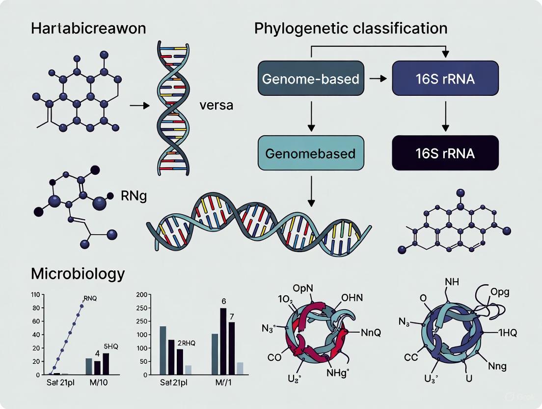
This article provides a comprehensive analysis of genome-based and 16S rRNA gene sequencing methods for microbial phylogenetic classification and identification.
Genome-Based vs. 16S rRNA Phylogenetic Classification: A Modern Guide for Microbial Researchers
Abstract
This article provides a comprehensive analysis of genome-based and 16S rRNA gene sequencing methods for microbial phylogenetic classification and identification. It explores the foundational principles of both approaches, detailing practical methodologies and their diverse applications in clinical, environmental, and industrial microbiology. The content addresses key technical challenges, including error correction, primer selection bias, and database limitations, while offering optimization strategies. A critical comparative evaluation examines the resolution, accuracy, and practical trade-offs of each method, supported by recent technological advancements in long-read sequencing. Designed for researchers, scientists, and drug development professionals, this review synthesizes current evidence to guide method selection and discusses future implications for biomedical research and clinical diagnostics.
The Core Principles: Unraveling 16S rRNA and Whole-Genome Phylogenetics
The 16S ribosomal RNA (rRNA) gene stands as one of the most pivotal molecular markers in the history of microbiology. Since Carl Woese's pioneering work in 1977, which utilized the gene to delineate the previously unknown domain of Archaea, the 16S rRNA gene has served as the cornerstone for bacterial identification and phylogenetic classification [1] [2]. This gene, approximately 1,500 base pairs in length, possesses a unique architecture of nine hypervariable regions (V1-V9) interspersed with conserved sequences, making it ideally suited for differentiating bacterial taxa while allowing for the design of universal primers [3]. Its universality across bacteria and archaea, combined with its functional constancy and molecular clock-like properties, established it as the "gold standard" for microbial taxonomy for decades [1] [2].
However, the rapid advancement of genome sequencing technologies and sophisticated computational methods has prompted a critical re-evaluation of the 16S rRNA gene's role in modern microbial taxonomy. This guide objectively examines the performance of 16S rRNA gene analysis against emerging genome-based approaches, synthesizing current experimental data to delineate their respective strengths, limitations, and optimal applications in research and diagnostic contexts.
Performance Comparison: 16S rRNA Gene vs. Genome-Based Classification
Extensive comparative studies have quantified the taxonomic resolution and reliability of 16S rRNA-based methods against genome-based approaches. The table below summarizes key performance metrics based on recent empirical evidence.
Table 1: Performance comparison of 16S rRNA gene sequencing versus genome-based classification methods
| Performance Metric | 16S rRNA Gene Sequencing | Genome-Based Classification |
|---|---|---|
| Species-level identification | 47-76% of sequences, depending on platform and region [4] | Nearly 100% with established thresholds (e.g., 95% ANI) [2] |
| Strain-level differentiation | Limited due to intragenomic heterogeneity [5] | High resolution using core genome SNPs [6] |
| Concordance with species phylogeny | 50.7% (intra-genus) to 73.8% (inter-genus) [1] | 93-100% (core genome phylogeny) [1] |
| Impact of HGT/Recombination | Subject to recombination/HGT, confounding phylogeny [1] [2] | Minimal when core genes are used with recombination filtering [1] |
| Influence of copy number variation | High potential to confound abundance metrics [1] | Not applicable (single-copy genes used) [1] |
| Required SNPs for 80% concordance | 690 ± 110 [1] | Not specified (inherently higher phylogenetic signal) |
The limitations of 16S rRNA gene analysis manifest particularly in complex taxonomic scenarios. Studies of the family Colwelliaceae revealed that phylogenetic positions remained ambiguous when classified solely based on 16S rRNA gene sequences, necessitating genome-based approaches for accurate taxonomic resolution [7]. Similarly, in non-pathogenic Yersinia, the 16S rRNA gene showed insufficient discriminatory power, with identical gene sequences found in genetically distinct species that were clearly separated by Average Nucleotide Identity (ANI) and core SNP analyses [6] [2].
Experimental Evidence: Key Studies and Methodologies
Phylogenetic Concordance Analysis
Experimental Protocol: A comprehensive phylogenomic study evaluated the strength of phylogenetic signal for the 16S rRNA gene by comparing it to core genome phylogenies at both intra-genus and inter-genus levels [1]. Researchers performed four intra-genus analyses (Clostridium, Legionella, Staphylococcus, and Campylobacter) and one inter-genus analysis of 41 core genera of the human gut microbiome. For each genus, representative strains were selected from RefSeq database with preference for closed genomes. Homologous gene clustering delineated single-copy core genes, which were aligned and concatenated to build species phylogenies. The 16S rRNA gene sequences were aligned separately, and phylogenies were constructed. Concordance between 16S rRNA gene trees and core genome trees was calculated as the proportion of matching bipartitions. Genes exhibiting evidence of recombination/HGT were identified and removed using multiple statistical approaches.
Key Findings: The 16S rRNA gene displayed notably low concordance with core genome phylogenies at the intra-genus level (average 50.7%), ranking among the lowest of all genes tested [1]. The gene exhibited clear evidence of recombination and horizontal gene transfer across multiple genera. Hypervariable regions showed even lower concordance than the full gene, with entropy masking providing little benefit. A critical finding was the logarithmic relationship between SNP count and concordance, revealing that approximately 690±110 SNPs are required for 80% concordance—far exceeding the average 16S rRNA gene SNP count of 254 [1].
Full-Length vs. Partial Gene Sequencing
Experimental Protocol: A landmark study evaluated the potential of full-length 16S rRNA gene sequencing to provide species- and strain-level resolution using in silico experiments and empirical sequencing [5]. Researchers downloaded non-redundant full-length 16S sequences from Greengenes database and trimmed them in silico to generate amplicons for different hypervariable regions. They then used the RDP classifier to calculate the frequency with which each sub-region could provide accurate species-level classification. For empirical validation, they performed PacBio Circular Consensus Sequencing (CCS) of a 36-species bacterial mock community, using multiple passes to generate high-fidelity reads. The resulting sequences were analyzed for intragenomic variation by comparison with known 16S copy variants in reference genomes.
Key Findings: The V4 region, commonly targeted in Illumina-based studies, performed worst, with 56% of in-silico amplicons failing to confidently match their correct species [5]. Different hypervariable regions showed significant taxonomic biases, with varying performance across bacterial phyla. Full-length 16S sequencing dramatically improved species-level discrimination, with PacBio CCS sequencing proving sufficiently accurate to resolve subtle nucleotide substitutions between intragenomic 16S gene copies. The study demonstrated that appropriate treatment of full-length 16S intragenomic copy variants enables taxonomic resolution at species and strain level [5].
Table 2: Species-level classification accuracy across sequencing platforms and target regions
| Sequencing Platform | Target Region | Species-Level Classification Rate | Key Limitations |
|---|---|---|---|
| Illumina MiSeq | V3-V4 | 47% [4] | Short reads limit discriminatory power |
| PacBio HiFi | Full-length (V1-V9) | 63% [4] | Higher cost; requires specialized analysis |
| Oxford Nanopore | Full-length (V1-V9) | 76% [4] | Higher error rate requires specific pipelines |
| In silico ideal | Full-length (V1-V9) | Nearly 100% [5] | Reference database quality dependent |
Taxonomic Reclassification Studies
Experimental Protocol: Multiple studies have employed genome-based taxonomic reclassification of bacterial groups that were poorly resolved by 16S rRNA gene analysis [6] [7]. A representative study on the family Colwelliaceae characterized four newly isolated species using a comprehensive taxogenomic framework [7]. Researchers analyzed genome-based indices including Average Nucleotide Identity (ANI), digital DNA-DNA hybridization (dDDH), and Average Amino Acid Identity (AAI) across all publicly available Colwelliaceae genomes. Genus-level AAI thresholds were established through repetitive clustering and evaluation strategies. 16S rRNA gene sequences were compared against genome-based phylogenies to identify discrepancies.
Key Findings: The analysis revealed that 16S rRNA gene sequences provided ambiguous phylogenetic positions for Colwelliaceae members [7]. Genome-based indices enabled the establishment of clear genus boundaries (AAI 74.07%-75.11%), leading to the proposal of 18 new genera and expanding the taxonomy from 6 to 24 genera. Similarly, in Yersinia, 34 out of 373 genomes had taxonomic affiliations based on core SNPs and ANI that did not match their GenBank classifications, which were based largely on 16S rRNA gene sequences [6]. These studies highlight the limitations of 16S rRNA gene phylogenies and support the use of taxogenomic approaches for higher taxonomic resolution.
Methodological Workflow: From 16S rRNA to Genome-Based Taxonomy
The following diagram illustrates the progressive refinement of microbial classification from traditional 16S rRNA approaches to modern genome-based methods, highlighting key decision points and analytical steps.
Table 3: Essential research reagents and computational resources for microbial taxonomy studies
| Category | Specific Tools/Reagents | Function/Application |
|---|---|---|
| Wet Lab Reagents | DNeasy PowerSoil Kit (QIAGEN) [4] | Microbial DNA extraction from complex samples |
| KAPA HiFi HotStart DNA Polymerase [4] | High-fidelity amplification of full-length 16S gene | |
| Nextera XT Index Kit [4] | Sample multiplexing for Illumina sequencing | |
| SMRTbell Express Template Prep Kit [4] | Library preparation for PacBio sequencing | |
| Primer Sets | 27F/1492R [7] [4] | Amplification of nearly full-length 16S rRNA gene |
| 341F/785R [4] | Targeting V3-V4 regions for Illumina sequencing | |
| Bioinformatic Tools | QIIME2 [3] [4] | Integrated analysis of 16S amplicon sequence data |
| DADA2 [3] [4] | Denoising and Amplicon Sequence Variant calling | |
| SPAdes/Unicycler [6] | Genome assembly from sequencing reads | |
| Snippy [6] | Core genome SNP identification and analysis | |
| Reference Databases | SILVA [4] | Curated database of aligned ribosomal RNA sequences |
| Greengenes [3] | 16S rRNA gene database with taxonomy information | |
| EzBioCloud [7] | Integrated database for prokaryote taxonomy identification |
The evidence synthesized in this guide clearly demonstrates that while the 16S rRNA gene remains a valuable tool for initial microbial surveys and continues to offer utility in clinical diagnostics where it demonstrates 60% diagnostic utility in confirmed infections [8], its limitations necessitate complementary genome-based approaches for definitive taxonomic classification. The gene's susceptibility to recombination, horizontal gene transfer, intragenomic heterogeneity, and limited phylogenetic signal at finer taxonomic scales constrains its standalone application in modern microbiology [1] [2].
The future of microbial taxonomy lies in integrated approaches that leverage the throughput and cost-effectiveness of 16S rRNA gene sequencing for initial surveys while employing genome-based methods for definitive taxonomic placement and phylogenetic inference. As sequencing technologies continue to advance and costs decrease, full-length 16S sequencing and targeted genome sequencing are poised to bridge the gap between these approaches, offering improved resolution while maintaining practical feasibility for diverse research and clinical applications [5] [4]. This integrated framework ensures that the 16S rRNA gene maintains its foundational role in microbiology while being augmented by genomic methods that provide the resolution required for precise taxonomic assignment and evolutionary inference.
The classification of prokaryotes is undergoing a fundamental paradigm shift, moving from a single-gene foundation to a whole-genome framework. For decades, the 16S rRNA gene has served as the cornerstone of bacterial identification and phylogenetic studies, providing a universal target for phylogenetic analysis. However, the rapidly expanding availability of whole-genome sequencing (WGS) has enabled the development of more robust, data-rich classification methods based on complete genetic information. This transition addresses critical limitations of 16S rRNA gene sequencing, including its inadequate resolution for closely related species and the challenges posed by intragenomic heterogeneity between multiple 16S copies within a single organism [9] [5].
Two genome-based methodologies have emerged as gold standards for species delimitation: Average Nucleotide Identity (ANI) and core genome Single Nucleotide Polymorphisms (core SNPs). These approaches leverage the comprehensive genetic content of organisms, providing unprecedented resolution for strain differentiation and taxonomic assignment. As the scientific community increasingly adopts these methods, understanding their technical implementation, comparative performance, and relationship to traditional 16S rRNA classification becomes essential for researchers across microbiology, genomics, and drug development. This guide provides a comprehensive comparison of these foundational genomic classification techniques, detailing their experimental protocols, applications, and performance metrics relative to established 16S rRNA methods.
Limitations of 16S rRNA Gene Sequencing
Resolution Constraints and Taxonomic Ambiguity
While 16S rRNA sequencing remains widely used for microbial community profiling, its limitations for precise taxonomic classification are well-documented. The gene's conservation pattern—alternating variable and conserved regions—creates inherent resolution boundaries that often prevent reliable discrimination at the species level [5]. Studies demonstrate that full-length 16S sequences (approximately 1,500 bp) provide significantly better taxonomic resolution than shorter hypervariable regions (e.g., V4, V3-V4), which are commonly targeted in Illumina-based sequencing approaches [5]. However, even full-length sequencing may fail to distinguish clinically distinct species.
Critical limitations include:
- High sequence similarity between distinct species: Some genetically separate species share nearly identical 16S sequences [6] [10].
- Intragenomic heterogeneity: Many bacteria contain multiple copies of the 16S rRNA gene with sequence variation, complicating classification and diversity estimates [5].
- Variable discriminatory power across taxa: Resolution thresholds differ significantly between bacterial groups, making universal identity cutoffs unreliable [11] [10].
Quantitative Resolution Thresholds Under Genome-Based Taxonomy
The emergence of genome-based taxonomy systems like the Genome Taxonomy Database (GTDB) has further clarified the limitations of 16S rRNA gene resolution. Under this framework, achieving species-level resolution typically requires clustering 16S sequences at a stringent 99% identity threshold, while genus-level resolution requires thresholds between 92-96% [11]. These findings underscore that historical assumptions about fixed similarity thresholds (e.g., 97% for species) are invalid in the genomic era.
Table 1: 16S rRNA Gene Resolution Thresholds Under GTDB Taxonomy
| Taxonomic Rank | Sequence Identity Threshold | Clustering Divergence |
|---|---|---|
| Species | ~99% | ~0.01 |
| Genus | 92-96% | 0.04-0.08 |
| Family | Variable across branches | No universal threshold |
Genome-Based Classification Methods
Average Nucleotide Identity (ANI)
Concept and Methodology
Average Nucleotide Identity calculates the average nucleotide sequence identity between homologous regions of two genomes, providing a robust, alignment-based measure of genomic relatedness. The method typically employs BLAST-based algorithms (ANIb) or k-mer based approaches (Mash) for rapid comparison [12]. ANI has become a standard metric for species demarcation, with a widely accepted threshold of 95-96% for species boundaries [12].
The experimental workflow for ANI analysis begins with quality-controlled whole-genome sequences, which are compared using specialized tools such as fastANI or the OrthoANI algorithm. These tools identify conserved genomic regions and calculate the average identity of aligned segments, providing a quantitative measure of evolutionary relatedness that correlates strongly with traditional DNA-DNA hybridization values but offers greater reproducibility and resolution [12].
Applications and Performance
ANI analysis has proven particularly valuable for clarifying taxonomic relationships within complex bacterial groups. In the Enterobacter cloacae complex, for example, ANI values provided definitive evidence for subspecies classification, resolving strains that appeared ambiguous using 16S rRNA sequencing alone [12]. Similarly, ANI has been instrumental in characterizing non-pathogenic Yersinia species, where 16S rRNA gene sequences showed insufficient variation to reliably distinguish between distinct species [6].
Table 2: ANI Thresholds for Taxonomic Delineation
| Taxonomic Relationship | ANI Value Range | Interpretation |
|---|---|---|
| Same species | ≥95-96% | Conspecific genomes |
| Different species | <95-96% | Genomically distinct species |
| Subspecies level | >98% | Intraspecific variation |
Core Genome Single Nucleotide Polymorphisms (core SNPs)
Concept and Methodology
Core genome SNP analysis identifies single nucleotide polymorphisms present in conserved genomic regions shared among all compared isolates. This method focuses on the most stable portions of the genome, excluding accessory genomic elements that may be horizontally transferred. The core genome represents the backbone of phylogenetic inheritance, making it ideal for reconstructing evolutionary relationships and transmission pathways.
The technical workflow involves:
- Whole-genome sequencing of multiple isolates using either short-read (Illumina) or long-read (PacBio, Nanopore) platforms
- Reference-based mapping using tools like Snippy or quality-controlled de novo assembly
- Identification of conserved core genomic regions and extraction of variable sites
- Phylogenetic reconstruction based on SNP patterns using maximum likelihood or neighbor-joining methods
Applications and Performance
Core SNP analysis provides the highest resolution for strain differentiation and epidemiological tracking. In studies of Microsporum canis, core SNP phylogenetics revealed multiple genotypes within the same species, enabling researchers to distinguish between strains of human and animal origin and trace zoonotic transmission patterns [13]. Similarly, for non-pathogenic Yersinia species, core SNP analysis generated phylogenetic trees that more accurately reflected evolutionary relationships compared to 16S rRNA-based phylogenies, which showed poor correlation with genome-wide data [6].
Direct Comparative Analysis: 16S rRNA vs. Genome-Based Methods
Resolution and Accuracy Comparison
Multiple studies have directly compared the taxonomic resolution of 16S rRNA gene sequencing versus whole-genome methods. The results consistently demonstrate the superior discriminatory power of genomic approaches:
Table 3: Method Comparison - 16S rRNA vs. Genome-Based Classification
| Performance Metric | 16S rRNA Sequencing | Whole-Gome Methods (ANI/core SNPs) |
|---|---|---|
| Species-level resolution | Limited, highly variable across taxa [5] [10] | High, consistent across diverse organisms [12] [6] |
| Strain differentiation | Generally not possible [5] | High resolution for epidemiological tracking [13] |
| Reference database issues | Inconsistent nomenclature, variable quality [10] | Standardized frameworks emerging (GTDB) [11] |
| Intragenomic heterogeneity | Complicates analysis, often overlooked [5] | Not applicable (whole-genome approach) |
| Computational requirements | Moderate | High (infrastructure and expertise needed) |
In one striking example from Yersinia research, identical 16S rRNA gene sequences were found in genomes of Y. intermedia and Y. rochesterensis that were clearly distinguished as separate species using both ANI and core SNP analyses [6]. This demonstrates how 16S-based identification can potentially group genetically distinct organisms, leading to misclassification.
Technical and Practical Considerations
While genome-based methods offer superior resolution, they present different practical considerations:
16S rRNA sequencing advantages include lower cost, simpler data analysis, established workflows, and applicability to complex microbial communities where whole-genome sequencing may be impractical. The method remains valuable for initial community profiling and identifying uncultivated organisms [14].
Whole-genome sequencing advantages encompass comprehensive genetic characterization, strain-level discrimination, functional gene assessment, and accurate phylogenetic reconstruction. The declining cost of sequencing has made WGS increasingly accessible for routine classification [14].
Experimental Protocols
ANI Analysis Workflow
Detailed Protocol
Genome Assembly and Quality Control
- Sequence bacterial isolates using Illumina, PacBio, or Nanopore platforms
- Perform de novo assembly using SPAdes (for Illumina data) or Flye (for long reads)
- Assess assembly quality using QUAST, checking for contiguity (N50), completeness, and contamination
Reference Selection
- Select appropriate reference genomes from databases such as GTDB or NCBI RefSeq
- Prioritize type strains when available for taxonomic comparisons
ANI Calculation
- Use fastANI for rapid k-mer based analysis or OrthoANI for BLAST-based alignment
- Command example:
fastANI -q query_genome.fna -r reference_genome.fna -o output.ani - Generate pairwise ANI matrix for multiple genomes
Interpretation
- Apply species boundary threshold (95-96% ANI)
- Identify conspecific groups and outliers
- Compare with additional genomic features (e.g., digital DDH) for comprehensive taxonomy
Core SNP Phylogeny Workflow
Detailed Protocol
Data Preparation and Mapping
- Obtain quality-filtered whole-genome sequencing reads
- Select appropriate reference genome (high-quality, closely related)
- Map reads using BWA-MEM or Bowtie2, then process with SAMtools
Variant Calling and Filtering
- Identify SNPs using Snippy, GATK, or SAMtools/BCFtools
- Apply quality filters: minimum mapping quality (Q30), read depth (>10x), and base quality
- Exclude repetitive regions and phage elements to avoid false positives
Core Genome Alignment
- Extract SNPs present in all isolates (core genome)
- Create concatenated SNP alignment using custom scripts or snippy-core
Phylogenetic Analysis
- Select appropriate substitution model using ModelTest-NG
- Construct maximum-likelihood tree with RAxML or IQ-TREE
- Assess branch support with bootstrapping (100-1000 replicates)
- Visualize and annotate trees using iTOL or FigTree
Research Reagent Solutions
Table 4: Essential Research Reagents and Tools for Genomic Taxonomy
| Category | Specific Tools/Reagents | Function |
|---|---|---|
| Sequencing Platforms | Illumina MiSeq/NovaSeq, PacBio Sequel, Oxford Nanopore | Whole-genome sequence data generation |
| Assembly Tools | SPAdes, Unicycler, Flye | De novo genome assembly from raw reads |
| ANI Analysis | fastANI, OrthoANI, pyani | Calculate average nucleotide identity between genomes |
| SNP Phylogenetics | Snippy, GATK, kSNP3 | Identify core SNPs and construct phylogenetic trees |
| Reference Databases | GTDB, NCBI RefSeq, SILVA | Curated genomic and 16S reference sequences |
| Quality Control | FastQC, Quast, CheckM | Assess sequence and assembly quality |
The transition from 16S rRNA gene sequencing to genome-based classification represents a fundamental advancement in microbial taxonomy. Methods based on Average Nucleotide Identity and core genome SNPs provide unprecedented resolution for species delineation and strain tracking, addressing critical limitations of single-gene approaches. While 16S rRNA sequencing retains utility for initial community profiling and studies of uncultivated organisms, its inadequate resolution for closely related species and susceptibility to database inaccuracies necessitate cautious interpretation.
The future of microbial classification lies in the integration of multiple genomic markers within standardized taxonomic frameworks like the Genome Taxonomy Database. As sequencing costs continue to decline and analytical tools become more accessible, genome-based approaches will increasingly become the default standard for definitive taxonomic assignment, particularly in clinical and regulatory contexts where accurate strain-level identification is essential. For researchers navigating this transition, understanding the technical requirements, performance characteristics, and appropriate applications of both 16S rRNA and genome-based methods is crucial for designing robust classification workflows and accurately interpreting microbial diversity.
The field of microbial classification has been fundamentally shaped by two powerful sequencing paradigms: targeted 16S rRNA gene sequencing and whole-genome analysis. For decades, 16S rRNA gene sequencing has served as the cornerstone of microbial ecology, providing a cost-effective method for profiling complex bacterial communities [15]. However, the rapidly evolving landscape of genome-based taxonomy now offers unprecedented resolution through techniques like whole-genome sequencing and shotgun metagenomics [7] [11]. This guide provides an objective comparison of these approaches, examining their performance characteristics, experimental requirements, and suitability for different research scenarios within the broader context of the ongoing shift from gene-centric to genome-centric classification systems.
Technical Foundations: Methodological Comparison
The fundamental distinction between these approaches lies in their scope—16S sequencing targets a single, highly conserved genetic marker, while whole-genome methods attempt to capture all genetic material in a sample.
Table 1: Core Technical Specifications of Sequencing Approaches
| Parameter | 16S rRNA Gene Sequencing | Shotgun Metagenomics | Whole-Genome Sequencing (Isolates) |
|---|---|---|---|
| Target Region | Variable regions of 16S rRNA gene (e.g., V3-V4, full-length) [16] [15] | All genomic DNA in sample [17] | Complete genome of isolated microbe |
| Taxonomic Resolution | Genus-level (typically), sometimes species [18] | Species to strain-level [17] [18] | Highest resolution (strain-level) |
| Functional Insights | Limited (predicted) [18] | Comprehensive (direct gene detection) [17] | Comprehensive (complete genetic repertoire) |
| Bias Sources | Primer selection, PCR amplification, rRNA copy number variation [16] [19] | DNA extraction efficiency, host DNA contamination [17] | Culture bias (for isolates) |
| Cost per Sample | Lower | Higher [18] | Moderate to High |
| Hands-on Time | Lower | Moderate to High | Moderate to High |
The following workflow diagram illustrates the fundamental procedural differences between these approaches:
Experimental Protocols in Practice
16S rRNA Gene Sequencing Workflow
The 16S rRNA gene sequencing protocol typically begins with careful sample preservation and DNA extraction. For human fecal samples, collection often involves using DNA/RNA shielding buffer with immediate freezing at -80°C [16]. DNA extraction utilizes specialized kits like the Quick-DNA HMW MagBead Kit, with DNA quality verified through fluorometry and spectrophotometry [16].
PCR Amplification: The critical amplification step uses primers targeting conserved regions of the 16S rRNA gene. Key primer sets include:
- Standard primers: 27F (5′-AGAGTTTGATCMTGGCTCAG-3′) and 1492R (5′-CGGTTACCTTGTTACGACTT-3′) [16] [7]
- Degenerate primers: Modified versions with increased degeneracy to capture broader taxonomic diversity (e.g., S-D-Bact-0008-c-S-20) [16]
PCR conditions typically involve: 25 cycles of 95°C for 20s, 51°C for 30s, and 65°C for 2 minutes using master mixes like LongAMP Taq 2x Master Mix [16]. For full-length 16S sequencing on nanopore platforms, the 16S Barcoding Kit from Oxford Nanopore Technologies is commonly employed [16].
Shotgun Metagenomic Sequencing
For shotgun sequencing, the same DNA extraction methods apply, but without targeted amplification. Instead, DNA is fragmented and prepared for sequencing using library prep kits like Illumina DNA Prep [15]. Critical considerations include:
- Sequencing depth: >500,000 reads per sample recommended to avoid skewed diversity metrics [17]
- Host DNA depletion: Particularly important for low-biomass samples where host DNA can dominate
- Quality control: Verification of DNA quantity and quality using fluorometry and bioanalyzer systems [19]
Whole-Genome Sequencing for Isolates
Genome-based classification of microbial isolates follows a distinct pathway:
Culturing and DNA Extraction: Pure cultures are established on appropriate media (e.g., marine agar for marine bacteria) [7], followed by high-quality DNA extraction using kits such as LaboPass bacterial genomic DNA isolation kit [7].
Genome Sequencing and Analysis: Sequencing generates complete genomes for analysis using multiple genomic indices:
- Average Nucleotide Identity (ANI): Both ANI-BLAST (ANIb) and ANI-MUMmer (ANIm) with species threshold ≥95-96% [20]
- Digital DNA-DNA Hybridization (dDDH): Species threshold ≥70% [20]
- Average Amino Acid Identity (AAI): Genus-level thresholds vary (e.g., 74-75% for Colwelliaceae) [7]
Performance Comparison: Experimental Data
Direct comparisons between 16S and shotgun sequencing reveal significant differences in detection capability and taxonomic resolution.
Table 2: Experimental Comparison of 16S vs. Shotgun Sequencing in Gut Microbiome Studies
| Performance Metric | 16S rRNA Sequencing | Shotgun Metagenomics | Experimental Context |
|---|---|---|---|
| Genera Detected | 288 genera | 288 genera + additional rare taxa [17] | Chicken GI tract [17] |
| Differential Abundance | 108 significant differences | 256 significant differences [17] | Caeca vs. crop comparison [17] |
| Sensitivity in Low Biomass | Lower detection rate | Higher detection rate; requires optimization [19] | Equine uterine microbiome [19] |
| Taxonomic Skewing | Affected by primer choice and rRNA copy number [16] [19] | Less affected by genetic copy number variation | Human fecal samples [16] |
| Technology-Specific Genera | Some genera only detected with 16S | Many genera only detected with shotgun [17] | Pediatric gut microbiome [18] |
Impact of Primer Selection in 16S Sequencing
Primer choice significantly influences 16S sequencing results. A comparison of conventional (27F-I) versus degenerate (27F-II) primers in human fecal samples revealed striking differences: the conventional primer revealed significantly lower biodiversity and an unusually high Firmicutes/Bacteriodetes ratio compared to the degenerate primer [16]. This demonstrates how technical choices in 16S protocols can dramatically impact biological interpretations.
Detection Limit Differences
The sensitivity advantage of shotgun sequencing becomes particularly evident in detecting rare taxa. One study found that shotgun sequencing identified 152 statistically significant abundance changes between gut compartments that 16S sequencing failed to detect, while 16S found only 4 changes missed by shotgun sequencing [17]. This disparity is largely attributed to the higher sampling depth possible with shotgun approaches.
Taxonomic Resolution: 16S rRNA versus Genome-Based Classification
The move toward genome-based taxonomy highlights limitations of 16S rRNA gene sequencing for precise taxonomic placement.
The GTDB Framework and 16S Divergence
The Genome Taxonomy Database (GTDB) initiative represents a fundamental shift from 16S-based to genome-based prokaryotic taxonomy [11]. Analysis of 16S sequence divergence within this framework reveals that:
- Species-level resolution requires clustering at ~99% identity (0.01 divergence) [11]
- Genus-level resolution requires thresholds of 92-96% identity (0.04-0.08 divergence) [11]
- Optimal thresholds vary significantly across phylogenetic branches, challenging fixed threshold approaches [11]
Case Study: Colwelliaceae Reclassification
A comprehensive revision of the family Colwelliaceae demonstrates the power of genome-based classification. Through analysis of genome-based indices (AAI, ANI, dDDH) across all available Colwelliaceae genomes, researchers expanded the taxonomy from 6 to 24 genera, proposing 18 new genera [7]. This reclassification was necessary because 16S rRNA gene sequences provided ambiguous phylogenetic positions, limiting accurate taxonomic resolution [7].
Species Delineation Challenges
The limitations of 16S sequencing for species-level identification are evident in cases like Micromonospora veneta and M. coerulea, which share 99.2% 16S rRNA gene similarity yet were confirmed as the same species through genomic metrics (AAI: 97.57%, ANI: 97.81%, dDDH: 85.0%) [20]. All values exceeded species thresholds, demonstrating that 16S similarity alone cannot reliably delineate species.
The Scientist's Toolkit: Essential Research Reagents
Table 3: Key Research Reagents and Their Applications
| Reagent/Kit | Application | Function | Example Use |
|---|---|---|---|
| Quick-DNA HMW MagBead Kit | DNA extraction | High molecular weight DNA purification | Human fecal samples [16] |
| 16S Barcoding Kit (ONT) | Library preparation | Targeted amplification of full-length 16S | Nanopore sequencing [16] |
| LongAMP Taq 2x Master Mix | PCR amplification | High-fidelity amplification of 16S | Degenerate primer protocols [16] |
| AllPrep DNA/RNA/miRNA Universal Kit | Nucleic acid extraction | Simultaneous DNA/RNA isolation | RNA-based microbiome studies [19] |
| ZymoBIOMICS Microbial Community DNA Standard | Quality control | Protocol validation and sensitivity testing | Low-biomass microbiome studies [19] |
| Marine Agar 2216 | Microbial culturing | Isolation of marine bacteria | Colwelliaceae isolation [7] |
The choice between targeted 16S rRNA sequencing and whole-genome approaches depends on research goals, budget, and sample type. 16S rRNA sequencing remains valuable for large-scale diversity studies where cost-effectiveness is paramount and genus-level resolution is sufficient. However, shotgun metagenomics provides superior taxonomic resolution, functional insights, and detection of rare taxa, despite higher costs and computational demands [17] [18]. For definitive taxonomic classification of isolates, genome-based methods using ANI, dDDH, and AAI provide the highest resolution and are becoming the gold standard [7] [11] [20].
The field continues to evolve with techniques like RNA-based 16S sequencing offering insights into active community members [19], and long-read technologies enabling full-length 16S sequencing with improved taxonomic resolution [16]. As genome databases expand and costs decrease, the integration of both targeted and whole-genome approaches will likely provide the most comprehensive understanding of microbial communities.
Strengths and Inherent Limitations of Each Foundational Approach
The accurate classification of microorganisms is fundamental to advancing our understanding of microbial ecology, evolution, and their roles in health and disease. For decades, 16S ribosomal RNA (rRNA) gene sequencing has served as the cornerstone of bacterial identification and phylogenetic analysis [21]. However, with the advent of high-throughput sequencing technologies, whole-genome sequencing (WGS) approaches have emerged as a powerful alternative, enabling genome-based phylogenetic classification with superior resolution [7] [22]. This guide provides an objective comparison of these two foundational approaches, framing the analysis within the broader thesis of genome-based versus 16S rRNA-based phylogenetic research. We summarize experimental data, detail key methodologies, and provide practical resources for researchers, scientists, and drug development professionals navigating the choice between these techniques.
Comparative Analysis of Foundational Approaches
The following tables summarize the core characteristics, performance metrics, and optimal use cases for 16S rRNA gene sequencing and whole-genome sequencing approaches.
Table 1: Core Characteristics and Technical Performance
| Feature | 16S rRNA Gene Sequencing | Whole-Genome Sequencing |
|---|---|---|
| Genetic Target | Single gene (~1,500 bp) with 9 hypervariable regions [5] | Entire genome, all genomic regions [14] |
| Taxonomic Resolution | Limited species/strain resolution; struggles with closely related taxa [23] [5] | High resolution to species and strain level; identifies subtle nucleotide substitutions [7] [5] |
| Primary Analytical Outputs | Operational Taxonomic Units (OTUs), Amplicon Sequence Variants (ASVs) | Average Nucleotide Identity (ANI), digital DNA-DNA Hybridization (dDDH), core-genome phylogeny [7] [23] |
| Key Quantitative Thresholds | Traditional: >97% similarity (species), >95% (genus) [5] | Genome-based: ~95-96% ANI for species demarcation; genus-level AAI thresholds vary (e.g., 74-75% in Colwelliaceae) [7] |
| Inherent Biases | PCR primer selection, variable region choice, copy number variation [5] [14] [24] | Genome size bias, reference database dependency, host DNA contamination [14] |
| Ability to Detect Non-Bacteria | Limited to bacteria and archaea | Comprehensive: bacteria, archaea, viruses, fungi, protozoa [14] |
Table 2: Application-Based Suitability and Data Characteristics
| Aspect | 16S rRNA Gene Sequencing | Whole-Genome Sequencing |
|---|---|---|
| Optimal Use Cases | Community profiling, diversity studies, targeted analysis, large cohort screenings | Strain-level discrimination, functional potential assessment, novel pathogen discovery, metagenomic association studies |
| Relative Cost | Lower cost, cost-effective for large-scale studies [14] | Higher cost, though becoming more affordable [14] |
| Computational Demand | Moderate, standardized pipelines | High, complex bioinformatics, extensive computational resources [14] |
| Sensitivity in Community Analysis | Reveals broad shifts but with limited resolution; gives greater weight to dominant bacteria [25] | More detailed snapshot in depth and breadth; detects rare taxa [14] [25] |
| Quantitative Accuracy | Skewed by variable 16S copy numbers (1-15+ per genome) [24] | Not affected by 16S copy number variation; enables alternative abundance estimates [24] |
Experimental Evidence and Validation Studies
Phylogenetic Resolution and Taxonomic Classification
Supporting Experimental Data: A 2025 study on the family Colwelliaceae demonstrated that a genome-based phylogenetic analysis using Average Amino Acid Identity (AAI) revealed the need for significant taxonomic revision. The research proposed expanding the taxonomy from 6 to 24 genera, a reassignment impossible with 16S rRNA data alone due to its limited resolution [7]. This genome-based approach provided a stable taxonomic framework superior to previous 16S-based classifications.
Similarly, an analysis of Oxalobacteraceae showed that phylogenomic trees and genomic similarity indices (ANI, percentage of conserved proteins) provided a clearer and more reliable classification system compared to previous studies that relied heavily on 16S rRNA gene analysis [22].
Community Analysis and Diversity Assessment
Supporting Experimental Data: A 2024 study comparing 16S rRNA and shotgun sequencing for human gut microbiota analysis found that 16S detects only part of the gut microbiota community revealed by shotgun. The 16S abundance data was sparser and exhibited lower alpha diversity. Furthermore, the two methods highly differed in lower taxonomic ranks, partially due to disagreements in reference databases [14].
Another study on freshwater microbial communities found that while 16S rRNA gene sequencing captured broad shifts in community diversity over time, it had limited resolution and lower sensitivity compared to metagenomic data. The metagenomic approach identified 1.5 times as many phyla and ~10 times as many genera as the 16S approach [25].
Impact of Intragenomic Heterogeneity
Supporting Experimental Data: Research on non-pathogenic Yersinia revealed significant intragenomic heterogeneity in 16S rRNA genes. Above 50% of complete genomes have four or more variants of the 16S rRNA gene. This heterogeneity can confound accurate species identification, as identical 16S rRNA gene sequences were found in genomes of different Yersinia species that were clearly distinguished by ANI and core SNP analyses [23].
A 2019 study in Nature Communications confirmed that high-throughput full-length 16S sequencing can resolve subtle nucleotide substitutions between intragenomic copies, demonstrating that modern analysis must account for this variation to achieve species and strain-level resolution [5].
Detailed Experimental Protocols
16S rRNA Gene Sequencing Workflow
Figure 1: 16S rRNA gene sequencing workflow involves targeted amplification of specific variable regions before sequencing.
Key Methodological Steps:
DNA Extraction: Genomic DNA is extracted from clinical or environmental samples using commercial kits (e.g., NucleoSpin Soil Kit, Dneasy PowerLyzer Powersoil kit) [14]. Automated nucleic acid extraction machines (QIAcube, Maxwell RSC, KingFisher) can streamline this process [26].
PCR Amplification: Variable regions of the 16S rRNA gene (e.g., V3-V4, V4, V1-V2) are amplified using universal primer sets (e.g., 27F/1492R) [7] [14]. The choice of variable region significantly impacts taxonomic resolution and bias [5].
Library Preparation and Sequencing: Amplified products are processed to specific fragment sizes, adapters are added, and amplicons are quantified and normalized prior to sequencing on platforms such as Illumina MiSeq or Ion Torrent [26] [14].
Bioinformatic Analysis:
- Sequence Processing: Tools like DADA2 are used for quality filtering, trimming, denoising, and merging paired-end reads [14].
- Taxonomic Assignment: Processed sequences are classified against reference databases (SILVA, Greengenes, RDP) using classifiers like the RDP classifier or BLASTN against custom databases [5] [14].
- Diversity Analysis: Clustering into Operational Taxonomic Units (OTUs) or Amplicon Sequence Variants (ASVs), followed by alpha and beta diversity analyses [5] [25].
Whole-Genome Sequencing for Phylogenetic Classification
Figure 2: Whole-genome sequencing workflow sequences all genomic material without targeted amplification, enabling comprehensive analysis.
Key Methodological Steps:
DNA Extraction and Library Preparation: Similar to 16S protocols, but without target-specific amplification. DNA is fragmented, and adapters are ligated for shotgun sequencing [14].
Sequencing: Performed using various platforms:
Bioinformatic Analysis:
- Quality Control and Host DNA Removal: Tools like FastQC and Bowtie2 (for filtering host sequences) [14].
- Genome Assembly: De novo assembly using tools like SPAdes, Unicycler, or reference-based assembly [23].
- Genome-Based Metrics Calculation:
- Average Nucleotide Identity (ANI): Calculated using tools like OrthoANI or FastANI for species demarcation [7].
- digital DNA-DNA Hybridization (dDDH): Calculated in silico for species boundary determination [7].
- Average Amino Acid Identity (AAI): Used for genus-level classification with taxon-specific thresholds [7].
- Phylogenomic Analysis: Construction of phylogenies based on core gene sets or single-copy orthologous genes [7] [22].
The Scientist's Toolkit: Essential Research Reagents and Solutions
Table 3: Key Research Reagents and Computational Tools
| Item | Type | Primary Function | Examples/Alternatives |
|---|---|---|---|
| DNA Extraction Kits | Wet-lab reagent | Isolation of high-quality genomic DNA from diverse sample types | NucleoSpin Soil Kit, Dneasy PowerLyzer Powersoil Kit [14] |
| PCR Primers for 16S | Wet-lab reagent | Amplification of specific 16S variable regions | 27F/1492R (full-length); V4-specific primers [7] [5] |
| Automated Nucleic Acid Extraction Systems | Laboratory equipment | Standardized, high-throughput DNA extraction | QIAcube (Qiagen), Maxwell RSC (Promega), KingFisher (Thermo Fisher) [26] |
| 16S Reference Databases | Bioinformatics resource | Taxonomic classification of 16S sequences | SILVA, Greengenes, RDP [5] [14] |
| Genome Reference Databases | Bioinformatics resource | Taxonomic and functional analysis of WGS data | NCBI RefSeq, GTDB, UHGG [14] |
| Taxonomic Classifiers | Bioinformatics tool | Assigning taxonomy to sequence data | RDP Classifier, DADA2, Kraken2, Bracken [5] [14] |
| Genome Analysis Tools | Bioinformatics tool | Calculation of genome-based metrics | OrthoANI, FastANI, TypeMat genomes for dDDH [7] |
| Phylogenetic Tree Construction Software | Bioinformatics tool | Building phylogenomic trees from sequence alignments | MEGA, RAxML, IQ-TREE [7] [24] |
The choice between 16S rRNA gene sequencing and whole-genome sequencing for phylogenetic classification depends on research goals, resources, and required resolution. 16S rRNA sequencing remains valuable for broad community profiling and large-scale studies where cost-effectiveness is paramount. However, its limitations in taxonomic resolution, sensitivity to primer choice, and bias from copy number variation must be considered. Whole-genome approaches provide superior resolution for species and strain discrimination, enable functional insights, and support robust taxonomic revisions, albeit at higher computational and financial costs.
As sequencing technologies continue to advance and costs decrease, the field is moving toward a integrated approach where each method is selected based on specific research questions. Future directions will likely involve combining the breadth of 16S surveys with the depth of genome-level analysis to achieve a more comprehensive understanding of microbial systems across diverse environments from clinical settings to natural ecosystems.
Methodologies in Action: From Lab Bench to Data Analysis
The 16S ribosomal RNA (rRNA) gene sequencing has served as the cornerstone of microbial ecology and phylogenetic studies for decades, providing insights into the composition of complex microbial communities that are difficult or impossible to culture. As a phylogenetic marker, the 16S rRNA gene offers unique advantages, including its universal distribution across bacteria and archaea, the presence of both highly conserved and variable regions, and its sufficient length for robust phylogenetic analysis [21]. However, the field currently stands at a crossroads, with researchers facing critical decisions in experimental design that significantly impact downstream results and interpretations.
This guide objectively compares the current landscape of 16S rRNA sequencing workflows, focusing on two fundamental choices: the selection of hypervariable regions and sequencing platforms. Within the broader context of genome-based versus 16S rRNA phylogenetic classification research, we examine how these technical decisions influence taxonomic resolution, diversity metrics, and ultimately, biological conclusions. Recent studies have highlighted that the selection of particular 16S rRNA hypervariable regions is a crucial step that introduces significant variability in study results [27], while simultaneous advancements in sequencing technologies from Illumina, PacBio, and Oxford Nanopore Technologies (ONT) have expanded the methodological toolbox available to researchers.
Hypervariable Region Selection: A Primary Consideration
The 16S rRNA gene comprises nine hypervariable regions (V1-V9) flanked by conserved sequences, with researchers typically targeting specific variable regions for amplification and sequencing due to technical limitations and cost considerations. The selection of which hypervariable region(s) to target represents a fundamental methodological decision that directly influences microbial community profiles.
Comparative Performance of Common Regions
Recent research has systematically evaluated the performance of different hypervariable regions in various study systems. In a longitudinal gut microbiome study of adolescent patients with anorexia nervosa (AN) and matched controls, researchers directly compared the V1V2 and V3V4 regions [27]. While dominant genera such as Bacteroides, Faecalibacterium, and Phocaeicola were consistently detected across both regions, significant differences emerged in diversity measures. The within-sample longitudinal alpha diversity varied between regions, with the Chao1 index values being significantly higher in the V1V2 region. Similarly, overall microbiome profiles based on beta diversity differed substantially between regions [27].
Bland-Altman analysis in the same study revealed a general lack of strong agreement between the two sequencing approaches, except for a few taxa including Faecalibacterium, Ruminococcus, Roseburia, Turicibacter, and Anaerotruncus. The authors concluded that while some results were similar across both hypervariable regions, most findings were sensitive to the chosen region, underscoring the importance of primer selection in microbiome studies [27].
Table 1: Comparison of Hypervariable Region Performance in Microbial Studies
| Hypervariable Region | Consistent Detection | Alpha Diversity | Beta Diversity | Taxonomic Agreement | Recommended Applications |
|---|---|---|---|---|---|
| V1-V2 | Dominant genera (Bacteroides, Faecalibacterium, Phocaeicola) | Higher Chao1 values in longitudinal studies [27] | Differs significantly from V3-V4 profiles [27] | Strong for some taxa (Faecalibacterium, Ruminococcus) but generally poor agreement with V3-V4 [27] | Genus-level resolution for specific taxa like Akkermansia [27] |
| V3-V4 | Dominant genera consistently detected [27] | Lower Chao1 values compared to V1-V2 in some studies [27] | Differs significantly from V1-V2 profiles [27] | Strong for some Firmicutes but generally poor agreement with V1-V2 [27] | General community profiling, standardized workflows |
| V4 | Similar dominant taxa at higher taxonomic levels | Varies by ecosystem | Samples cluster less clearly by source (e.g., soil type) [28] | Varies across platforms | Illumina-focused studies, large-scale comparisons |
| Full-length (V1-V9) | Highest consistency for dominant and rare taxa | Most comprehensive diversity assessment | Clear sample clustering by source (e.g., soil type) [28] | Excellent cross-platform agreement when quality-controlled | Species-level resolution, biomarker discovery [29] |
The variable performance across hypervariable regions extends beyond human gut studies. Research in soil microbiomes demonstrated that the V4 region alone showed limited ability to cluster samples according to soil type, unlike fuller gene regions [28]. This suggests that different ecosystems may require region-specific optimization for optimal characterization.
Sequencing Platform Technologies: A Comparative Analysis
The evolution of sequencing technologies has dramatically expanded options for 16S rRNA sequencing, with second-generation (Illumina) and third-generation (PacBio, ONT) platforms offering distinct trade-offs in read length, accuracy, throughput, and cost.
Platform-Specific Performance Metrics
Recent comparative studies have quantified the performance differences across major sequencing platforms. In a study comparing Illumina, PacBio, and ONT for sequencing rabbit gut microbiota, researchers found notable differences in taxonomic resolution [4]. At the species level, ONT and PacBio exhibited superior resolution (76% and 63% respectively) compared to Illumina (47%). However, a significant limitation emerged across all platforms, with most sequences classified to species level being labeled as "uncultured_bacterium," indicating persistent challenges in comprehensive species-level identification [4].
Table 2: Sequencing Platform Technical Specifications and Performance
| Platform | Read Length | Target Region | Error Rate | Species-Level Resolution | Key Advantages | Key Limitations |
|---|---|---|---|---|---|---|
| Illumina | Short (300-600 bp) | Single hypervariable regions (e.g., V3-V4, V4) [30] | Low (<0.1%) [30] | Limited (47%) [4] | High accuracy, high throughput, low cost per sample | Short reads limit species-level resolution, amplification biases |
| PacBio | Long (full-length 16S) | V1-V9 (full-length) [4] | Very low (<0.1%) with HiFi mode [28] | Moderate (63%) [4] | High-fidelity long reads, excellent species-level discrimination | Higher cost, lower throughput, complex data processing |
| Oxford Nanopore | Long (full-length 16S) | V1-V9 (full-length) [4] [29] | Moderate (1-5%) with latest chemistry [29] | High (76%) [4] | Real-time sequencing, low initial cost, long reads enable species ID | Higher error rate, requires specific bioinformatic tools |
The analytical implications of platform selection extend beyond simple resolution metrics. In respiratory microbiome studies, Illumina captured greater species richness, while community evenness remained comparable between Illumina and ONT platforms. Beta diversity differences were more pronounced in complex pig microbiome samples compared to human samples, suggesting that sequencing platform effects are context-dependent [30]. Taxonomic profiling revealed that Illumina detected a broader range of taxa, while ONT exhibited improved resolution for dominant bacterial species [30].
Diagnostic and Biomarker Applications
The clinical implications of platform selection are particularly significant in diagnostic and biomarker discovery applications. A 2025 study demonstrated that ONT sequencing identified more specific bacterial biomarkers for colorectal cancer than those obtained with Illumina, including Parvimonas micra, Fusobacterium nucleatum, Peptostreptococcus stomatis, and Bacteroides fragilis [29]. This enhanced detection capability facilitated colorectal cancer prediction through machine learning with an AUC of 0.87 using 14 species, highlighting the translational potential of long-read sequencing for clinical biomarker development [29].
In clinical diagnostics, ONT sequencing demonstrated a higher positivity rate (72%) compared to Sanger sequencing (59%) for pathogen detection in culture-negative samples, with improved detection of polymicrobial infections [31]. ONT successfully identified fastidious pathogens like Borrelia bissettiiae in joint fluid that was missed by Sanger sequencing, underscoring its clinical utility for difficult-to-diagnose infections [31].
Experimental Protocols and Methodologies
Standardized protocols are essential for reproducible 16S rRNA sequencing across different platforms and research groups. Below, we outline the core methodological approaches for each major sequencing technology.
Library Preparation and Sequencing
Illumina Protocol (V3-V4 Region)
- Primers: 341F (CCTACGGGNGGCWGCAG) and 805R (GACTACHVGGGTATCTAATCC) or similar [32]
- Amplification: 25-30 cycles with annealing temperature ~55-60°C
- Library Preparation: Use of platform-specific kits (e.g., QIAseq 16S/ITS Region Panel)
- Sequencing: Illumina MiSeq or NextSeq for 2×300 bp paired-end reads [30]
PacBio Protocol (Full-Length 16S)
- Primers: 27F (AGRGTTTGATCMTGGCTCAG) and 1492R (GGTTACCTTGTTACGACTT) [4]
- Amplification: 27-30 cycles with annealing at ~57°C
- Library Preparation: SMRTbell Express Template Prep Kit with barcoding for multiplexing
- Sequencing: Sequel II system with 10-hour movie time, leveraging Circular Consensus Sequencing (CCS) for high accuracy [4] [28]
Oxford Nanopore Protocol (Full-Length 16S)
- Primers: 27F and 1492R with native barcoding [28] [29]
- Amplification: 35-40 cycles with annealing at ~55°C
- Library Preparation: 16S Barcoding Kit (SQK-16S114)
- Sequencing: MinION Mk1C with R10.4.1 flow cells, 72-hour sequencing recommended [30]
Bioinformatic Processing
Bioinformatic pipelines vary significantly by platform due to fundamental differences in data structure and error profiles:
Illumina Data Processing
- Quality control with FastQC and adapter trimming with Cutadapt [30]
- Denoising with DADA2 for Amplicon Sequence Variants (ASVs) [27]
- Taxonomic assignment against SILVA or Greengenes2 databases [27]
PacBio Data Processing
- Circular Consensus Sequencing (CCS) read generation for high fidelity
- Demultiplexing and quality filtering
- Denoising with DADA2 or similar tools for ASVs [4]
Oxford Nanopore Data Processing
- Basecalling with Dorado (sup, hac, or fast models) [29]
- Demultiplexing and adapter trimming
- Taxonomic classification with Emu, EPI2ME Fastq 16S, or Spaghetti [4] [31] [29]
Figure 1: Comprehensive 16S rRNA Sequencing and Analysis Workflow. The diagram outlines the key steps in 16S rRNA sequencing, from sample processing through platform-specific sequencing to bioinformatic analysis.
The Genome-Based Classification Context
The expanding use of 16S rRNA sequencing must be contextualized within the broader framework of microbial systematics, where genome-based classification is increasingly becoming the gold standard. The limitations of 16S rRNA gene analysis have prompted a shift toward whole-genome approaches for definitive taxonomic placement.
Limitations of 16S rRNA in Phylogenetic Classification
Research on the family Colwelliaceae demonstrates the ambiguous phylogenetic positions that can result from classification based solely on 16S rRNA gene sequences [7]. While 16S rRNA can reliably distinguish organisms at the genus level across major bacterial phyla, it frequently lacks resolution for precise species-level classification, particularly for closely related taxa [21]. Microheterogeneity in 16S rRNA gene sequences within a species is common, and the proliferation of species names based on minimal genetic and phenotypic differences raises communication difficulties [21].
The Emergence of Taxogenomics
Taxogenomics, which integrates whole-genome analyses with traditional taxonomic methods, has emerged as a powerful approach for resolving taxonomic ambiguities. In the Colwelliaceae study, researchers employed genome-based indices including Average Nucleotide Identity (ANI), digital DNA-DNA Hybridization (dDDH), and Average Amino Acid Identity (AAI) to establish robust genus-level thresholds ranging from 74.07% to 75.11% [7]. This approach enabled the reclassification of 47 species and the proposal of 18 new genera, expanding the taxonomy from 6 to 24 genera—a resolution unattainable through 16S rRNA analysis alone [7].
The 16S rRNA gene sequencing remains valuable for initial taxonomic surveys, community profiling, and studies requiring high-throughput analysis of multiple samples. However, for definitive taxonomic placement, particularly at the species and strain levels, genome-based approaches provide superior resolution. This hierarchical understanding—using 16S rRNA for broad community assessment and reserving whole-genome methods for precise taxonomic assignment—represents current best practices in microbial systematics.
The Scientist's Toolkit: Essential Research Reagents and Materials
Table 3: Essential Research Reagents and Materials for 16S rRNA Sequencing
| Item | Function | Examples/Specifications |
|---|---|---|
| DNA Extraction Kit | Isolation of high-quality microbial DNA from complex samples | DNeasy PowerSoil Kit (QIAGEN), Quick-DNA Fecal/Soil Microbe Microprep Kit (Zymo Research) [4] [28] |
| 16S rRNA Primers | Amplification of target regions through PCR | 27F/1492R (full-length), 341F/805R (V3-V4), 515F/806R (V4) [27] [32] |
| PCR Enzymes | Robust amplification of 16S rRNA genes | KAPA HiFi HotStart DNA Polymerase (PacBio), Q5 High-Fidelity DNA Polymerase [4] |
| Sequencing Library Prep Kits | Platform-specific library preparation | QIAseq 16S/ITS Region Panel (Illumina), SMRTbell Express Template Prep Kit (PacBio), 16S Barcoding Kit (ONT) [4] [30] |
| Taxonomic Reference Databases | Classification of sequences into taxonomic units | SILVA, Greengenes2, Emu Default Database [27] [29] |
| Bioinformatic Tools | Processing, denoising, and analyzing sequence data | DADA2 (Illumina/PacBio), Emu (ONT), QIIME2, EPI2ME [27] [4] [29] |
The selection of hypervariable regions and sequencing platforms for 16S rRNA studies represents a critical decision point that directly influences research outcomes and biological interpretations. Short-read Illumina platforms targeting specific hypervariable regions offer cost-effective solutions for large-scale genus-level surveys, while long-read technologies from PacBio and ONT provide enhanced species-level resolution through full-length 16S rRNA sequencing.
As the field progresses, researchers must align their methodological choices with specific research objectives, recognizing that 16S rRNA sequencing exists within a broader taxonomic framework increasingly dominated by genome-based approaches. The integration of 16S rRNA data for community profiling with whole-genome methods for definitive taxonomic placement represents the path forward for comprehensive microbial community analysis.
Future developments will likely focus on improving the accuracy of long-read sequencing, reducing costs, enhancing reference databases, and developing integrated bioinformatic pipelines that leverage the complementary strengths of multiple sequencing technologies. Such advancements will further solidify the role of 16S rRNA sequencing as an essential tool in the microbial ecologist's toolkit while properly contextualizing its capabilities and limitations within the broader landscape of microbial systematics.
The classification of prokaryotes is undergoing a fundamental transformation, moving from a reliance on single-gene analysis to comprehensive genome-based techniques. For decades, 16S ribosomal RNA (rRNA) gene sequencing has served as the cornerstone of microbial identification and phylogenetic classification [21]. While this method revolutionized microbiology by providing a universal phylogenetic framework, its limitations for distinguishing between closely related species have become increasingly apparent [2]. Genome-based approaches, including Whole Genome Sequencing (WGS), Average Nucleotide Identity (ANI) calculation, and core-genome phylogeny, now offer unprecedented resolution for taxonomic classification and evolutionary studies, providing robust alternatives to 16S rRNA-based methods [7] [23] [33].
The limitations of 16S rRNA stem from its evolutionary rigidity and high sequence conservation between distinct species [2]. Studies have documented over 175 cases where two genomically distinct species, validated by ANI values well below the 95% species threshold, shared essentially identical 16S rRNA sequences (>99.9% identity) [2]. This resolution gap has driven the adoption of whole-genome techniques, which are increasingly accessible and form the basis for modern polyphasic taxonomy, providing greater accuracy for clinical diagnostics, biotechnology prospecting, and evolutionary studies [7] [34].
Comparative Analysis of Classification Techniques
Table 1: Comparison of Key Microbial Classification Techniques
| Feature | 16S rRNA Gene Sequencing | Average Nucleotide Identity (ANI) | Core-Genome Phylogeny |
|---|---|---|---|
| Genetic Basis | Single gene (~1,550 bp) with conserved and variable regions [21] | Genome-wide comparison of all shared genomic regions [35] | Analysis of hundreds to thousands of conserved core genes [33] [36] |
| Resolution Power | Limited to genus level; often fails at species/strain level [2] | High resolution at species level (95% threshold) [35] | Highest resolution for strain-to-species level and beyond [33] |
| Quantitative Threshold | ~98.7% sequence similarity for same species [23] | 95% ANI for species demarcation [35] | No universal % threshold; based on phylogenetic tree topology |
| Key Limitations | Evolutionary rigidity; intragenomic heterogeneity; horizontal gene transfer issues [23] [2] | Requires genome sequences; computationally intensive for large datasets [35] | Most computationally intensive; requires high-quality genomes [33] |
| Best Applications | Initial identification; phylogenetic studies of diverse taxa; clinical rapid screening [21] | Definitive species delineation; reclassification studies [7] [34] | High-resolution evolutionary studies; outbreak investigation [33] |
Experimental Protocols for Genome-Based Techniques
Whole Genome Sequencing and Assembly
Isolation and DNA Extraction: The process begins with cultivating pure cultures on appropriate media (e.g., Marine Agar 2216 for marine bacteria) [7]. High-quality genomic DNA is extracted using commercial kits, with quality and quantity verified through fluorometry and gel electrophoresis [36].
Library Preparation and Sequencing: For Illumina platforms, fragment libraries (200-300 bp) are prepared. Sequencing generates paired-end reads with substantial depth (e.g., 129- to 388-fold coverage) [36].
Genome Assembly and Quality Control: Reads are assembled into scaffolds using tools like SOAPdenovo or Unicycler [7] [36]. Assembly quality is assessed using completion scores from tools like CheckM; low-quality genomes are excluded from analysis [37]. The resulting assemblies are annotated using RAST or similar platforms to identify coding sequences [36].
Average Nucleotide Identity (ANI) Calculation
ANI quantifies nucleotide-level identity between two genomes by comparing all orthologous regions shared between them [35]. Two primary methods are used:
BLAST-based ANI (ANIb): The query genome is fragmented into 1020-nucleotide chunks, which are searched against the subject genome using BLASTN. The ANI value is the mean identity of all BLAST matches that show more than 30% overall sequence identity over an alignable region of at least 70% [35].
MUMmer-based ANI (ANIm): This method uses the MUMmer software package, which employs suffix trees to find maximal unique matches between genomes as alignment anchors. This approach is typically faster than BLAST-based methods [35].
The established species boundary is 95% ANI, which corresponds to the traditional 70% DNA-DNA hybridization cutoff [35]. For genus-level classification, Average Amino Acid Identity (AAI) thresholds between 74.07% and 75.11% have been proposed for specific bacterial families like Colwelliaceae [7].
Core-Genome Phylogeny Construction
Core Genome Identification: The core genome consists of genes common to all taxa under analysis. Gene families are typically defined using a threshold of >50% amino acid identity over >50% of the sequence length [36]. For 35 Escherichia and Shigella genomes, this approach identified a core genome of 2,159,296 aligned nucleotides [33].
SNP Identification and Alignment: The core genome alignment is used to identify single nucleotide polymorphisms (SNPs). In the PhaME workflow, these SNPs are parsed to coding or non-coding regions and classified as synonymous or non-synonymous [33].
Phylogenetic Tree Construction: A maximum likelihood phylogeny is constructed from the core SNPs or concatenated core gene sequences using software like PHYML with appropriate models (e.g., WAG) and bootstrap resampling (e.g., 500 iterations) to assess node support [33] [36].
The Scientist's Toolkit: Essential Research Reagents and Computational Tools
Table 2: Key Reagents and Tools for Genome-Based Taxonomic Studies
| Category | Item | Specific Example | Function/Application |
|---|---|---|---|
| Growth Media | Marine Agar 2216 | BD Biosciences [7] | Cultivation of marine bacteria |
| M17 Broth | Oxoid Ltd [36] | Cultivation of Lactococcus species | |
| DNA Extraction | Bacterial DNA Kit | OMEGA D3350-02 [36] | High-quality genomic DNA isolation |
| LaboPass Kit | Cosmo Gentech [7] | PCR-ready DNA extraction | |
| Sequencing | Illumina Platform | HiSeq 2000 [36] | Whole genome sequencing |
| Sanger Sequencing | - | 16S rRNA gene verification [7] | |
| Bioinformatics Tools | JSpecies | - | ANIb and ANIm calculations [35] |
| PhaME | - | Core-genome SNP phylogeny [33] | |
| RAST | - | Genome annotation [36] | |
| CheckM | - | Genome completion assessment [37] |
The paradigm shift from 16S rRNA to genome-based classification represents more than just a technological upgrade—it fundamentally enhances how we understand microbial diversity and evolution. While 16S rRNA retains value for initial identification and diversity surveys, whole-genome approaches provide the necessary resolution for accurate species delineation and robust phylogenetic inference [23] [2].
The future of microbial taxonomy lies in polyphasic approaches that integrate multiple genomic indices (ANI, dDDH, AAI) with core-genome phylogeny and phenotypic data [34]. As sequencing costs continue to decline and computational tools become more accessible, genome-based classification will transition from specialized reference laboratories to routine use, ultimately enabling more accurate disease diagnosis, refined bioprospecting efforts, and a deeper understanding of microbial evolution and ecosystem function [7] [34]. This integrated framework promises to resolve long-standing taxonomic uncertainties and reveal previously hidden microbial diversity across environments from deep-sea sediments to human microbiomes.
The accurate and timely identification of pathogens is a cornerstone of effective clinical diagnostics and treatment. For decades, 16S ribosomal RNA (rRNA) gene sequencing has served as the primary molecular method for bacterial identification and phylogenetic classification [21] [9]. However, within the context of a broader thesis on classification research, the comparative performance of 16S rRNA-based methods against full genome-based techniques is a critical frontier. Genome-based phylogenetic classification, leveraging analyses such as Average Nucleotide Identity (ANI) and core genome Single Nucleotide Polymorphisms (SNPs), offers a fundamentally different approach with potentially superior resolution [23] [2]. This guide objectively compares the performance, applications, and limitations of 16S rRNA and genome-based methods for pathogen identification from complex clinical samples, providing researchers and drug development professionals with a data-driven framework for selecting appropriate methodologies.
Performance Comparison: 16S rRNA vs. Genome-Based Identification
The choice between 16S rRNA and whole-genome sequencing (WGS) involves trade-offs between resolution, cost, speed, and technical feasibility. The table below summarizes the core characteristics of each approach based on current literature.
Table 1: Comparative performance of 16S rRNA and genome-based identification methods
| Feature | 16S rRNA Gene Sequencing | Whole-Genome Sequencing (WGS) |
|---|---|---|
| Genetic Target | Single gene (~1,500 bp) with variable and conserved regions [21] [9] | Entire genome (millions of base pairs) [23] [38] |
| Primary Analytical Methods | Sequence similarity scoring (e.g., BLAST) against reference databases [39] | Average Nucleotide Identity (ANI), core-genome SNP analysis [23] |
| Species-Level Resolution | Variable; often inadequate for closely related species [40] [2] | High; considered the gold standard for species delineation [23] [2] |
| Quantitative Definition of Species | No consensus (commonly cited threshold <97% similarity may indicate new species) [40] | Yes (ANI <95% often indicates separate species) [2] |
| Impact of Intragenomic Heterogeneity | High; multiple, divergent gene copies within a genome can confound identification [23] [2] | Low; analysis is based on the entire genomic landscape, mitigating single-gene effects |
| Typical Turnaround Time | ~24 hours with optimized nanopore workflows [41] | Generally longer due to higher computational burden for assembly and analysis [38] |
| Key Limitation | Poor discriminatory power for some genera; identical sequences in distinct species [40] [2] | Higher cost and computational complexity; lack of universal analysis pipelines [38] |
Experimental Data and Protocol Analysis
Key Findings from Comparative Studies
Recent studies directly comparing these methods provide compelling quantitative data on their relative performance.
Table 2: Experimental results from direct comparative studies
| Study Focus | 16S rRNA Performance | Genome-Based Performance | Citation |
|---|---|---|---|
| Identification of Non-pathogenic Yersinia | Phylogenetic tree based on 16S rRNA did not represent true phylogenetic relationships between species. Identical 16S sequences were found in genetically distinct species (Y. intermedia and Y. rochesterensis). | Core SNP and ANI analysis provided correct species identification and phylogeny, resolving the discrepancies found with 16S rRNA. | [23] |
| Theoretical Species Discrimination | A 16S rRNA similarity score of >97% does not guarantee species-level identity and may require DNA-DNA hybridization for confirmation. | ANI values of ≥95% are widely accepted as a robust genomic standard for species boundaries. | [40] [2] |
| Clinical Diagnostic Accuracy | Provides genus identification in >90% of cases, but species-level identification is lower (65 to 83%). | Recognized as the definitive method for strain typing and resolving ambiguous identifications from other methods. | [40] [9] |
| Impact of Sequencing Technology | Short-read (Illumina) of hypervariable regions often limits resolution to genus level. Full-length 16S sequencing with long-read (Nanopore) improves species-level identification [41] [42]. | Short- or long-read WGS provides the highest resolution regardless of technology, though long-reads simplify genome assembly. | [38] [41] |
Detailed Experimental Protocols
To ensure reproducibility, here are the detailed methodologies from key cited experiments.
Protocol 1: Full-Length 16S rRNA Sequencing for Pneumonia Pathogen Identification [39] This protocol was designed for high-specificity detection of pneumonia pathogens from complex samples.
- Primer Design and Database Curation: Specific primers were designed to flank the 16S rRNA gene. A local BLAST database was created using consensus sequences from 37 pneumonia-causing bacteria and 4 α-hemolytic streptococci.
- Library Preparation and Sequencing: Genomic DNA is extracted from clinical samples (e.g., sputum). The full-length 16S rRNA gene is amplified by PCR. Sequencing libraries are prepared and sequenced on an Illumina MiSeq platform.
- Bioinformatic Analysis: A custom BLAST wrapper program, Cheryblast + ob, classifies each sequencing read. The algorithm is designed to accommodate intra-species variation and critically distinguish S. pneumoniae from other oral streptococci.
- Validation: The method was validated using 20,309 copies of 16S rRNA from 41 species, achieving a sensitivity of >0.996 and specificity of 1.000. It was also tested on artificial DNA mixtures to simulate patient samples.
Protocol 2: Genome-Based Identification of Non-pathogenic Yersinia [23] This protocol uses whole-genome sequencing to resolve the limitations of 16S rRNA identification.
- Genome Sequencing and Assembly: Genomic DNA from Yersinia strains is sequenced using next-generation sequencing platforms (e.g., Illumina, IonTorrent). Reads are assembled into draft genomes using assemblers like SPAdes or Unicycler.
- Average Nucleotide Identity (ANI) Analysis: The assembled genome of a query strain is compared to reference genomes. ANI is calculated as the percentage of identical nucleotides in the aligned genomic regions. A value below approximately 95% indicates separate species.
- Core Genome SNP (Single Nucleotide Polymorphism) Analysis: The core genome (set of genes shared by all strains under study) is identified. SNPs in the core genome are then called and used to build a high-resolution phylogenetic tree.
- Species Assignment: Strains are identified and grouped based on the consensus of ANI values and core-genome SNP phylogeny, which can correct misidentifications based on 16S rRNA alone.
The logical relationship and output of this comparative analysis is summarized in the workflow below.
The Scientist's Toolkit: Essential Research Reagents and Materials
Successful implementation of these diagnostic workflows relies on specific reagents and platforms.
Table 3: Key research reagent solutions for pathogen identification genomics
| Item | Function | Specific Examples / Notes |
|---|---|---|
| Broad-Range 16S PCR Primers | Amplification of the 16S rRNA gene from a wide spectrum of bacteria for sequencing. | 27F/1492R primer pair is conventional; degenerate primers (e.g., 27F-II) can reduce bias in complex samples [42]. |
| Long-Amp PCR Master Mix | Efficient amplification of long targets, such as the full-length ~1,500 bp 16S gene. | Essential for preparing libraries for nanopore sequencing [41] [42]. |
| Next-Generation Sequencers | Platforms for high-throughput DNA sequencing. | Short-read: Illumina MiSeq (high accuracy). Long-read: Oxford Nanopore MinION (portable, long reads); PacBio (long reads) [38] [41]. |
| Bioinformatics Software | Tools for analyzing sequence data. | BLAST: For 16S sequence similarity searches [39]. SPAdes/Unicycler: For WGS genome assembly [23]. FastANI: For calculating Average Nucleotide Identity [23] [2]. |
| Curated Reference Databases | Essential for accurate taxonomic assignment of sequenced reads. | GenBank: Extensive but requires careful curation. Specialized 16S databases: e.g., RDP, SILVA. Species-specific genome databases: Crucial for ANI analysis [23] [9]. |
The evolution of pathogen identification is characterized by a transition from a single-gene to a whole-genome paradigm. While 16S rRNA sequencing remains a powerful, cost-effective tool for genus-level identification and broad microbial community profiling, its limitations in species-level resolution are inherent and significant [40] [2]. Whole-genome sequencing, through ANI and core-genome SNP analysis, provides a definitive and high-resolution framework for species delimitation and strain typing, effectively acting as an arbiter for ambiguous 16S rRNA classifications [23]. For researchers and drug development professionals, the choice is not necessarily one of outright replacement but of strategic application. 16S rRNA is sufficient for many diagnostic and exploratory purposes, but WGS is indispensable for outbreak investigation, understanding microbial evolution, and definitively characterizing novel or closely related pathogens. The ongoing development of portable, real-time WGS technologies promises to further integrate genomic-level accuracy into routine clinical diagnostics [38] [43].
The 16S ribosomal RNA (16S rRNA) gene has served as the cornerstone of microbial ecology for decades, providing a universal phylogenetic marker for characterizing bacterial communities across diverse ecosystems [9] [21]. This approximately 1,500-base-pair gene contains a unique mosaic of both highly conserved regions, which enable broad bacterial targeting, and nine hypervariable regions (V1-V9), which provide the taxonomic resolution necessary for differentiating microbial taxa [21] [5]. The fundamental technique involves PCR amplification of this gene from community DNA, followed by high-throughput sequencing and bioinformatic analysis to reveal the taxonomic composition of samples ranging from the human gut to complex environmental matrices like soil [44].
The central challenge in 16S rRNA sequencing has historically been the technological compromise between sequencing length and throughput. While early Sanger sequencing could generate long, accurate reads, it was prohibitively expensive for large-scale studies [5]. The advent of next-generation sequencing (NGS) platforms like Illumina revolutionized the field by enabling high-throughput analysis but limited researchers to sequencing short fragments (300-600 bp) covering only a few variable regions, which constrained taxonomic resolution [5] [45]. Today, third-generation sequencing technologies from PacBio and Oxford Nanopore Technologies (ONT) have overcome this limitation by enabling high-throughput sequencing of the full-length 16S rRNA gene, providing superior taxonomic resolution while maintaining the depth required for complex microbiome studies [46] [28] [45].
This guide objectively compares the current sequencing platforms and methodologies for 16S rRNA-based microbiome profiling, framing this analysis within the broader thesis of how targeted 16S rRNA approaches complement and contrast with whole-genome metagenomic strategies for phylogenetic classification.
Comparative Analysis of Sequencing Platforms and Methodologies
Performance Comparison of Major Sequencing Platforms
Table 1: Comparative performance of sequencing platforms for 16S rRNA microbiome analysis
| Platform | Read Length | Target Region | Species-Level Resolution | Key Strengths | Key Limitations |
|---|---|---|---|---|---|
| Illumina | Short-read (300-600 bp) | V3-V4, V4, etc. | Limited (~55% of reads) [45] | High accuracy (>99.9%), low cost per sample, established pipelines | Limited to partial gene, lower taxonomic resolution |
| PacBio (Sequel IIe) | Full-length (~1500 bp) | V1-V9 | High (~74% of reads) [45] | High-fidelity (HiFi) reads, exceptional accuracy with CCS, excellent for complex communities | Higher cost per sample, lower throughput than Illumina |
| Oxford Nanopore (MinION) | Full-length (~1500 bp) | V1-V9 | High (comparable to PacBio) [46] [28] | Real-time sequencing, portable, rapidly improving accuracy (>99%) with R10.4.1 flow cells [28] | Slightly higher error rates than PacBio, requires specialized analysis tools |
Table 2: Taxonomic resolution across different 16S rRNA variable regions (in-silico analysis)
| Target Region | Species-Level Classification Rate | Taxonomic Biases | Recommended Applications |
|---|---|---|---|
| V4 | 44% [5] | Least discriminatory region | General diversity surveys (when budget constrained) |
| V1-V3 | ~80% [5] | Poor for Proteobacteria [5] | Oral, gut, and saliva microbiomes [47] |
| V3-V5 | ~70% [5] | Poor for Actinobacteria [5] | Human microbiome projects |
| V6-V9 | ~75% [5] | Best for Clostridium and Staphylococcus [5] | Gut microbiome studies |
| Full-length (V1-V9) | >95% [5] | Most balanced taxonomic profile | All applications requiring species-level resolution |
Impact of Primer Selection and Experimental Design
Primer selection critically influences amplification bias and taxonomic resolution in 16S rRNA studies. A 2025 comparative analysis of human oropharyngeal swabs demonstrated that primer degeneracy significantly impacts microbial community composition and diversity estimates [46]. The study found that a more degenerate primer set (27F-II) yielded significantly higher alpha diversity (Shannon index: 2.684 vs. 1.850; p < 0.001) and detected a broader range of taxa across all phyla compared to the standard 27F primer (27F-I) [46]. The taxonomic profiles generated with 27F-II also showed stronger correlation with reference datasets (Pearson's r = 0.86, p < 0.0001) than those generated with 27F-I (r = 0.49, p = 0.06) [46].
Recent methodological innovations include concatenation-based approaches for analyzing dual 16S rRNA amplicon reads. A 2025 study demonstrated that direct joining (DJ) of paired-end reads from both V1-V3 and V6-V8 regions improved taxonomic resolution and functional predictions compared to traditional merging approaches, better bridging the gap between amplicon sequencing and whole metagenome sequencing [47]. This approach proved particularly valuable for detecting rare taxa and improving accuracy in gut microbiome studies involving ulcerative colitis patients [47].
Differential Abundance Methodologies
A comprehensive 2022 evaluation of 14 differential abundance (DA) testing methods across 38 datasets revealed substantial variability in results depending on the chosen methodology [48]. The study found that different DA tools identified drastically different numbers and sets of significant amplicon sequence variants (ASVs), with results further dependent on data pre-processing steps such as rarefaction and prevalence filtering [48]. For many tools, the number of features identified correlated with aspects of the data, such as sample size, sequencing depth, and effect size of community differences [48]. The evaluation concluded that ALDEx2 and ANCOM-II produced the most consistent results across studies and agreed best with the intersect of results from different approaches [48].
Experimental Protocols for Key Comparative Studies
Protocol 1: Comparative Evaluation of Sequencing Platforms for Soil Microbiome
A 2025 study provided a detailed protocol for comparing Illumina, PacBio, and Oxford Nanopore technologies for soil microbiome analysis [28]:
Sample Collection and DNA Extraction:
- Soil samples were collected from three distinct soil types (chernozem) at two depth layers (0-10 cm and 10-20 cm)
- Samples were passed through a 1 mm sieve under sterile conditions and stored at -20°C until DNA extraction
- DNA was extracted using the Quick-DNA Fecal/Soil Microbe Microprep kit (Zymo Research)
- DNA quantification was performed using Qubit 4 Fluorometer, with quality assessed by 1% agarose gel electrophoresis
PacBio Sequel IIe Sequencing:
- Full-length 16S rRNA gene amplification from 5 ng genomic DNA using universal primers: 5'-GCATC/barcode/AGRGTTYGATYMTGGCTCAG-3' and 5'-GCATC/barcode/RGYTACCTTGTTACGACTT-3'
- PCR conditions: 30 cycles of 95°C for 30 s, 57°C for 30 s, and 72°C for 60 s
- Library preparation with SMRTbell Prep Kit 3.0 following PacBio's 16S SMRTbell protocol
- Sequencing on PacBio Sequel IIe system with 10-hour movie time
Oxford Nanopore MinION Sequencing:
- PCR amplification using primers 27F (AGAGTTTGATYMTGGCTCAG) and 1492R (GGTTACCTTGTTAYGACTT)
- Amplicon purification with KAPA HyperPure Beads (Roche)
- Library preparation with Native Barcoding Kit 96
- Sequencing on MinION platform with R10.4.1 flow cells
Bioinformatic Analysis:
- Sequencing depth normalized across platforms (10,000, 20,000, 25,000, and 35,000 reads per sample)
- Standardized bioinformatics pipelines tailored to each platform
- Taxonomic classification using SILVA database
- Alpha and beta diversity metrics calculated for cross-platform comparison
Protocol 2: Comparison of Illumina and PacBio for Human Microbiome Samples
A 2024 study compared Illumina and PacBio sequencing for human microbiome samples using this experimental approach [45]:
Sample Collection:
- Fecal samples, saliva, and subgingival plaque collected from 9 volunteers
- Fecal samples preserved in RNAlater (1:1 ratio)
- Saliva collected as unstimulated saliva (2 ml)
- Subgingival plaque collected with paper points placed in periodontal pockets for 1 minute
DNA Isolation:
- Bacterial pellets obtained from all sample types via centrifugation
- DNA extraction using standardized protocol with PBS resuspension
Illumina MiSeq Sequencing (V3-V4 regions):
- Amplification of V3-V4 regions using primers from Klindworth et al., 2013
- Sequencing on Illumina MiSeq platform
PacBio Sequel II Sequencing (Full-length 16S):
- Amplification of full-length 16S rRNA gene using primers 27F and 1492R
- Sequencing on PacBio Sequel II system following standard protocol
Analysis:
- Taxonomic assignment compared between platforms
- Proportion of reads assigned to genus and species level calculated
- Statistical analysis of differential abundance with multiple testing correction
Research Reagent Solutions and Experimental Tools
Table 3: Essential research reagents and kits for 16S rRNA microbiome studies
| Reagent/Kits | Specific Examples | Application Function |
|---|---|---|
| DNA Extraction Kits | Quick-DNA Fecal/Soil Microbe Microprep Kit (Zymo Research) [28]; FastDNA SPIN kits (MP Biomedicals) [44] | Efficient lysis and isolation of microbial DNA from complex matrices like soil and feces |
| PCR Amplification Kits | KAPA HiFi HotStart ReadyMix (Roche) [45] | High-fidelity amplification of 16S rRNA gene regions with minimal bias |
| Library Preparation Kits | SMRTbell Prep Kit 3.0 (PacBio) [28]; Native Barcoding Kit 96 (Oxford Nanopore) [28] | Preparation of sequencing libraries with sample-specific barcodes for multiplexing |
| Mock Communities | ZymoBIOMICS Gut Microbiome Standard (D6331) [28]; ZIEL-II mock community [47] | Quality control and benchmarking of entire workflow from DNA extraction to bioinformatic analysis |
| Quantification Tools | Qubit Fluorometer (Thermo Fisher) [28]; Fragment Analyzer (Agilent) [28] | Accurate quantification of DNA concentration and assessment of fragment size distribution |
Methodological Decision Framework and Experimental Workflows
The comparative data presented in this guide demonstrates that full-length 16S rRNA sequencing using third-generation platforms provides taxonomic resolution that approaches the discriminatory power required for species-level analysis, historically only achievable through whole-genome sequencing [5] [45]. While metagenomic sequencing remains essential for functional profiling and strain-level discrimination, full-length 16S rRNA sequencing offers a cost-effective alternative for large-scale taxonomic surveys, particularly when studying complex microbial communities with high diversity [5].
The future of 16S rRNA sequencing in microbiome research will likely involve increased adoption of full-length sequencing as costs decrease and analytical methods improve. Integration of multiple variable regions through concatenation approaches [47] and careful selection of degenerate primers [46] will further enhance taxonomic resolution. Additionally, the development of portable sequencing solutions using Oxford Nanopore technology enables real-time microbiome analysis in field settings, opening new possibilities for environmental monitoring and point-of-care diagnostics [28] [44].
As the field progresses, 16S rRNA sequencing will continue to serve as a foundational tool in microbiome research, complementing rather than competing with whole-metagenome approaches. By providing a balanced perspective on the strengths and limitations of current methodologies, this guide aims to empower researchers to select the most appropriate strategies for their specific research questions in microbial ecology.
Application in Forensic Science and Industrial Strain Screening
The field of microbial classification is dominated by two principal approaches: targeted 16S ribosomal RNA (rRNA) gene sequencing and whole-genome sequencing (WGS). The 16S rRNA gene, a component of the small ribosomal subunit, has served as the "gold standard" for bacterial identification and phylogenetic studies for decades due to its universal distribution and conserved nature [49] [2]. However, within the context of modern forensic science and industrial strain screening, a critical evaluation of its performance against genome-based classification methods is essential. This guide objectively compares these methodologies, examining their performance characteristics through experimental data, with particular emphasis on their applications in human identification, trace evidence analysis, and high-resolution strain discrimination.
The central thesis framing this comparison posits that while 16S rRNA gene sequencing provides a cost-effective and standardized entry point for taxonomic classification, whole-genome methods offer superior resolution for species- and strain-level discrimination, albeit often at greater computational and financial cost. Emerging evidence suggests that the evolutionary dynamics of the 16S rRNA gene itself may limit its discriminatory power for closely related taxa, challenging its status as an infallible marker [50] [2]. This guide synthesizes current experimental data to evaluate the practical implications of this genomic dichotomy for researchers and practitioners.
Methodological Foundations: Core Technologies and Workflows
16S rRNA Gene Sequencing Approaches
The application of 16S rRNA gene sequencing encompasses several distinct laboratory and bioinformatic protocols, each with specific performance characteristics. The fundamental steps involve targeted amplification of all or part of the 16S rRNA gene, sequencing, and subsequent taxonomic classification against reference databases.
Partial Gene Sequencing (V3-V4 Region): This widespread approach utilizes short-read sequencing platforms (e.g., Illumina MiSeq) to amplify and sequence hypervariable regions, typically the V3-V4 regions, which generate ~460 base pair amplicons. Bioinformatic processing involves quality filtering, clustering into Operational Taxonomic Units (OTUs) or denoising into Amplicon Sequence Variants (ASVs) using tools like DADA2 within pipelines such as QIIME2 [29] [51]. A typical protocol uses primer sets 341F (5’-CCTACGGGNGGCWGCAG-3’) and 806R (5’-GACTACHVGGGTATCTAATCC-3’) with 25-30 PCR cycles [51]. While cost-effective and high-throughput, its main limitation is restricted taxonomic resolution, often capping at the genus level.
Full-Length Gene Sequencing (V1-V9 Region): Advanced long-read sequencing technologies from Oxford Nanopore Technologies (ONT) and PacBio enable sequencing of the entire ~1500 bp 16S rRNA gene. The ONT method, for example, may use the SQK-SLK109 library prep kit with sequencing on GridION or MinION devices using R9.4.1 or newer R10.4.1 flow cells, which improve accuracy [31] [29]. Bioinformatic analysis employs specialized tools like Emu or NanoClust to handle the characteristic error profile of long reads [29]. This approach provides species-level resolution by capturing all variable regions, making it significantly more powerful than partial gene sequencing [29] [51].
Whole Genome Sequencing (WGS) Approaches
Whole Genome Sequencing circumvents PCR amplification biases by sequencing random fragments of the entire genomic DNA, providing a comprehensive view of an organism's genetic content.
Shotgun Metagenomics: This WGS approach sequences all DNA in a sample without targeted amplification. Laboratory protocols involve mechanical shearing of DNA, library preparation with platform-specific adapters (e.g., Illumina Nextera XT), and high-throughput sequencing [52]. Computational analysis involves quality control, removal of host DNA (in clinical samples), and taxonomic profiling using tools like Kraken2 or MetaPhlAn, which map reads to comprehensive genomic databases [52]. This method allows for simultaneous taxonomic profiling and functional potential assessment but requires greater sequencing depth and computational resources.
Core Genome Phylogeny: This method represents the gold standard for phylogenetic resolution. It involves sequencing complete microbial genomes, identifying single-copy genes shared across all taxa under study (the "core genome"), and constructing phylogenies from concatenated alignments of these genes [50]. This approach eliminates issues of horizontal gene transfer and provides a robust species phylogeny against which individual gene trees (like 16S rRNA) can be evaluated for concordance [50].
Table 1: Key Research Reagent Solutions for Microbial Sequencing
| Reagent/Kit | Application | Function | Example Use-Case |
|---|---|---|---|
| QIAamp PowerFecal Pro DNA Kit | DNA Extraction | Isolation of high-quality microbial DNA from complex samples | Standardized extraction from forensic swabs or fecal samples [51] |
| KAPA HiFi HotStart ReadyMix | PCR Amplification | High-fidelity amplification of target genes | Full-length 16S rRNA amplification with minimal errors [51] |
| Oxford Nanopore SQK-SLK109 | Library Preparation | Prepares DNA libraries for long-read sequencing | Full-length 16S rRNA sequencing on GridION/MinION [31] |
| Nextera XT DNA Library Prep Kit | Library Preparation | Prepares DNA libraries for Illumina short-read sequencing | Shotgun metagenomics or 16S V3-V4 sequencing [51] |
| AMPure XP/PB Beads | Sample Purification | Size-selective purification of nucleic acids | Post-PCR clean-up and library size selection [51] |
Comparative Experimental Workflows
The following diagram illustrates the key decision points and procedural differences between 16S rRNA and whole-genome sequencing approaches for strain identification and screening.
Performance Comparison: Resolution, Accuracy, and Application Efficacy
Taxonomic Resolution and Concordance with Genomic Truth
Experimental data consistently demonstrates that the resolution achievable by 16S rRNA gene sequencing is intrinsically limited compared to whole-genome methods. A critical phylogenomic study evaluating concordance between 16S rRNA gene trees and core genome phylogenies revealed significant discordance at the intra-genus level. The 16S rRNA gene showed one of the lowest levels of concordance with the core genome phylogeny (averaging 50.7% across Clostridium, Legionella, Staphylococcus, and Campylobacter), and even its hypervariable regions performed worse [50]. The study identified that the 16S rRNA gene is frequently subject to horizontal gene transfer and recombination, further complicating its phylogenetic signal [50].
The limitation is quantitively evident in species discrimination. Research shows that two clearly distinct species with only ~82.5% Average Nucleotide Identity (ANI) at the whole-genome level can share essentially identical 16S rRNA gene sequences (>99.9% identity) [2]. An analysis of over 1,200 species across 15 bacterial genera identified more than 175 instances of well-differentiated species possessing nearly identical 16S rRNA copies, challenging its reliability as a species-specific marker [2].
Table 2: Comparative Performance in Taxonomic Classification
| Performance Metric | 16S V3-V4 Sequencing | Full-Length 16S Sequencing | Whole Genome Shotgun (WGS) |
|---|---|---|---|
| Theoretical Resolution | Genus-level | Species- to Strain-level | Strain- to Single-Nucleotide level |
| Concordance with Core Genome | Not Reported | ~50-74% [50] | 100% (by definition) |
| Species Discrimination Accuracy | Low for closely related taxa (e.g., E. coli vs. Shigella) [51] | Moderate to High | Very High [52] |
| Error from Multi-copy Operons | High (inflates abundance) [50] | High (inflates abundance) [50] | Minimal (single-copy genes used) |
| Ability to Detect New Species | Limited; relies on 16S database | Improved | High; can calculate ANI [52] |
Application-Specific Performance in Forensic and Industrial Contexts
Forensic Individual Identification and Trace Evidence
In forensic science, the human microbiome serves as a unique identifier, with skin, oral, and gut communities providing individualized microbial fingerprints [49]. The performance of sequencing methods directly impacts the evidential value.
A primary application is matching touch DNA or skin microbiome traces to individuals. One study demonstrated that skin microbiome profiling with supervised learning could classify individuals with up to 100% accuracy using stable clade-specific markers [49]. Furthermore, research on the "touch microbiome" has shown that core skin taxa and unique donor-characterizing taxa can be identified from fingerprints on surfaces, even after 30 days, offering potential when human DNA fails [49].
For body fluid identification, the oral microbiome in saliva has high forensic value. While a random forest model based on 16S data could distinguish saliva from different regions at the genus level, the error rate underscores the need for higher-resolution methods [49]. Direct experimental comparison using controlled reference samples found that WGS "allows for better taxonomic annotation of microbiomes in comparison to 16S," and concluded that "using 16S data for metagenomic investigations can lead to conclusions that are incorrect" [52].
Biomarker Discovery and Clinical Diagnostics
The ability to discover precise, disease-specific bacterial biomarkers is crucial for both clinical diagnostics and industrial strain screening. A 2025 study on colorectal cancer (CRC) compared Illumina V3-V4 sequencing to ONT full-length 16S sequencing [29]. While genus-level abundance correlated well between methods (R² ≥ 0.8), full-length 16S sequencing identified more specific CRC biomarkers, including Parvimonas micra, Fusobacterium nucleatum, and Peptostreptococcus anaerobius [29]. Consequently, a predictive model using full-length 16S data achieved a significantly higher Area Under the Curve (AUC) of 86.98% compared to 70.27% for the V3-V4 model [29].
Similar results were reported in a study on metabolic liver disease in children, where the predictive model based on full-length 16S data (AUC: 86.98%) significantly outperformed the model based on V3-V4 data (AUC: 70.27%) [51]. This demonstrates that enhanced taxonomic resolution directly translates to improved diagnostic and screening performance.
In clinical diagnostics, a study of 101 culture-negative samples found that 16S rRNA gene sequencing via ONT had a higher positivity rate for clinically relevant pathogens (72%) compared to Sanger sequencing (59%). ONT was particularly superior in detecting polymicrobial infections (13 samples vs. 5 with Sanger) and identified a rare pathogen, Borrelia bissettiiae, in a joint fluid sample that Sanger sequencing missed [31].
Table 3: Performance in Practical Application Scenarios
| Application Scenario | 16S rRNA Sequencing Performance | WGS Performance | Supporting Evidence |
|---|---|---|---|
| Soil Trace Evidence | Can link evidence to crime scene using bacterial/fungal profiles [49] | Presumed superior but less studied for forensic soil | Evidence soil associated with correct habitat 99% of the time with 1 mg [49] |
| Infectious Disease Diagnostics | Positivity rate: 72% (ONT), 59% (Sanger); good for polymicrobial detection [31] | Higher accuracy expected; not directly compared in study | ONT detected 13 polymicrobial samples vs. 5 for Sanger [31] |
| Disease Biomarker Discovery | Full-length 16S identifies more specific biomarkers than V3-V4 [29] | Considered the most comprehensive approach | FL16S AUC for CRC: 86.98% vs. V3-V4 70.27% [29] [51] |
| Strain-level Discrimination | Limited by evolutionary stasis and HGT of 16S gene [2] | High; enabled by core genome phylogenies & ANI | >175 cases of distinct species with identical 16S [2] |
The comparative analysis of 16S rRNA gene sequencing and whole-genome methods reveals a clear trade-off between practicality and resolution. For forensic applications and industrial strain screening where the highest discriminatory power is essential—such as linking a suspect to a crime scene with absolute certainty or identifying a proprietary industrial strain—whole-genome sequencing and core genome phylogeny represent the unequivocal gold standard. They overcome the fundamental limitations of 16S rRNA, including its evolutionary rigidity, horizontal gene transfer, and multi-copy operon issues [50] [2].
However, full-length 16S rRNA sequencing, particularly with third-generation technologies, presents a powerful intermediate solution. It offers a significant improvement in taxonomic resolution over short-read partial gene sequencing at a lower cost and complexity than WGS, making it highly suitable for large-scale screening and biomarker discovery where absolute strain-level discrimination is not critical [29] [51].
The future of microbial classification in these fields lies in method selection guided by the specific requirement for resolution. For a definitive identification, WGS is indispensable. For population-level studies and initial screening, full-length 16S sequencing provides a robust and cost-effective tool. As sequencing costs continue to fall and bioinformatic tools become more sophisticated, the integration of these approaches—using 16S for broad surveys and WGS for definitive confirmation—will provide the most powerful framework for advancing forensic microbiology and industrial strain screening.
Navigating Technical Challenges and Optimizing Protocols
For decades, the 16S ribosomal RNA (rRNA) gene has served as the cornerstone of microbial classification and phylogenetic studies, providing a universal framework for characterizing bacterial and archaeal communities. This approximately 1,500-base-pair gene contains nine hypervariable regions (V1-V9) flanked by conserved sequences, making it ideal for primer design and taxonomic discrimination [53]. However, as microbiome research has advanced toward more precise quantitative applications in drug development and clinical diagnostics, significant limitations have emerged that challenge the reliability of 16S rRNA-based classification. Three technical challenges—primer bias, chimeric sequence formation, and intragenomic heterogeneity—consistently introduce artifacts that can compromise data interpretation and cross-study comparisons [53] [54] [55].
The growing recognition of these limitations has accelerated a paradigm shift toward genome-based phylogenetic classification, which offers superior resolution and accuracy for microbial identification [23] [56]. This comparison guide objectively evaluates the performance of 16S rRNA sequencing against genome-based methods, providing researchers with experimental data and protocols to navigate the trade-offs between these approaches. Through systematic benchmarking using mock communities and clinical samples, we quantify how technical artifacts influence microbial diversity estimates and taxonomic classification, offering practical solutions for mitigating these issues in research and drug development applications.
Experimental Protocols and Benchmarking Approaches
Standardized Mock Community Designs
To objectively evaluate methodological performance, researchers employ synthetic microbial communities with known composition. These mock communities span a complexity gradient from simple (15-20 strains) to highly complex (227 bacterial strains), enabling systematic assessment of method accuracy [54]. The HC227 mock community, comprising 227 bacterial strains from 197 species, represents the most challenging benchmark currently available [54]. For gut microbiome studies, the ZymoBIOMICS Gut Microbiome Standard provides a validated reference containing 19 bacterial and archaeal strains representative of human gastrointestinal microbiota [57].
Protocol 1: Mock Community Validation
- Obtain commercially available mock communities or create custom mixes from quantified genomic DNA
- Include organisms representing the phylogenetic diversity expected in experimental samples
- Process mock samples in parallel with experimental samples through all steps (DNA extraction, amplification, sequencing)
- Compare observed composition to expected composition using quantitative metrics (e.g., relative abundance correlation, sensitivity/specificity)
Bioinformatics Benchmarking Frameworks
Comparative evaluations of bioinformatic pipelines utilize unified preprocessing steps to isolate the effects of clustering and denoising algorithms from other variables [54]. Performance metrics include error rates, microbial composition accuracy, over-merging/over-splitting tendencies, and diversity analysis fidelity.
Protocol 2: Pipeline Performance Assessment
- Subsample sequences to standardized depth (e.g., 30,000 reads per sample)
- Apply multiple clustering/denoising algorithms to identical quality-filtered datasets
- Compare output against reference sequences from mock communities
- Quantify false positive (over-splitting) and false negative (over-merging) rates
- Evaluate computational efficiency and scalability
Table 1: Key Research Reagent Solutions for 16S rRNA Studies
| Reagent/Resource | Function | Example Products/References |
|---|---|---|
| Mock Communities | Method validation and quality control | ZymoBIOMICS Gut Microbiome Standard, HC227 community [54] [57] |
| Reference Databases | Taxonomic classification | GreenGenes, SILVA, RDP, GRD, LTP [53] |
| Clustering Algorithms | OTU generation | UPARSE, DGC, Average Neighborhood, Opticlust [54] |
| Denoising Algorithms | ASV generation | DADA2, Deblur, MED, UNOISE3 [54] |
| Chimera Detection Tools | Artifact identification | Chimera Slayer, Uchime2_ref, Bellerophon, Pintail [58] [55] |
Primer Bias: Variable Region Selection Dramatically Impacts Taxonomic Profiles
Experimental Evidence of Region-Specific Bias
The selection of hypervariable regions targeted for amplification introduces substantial bias in microbial community profiles, with different primer pairs recovering distinct taxonomic compositions from identical samples [53] [27]. In a systematic comparison of seven commonly used primer pairs (V1-V2, V1-V3, V3-V4, V4, V4-V5, V6-V8, and V7-V9), researchers observed primer-specific rather than donor-specific clustering of human stool samples, with differences becoming more pronounced at finer taxonomic resolutions [53].
Critical findings from comparative studies include:
- Differential Taxon Detection: Specific bacterial taxa are systematically underrepresented or missed entirely with certain primer pairs. For example, Bacteroidetes is not detected using primers 515F-944R (targeting V4-V5), while Verrucomicrobia is only captured with specific primer combinations [53].
- Diversity Metric Variability: Within-sample alpha diversity measures (e.g., Chao1 index) show significant variation between regions, with V1-V2 typically yielding higher richness estimates than V3-V4 in gut microbiome studies [27].
- Database Interaction Effects: Taxonomic classification accuracy depends on interactions between primer choice and reference database, with nomenclatural differences between databases further complicating cross-study comparisons [53].
In Silico Primer Validation Methodology
Comprehensive primer evaluation begins with in silico analysis of coverage and specificity across target microbiomes. A systematic assessment of 57 commonly used 16S rRNA primer sets against the SILVA database revealed significant limitations in "universal" primers, with many failing to adequately capture microbial diversity due to unexpected variability in traditionally conserved regions [57].
Protocol 3: In Silico Primer Validation
- Retrieve primer sequences from literature or commercial sources
- Use alignment tools (TestPrime, ecoPCR) to evaluate coverage against reference databases
- Calculate amplification efficiency for target taxonomic groups
- Identify potential mismatches in binding regions that may cause amplification bias
- Select primer pairs achieving ≥70% coverage across dominant phyla and ≥90% coverage for key genera [57]
Table 2: Performance Comparison of Commonly Used 16S rRNA Primer Pairs
| Target Region | Primer Pair | Reported Coverage | Strengths | Limitations |
|---|---|---|---|---|
| V1-V2 | 27F-338R | Variable by database | High resolution for Akkermansia [27] | Lower diversity estimates in some gut studies |
| V3-V4 | 341F-785R | 70-90% for gut phyla | Balanced performance for gut microbiota [53] | Misses some Bacteroidetes members |
| V4 | 515F-806R | >90% for many environments | Widely standardized, good for general diversity | Limited resolution for closely related species |
| V4-V5 | 515F-944R | ~80% for gut phyla | Extended coverage | Fails to detect Bacteroidetes [53] |
| V7-V9 | 1115F-1492R | Variable | Useful for specific environments | Poor coverage of key gut taxa |
Diagram 1: Primer bias origins and mitigation strategies. Different variable regions introduce specific taxonomic biases that can be addressed through complementary approaches.
Chimeras: PCR Artifacts That Inflate Apparent Diversity
Chimera Formation Mechanisms and Rates
Chimeras are hybrid sequences formed during PCR amplification when an incomplete DNA extension product from one template acts as a primer on another, related template in subsequent amplification cycles [58] [55]. These artificial sequences do not exist in nature but are falsely interpreted as novel organisms, thereby inflating apparent microbial diversity. Studies estimate that as many as 30% of sequences from mixed-template environmental samples may be chimeric, with rates exceeding 70% for less-abundant species [55].
Factors influencing chimera formation include:
- Template Abundance: Rare templates are more susceptible to chimera formation, as they are more likely to be used as secondary targets for incomplete extension products from abundant templates [55].
- Sequence Similarity: Templates with higher sequence identity more readily form chimeras due to easier cross-annealing of partial extension products [55].
- PCR Conditions: Increased cycle numbers, poor polymerase processivity, and template damage all elevate chimera formation rates [55].
Chimera Detection Tool Performance
Numerous algorithms have been developed to identify chimeric sequences, with varying sensitivity and specificity characteristics. Benchmarking studies using simulated chimeras reveal significant differences in detection capabilities, particularly for chimeras formed between closely related parent sequences [55].
Protocol 4: Comprehensive Chimera Detection
- Apply multiple chimera detection algorithms (e.g., Uchime2_ref, Chimera Slayer, DECIPHER)
- Use reference database mode when possible for improved sensitivity
- Validate detection parameters using mock communities
- Remove identified chimeras prior to downstream analysis
- Report chimera rates as a quality control metric
Critical findings from chimera detection benchmarking:
- Uchime2_ref (used by NCBI) identifies chimeras >3% diverged from closest parent, effectively reducing spurious OTUs that degrade diversity estimates [58].
- Chimera Slayer demonstrates superior sensitivity for detecting chimeras with minimal parent sequence divergence (4% or less), recognizing >87% of such chimeras with a 1.6% false positive rate [55].
- Algorithm Limitations: Most tools show reduced sensitivity for chimeras formed from closely related parents (intra-genus), with BellerophonGG requiring at least 13% parent divergence to achieve 50% detection sensitivity [55].
Table 3: Performance Comparison of Chimera Detection Algorithms
| Algorithm | Sensitivity for Close Relatives | False Positive Rate | Strengths | Implementation |
|---|---|---|---|---|
| Chimera Slayer | >87% for ≥4% divergence | 1.6% | Best for intra-genus chimeras | Mothur, standalone |
| Uchime2_ref | >90% for ≥3% divergence | <2% | NCBI standard, balanced performance | VSEARCH, USEARCH |
| BellerophonGG | ~50% for ≥13% divergence | 7.1% | Integrated in GreenGenes | GreenGenes pipeline |
| WigeoN (Pintail) | Intermediate sensitivity | ~3% | General anomaly detection | Standalone |
Intragenomic Heterogeneity: Multiple Gene Copies Complicate Taxonomic Resolution
Prevalence and Impact on Classification
Intragenomic heterogeneity refers to the presence of multiple, non-identical 16S rRNA gene copies within a single organism, creating challenges for precise taxonomic classification. Contrary to early assumptions of sequence identity between copies, systematic studies reveal that approximately 6.9% of Streptomyces strains carry heterogeneous 16S rRNA genes, with some strains containing up to 14 heterogeneous loci within the hypervariable α region [59]. This heterogeneity is not rare but rather a common feature across diverse bacterial taxa.
In Yersinia species, complete genome analyses demonstrate that above 50% of genomes have four or more variants of the 16S rRNA gene, with average intragenomic homology of 98.76% and maximum variability reaching 2.85% [23]. This variation introduces substantial noise in taxonomic classification, particularly for closely related species. In critical cases, identical 16S rRNA gene sequences are found in genomes of distinct Yersinia species (Y. intermedia and Y. rochesterensis) that are clearly separated by whole-genome analyses [23].
Mechanisms Generating Heterogeneity
Two distinct mechanisms contribute to 16S rRNA gene heterogeneity:
- Replication Errors: Strains with minimal heterogeneity (<2 heterogeneous bases) primarily show transitional substitutions consistent with misincorporation during DNA replication [59].
- Horizontal Gene Transfer: Strains with extensive heterogeneity (≥5 heterogeneous bases) predominantly exhibit transversional substitutions, suggesting acquisition through horizontal gene transfer events [59].
This heterogeneity has practical implications for clustering approaches, as denoising algorithms that generate amplicon sequence variants (ASVs) may over-split sequences from the same organism into multiple taxonomic units, thereby inflating diversity estimates [54].
Diagram 2: Impact of intragenomic heterogeneity on ASV generation. Sequence variation between multiple 16S rRNA gene copies within a single organism can lead to over-splitting during bioinformatic processing.
Genome-Based Classification: A Superior Alternative for Precision Applications
Whole Genome Sequencing for Taxonomic Identification
Genome-based identification (Genome-ID) involves sequencing and analyzing the entire genome of microorganisms, providing a comprehensive genetic profile that surpasses the limited resolution of 16S rRNA gene sequencing [56]. This approach offers several distinct advantages for phylogenetic classification and microbial identification:
- Highest Taxonomic Resolution: Genome sequencing distinguishes not only species and strains but also provides insights into sub-strain level variation [23] [56].
- Functional Characterization: Beyond taxonomy, genome sequencing reveals functional potential, including virulence factors, antimicrobial resistance genes, and metabolic capabilities [56].
- Independence from PCR Biases: Whole-genome approaches avoid amplification biases associated with primer selection and variable region choice [23].
Genomic Benchmarking Reveals 16S Limitations
Comparative studies using whole-genome sequences as reference standards have quantified the limitations of 16S rRNA-based classification. In non-pathogenic Yersinia, genome-based analyses (core SNPs and Average Nucleotide Identity) revealed misidentification of 34 genomes that had been incorrectly classified using 16S rRNA sequences in GenBank [23]. Phylogenetic trees reconstructed from 16S rRNA genes showed significant discordance with trees based on core genome SNPs, failing to accurately represent evolutionary relationships between closely related Yersinia species [23].
Protocol 5: Genome-Based Microbial Identification
- Sequence complete or draft genomes using long-read or hybrid approaches
- Calculate Average Nucleotide Identity (ANI) against reference genomes
- Identify core genes and construct phylogenetic trees from concatenated sequences
- Perform functional annotation of identified genes
- Use established cutoffs (e.g., ≥95% ANI for species designation)
Table 4: Comprehensive Comparison: 16S rRNA vs. Genome-Based Identification
| Parameter | 16S rRNA-ID | Genome-ID |
|---|---|---|
| Genetic Basis | Single gene (~1,500 bp) | Complete genome (millions of bp) |
| Taxonomic Resolution | Genus to species level | Species to strain level |
| Information Scope | Evolutionary history, taxonomy | Complete genetic blueprint, functional potential |
| PCR Bias | Significant - primer and region dependent | Minimal - not amplification dependent |
| Intragenomic Heterogeneity Impact | Major - complicates classification | Minimal - genome provides context |
| Chimera Formation | Significant concern (up to 30% of sequences) | Not applicable |
| Cost and Throughput | Low cost, high throughput | Higher cost, moderate throughput |
| Reference Databases | Multiple with nomenclature issues (GreenGenes, SILVA, RDP) | Larger, more standardized (RefSeq, GenBank) |
| Applications | Microbial ecology, diversity surveys, community profiling | Comparative genomics, pathogen characterization, functional studies |
Integrated Solutions and Future Perspectives
Mitigation Strategies for 16S rRNA Limitations
While genome-based approaches offer superior resolution, 16S rRNA sequencing remains valuable for large-scale ecological studies where cost and throughput are primary considerations. Several strategies can mitigate the limitations of 16S rRNA sequencing:
- Multi-Region Amplification: Combining data from multiple variable regions improves coverage and reduces primer bias [53] [57].
- Mock Community Integration: Including mock communities in every sequencing run enables quality control and normalization across studies [53] [54].
- Updated Reference Databases: Using comprehensively curated databases with standardized nomenclature improves taxonomic classification accuracy [53] [23].
- Algorithm Selection: Choosing appropriate clustering/denoising methods based on study goals—OTU methods for reducing errors (UPARSE performs best) or ASV methods for reproducible variant calling (DADA2 performs best) [54].
Hybrid Approaches for Enhanced Classification
Emerging methodologies combine the throughput of 16S rRNA sequencing with the precision of genome-based classification:
- Phylogenetic Placement: Reference trees constructed from genome sequences can be used to place 16S rRNA sequences in their proper evolutionary context.
- Targeted Metagenomics: Sequencing multiple phylogenetic marker genes (e.g., 16S, 23S, rpoB) provides improved resolution while maintaining cost efficiency.
- Binning Validation: Metagenome-assembled genomes can be validated using 16S rRNA sequences from the same samples.
For drug development and clinical applications where precise microbial identification is critical, genome-based approaches provide the necessary resolution to distinguish pathogenic from commensal strains and identify antimicrobial resistance markers. However, for large-scale biomarker discovery and ecological monitoring, 16S rRNA sequencing—when carefully controlled for its limitations—remains a valuable tool in the microbial analysis toolkit.
As sequencing costs continue to decline and bioinformatic methods improve, the field is moving toward an integrated framework where 16S rRNA sequencing provides broad ecological patterns that are validated and refined through targeted genome-based analyses of key taxa of interest.
The choice between whole-genome sequencing and 16S rRNA gene analysis represents a fundamental decision in microbial phylogenetics, with significant implications for research outcomes, resource allocation, and interpretive accuracy. While whole-genome sequencing offers theoretically comprehensive genetic information, it introduces substantial challenges in computational requirements, operational costs, and analytical complexity. Conversely, 16S rRNA sequencing provides a cost-effective, standardized alternative but faces limitations in taxonomic resolution and database-related inconsistencies. This comparison guide objectively evaluates these competing approaches through systematic analysis of experimental data, quantifying their performance across multiple dimensions including taxonomic accuracy, computational efficiency, and operational practicality. By framing this comparison within the broader thesis of genome-based versus 16S rRNA phylogenetic classification, we provide researchers, scientists, and drug development professionals with evidence-based guidance for selecting appropriate methodologies based on specific research objectives and resource constraints.
The 16S rRNA gene has served as a cornerstone of microbial phylogenetics for decades due to its universal distribution among prokaryotes, functional constancy, and appropriate evolutionary characteristics [60]. This ~1500 base pair gene contains nine hypervariable regions (V1-V9) that provide species-specific signatures interspersed with conserved regions that enable primer binding and phylogenetic comparison [5]. While technological limitations historically restricted sequencing to specific variable regions, advances in third-generation sequencing now enable full-length 16S rRNA analysis, potentially bridging the resolution gap between traditional 16S approaches and whole-genome methods [61] [42].
Performance Comparison: Taxonomic Resolution Across Methodologies
Relative Taxonomic Classification Accuracy
Table 1: Comparative Taxonomic Resolution Across Genomic and 16S rRNA Approaches
| Methodology | Species-Level Resolution | Genus-Level Resolution | Strain-Level Discrimination | Technical Limitations |
|---|---|---|---|---|
| Whole-Genome Sequencing | 90-98% [5] | 95-99% [5] | High (via SNP analysis) [5] | High computational demands, cost-prohibitive at scale [62] |
| Full-Length 16S rRNA | 70-85% [5] [61] | 90-95% [5] [61] | Limited (via intragenomic copy variation) [5] | Primer selection bias, database gaps [42] |
| 16S Sub-Regions (V1-V3) | 50-65% [5] [61] | 85-90% [61] | Not achievable [5] | Region-specific taxonomic bias [5] |
| 16S Sub-Regions (V4) | 30-45% [5] | 80-85% [5] | Not achievable [5] | Lowest resolution among variable regions [5] |
Experimental data from systematic evaluations demonstrates that full-length 16S rRNA sequencing achieves superior taxonomic resolution compared to sub-region targeting. One comprehensive analysis revealed that the V4 region failed to confidently classify 56% of sequences at the species level, while full-length sequences successfully classified nearly all sequences to the correct species [5]. Different variable regions exhibit distinct taxonomic biases, with V1-V3 performing poorly for Proteobacteria and V3-V5 showing limitations for Actinobacteria [5]. For skin microbiome studies, the V1-V3 region provides resolution comparable to full-length sequencing, making it a practical choice when technical constraints prevent full-length analysis [61].
Classification Performance Across Tools and Databases
Table 2: Performance Metrics of Taxonomic Classification Tools Using 16S rRNA Data
| Tool | Recall at Genus Level | Precision at Genus Level | Computational Performance | Optimal Database Pairing |
|---|---|---|---|---|
| QIIME 2 | 67-79.5% [63] | Moderate [63] | Highest (CPU time and memory usage almost 2× and 30× higher than MAPseq) [63] | SILVA for human gut and soil; Greengenes for ocean [63] |
| MAPseq | Lower than QIIME 2 [63] | Highest (miscall rates <2%) [63] | Lowest resource requirements [63] | SILVA [63] |
| mothur | Lower than QIIME 2 [63] | Moderate [63] | Moderate [63] | SILVA generally outperforms Greengenes [63] |
Benchmarking studies using simulated datasets representing human gut, ocean, and soil environments reveal significant performance differences among taxonomic classification tools. QIIME 2 achieves the highest recall at genus and family levels but requires substantially greater computational resources [63]. MAPseq demonstrates exceptional precision with consistently low miscall rates below 2% and significantly reduced computational overhead [63]. Database selection further influences classification accuracy, with SILVA generally providing higher recall than Greengenes, though performance varies by ecosystem [63].
Experimental Protocols and Methodologies
Full-Length 16S rRNA Sequencing Protocol
The PacBio Sequel II system enables high-fidelity full-length 16S rRNA sequencing through Circular Consensus Sequencing (CCS), which minimizes random sequencing errors through multiple passes of the same template [5] [61]. The standard protocol encompasses:
DNA Extraction: Using the PowerSoil DNA Isolation kit or similar systems to obtain high-quality microbial DNA from various sample types [61].
PCR Amplification: Employing universal primers 27F (AGRGTTTGATYNTGGCTCAG) and 1492R (TASGGHTACCTTGTTASGACTT) targeting the nearly full-length 16S rRNA gene. The reaction system includes 15 μL PCR Master Mix, 3 μL mixed primers, 1.5 μL genomic DNA, and 10.5 μL nuclease-free water [61]. Cycling conditions comprise initial denaturation at 95°C for 2 minutes, followed by 25 cycles of denaturation (98°C for 10s), annealing (55°C for 30s), and extension (72°C for 90s), with a final extension at 72°C for 2 minutes [61].
Library Preparation: Utilizing the SMRTbell Template Prep Kit for damage repair, end repair, and adapter ligation [61]. PCR products are purified with AMPure PB magnetic beads, with quality assessment via Agilent 2100 bioanalyzer and quantification by Qubit fluorometry [61].
Sequencing: Library primer and polymerase attachment followed by sequencing on the PacBio Sequel II system. The SMRT Link Analysis software converts BAM files to CCS sequences with minimum parameters of ≥5 passes and ≥0.99 predicted accuracy [61].
Taxonomic Classification and Analysis Workflow
Following sequencing, the taxonomic classification process involves:
Sequence Processing: Demultiplexing CCS sequences using lima v1.7.0, followed by primer removal and quality filtering with Cutadapt v1.9.1 to select sequences between 1,200-1,650 bp [61].
Reference Database Alignment: Comparison against curated 16S rRNA databases (SILVA, Greengenes, RDP) using alignment tools [63] [5]. The RDP classifier provides taxonomic assignments based on Bayesian classification algorithms [63].
Intragenomic Variation Analysis: Resolving 16S gene copy variants within single genomes to enable strain-level discrimination [5]. This involves identifying single-nucleotide polymorphisms that represent genuine intragenomic variation rather than sequencing artifacts [5].
Experimental validation studies demonstrate that ten passes in CCS sequencing can minimize combined errors to a frequency of <1.0%, sufficient to resolve subtle nucleotide substitutions between intragenomic 16S gene copies [5].
Computational and Cost Considerations
Resource Requirements Comparison
Table 3: Computational Resource and Cost Analysis
| Methodology | Compute Requirements | Storage Needs | Approximate Cost per Sample | Infrastructure Demands |
|---|---|---|---|---|
| Whole-Genome Sequencing | 518 core hours per genome for read mapping; additional 200 core hours for variant calling [62] | 30-50 TB per week per HiSeq X 10 system [62] | $800 reagents + $137 hardware + $35-80 compute [62] | 1,450-core cluster needed to support one HiSeq X 10 system [62] |
| Full-Length 16S rRNA | Significantly lower than WGS; QIIME 2 requires 2× CPU time and 30× memory vs. MAPseq [63] | Moderate (depends on sample volume) | Primarily reagent and sequencing costs | Desktop to moderate cluster depending on scale [64] |
| 16S Sub-Regions | Lowest; MAPseq shows optimal computational performance [63] | Minimal | Cost-effective for large-scale studies | Single-CPU desktop sufficient [64] |
The computational burden of whole-genome sequencing creates significant infrastructure challenges. A single HiSeq X 10 system producing 340 genomes weekly requires approximately 175,000 CPU core hours for read mapping alone, costing $8,800-$21,000 weekly at standard rates [62]. This demands a dedicated 90-node cluster (1,450 cores) to maintain processing throughput, representing approximately $450,000 in capital investment [62]. Storage requirements are equally substantial, with 30-50TB of raw data generated weekly, necessitating significant storage infrastructure [62].
In contrast, 16S rRNA sequencing methodologies offer substantially reduced computational demands. While performance varies among tools, all 16S analysis pipelines complete significantly faster than whole-genome processing, with some analyses completing on a single-CPU desktop in under three hours [64]. This dramatically reduces both infrastructure requirements and operational costs, making 16S methodologies accessible to smaller laboratories without specialized computational resources.
The Scientist's Toolkit: Essential Research Reagents and Materials
Table 4: Essential Research Reagents and Materials for Genomic Classification Studies
| Item | Function | Application Notes |
|---|---|---|
| PowerSoil DNA Isolation Kit | High-quality microbial DNA extraction from complex samples | Effective for diverse sample types including soil, skin, and fecal samples [61] |
| SMRTbell Template Prep Kit | Library preparation for PacBio sequencing | Enables long-read amplicon sequencing with minimal fragmentation [61] |
| 16S Barcoding Kit (ONT) | Contains standard primers for full-length 16S amplification | Includes 27F/1492R primers; may exhibit primer bias [42] |
| Degenerate 27F-II Primer | Improved coverage of diverse bacterial taxa | Reduces amplification bias in complex communities [42] |
| AMPure PB Beads | PCR product purification and size selection | Critical for obtaining high-quality sequencing libraries [61] |
| SILVA Database | Curated 16S rRNA reference database | Generally provides higher recall than Greengenes [63] |
| QIIME 2 Platform | Integrated microbiome analysis pipeline | Highest recall but substantial computational requirements [63] |
Selection of appropriate reagents and reference databases significantly impacts classification accuracy. Studies demonstrate that primer choice dramatically influences perceived microbial diversity, with degenerate primers (27F-II) revealing significantly higher biodiversity compared to standard primers (27F-I) included in commercial kits [42]. Similarly, database selection affects taxonomic assignment accuracy, with SILVA generally providing higher recall than Greengenes, though optimal database choice depends on the specific ecosystem under study [63].
The choice between whole-genome and 16S rRNA-based phylogenetic classification involves balancing resolution requirements against practical constraints. Whole-genome sequencing provides unparalleled resolution and strain-level discrimination but imposes substantial computational burdens and costs that may be prohibitive for large-scale studies. Full-length 16S rRNA sequencing with third-generation platforms offers a compelling intermediate approach, delivering species-level resolution for most applications while remaining computationally tractable. For projects prioritizing high-throughput analysis or facing technical constraints, targeted 16S sub-regions (particularly V1-V3) provide genus-level classification with minimal infrastructure requirements.
Researchers should select classification tools based on specific precision and recall requirements, with QIIME 2 optimal for maximal taxonomic recovery and MAPseq preferable for high-precision applications with computational constraints. Database selection should be tailored to the specific ecosystem under investigation, with SILVA generally preferred except for marine environments where Greengenes may outperform. Through strategic methodology selection informed by these comparative data, researchers can optimize their phylogenetic classification approach to balance analytical depth with practical implementation constraints.
The choice of wet-lab protocols is a critical, yet often overlooked, factor determining the success of downstream genomic analyses. In the modern context of genome-based phylogenetic classification, the integrity of DNA from the moment of extraction through library preparation directly influences the reliability of data used to build taxonomic trees. While the scientific community is increasingly moving away from 16S rRNA gene sequencing alone due to its limited discriminatory power for closely related species and its susceptibility to intragenomic heterogeneity, this transition is wholly dependent on the quality of the input DNA [7] [6]. Genome-based taxonomy, which relies on metrics such as Average Nucleotide Identity (ANI) and core-genome phylogenies, requires high-quality, high-molecular-weight DNA to generate complete and uncontaminated assemblies [7] [22]. Consequently, optimizing wet-lab protocols is not merely a procedural concern but a foundational step in ensuring that genomic data accurately reflects biological reality, enabling robust phylogenetic comparisons and trustworthy taxonomic classifications.
A Systematic Comparison of DNA Extraction and Library Preparation Methods
The journey from a raw sample to a sequenced library involves several key steps, each introducing potential bias. The following sections provide a detailed, evidence-based comparison of common methodologies.
DNA Extraction Methods
DNA extraction methods are designed to balance the efficient recovery of DNA from a sample with the removal of inhibitors that can hamper subsequent steps. The optimal choice often depends on the sample type, particularly its level of DNA degradation and the complexity of the surrounding matrix.
Table 1: Comparison of DNA Extraction Methods
| Method | Key Principle | Sample Suitability | Performance Highlights | Key Considerations |
|---|---|---|---|---|
| QG Method (Rohland & Hofreiter, 2007) [65] [66] | Silica-based binding with a high-concentration guanidinium thiocyanate buffer [65]. | Fresh tissues, microbial cultures, moderate-quality specimens. | Effective DNA release with minimal PCR inhibitors [65]. | Can be outperformed by other methods for highly degraded samples [65]. |
| PB Method (Dabney et al., 2013) [65] | Uses a sodium acetate, isopropanol, and guanidinium HCl buffer to enhance binding of short DNA fragments [65]. | Ancient DNA, formalin-fixed specimens, and other highly degraded samples [65]. | Superior recovery of DNA fragments shorter than 50 bp [65]. | Specifically optimized for short fragments. |
| Patzold (P) Method (Magnetic Bead-Based) [66] | Uses a commercial kit (e.g., Monarch PCR & DNA Clean-up Kit) with magnetic beads to bind and purify DNA [66]. | Museum specimens, high-throughput applications [66]. | Amenable to automation and scaling; effective for fragmented DNA. | Performance can be comparable to the Rohland method for museum specimens [66]. |
Library Preparation Protocols
Library preparation converts the purified DNA into a format compatible with sequencing platforms. The choice between single-stranded and double-stranded methods is particularly consequential for suboptimal samples.
Table 2: Comparison of Library Preparation Methods
| Method | Type | Principle | Best For | Advantages | Disadvantages |
|---|---|---|---|---|---|
| Double-Stranded Library (DSL) [65] | Double-stranded | DNA molecules are end-repaired and ligated to double-stranded adapters [65]. | High-quality, modern DNA. | Widely used; established protocol [65]. | Lower conversion efficiency of fragmented DNA; can increase clonality [65]. |
| Single-Stranded Library (SSL) [65] | Single-stranded | DNA is denatured into single strands before adapter ligation, capturing more molecules [65]. | Degraded DNA (ancient DNA, museum specimens) [65]. | Higher conversion efficiency of short, damaged DNA fragments [65]. | Historically more expensive and time-consuming [65]. |
| Santa Cruz Reaction (SCR) [66] | Single-stranded | A DIY SSL method that reduces cost and processing time [65] [66]. | High-throughput studies of degraded DNA (e.g., museum collections) [66]. | Cost-effective; easily implemented at high throughput; most effective for retrieving degraded DNA [66]. | A relatively recent protocol with potentially limited adoption. |
| Automated Systems (e.g., Tecan MagicPrep) [67] | Varies | A commercial automated solution for library preparation [67]. | Clinical laboratories, routine microbial WGS requiring high efficiency [67]. | Reduces hands-on time by ~5 hours per run; improves workflow efficiency [67]. | Initial investment cost; may offer less flexibility than manual methods. |
Experimental Data: Protocol Performance in Practice
Independent studies have quantified the performance of these methods, providing a basis for informed selection.
Table 3: Experimental Performance Data from Recent Studies
| Study Context | Compared Methods | Key Quantitative Findings |
|---|---|---|
| Ancient Dental Calculus (Wright et al., 2025) [65] [68] | DNA Extraction: QG vs. PBLibrary Prep: DSL vs. SSL | - No single protocol or combination outperformed all others across all metrics (fragment length, endogenous content, microbial composition).- Protocol effectiveness was highly dependent on sample preservation state [65]. |
| Museum Specimens (Collections-based genomics, 2025) [66] | DNA Extraction: Rohland (R) vs. Patzold (P)Library Prep: NEB vs. IDT vs. SCR | - DNA extraction methods did not differ significantly in DNA yield.- The SCR library build was the most effective at retrieving degraded DNA and was easily implemented at high throughput for low cost [66]. |
| Clinical Microbial WGS (UCLA Evaluation, 2024) [67] | Manual (Nextera DNA Flex) vs. Automated (Tecan MagicPrep) | - MagicPrep produced higher library concentrations with smaller sizes, and correspondingly higher molarity.- Sequence quality metrics and variant calling showed 100% concordance with the reference method [67]. |
The Broader Context: Wet-Lab Choices and the Shift to Genome-Based Taxonomy
The optimization of wet-lab protocols is not an end in itself but a crucial enabler for the paradigm shift from 16S rRNA-based to genome-based phylogenetic classification.
- The Limitations of 16S rRNA: While the 16S rRNA gene has been a cornerstone of microbial taxonomy, it has significant limitations. Its high sequence similarity between some species and heterogeneity within copies at the intragenomic level can blur taxonomic lines, making accurate identification difficult [6]. For instance, studies on the family Colwelliaceae and the genus Yersinia have found that phylogenetic trees based on 16S rRNA often differ from those based on core-genome single-nucleotide polymorphisms (SNPs) and ANI, and do not accurately represent true phylogenetic relationships [7] [6].
- The Power of Whole Genomes: Genome-based classification provides a much higher resolution. It leverages indices like ANI, digital DNA-DNA hybridization (dDDH), and Average Amino Acid Identity (AAI) to define taxonomic ranks with greater accuracy [7]. This approach has led to major taxonomic revisions, such as the proposal to expand the family Colwelliaceae from 6 to 24 genera based on established genus-level AAI thresholds [7].
- The Critical Link: The success of this genome-based approach is entirely dependent on the quality of the genomic data, which is a direct product of wet-lab protocols. For example, the recovery of over 15,000 previously undescribed microbial species from complex terrestrial habitats was made possible by deep long-read sequencing and a customized bioinformatic workflow (
mmlong2) designed to handle such complexity [69]. This would not have been feasible with suboptimal DNA or poorly constructed libraries. Furthermore, efforts to link 16S rRNA gene sequences to the modern Genome Taxonomy Database (GTDB) highlight that accurate taxonomic assignment from 16S data requires adaptive, rather than fixed, clustering thresholds, a process that itself relies on high-quality genomic reference data [11].
Essential Research Reagent Solutions
The following table details key reagents and their functions that are fundamental to the protocols discussed in this guide.
Table 4: Key Research Reagent Solutions and Their Functions
| Reagent / Kit | Primary Function in Workflow |
|---|---|
| Binding Buffer D (Rohland method) [66] | A key component in silica-based DNA extraction, facilitating the binding of DNA to silica beads or columns in the presence of chaotropic salts. |
| Silica Beads/Magnetic Beads | Provide a solid-phase matrix for DNA binding and purification, allowing for the separation of DNA from contaminants and inhibitors through washing steps. |
| Proteinase K | An enzyme used in the lysis step to digest proteins and degrade nucleases, thereby liberating DNA and preventing its degradation. |
| SPRI (Solid Phase Reversible Immobilization) Beads | Used for size-selective cleanup and purification of DNA fragments during library preparation, such as post-ligation and post-amplification. |
| Universal Indexing Primers (e.g., from Illumina, NEB) | Short, adapter-compatible oligonucleotides containing unique barcode sequences that allow for the multiplexing of multiple samples in a single sequencing run. |
| AmpliTaq Gold Mastermix | A uracil-tolerant PCR enzyme mix crucial for amplifying ancient DNA or damaged historical DNA, which often contains uracils resulting from cytosine deamination. |
Visualizing the Experimental Workflow
The following diagram illustrates a generalized, optimized workflow for handling challenging samples, integrating the most effective methods discussed above.
The path from DNA to data is paved with technical decisions that profoundly impact the biological conclusions one can draw. As this guide demonstrates, there is no single "best" protocol for DNA extraction and library preparation. The optimal choice is a deliberate one, contingent on the sample's preservation state, the specific research objectives, and the desired balance between data quality and throughput. The clear trend in microbial systematics is the move toward genome-based classification, which offers unparalleled resolution but demands high-quality genomic input. By carefully optimizing wet-lab protocols—selecting a specialized extraction method for degraded samples, choosing a cost-effective single-stranded library prep like SCR for high-throughput historical DNA projects, or implementing automation for clinical efficiency—researchers can ensure their foundational data is robust. This, in turn, empowers the generation of reliable, high-resolution genomic insights, solidifying the taxonomic framework upon which modern microbiology and drug discovery depend.
The field of microbial phylogenetics and classification is undergoing a fundamental paradigm shift, moving from reliance on the 16S rRNA gene toward comprehensive genome-based analyses. For decades, the 16S rRNA gene has served as the "gold standard" for bacterial identification and phylogenetic placement due to its universal presence and conserved nature [21]. However, this approach suffers from limited resolution, an inability to distinguish between closely related species, and sensitivity to sequencing errors and technical biases [7] [54]. The advent of accessible whole-genome sequencing has enabled a new era of taxogenomics—using whole-genome analyses to resolve taxonomic ambiguities [7]. This guide provides a comparative analysis of modern bioinformatic pipelines, error correction methods, and database curation practices, framing them within the core thesis that genome-based classification is superseding 16S rRNA methods for precise phylogenetic analysis and species identification.
Comparative Performance of Bioinformatics Pipelines
Genome Assembly Tools
Table 1: Benchmarking of Hybrid De Novo Genome Assemblers for Human WGS Data
| Assembler | Type | Key Metric (QUAST) | BUSCO Completeness | Computational Cost | Best Use Case |
|---|---|---|---|---|---|
| Flye | Long-read only | Outperformed all assemblers | High | Moderate | General eukaryotic assemblies |
| Flye + Ratatosk | Hybrid | Optimal results | High | High | Most accurate human assemblies |
| Racon + Pilon | Polishing scheme | Improved assembly accuracy & continuity | Enhanced | High (two rounds) | Post-assembly polishing |
| MEGAHIT | - | - | - | - | Metagenomic assemblies |
| rnaSPAdes | - | - | - | - | RNA sequencing data |
The performance of assembly pipelines is context-dependent. For human whole-genome sequencing (WGS) data, a benchmark of 11 pipelines demonstrated that Flye, a long-read assembler, outperformed others, especially when combined with Ratatosk for error-correcting long reads [70]. Polishing, particularly two rounds of Racon followed by Pilon, was critical for achieving the highest assembly accuracy and continuity [70]. For specialized applications, the choice of assembler is crucial. In the analysis of viral metagenomes from nosocomial outbreaks, coronaSPAdes specifically outperformed other assemblers (MEGAHIT, rnaSPAdes, rnaviralSPAdes) for seasonal coronaviruses, generating more complete data and covering a higher percentage of the viral genome [71].
Metagenomic Classification Tools
Table 2: Performance of Metagenomic Classification Tools for Pathogen Detection
| Tool | Detection Limit | Reported F1-Score | Strengths | Limitations |
|---|---|---|---|---|
| Kraken2/Bracken | 0.01% | Consistently highest | Broad sensitivity, high accuracy | - |
| Kraken2 | 0.01% | High | Broad detection range | Slightly lower accuracy than Bracken-enhanced |
| MetaPhlAn4 | ~0.1% | Variable, performed well | Valuable for specific, known pathogens | Limited detection at very low abundances |
| Centrifuge | >0.01% | Lowest | - | Underperformed across food matrices |
In metagenomic studies, the selection of a classification tool significantly impacts pathogen detection capabilities. A benchmarking study using simulated metagenomes to detect foodborne pathogens found that Kraken2/Bracken achieved the highest classification accuracy and broadest sensitivity, correctly identifying pathogen sequences down to a 0.01% relative abundance [72]. MetaPhlAn4 also performed well but was limited in detecting pathogens at the lowest abundance levels (0.01%), making it suitable for scenarios where pathogen prevalence is higher [72]. Centrifuge exhibited the weakest performance across tested conditions [72].
16S rRNA Amplicon Analysis: OTU vs. ASV Methods
Table 3: Comparison of Clustering and Denoising Algorithms for 16S rRNA Amplicon Data
| Algorithm | Method | Reported Error Rate | Tendency | Closest to Intended Community |
|---|---|---|---|---|
| DADA2 | Denoising (ASV) | Low | Over-splitting | Yes |
| Deblur | Denoising (ASV) | Low | Over-splitting | - |
| UNOISE3 | Denoising (ASV) | Low | Over-splitting | - |
| UPARSE | Clustering (OTU) | Lowest | Over-merging | Yes |
| Opticlust | Clustering (OTU) | Low | Over-merging | - |
For 16S rRNA amplicon sequencing, methods fall into two categories: clustering-based (OTUs) and denoising-based (ASVs). A comprehensive benchmarking analysis using a complex mock community of 227 bacterial strains revealed a key trade-off [54]. ASV algorithms, led by DADA2, produce a consistent output with low error rates but suffer from over-splitting (generating multiple variants from a single biological sequence). In contrast, OTU algorithms, particularly UPARSE, achieve clusters with the lowest error rates but with more over-merging (lumping distinct biological sequences together) [54]. Both DADA2 and UPARSE showed the closest resemblance to the intended mock community structure in diversity analyses [54].
Error Correction: Strategies and Benchmarking
Sequencing Technologies and Error Profiles
Table 4: Error Profiles and Correction Strategies for Long-Read Sequencing Technologies
| Technology | Primary Error Type | Initial Error Rate | Primary Correction Strategy | Post-Correction Accuracy |
|---|---|---|---|---|
| PacBio (HiFi) | Stochastic | ~15% (single pass) | Circular Consensus Sequencing (CCS) | < 1% (QV > 30) |
| Nanopore | Systematic (Homopolymers) | 7-10% | Deep Learning Models (Bonito, Guppy) & R10 Chip | High (varies with depth & tools) |
Understanding the fundamental error profiles of sequencing technologies is essential for selecting appropriate correction strategies. PacBio errors are predominantly stochastic, arising from limitations in fluorescence signal detection. The company's HiFi mode employs Circular Consensus Sequencing (CCS), which sequences the same DNA molecule multiple times to generate highly accurate consensus reads (HiFi reads), reducing the error rate to less than 1% [73]. In contrast, Nanopore errors are largely systematic, concentrated in homopolymeric regions due to biases in current signal recognition. Its correction strategy relies on a combination of hardware improvements (e.g., the dual-reader head R10 chip) and deep learning-based base-calling algorithms (e.g., Bonito, Guppy) [73].
Computational Error-Correction Methods
A benchmarking study of computational error-correction methods for next-generation sequencing data revealed that no single method performs best across all data types [74]. The study, which used a UMI-based gold standard to evaluate tools like Coral, Bless, Fiona, and Lighter, found that method performance varies substantially based on the dataset's heterogeneity [74]. The "gain" metric is critical for evaluating these tools, representing the balance between true positive corrections and false positive alterations. A gain of 1.0 indicates the tool corrected all errors without introducing new mistakes, while a negative gain implies the tool introduced more errors than it fixed [74]. The efficacy of these tools is also influenced by parameters like k-mer size, with increased k-mer size typically offering improved accuracy [74].
Experimental Protocols for Benchmarking
Protocol 1: Taxogenomic Framework for Phylogenetic Revision
This protocol, derived from a study that reclassified the family Colwelliaceae, outlines a genome-based method for phylogenetic analysis and genus delineation [7].
- Genome Acquisition and Sequencing: Isolate strains and extract genomic DNA. Perform whole-genome sequencing using a combination of short-read (Illumina) and/or long-read (PacBio, Nanopore) technologies to ensure high coverage and quality.
- Genome Assembly and Annotation: Assemble raw reads into contigs using an appropriate hybrid or long-read assembler (see Table 1). Annotate genomes to identify coding sequences.
- Calculation of Genome-Based Indices: Calculate key genomic metrics for all isolates and publicly available reference genomes:
- Average Nucleotide Identity (ANI): Determines species-level relatedness.
- Digital DNA-DNA Hybridization (dDDH): Complements ANI for species demarcation.
- Average Amino Acid Identity (AAI): Used to establish genus-level thresholds through repetitive clustering algorithms.
- Phylogenetic Tree Construction: Construct a robust phylogenetic tree based on whole-genome alignments or a core set of conserved genes, rather than the 16S rRNA gene alone.
- Taxonomic Re-evaluation: Apply the calculated genus-level AAI thresholds (e.g., 74.07% to 75.11% for Colwelliaceae) to re-evaluate existing species placements and propose new genera or species as supported by the genomic data [7].
Diagram 1: Taxogenomic Phylogenetic Revision Workflow
Protocol 2: Benchmarking 16S rRNA Amplicon Processing Tools
This protocol provides a framework for objectively evaluating OTU-clustering and ASV-denoising algorithms using a mock microbial community [54].
- Mock Community Selection: Utilize a complex, well-defined mock community with a known composition (e.g., the 227-strain community from the study).
- Sequencing and Data Preprocessing: Sequence the V3-V4 or V4 region of the 16S rRNA gene on an Illumina MiSeq platform. Perform unified preprocessing steps for all datasets to ensure a fair comparison:
- Strip primer sequences.
- Merge paired-end reads.
- Quality filtering (discard reads with ambiguous characters, optimize maximum expected error rate).
- Subsample to a standardized number of reads per sample (e.g., 30,000).
- Algorithm Application: Process the preprocessed data through a suite of clustering (e.g., UPARSE, Opticlust) and denoising (e.g., DADA2, Deblur, UNOISE3) tools.
- Performance Evaluation: Compare the output of each tool against the known composition of the mock community using several metrics:
- Error Rate: The number of erroneous sequences introduced.
- Over-splitting/Merging: The tendency to split one biological sequence into multiple OTUs/ASVs or merge distinct sequences into one.
- Alpha and Beta Diversity: How well the resulting community structure matches the expected diversity.
Diagram 2: 16S rRNA Tool Benchmarking with Mock Community
Table 5: Key Reagents and Resources for Phylogenetic and Metagenomic Studies
| Item | Function/Description | Example Use Case |
|---|---|---|
| Marine Agar 2216 | Culture medium for isolating marine bacteria. | Isolation of novel Colwelliaceae strains from marine sediment [7]. |
| Universal 16S rRNA Primers (27F/1492R) | PCR amplification of the ~1500 bp 16S rRNA gene for initial identification. | Preliminary phylogenetic placement of bacterial isolates [7]. |
| Unique Molecular Identifiers (UMIs) | Short random nucleotide sequences used to tag individual DNA molecules before amplification. | Creating gold-standard error-free datasets for benchmarking error-correction tools [74]. |
| High-Fidelity DNA Polymerase | Enzyme with proofreading activity for accurate PCR amplification. | Generating amplicons for 16S rRNA sequencing with minimal introduction of errors. |
| SILVA Database | A comprehensive, curated database of aligned ribosomal RNA sequences. | Taxonomic classification of 16S rRNA amplicon sequences [54]. |
| Mock Microbial Communities | Genomic DNA mixtures from known bacterial strains. | Benchmarking and validating bioinformatic pipelines for amplicon and metagenomic analysis [54]. |
The evolution from 16S rRNA gene sequencing to genome-based classification represents a significant leap forward in microbial systematics. This guide has demonstrated that while 16S rRNA sequencing remains a valuable tool for initial surveys, its limitations in resolution and susceptibility to technical artifacts are profound. The future of high-resolution phylogenetic classification lies in taxogenomic approaches that leverage whole-genome data through robust pipelines like Flye+Racon+Pilon for assembly and rely on metrics like ANI and AAI for taxonomic demarcation. For metagenomic applications, Kraken2/Bracken offers superior sensitivity for pathogen detection, while for 16S amplicon studies, the choice between DADA2 (ASVs) and UPARSE (OTUs) involves a conscious trade-off between over-splitting and over-merging. Successful implementation requires careful selection of error correction strategies tailored to the sequencing technology—PacBio HiFi for intrinsic accuracy or Nanopore with deep learning correction for real-time applications. By adopting the optimized pipelines and rigorous benchmarking protocols outlined herein, researchers can achieve a more accurate and comprehensive understanding of microbial phylogeny and diversity.
The Impact of Long-Read Sequencing (PacBio, ONT) on Resolution and Error Rates
In the field of genomic research, the choice between genome-based phylogenetic classification and 16S rRNA-based methods has long been influenced by the capabilities and limitations of available sequencing technologies. The advent of long-read sequencing, primarily driven by Pacific Biosciences (PacBio) and Oxford Nanopore Technologies (ONT), is fundamentally reshaping this landscape. These third-generation sequencing technologies provide unprecedented access to genomic and transcriptomic data by generating reads that are thousands to millions of bases long. This capability is crucial for resolving complex genomic regions, detecting structural variations, and achieving precise taxonomic classification, thereby directly addressing the core trade-offs between resolution and scalability inherent in phylogenetic research. This guide objectively compares the performance of PacBio and ONT technologies, with a specific focus on their impact on resolution and error rates within the context of modern genomic and 16S rRNA-based studies.
Technological Foundations and Sequencing Principles
Understanding the distinct operational principles of PacBio and ONT technologies is essential for interpreting their output data, inherent error profiles, and optimal application scenarios.
PacBio Single Molecule Real-Time (SMRT) Sequencing: This technology utilizes zero-mode waveguides (ZMWs)—nanoscale holes that contain a single DNA polymerase molecule. As the polymerase incorporates fluorescently-labeled nucleotides into the DNA template, each incorporation event emits a light pulse that is detected in real-time. The key to its high accuracy is the HiFi (High Fidelity) read mode, which uses circular consensus sequencing (CCS). In this mode, a single DNA molecule is sequenced repeatedly through circularization, generating multiple subreads that are consolidated into one highly accurate consensus read with an accuracy exceeding 99.9% [75] [76] [73].
Oxford Nanopore Technologies (ONT) Sequencing: ONT is based on the electrophoresis of DNA or RNA molecules through protein nanopores. An applied voltage drives the nucleic acid strand through the pore. As each nucleotide or k-mer passes through, it causes a characteristic disruption in the ionic current. This change in current is measured and decoded in real-time to determine the sequence. A significant advantage of this method is its ability to directly sequence native DNA and RNA, which allows for the direct detection of epigenetic modifications like 5mC and 5hmC without prior chemical treatment [75] [77] [76].
The following diagram illustrates the core signaling pathways and logical relationships of these two technologies.
Comparative Performance: Resolution, Accuracy, and Data Output
The differing principles of PacBio and ONT lead to distinct performance profiles, particularly in read length, accuracy, and data output, which directly influence their suitability for various research applications.
Table 1: Performance and Technical Specifications Comparison
| Feature | PacBio HiFi Sequencing | ONT Nanopore Sequencing |
|---|---|---|
| Sequencing Principle | Fluorescent detection in Zero-Mode Waveguides (ZMWs) [76] | Nanopore current sensing [76] |
| Typical Read Length | 10–20 kb (HiFi reads) [76] | 20 kb to >1 Mb (ultra-long reads) [75] [76] |
| Raw Read Accuracy | ~85% (single pass) [76] | ~93.8% (R10 chip) [76] |
| Consensus Read Accuracy | >99.9% (Q20-Q30+) via HiFi CCS [75] [77] [78] | ~99.996% (requires high coverage & post-processing) [76] |
| Primary Error Type | Random errors (stochastic) [73] | Systematic errors (e.g., in homopolymer regions) [73] |
| DNA Modification Detection | Yes (5mC, 6mA), without bisulfite treatment [75] | Yes (5mC, 5hmC, 6mA), direct detection [75] [77] |
| Throughput per Run | 60-120 Gb (e.g., Revio, Vega systems) [75] | Up to 1.9 Tb (PromethION) [76] |
| Run Time | ~24 hours [75] | Up to 72 hours [75] |
| Real-Time Data Analysis | No | Yes [76] [78] |
Table 2: Application-Based Performance in Phylogenetic Research
| Application | PacBio HiFi Sequencing | ONT Nanopore Sequencing |
|---|---|---|
| Full-Length 16S rRNA Analysis | High-resolution species-level classification [28] [79] | Good genus-level resolution; species-level improving with new chemistry [28] [80] |
| De Novo Genome Assembly | High-quality, contiguous assemblies due to high accuracy [77] [78] | Lower contiguity due to higher error rates, but improved by ultra-long reads [77] |
| Structural Variant Detection | High precision in calling SVs, indels [75] [76] | Effective for large SVs; can struggle with precise indel calling in repeats [75] [76] |
| Metagenomic Profiling | Slightly superior in detecting low-abundance taxa [28] | Excellent for real-time, on-site pathogen surveillance [75] [77] |
| Portability / Field Sequencing | Not available; lab-bound systems [75] | Excellent (e.g., MinION, Mk1D) [77] [76] |
Experimental Protocols for Comparative Evaluation
To objectively assess the performance of these technologies in a relevant context, the following is a generalized experimental protocol adapted from recent comparative studies, particularly those focusing on 16S rRNA and whole-genome analyses [28] [80].
Protocol: Comparative Analysis of Bacterial Diversity Using Full-Length 16S rRNA Sequencing
Objective: To evaluate and compare the performance of PacBio and ONT in profiling bacterial community composition and achieving taxonomic classification down to the species level.
The Scientist's Toolkit: Key Research Reagents and Materials
| Item | Function in the Protocol |
|---|---|
| ZymoBIOMICS Gut Microbiome Standard (D6331) | A defined microbial community standard used as a positive control to assess accuracy and bias in taxonomic classification [28]. |
| Quick-DNA Fecal/Soil Microbe Microprep Kit | Used for standardized extraction of high-quality microbial genomic DNA from complex samples [28]. |
| PacBio SMRTbell Prep Kit 3.0 | Library preparation kit for PacBio platforms, used to create SMRTbell libraries for sequencing on the Sequel IIe system [28]. |
| ONT 16S Barcoding Kit (SQK-16S024) | Library preparation kit for amplifying and barcoding the full-length 16S rRNA gene for multiplexed ONT sequencing [80]. |
| ONT Flongle / MinION Flow Cells (R10.4.1) | Disposable flow cells containing nanopores. The R10.4.1 chemistry improves accuracy, especially in homopolymer regions [28] [80]. |
| Dorado Basecaller | ONT's high-accuracy, deep learning-based software for converting raw electrical signal data (FAST5/POD5) into nucleotide sequences (FASTQ) [77]. |
Methodology:
- Sample Preparation and DNA Extraction:
- Collect biological samples (e.g., soil, human gut microbiota) with multiple biological replicates. Use a standardized DNA extraction kit (e.g., Quick-DNA Fecal/Soil Microbe Microprep Kit) to ensure high-quality, pure genomic DNA [28].
- PCR Amplification:
- Library Preparation and Sequencing:
- PacBio: Prepare the library using the SMRTbell Prep Kit and sequence on a Sequel IIe system with a 10-hour movie time [28].
- ONT: Prepare the library using the Native Barcoding Kit and sequence on a MinION or GridION platform using an R10.4.1 flow cell. Perform basecalling in real-time or post-run using high-accuracy models (e.g., Dorado
sup@v5.0) [77] [80].
- Bioinformatic Processing:
- PacBio Data: Process subreads through the Circular Consensus Sequencing (CCS) algorithm to generate HiFi reads. Demultiplex and trim adapters/barcodes.
- ONT Data: Demultiplex reads and perform optional error-correction polishing with tools like
Medaka[77]. - For both platforms, use the same downstream bioinformatic pipeline: cluster reads into Amplicon Sequence Variants (ASVs), assign taxonomy against a reference database (e.g., SILVA), and calculate alpha- and beta-diversity metrics. Normalize sequencing depth across all samples for a fair comparison [28].
The workflow for this comparative experiment is summarized below.
The choice between PacBio and ONT is not a matter of one technology being universally superior, but rather of selecting the right tool for the specific research question, guided by their respective impacts on resolution and error rates.
Choosing for High Resolution and Accuracy: For applications where the highest possible base-level accuracy is paramount—such as generating reference-grade genome assemblies, identifying rare genetic variants in rare disease research, or conducting precision transcriptome analysis—PacBio HiFi sequencing is often the preferred choice [75] [81] [78]. Its circular consensus model systematically reduces random errors, providing a level of precision that is critical for definitive conclusions in clinical and pharmaceutical development settings [73].
Choosing for Flexibility, Speed, and Longest Reads: When the research demands real-time data analysis, portability for field deployment, or the ability to span extremely long, complex repetitive regions, ONT holds a distinct advantage [77] [76] [78]. Its rapid turnaround time has proven invaluable for the real-time genomic surveillance of pathogens during outbreaks, such as Ebola and SARS-CoV-2 [77]. The ability to generate ultra-long reads (over 1 Mb) makes it powerful for resolving complex structural variations and improving genome assembly contiguity.
In conclusion, both PacBio and ONT have significantly advanced the field of phylogenetics by mitigating the historical trade-off between read length and accuracy. PacBio excels in delivering exceptional accuracy for definitive variant calling, while ONT offers unparalleled flexibility and real-time insights. The ongoing innovation in chemistry and basecalling algorithms for both platforms promises to further enhance their capabilities, solidifying the role of long-read sequencing as a cornerstone of genome-based and 16S rRNA phylogenetic classification research.
A Critical Comparative Analysis: Resolution, Accuracy, and Cost-Efficiency
The accurate classification of microorganisms down to the species and strain level is a cornerstone of modern microbial research, impacting fields from diagnostics to drug discovery. For decades, 16S rRNA gene sequencing has been the standard workhorse for bacterial identification and phylogenetic studies. However, with the advent of more accessible whole-genome sequencing, genome-based methods like Average Nucleotide Identity (ANI) and core-genome Single Nucleotide Polymorphism (SNP) analysis are challenging this paradigm. Framed within the broader thesis of genome-based versus 16S rRNA phylogenetic classification, this guide provides an objective comparison of these methodologies, empowering researchers to select the optimal tool for their specific resolution requirements.
Fundamental Principles and Technical Foundations
16S rRNA Gene Sequencing
The 16S ribosomal RNA gene is a highly conserved housekeeping gene present in all bacteria and archaea. Its structure, consisting of nine hypervariable regions (V1-V9) flanked by conserved sequences, makes it an ideal target for phylogenetic analysis and taxonomic classification [40]. The traditional approach involves amplifying and sequencing one or more of these variable regions, then comparing the resulting sequences to curated databases to assign taxonomic identity. A widely accepted (though often flawed) historical rule of thumb states that >97% sequence similarity indicates organisms belong to the same species [40] [5].
Genome-Based Taxonomic Methods
Genome-based methods leverage data from entire bacterial genomes, moving beyond a single gene to provide a comprehensive genetic overview.
- Average Nucleotide Identity (ANI): Calculates the average nucleotide identity of homologous regions shared between two genomes, with a typical species demarcation threshold of 94-96% [82].
- Core Genome SNP Analysis: Identifies single nucleotide polymorphisms across the core genome—the set of genes shared by all members of a taxonomic group—to provide high-resolution phylogenetic analysis [23].
- Digital DNA-DNA Hybridization (dDDH): A computational simulation of the wet-lab DNA-DNA hybridization technique that serves as the historical gold standard for species definition [82].
Table 1: Key Characteristics of 16S rRNA and Genome-Based Methods
| Feature | 16S rRNA Gene Sequencing | Genome-Based Methods |
|---|---|---|
| Genetic Basis | Single, highly conserved gene | Entire genome or core set of genes |
| Species Definition | >97% sequence similarity (often unreliable) | 94-96% ANI or ≥70% dDDH |
| Primary Output | Taxonomic assignment based on sequence similarity | Genomic similarity metrics and phylogenetic trees |
| Key Limitation | Low discriminatory power for closely related species; intragenomic variation | Requires high-quality genome assemblies; more computationally intensive |
Comparative Analytical Performance
Resolution at Species and Strain Levels
The fundamental limitation of 16S rRNA sequencing is its insufficient resolution for reliable species-level identification, let alone strain discrimination.
- Species-Level Discordance: A comprehensive study on Streptomyces revealed that 16S rRNA sequences often do not map accurately to genome-based taxonomy. Distinct species can share identical full-length 16S sequences, while isolates of the same species can possess different 16S sequences [82]. In the Yersinia genus, 16S rRNA gene analysis could not distinguish between Y. intermedia and Y. rochesterensis, which were clearly resolved by ANI and core SNP analysis [23].
- Strain-Level Variation: Full-length 16S sequencing can detect intragenomic copy variants—slightly different copies of the 16S gene within a single organism. While this was once considered noise, it is now recognized that these variants can reflect strain-level variation. However, they still cannot reliably replace whole-genome methods for strain tracking [5].
Impact on Phylogenetic Classification
The phylogenetic trees generated from 16S rRNA sequences frequently disagree with those built from whole-genome data. In the case of Yersinia, the phylogenetic tree based on 16S rRNA genes was not consistent with the tree generated from core SNPs of the genomes, failing to represent the true evolutionary relationships between species [23]. This indicates that the 16S gene's evolutionary history does not always reflect the species' overall genomic evolution.
Technical Biases and Limitations
- Intragenomic Heterogeneity: Many bacterial genomes contain multiple copies of the 16S rRNA gene, which can exhibit sequence variation. One study found over 50% of complete Yersinia genomes had four or more variants of the 16S gene [23]. This heterogeneity complicates sequencing and interpretation.
- Primer and Region Selection Bias: The choice of which hypervariable region(s) to sequence significantly impacts the results. Studies show that different regions (e.g., V1-V2 vs. V3-V4) can yield different taxonomic profiles and diversity estimates from the same sample [27]. No single region can capture the full taxonomic information present in the entire gene [5].
Table 2: Quantitative Comparison of Method Performance
| Performance Metric | 16S rRNA Gene Sequencing | Genome-Based Methods |
|---|---|---|
| Species-Level ID Accuracy | 65-83% [40] | Resolves species with >94% ANI [82] |
| Genus-Level ID Accuracy | >90% [40] | High (near 100% when genus is defined) [23] |
| Impact of Intragenomic Variation | High (1-21 gene copies/genome) [19] | Low (analyzes whole genome) |
| Ability to Detect Mixed Communities | Good, but may miss rare taxa | Excellent with sufficient sequencing depth [17] |
Experimental Protocols and Workflows
16S rRNA Gene Sequencing Workflow
The following diagram illustrates the standard workflow for bacterial identification using 16S rRNA gene sequencing, incorporating both Sanger and next-generation sequencing (NGS) approaches.
Key Experimental Steps for 16S rRNA Sequencing [26] [80]:
- DNA Extraction: Genomic DNA is isolated from bacterial colonies or directly from clinical/environmental samples using commercial kits (e.g., Quick-DNA Fungal/Bacterial Miniprep kit) or conventional protocols like boil-prep.
- PCR Amplification: Specific hypervariable regions of the 16S rRNA gene (e.g., V1-V2, V3-V4, or V4) are amplified using universal primers. The choice of primer pair is critical and introduces bias.
- Library Preparation: For NGS, amplified products are barcoded (multiplexed) and adapted for the sequencing platform.
- Sequencing:
- Sanger Sequencing: Traditionally used for the first ~500 bp (e.g., V1-V3 regions). Low throughput, slower turnaround [80].
- Next-Generation Sequencing (NGS): Illumina platforms for short-read sequencing of single or multiple variable regions. Higher throughput but limited by read length [26].
- Long-Read Sequencing: Oxford Nanopore Technologies (ONT) or PacBio for full-length (~1500 bp) 16S gene sequencing. Provides higher taxonomic resolution than short-read methods [80] [5].
- Bioinformatic Analysis: Sequences are quality-filtered, denoised, and clustered into Operational Taxonomic Units (OTUs) or Amplicon Sequence Variants (ASVs). Taxonomy is assigned by comparing sequences to reference databases (e.g., Greengenes, SILVA, RDP) [27].
Genome-Based Identification Workflow
The workflow for genome-based taxonomic identification relies on data generated from Whole Genome Sequencing (WGS).
Key Experimental Steps for Genome-Based Identification [23]:
- DNA Extraction & WGS: High-molecular-weight genomic DNA is extracted and sequenced using short-read (Illumina), long-read (ONT, PacBio), or hybrid approaches.
- Genome Assembly: Sequencing reads are assembled into contiguous sequences (contigs) or complete genomes using assemblers like SPAdes or Unicycler.
- Genome-Based Analysis:
- Average Nucleotide Identity (ANI): Uses tools like OrthoANI to compare the query genome to a database of type strain genomes. ≥95-96% ANI is the accepted species boundary.
- Core Genome SNP Analysis: A core genome multi-locus sequence typing (cgMLST) scheme identifies SNPs in shared genes to build high-resolution phylogenies for strain tracking and outbreak investigation.
- Digital DNA-DNA Hybridization (dDDH): Calculates genome-to-genome distances, with ≥70% similarity indicative of the same species.
The Scientist's Toolkit: Essential Research Reagents and Solutions
Table 3: Key Reagents and Kits for Taxonomic Identification
| Item Name | Function/Application | Example Products/Citations |
|---|---|---|
| DNA Extraction Kits | Isolation of high-quality genomic DNA from bacterial cultures or low-biomass samples. | AllPrep DNA/RNA/miRNA Universal Kit [19], Quick-DNA Fungal/Bacterial Miniprep Kit [80] |
| 16S PCR Primers | Amplification of specific hypervariable regions of the 16S rRNA gene. | 27F/338R (V1-V2), 341F/805R (V3-V4), 515F/806R (V4) [19] [27] |
| 16S Sequencing Kits | Library preparation and barcoding for targeted 16S sequencing. | MicroSEQ 500 16S rDNA PCR kit (Sanger) [80], 16S Barcoding Kit (Oxford Nanopore) [80] |
| Positive Control DNA | Verification of PCR and sequencing efficacy, especially critical for low-biomass samples. | ZymoBIOMICS Microbial Community DNA Standard [19] |
| Bioinformatics Databases | Reference databases for taxonomic assignment of 16S sequences or whole genomes. | 16S: Greengenes, SILVA, RDP [27]. Genomes: NCBI RefSeq, GTDB. Specialized: SmartGene 16S Centroid database [80] |
| Bioinformatics Software | Tools for analysis, from raw data processing to final taxonomic classification. | 16S: QIIME2, MOTHUR, DADA2 [27]. Genomes: SPAdes/Unicycler (assembly), FastANI (ANI), CFSAN SNP Pipeline [23] |
The evidence clearly demonstrates that while 16S rRNA gene sequencing remains a valuable tool for rapid, cost-effective genus-level profiling and community diversity assessment, its utility for definitive species-level and strain-level resolution is limited. Genome-based methods like ANI and core-genome SNP analysis provide superior resolution, accuracy, and reliability for species delineation and strain tracking, albeit at a higher cost and computational burden.
For researchers and drug development professionals, the choice between methods should be guided by the specific question:
- Use 16S rRNA sequencing for initial community characterization, high-throughput population surveys, or when budget and computational resources are constrained.
- Employ genome-based methods when accurate species identification is critical (e.g., pathogen diagnosis, defining novel species), when strain-level discrimination is needed (e.g., outbreak investigation), or when building robust phylogenetic trees.
The future of microbial taxonomy and phylogenetics is undoubtedly genome-centric. As sequencing costs continue to fall and bioinformatic tools become more user-friendly, genome-based methods are poised to become the new gold standard for precise bacterial classification.
The genus Yersinia, a member of the Enterobacteriaceae family, presents a significant challenge for microbial classification systems due to the complex evolutionary relationships between its pathogenic and non-pathogenic species [83] [84]. While three species—Y. pestis, Y. pseudotuberculosis, and Y. enterocolitica—are well-characterized human pathogens, the remaining species (including Y. frederiksenii, Y. intermedia, Y. kristensenii, Y. bercovieri, Y. massiliensis, Y. mollaretii, Y. rohdei, and Y. aldovae) are generally considered non-pathogenic but have been associated with occasional human infections [83]. This biological reality creates a pressing need for precise discrimination techniques, as the acquisition of virulence genes through horizontal gene transfer can potentially enable non-pathogenic strains to become pathogenic [83]. The limitations of conventional phenotypic identification methods have led to increased reliance on genotypic approaches, with 16S rRNA gene sequencing emerging as a fundamental tool for bacterial taxonomy [21]. However, as this case study will demonstrate, the resolution provided by different sequencing technologies and methodologies varies significantly, potentially leading to conflicting taxonomic assignments that impact both clinical diagnostics and evolutionary studies.
Experimental Approaches: Platform Comparisons and Methodologies
Sequencing Platform Performance Evaluation
Recent comparative studies have systematically evaluated the performance of major sequencing platforms for microbial community analysis. A 2025 study directly compared 16S rRNA gene sequencing using Illumina (targeting V4 and V3-V4 regions), PacBio (full-length and trimmed regions), and Oxford Nanopore Technologies (ONT, full-length) for assessing bacterial diversity in soil microbiomes [28]. The experimental design incorporated three distinct soil types with three independent biological replicates per sample, enhancing the statistical robustness of the findings. To ensure comparability, sequencing depth was normalized across platforms at 10,000, 20,000, 25,000, and 35,000 reads per sample, and standardized bioinformatics pipelines were applied tailored to each platform [28].
Another comprehensive study from 2021 compared five next-generation sequencers (MiSeq, IonTorrent, MGIseq-2000, Sequel II, and MinION) using various 16S rRNA gene primer pairs to analyze mock microbial communities [85]. This research utilized eight probiotic strains pooled as mock communities, with genomic DNA quantified by droplet digital PCR (ddPCR) to ensure precise mixture ratios. The study evaluated multiple variable regions (V1-V2, V3, V4, and V1-V3) to assess both platform-dependent and primer-dependent biases in microbial profiling [85].
Bioinformatics and Data Analysis Pipelines
The analytical approaches for these comparative studies involved sophisticated bioinformatics processing. The 2025 soil microbiome study applied standardized pipelines specifically tailored to each sequencing platform to ensure fair comparison [28]. For the mock community analysis, researchers used the MOTHUR pipeline to process sequences, including steps for removing unnecessary sequences, alignment, classification, and calculating sequencing error rates [85].
The emergence of specialized genomic databases has further enhanced analytical capabilities for complex genera like Yersinia. YersiniaBase, a dedicated genomic resource, provides tools for comparative analysis of Yersinia strains, including a Pairwise Genome Comparison tool (PGC), Pathogenomics Profiling Tool (PathoProT), and YersiniaTree for phylogenetic construction [83] [84]. As of 2015, this database contained 232 genome sequences across twelve Yersinia species, with approximately 90% belonging to Y. pestis [83]. The database employs RAST (Rapid Annotation using Subsystem Technology) for consistent genome annotation and PSORTb for predicting protein subcellular localization [83].
Results: Comparative Performance Data for Taxonomic Resolution
Sequencing Platform Accuracy and Bias Assessment
The comparative evaluation of sequencing platforms revealed significant differences in their performance for taxonomic classification:
Table 1: Comparison of Sequencing Platform Performance for Microbial Profiling
| Sequencing Platform | Read Length | Key Strengths | Key Limitations | Best Application |
|---|---|---|---|---|
| PacBio (Sequel II) | Full-length 16S (~1500 bp) | High accuracy (>99.9%) with CCS; exceptional species-level identification [28] | Higher cost; requires circular consensus sequencing [28] | Reference-grade taxonomy; strain-level discrimination [5] |
| Oxford Nanopore (MinION) | Full-length 16S (~1500 bp) | Real-time data processing; rapidly improving accuracy (>99%) [28] | Higher inherent error rates despite improvements [28] | Rapid field analysis; longitudinal studies |
| Illumina (MiSeq) | Short-read (150-300 bp) | High throughput; low per-base cost; established protocols [5] | Limited to variable regions; ambiguous taxonomic assignments [28] | High-throughput community profiling |
| Ion Torrent | Short-read (200-400 bp) | Fast run times; competitive cost | Higher error rates in homopolymer regions [85] | Diagnostic screening |
The 2025 soil microbiome study demonstrated that ONT and PacBio provided comparable bacterial diversity assessments, with PacBio showing slightly higher efficiency in detecting low-abundance taxa [28]. Despite differences in sequencing accuracy, ONT produced results that closely matched those of PacBio, suggesting that ONT's inherent sequencing errors do not significantly affect the interpretation of well-represented taxa [28]. Both long-read platforms enabled clear clustering of samples based on soil type, with the notable exception of the V4 region alone, which failed to demonstrate soil-type clustering (p = 0.79) [28].
The mock community analysis revealed significant platform-dependent biases, with short-read platforms (MiSeq, IonTorrent, and MGIseq-2000) generally showing lower bias than long-read platforms (Sequel II and MinION) in some configurations [85]. The study also identified substantial primer-dependent bias, with the V1-V2 and V3 regions providing microbial profiles most similar to the original mock community ratios, while the V1-V3 region showed relatively biased representation [85].
Impact of Target Region Selection on Taxonomic Resolution
The choice of 16S rRNA gene region significantly influences taxonomic resolution:
Table 2: Performance of 16S rRNA Gene Regions for Taxonomic Classification
| Target Region | Species-Level Classification Accuracy | Taxonomic Biases | Recommended Applications |
|---|---|---|---|
| Full-length (V1-V9) | Highest (near 100% for most species) [5] | Minimal overall bias | Reference methods; strain discrimination |
| V1-V3 | Moderate to high | Poor for Proteobacteria [5] | General community analysis |
| V3-V5 | Moderate | Poor for Actinobacteria [5] | Specific phylum-focused studies |
| V4 | Lowest (56% failed species-level classification) [5] | General underperformance across taxa | Not recommended for species-level ID |
| V6-V9 | Variable | Best for Clostridium and Staphylococcus [5] | Genus-specific targeting |
Research from 2019 demonstrated that targeting 16S variable regions with short-read sequencing platforms cannot achieve the taxonomic resolution afforded by sequencing the entire (~1500 bp) gene [5]. In silico experiments revealed that the V4 region performed particularly poorly, with 56% of in-silico amplicons failing to confidently match their sequence of origin at the species level [5]. By contrast, using full-length sequences enabled correct species-level classification for nearly all sequences [5].
Different variable regions also exhibited substantial taxonomic biases. The V1-V2 region performed poorly for classifying sequences belonging to the phylum Proteobacteria, while the V3-V5 region showed limitations with Actinobacteria [5]. These biases have significant implications for analyzing complex samples where multiple bacterial phyla are present.
The Scientist's Toolkit: Essential Research Reagents and Materials
Table 3: Key Research Reagent Solutions for Yersinia Taxonomy Studies
| Reagent/Material | Function | Application Notes |
|---|---|---|
| Quick-DNA Fecal/Soil Microbe Microprep Kit (Zymo Research) | DNA extraction from complex samples | Optimal for environmental and clinical isolates [28] |
| ZymoBIOMICS Gut Microbiome Standard | Mock community control | Validates entire workflow from extraction to analysis [28] |
| SMRTbell Prep Kit 3.0 (PacBio) | Library preparation for long-read sequencing | Enables circular consensus sequencing [28] |
| Native Barcoding Kit 96 (Oxford Nanopore) | Multiplexed library preparation | Allows real-time sequencing of full-length 16S [28] |
| GenElute Bacterial Genomic DNA Kit (Sigma-Aldrich) | DNA extraction from pure cultures | Ideal for reference strain preparation [85] |
| QX200 Droplet Digital PCR System (Bio-Rad) | Absolute quantification of DNA | Ensures precise mock community ratios [85] |
Visualizing Experimental Workflows and Taxonomic Resolution Pathways
Comparative Genomics Workflow for Yersinia Speciation
This workflow illustrates the critical decision points in sequencing platform selection and their impact on downstream taxonomic resolution. The pathway divergence demonstrates how methodological choices directly influence the ability to resolve complex taxa like non-pathogenic Yersinia species.
Discussion: Integrating Genomic and 16S rRNA Data for Taxonomic Resolution
The Impact of Intragenomic 16S Copy Variation
A critical consideration in high-resolution taxonomic studies is the presence of intragenomic variation between 16S rRNA gene copies. Modern full-length sequencing platforms are sufficiently accurate to resolve subtle nucleotide substitutions that exist between intragenomic copies of the 16S gene [5]. This variation, previously considered noise, actually provides valuable information for strain-level discrimination. Appropriate treatment of full-length 16S intragenomic copy variants has the potential to provide taxonomic resolution of bacterial communities at species and strain level [5]. This is particularly relevant for Yersinia species, where subtle genetic differences may distinguish pathogenic from non-pathogenic strains.
The limitations of short-read sequencing become apparent when considering that commonly used variable regions contain insufficient phylogenetic information to distinguish closely related species. For example, the V4 region—one of the most commonly targeted in Illumina-based studies—shows the lowest species-level discrimination power [5]. This technical limitation directly impacts the ability to resolve complex taxa like non-pathogenic Yersinia, potentially leading to conflicting results between studies using different methodological approaches.
Primer Selection and Database Considerations
The conservation of primer binding sites presents another challenge for comprehensive taxonomic profiling. A 2025 systematic evaluation of 57 commonly used 16S rRNA primer sets revealed significant limitations in widely used "universal" primers, which often fail to capture full microbial diversity due to unexpected variability in traditionally conserved regions [57]. This study identified substantial intergenomic variation, challenging assumptions about 16S rRNA gene conservation and emphasizing the need for tailored primer design informed by comprehensive sequence databases [57].
Database selection further influences taxonomic classification accuracy. Discrepancies between intergenomic patterns in NCBI and SILVA databases highlight the impact of database choices on taxonomic classification [57]. Specialized resources like YersiniaBase address this challenge for specific genera by providing curated genomic data and comparative analysis tools [83]. The integration of such specialized resources with appropriate sequencing technologies creates a powerful framework for resolving taxonomic conflicts.
This case study demonstrates that resolving complex taxa like non-pathogenic Yersinia requires careful consideration of multiple methodological factors. The conflicting results often observed in taxonomic studies frequently stem from technical limitations rather than biological reality. Sequencing platform selection, target region choice, primer design, and database curation all significantly impact taxonomic resolution.
The integration of full-length 16S rRNA sequencing with whole-genome comparative analysis represents the most robust approach for discriminating closely related species and strains. As sequencing technologies continue to evolve, with ONT platforms achieving progressively higher accuracy and PacBio refining its circular consensus sequencing, the limitations currently associated with long-read platforms are likely to diminish. Meanwhile, the development of specialized genomic resources like YersiniaBase provides essential infrastructure for comparative analysis of pathogenicity markers and evolutionary relationships.
For researchers investigating complex bacterial taxa, a multi-pronged approach utilizing full-length 16S sequencing for community profiling followed by targeted whole-genome sequencing of isolates of interest offers the most comprehensive strategy. This integrated methodology enables both broad community context and precise strain-level discrimination, effectively resolving the conflicting results that often arise from more limited methodological approaches. As our technical capabilities continue to advance, so too will our understanding of the subtle genetic differences that distinguish pathogenic and non-pathogenic members of clinically relevant bacterial genera.
Benchmarking Accuracy and Sensitivity in Mixed Microbial Communities
The accurate characterization of mixed microbial communities is fundamental to advancements in human health, environmental science, and biotechnology. The choice of sequencing technology and analytical approach significantly impacts the resolution, accuracy, and biological interpretation of microbiome data. This guide provides a comparative analysis of 16S rRNA gene-based and genome-based (shotgun metagenomic) phylogenetic classification methods, focusing on their performance in benchmarking studies using synthetic and complex natural communities. The central thesis underpinning this comparison is that while 16S rRNA sequencing offers a cost-effective tool for broad taxonomic surveys, whole-genome approaches provide superior resolution and functional insights, with emerging technologies like long-read sequencing bridging the gap between these paradigms.
The critical need for rigorous benchmarking is highlighted by studies demonstrating that the same samples processed with different techniques can yield substantially different taxonomic profiles [86] [14]. These discrepancies arise from fundamental methodological differences in genomic target, sequencing chemistry, and bioinformatic processing. By examining experimental data from controlled mock communities and real-world samples, this guide aims to equip researchers with the evidence needed to select the optimal methodology for their specific research context.
Comparative Analysis of Sequencing Technologies
The two primary sequencing platforms for microbiome analysis are Illumina (short-read) and Oxford Nanopore Technologies (ONT; long-read). Each possesses distinct technical characteristics that influence their application in microbial community profiling.
Table 1: Comparison of Sequencing Technologies for Microbiome Analysis
| Feature | Illumina (Short-Read) | Oxford Nanopore (Long-Read) |
|---|---|---|
| Typical 16S Target | Partial gene (e.g., V3-V4, ~300-500 bp) | Full-length 16S gene (~1,500 bp) |
| Read Length | Short (~300 bp) | Long (full-length 16S and beyond) |
| Base Error Rate | Low (<0.1%) [30] | Historically higher (5-15%), but improving [30] |
| Taxonomic Resolution | Genus-level reliable; species-level limited [86] [30] | Enhanced species-level and sometimes strain-level resolution [86] [30] |
| Primary Advantage | High accuracy, low cost per sample, high throughput | Long reads, portability, real-time sequencing |
| Primary Disadvantage | Limited phylogenetic resolution | Higher raw error rate requiring computational correction |
| Best Suited For | Large-scale population studies, genus-level profiling [30] | Studies requiring species-level ID, field applications [30] |
A benchmark study analyzing a real-world tuatara dataset found that Nanopore reads, processed with various bioinformatic approaches, provided higher accuracy in assigning taxonomy to a mock community than any technique combination with Illumina [86]. Furthermore, the top 10 genera assigned to the real-world database varied substantially across technique combinations, differing more by the taxonomy database used than by either the bioinformatic approach or the sequencing technology itself [86]. In respiratory microbiome studies, Illumina captures greater species richness, while ONT provides improved resolution for dominant bacterial species, with significant platform-specific biases in differential abundance [30].
Benchmarking with Synthetic Microbial Communities
Synthetic communities (SynComs) of known composition are the gold standard for empirically benchmarking the accuracy, sensitivity, and specificity of microbial profiling methods.
Experimental Protocols for Benchmarking
A critical benchmarking study for virus-host linkage used a SynCom composed of four marine bacterial strains and nine phages with known interactions [87]. The standard Hi-C proximity ligation protocol for identifying virus-host pairs was evaluated using this controlled community. The initial analysis showed poor specificity (26%), despite 100% sensitivity, meaning nearly three-quarters of the inferred linkages were incorrect [87]. However, bioinformatic optimization using Z-score filtering (Z ≥ 0.5) dramatically improved specificity to 99%, albeit with a reduction in sensitivity to 62% [87]. This study also established a detection limit, as reproducibility was poor below a minimal phage abundance of 10^5 PFU/mL [87].
For standard microbiome profiling, a common protocol involves using the ZymoBIOMICS Microbial Community Standard (e.g., #D6300 or #D6305), which contains a defined mix of bacterial species [86] [19] [88]. The general workflow is as follows:
- DNA Extraction: Co-extract DNA from the mock community and experimental samples using standardized kits (e.g., QIAamp Fast DNA Stool Kit, NucleoSpin Soil Kit) to minimize batch effects [86] [14].
- PCR Amplification (for 16S):
- Library Preparation & Sequencing: Follow manufacturer protocols for the respective platforms (e.g., Illumina MiSeq 2x300 bp chemistry; ONT GridION with 16S Barcoding Kit SQK-RAB204) [86].
- Bioinformatic Analysis: Process reads through standardized pipelines (e.g., DADA2 in QIIME2 for Illumina; EPI2ME or Emu for Nanopore) and assign taxonomy against reference databases (SILVA, GTDB, NCBI) [86] [30].
Key Findings from Mock Community Studies
Benchmarking against a mock community revealed that Nanopore, despite its higher per-base error rate, can achieve higher taxonomic accuracy than Illumina, likely due to the phylogenetic information contained in the full-length 16S rRNA gene [86]. However, another study on respiratory microbiomes found that Illumina captured greater species richness than Nanopore, suggesting that the optimal platform may depend on the specific microbial community being analyzed [30]. These findings underscore the non-trivial nature of platform selection and the necessity of using mock communities to validate specific laboratory and analytical workflows.
16S rRNA vs. Shotgun Metagenomic Sequencing
Moving beyond the sequencing platform, the choice between targeting the 16S rRNA gene and sequencing all microbial DNA (shotgun metagenomics) is a fundamental decision.
Table 2: 16S rRNA Gene Sequencing vs. Shotgun Metagenomics
| Feature | 16S rRNA Gene Sequencing | Shotgun Metagenomic Sequencing |
|---|---|---|
| Genomic Target | Single, highly conserved gene | All genomic DNA in a sample |
| Taxonomic Resolution | Typically genus-level, some species [14] | Species-level and strain-level possible [14] |
| Functional Insight | Limited to inference | Direct profiling of metabolic pathways |
| Quantitative Bias | Affected by rRNA gene copy number variation [19] | Less biased, though not perfectly quantitative |
| Host DNA Contamination | Minimal issue due to targeted amplification | Major issue in host-dominated samples (e.g., tissue) |
| Cost & Computational Load | Lower cost and computational requirements [14] | Higher cost and intensive bioinformatics [14] |
| Sparsity | Sparse abundance data [14] | Less sparse data |
| Key Limitation | Cannot differentiate dead/live cells; database disagreements [89] [14] | Reliance on incomplete reference databases [14] |
A direct comparison using 156 human stool samples found that 16S sequencing detects only a portion of the gut microbiota community revealed by shotgun sequencing, with 16S data being sparser and exhibiting lower alpha diversity [14]. The two methods highly differed at lower taxonomic ranks, partially due to disagreements between their respective reference databases (e.g., SILVA for 16S vs. GTDB/RefSeq for shotgun) [14]. When considering only the taxa shared by both methods, their abundance was positively correlated. It is important to note that 16S sequencing does not differentiate between viable and dead bacterial cells, a significant drawback for food safety and clinical applications where viability matters [89].
Impact of Bioinformatics and Reference Databases
The analytical pipeline is as critical as the wet-lab procedure. The choice of bioinformatic algorithm and reference database profoundly impacts results.
Bioinformatics Workflows
A study comparing three bioinformatic analyses for the 16S-23S rRNA region found that de novo assembly followed by BLAST against an in-house database was superior, resulting in a turnaround time of 2 hours and 5 minutes with 80% sensitivity [88]. This was approximately 2 hours faster than operational taxonomic unit (OTU) clustering (70% sensitivity) and 4.5 hours faster than a mapping-based approach (60% sensitivity) [88]. For standard 16S data, the DADA2 algorithm is widely used for inferring amplicon sequence variants (ASVs) [86] [30] [14].
Reference Databases and Taxonomic Disagreements
The high similarity of 16S rRNA gene sequences between some species and heterogeneity within copies at the intragenomic level can limit discriminatory power [23]. A study on non-pathogenic Yersinia demonstrated that the phylogenetic tree based on 16S rRNA genes differed from the tree based on core single-nucleotide polymorphisms (SNPs) of the genomes and did not represent the true phylogenetic relationship between the species [23]. Identical 16S sequences were found in genomes of Y. intermedia and Y. rochesterensis that were clearly distinct based on whole-genome average nucleotide identity (ANI) and core SNP analysis [23]. This highlights a fundamental limitation of 16S-based phylogeny.
The Scientist's Toolkit: Essential Reagents and Materials
Table 3: Key Research Reagent Solutions for Microbiome Studies
| Item | Function | Example Products & Kits |
|---|---|---|
| Mock Community | Benchmarking accuracy and sensitivity of workflows | ZymoBIOMICS Microbial Community Standard (D6300/D6305) [86] [19] [88] |
| DNA Extraction Kit | Co-isolates microbial DNA from complex samples | QIAamp Fast DNA Stool Kit [86], NucleoSpin Soil Kit [14], DNeasy PowerLyzer Powersoil Kit [14] |
| 16S PCR Primers | Amplifies target hypervariable region for sequencing | Illumina: 341F-785R (V3-V4) [86];Nanopore: ONT27F-ONT1492R (full-length) [86] |
| Library Prep Kit | Prepares amplicons or DNA for sequencing | Illumina: QIAseq 16S/ITS Region Panel [30];Nanopore: 16S Barcoding Kit SQK-RAB204/SQK-16S114 [86] [30] |
| Bioinformatics Pipelines | Processes raw data into taxonomic/functional profiles | DADA2/QIIME2 [86] [30] [14], EPI2ME [86] [30], nf-core/ampliseq [30] |
| Taxonomy Databases | Reference for classifying sequences | SILVA [86] [30] [14], GTDB [86], GreenGenes2 [86], NCBI RefSeq [86] [14] |
The benchmarking data presented in this guide lead to several conclusive recommendations. For researchers requiring a cost-effective, high-throughput method for broad taxonomic surveys at the genus level, 16S rRNA sequencing with Illumina remains a robust choice, particularly for large cohort studies or low-biomass samples. When the research question demands species-level resolution, strain tracking, or functional gene profiling, shotgun metagenomics is the superior, albeit more resource-intensive, option. Oxford Nanopore sequencing emerges as a powerful alternative when long reads are critical for resolving complex taxonomy or when rapid, real-time results are needed.
The most reliable strategy for any microbiome study is to align the methodology with the specific research objective. Future directions point toward hybrid approaches that leverage the strengths of multiple technologies, such as using Illumina for deep community sampling and Nanopore for resolving full-length markers or plasmids. Furthermore, the consistent use of synthetic mock communities and standardized bioinformatic pipelines is non-negotiable for ensuring the accuracy, reproducibility, and comparability of microbiome research across studies.
Evaluating Turnaround Time and Cost-Benefit for Clinical and Research Settings
The choice between whole-genome sequencing and 16S ribosomal RNA (rRNA) gene sequencing represents a fundamental methodological crossroads in microbial classification research. While whole-genome sequencing provides comprehensive genetic information enabling high-resolution strain typing and functional gene analysis, 16S rRNA sequencing offers a targeted, cost-effective approach for taxonomic classification and diversity studies [90] [15]. This comparison guide objectively evaluates the operational parameters—specifically turnaround time and cost-benefit ratios—of these approaches within clinical diagnostic and research environments. The 16S rRNA gene, approximately 1500 base pairs long, contains nine variable regions interspersed between conserved regions, providing a reliable genetic marker for phylogenetic classification [5] [15]. Despite the rising prominence of shotgun metagenomics, 16S rRNA sequencing remains widely deployed due to its lower cost, simpler workflow, and established bioinformatics pipelines, though with recognized limitations in species-level resolution and functional prediction capability [90].
Technical Comparison: Methodologies and Performance Metrics
Experimental Protocols for 16S rRNA Sequencing
The workflow for 16S rRNA sequencing involves standardized wet-lab and computational procedures. For short-read sequencing (e.g., Illumina platforms), the typical protocol targets hypervariable regions V3-V4 using primers 341F (5′-CCTACGGGNGGCWGCAG-3′) and 805R (5′-GACTACHVGGGTATCTAATCC-3′) [15]. For full-length sequencing (e.g., Oxford Nanopore Technologies, PacBio), the entire ~1500 bp gene is amplified using primers 27F (5′-AGAGTTTGATCMTGGCTCAG-3′) and 1492R (5′-CGGTTACCTTGTTACGACTT-3′) [91] [16]. Notably, primer selection critically impacts taxonomic representation; studies demonstrate that optimized, more degenerate primers (e.g., 27F-II with sequences 5′-TTTCTGTTGGTGCTGATATTGCAGRGTTYGATYMTGGCTCAG-3′) significantly improve detection of taxa like Bifidobacterium compared to conventional primers [91] [16].
Standardized Wet-Lab Protocol:
- DNA Extraction: Use bead-beating or enzymatic lysis protocols (e.g., DNeasy PowerSoil Kit) for mechanical disruption of diverse cell walls [92] [88].
- PCR Amplification: Employ 25-35 cycles with high-fidelity polymerase to minimize amplification bias [91].
- Library Preparation: Clean amplicons and attach sequencing adapters/barcodes (e.g., via Illumina DNA Prep or ONT 16S Barcoding Kit) [15].
- Sequencing: Execute on appropriate platform (Illumina for short-read; Nanopore/PacBio for long-read).
Bioinformatics Analysis Workflow:
- Quality Filtering: Remove low-quality reads and trim adapters (tools: Trimmomatic, Cutadapt).
- Deduplication/Optional Denoising: Identify unique sequences or correct sequencing errors (DADA2, Deblur).
- Taxonomic Assignment: Classify sequences against reference databases (GreenGenes, SILVA, NCBI) [92] [5].
- Diversity Analysis: Calculate α-diversity (within-sample) and β-diversity (between-sample) metrics (QIIME2, Mothur).
Comparative Performance Data
Experimental studies provide quantitative comparisons between methodological approaches. One systematic evaluation of 16S-23S rRNA region sequencing compared three bioinformatics approaches, finding that de novo assembly followed by BLAST achieved 80% sensitivity with a 2-hour 5-minute computational time, outperforming operational taxonomic unit (OTU) clustering (70% sensitivity, ~4 hours) and mapping approaches (60% sensitivity, ~6.5 hours) [92] [88]. Full-length 16S rRNA sequencing demonstrates superior resolution, with one study reporting accurate species-level classification for 7 out of 10 mock community species (90% accuracy for specific genera), significantly exceeding the performance of V3-V4 short-read sequencing [91].
Table 1: Performance Comparison of Sequencing and Analysis Methods
| Methodological Aspect | Comparison Metrics | Performance Data | Experimental Context |
|---|---|---|---|
| Target Region (16S) | Species-level classification accuracy | V4 region: 56% failure rate [5] | In-silico experiment using Greengenes database |
| Full-length (V1-V9): Near-perfect classification [5] | |||
| Bioinformatics Analysis | Sensitivity/Turnaround Time | De novo assembly + BLAST: 80% sensitivity, 2h 5m [92] | 16S-23S rRNA region sequencing of clinical samples [88] |
| OTU clustering: 70% sensitivity, ~4h [92] | |||
| Sequencing Technology | Taxonomic Resolution | Full-length: Species-level resolution for most taxa [91] | Mock community and human fecal samples [91] |
| Short-read (V3-V4): Genus-level resolution, misclassification common [91] | |||
| Primer Selection | Taxonomic Bias | Conventional 27F primer: Underrepresentation of Bifidobacterium [91] | Human fecal samples comparing primer sets [16] |
| Degenerate 27F-II primer: Improved diversity detection [16] |
Table 2: Operational Comparison for Clinical and Research Settings
| Parameter | 16S rRNA Sequencing | Shotgun Metagenomics | Traditional Culture |
|---|---|---|---|
| Typical Turnaround Time | 2-3 days (including analysis) [26] | 5-7 days (including complex analysis) | 2-5 days (fast-growing organisms) to weeks (slow-growers) [26] |
| Cost Per Sample | Low to Moderate | High | Low (but labor-intensive) |
| Key Strengths | Cost-effective community profiling; Well-standardized protocols; Culture-free [93] | Strain-level resolution; Functional gene analysis; Detection of viruses/eukaryotes | Gold standard for viability; Antibiotic susceptibility testing [26] |
| Key Limitations | Limited species/strain resolution; Cannot detect non-bacterial microbes; Primer bias [5] [90] | High cost; Complex data analysis; Computationally intensive | Misses unculturable organisms; Slow turnaround; Bias for fast-growers [26] |
| Optimal Application | Large-scale diversity studies; Initial pathogen screening; Community composition analysis | Outbreak investigation; Functional potential assessment; Comprehensive pathogen detection | Clinical diagnostics when viability matters; Antibiotic stewardship |
Turnaround Time Analysis
Turnaround time encompasses both laboratory processing and computational analysis. For 16S rRNA sequencing, the wet-lab component requires approximately 24-48 hours (DNA extraction, amplification, library preparation), while sequencing runs vary from 8-72 hours depending on platform and throughput requirements [26] [91]. The emerging nanopore sequencing technology (MinION) significantly reduces sequencing time through real-time data streaming, enabling completion in under two hours for rapid diagnostics [91]. However, comprehensive bioinformatics analysis adds substantial processing time, with different computational approaches requiring 2-6.5 hours for completion [92].
In clinical settings, 16S rRNA sequencing offers substantial time savings compared to culture-based identification, particularly for slow-growing (e.g., Mycobacteria) or fastidious organisms that require extended incubation [26] [92]. One study documented successful bacterial identification directly from heart valve tissues within 3 days using 16S-23S rRNA sequencing, compared to 5-9 days for culture-based approaches [88]. The methodological transition from Sanger sequencing to next-generation platforms has dramatically improved throughput, enabling parallel processing of hundreds of samples in a single run [26].
Cost-Benefit Considerations Across Settings
Clinical Diagnostics Context
In clinical microbiology laboratories, 16S rRNA sequencing provides maximum benefit when applied to culture-negative infections or polymicrobial specimens where traditional methods fail [26] [92]. The cost-benefit analysis favors 16S sequencing in scenarios where rapid pathogen identification directly influences antimicrobial therapy decisions, potentially reducing hospital stays and optimizing antibiotic usage [26] [93]. While the initial instrumentation investment is substantial (NGS platforms, computational infrastructure), the per-sample cost decreases significantly with batch processing [26]. Middle-income countries face particular challenges in implementing these technologies due to equipment costs, maintenance requirements, and need for specialized expertise [26].
Research Applications
For large-scale microbiome studies (e.g., human gut, environmental monitoring), 16S rRNA sequencing remains the most cost-effective method for characterizing microbial community structure across thousands of samples [94] [90]. The technique enables hypothesis generation about community dynamics before committing to more expensive shotgun metagenomics. However, functional inference tools (PICRUSt2, Tax4Fun2) that predict metabolic capabilities from 16S data show limited accuracy in detecting health-related functional changes, suggesting cautious interpretation is warranted [90]. The cost savings of 16S sequencing must be balanced against its limited resolution for distinguishing closely related species (e.g., Escherichia coli versus Shigella spp., Streptococcus mitis group members) that may have critical functional differences in research contexts [92] [5].
Methodological Visualizations
Experimental Workflow Comparison
Experimental Workflow Comparison
Decision Pathway for Method Selection
Decision Pathway for Method Selection
Essential Research Reagent Solutions
Table 3: Key Research Reagents and Materials for 16S rRNA Sequencing
| Reagent/Material | Function | Examples & Specifications |
|---|---|---|
| DNA Extraction Kits | Cell lysis and nucleic acid purification from complex samples | DNeasy PowerSoil (Qiagen), PureLink Genomic DNA Mini Kit [92] [88] |
| 16S Amplification Primers | Target-specific amplification of variable regions | 27F/1492R (full-length); 341F/805R (V3-V4); Optimized degenerate primers [91] [16] [15] |
| High-Fidelity Polymerase | Accurate PCR amplification with minimal bias | LongAmp Taq Master Mix, Q5 Hot Start High-Fidelity DNA Polymerase [16] |
| Library Preparation Kits | Adapter ligation and barcoding for multiplexing | 16S Barcoding Kit (ONT), Illumina DNA Prep [91] [15] |
| Quantitation Assays | Precise DNA measurement pre-sequencing | Qubit Fluorometer, Quantus Fluorometer [92] [16] |
| Bioinformatics Tools | Data processing, taxonomy assignment, visualization | QIIME2, Mothur, DADA2, SILVA/GreenGenes databases [92] [5] [90] |
The selection between genome-based and 16S rRNA phylogenetic classification methods involves strategic trade-offs between resolution, turnaround time, and cost efficiency. 16S rRNA sequencing provides compelling advantages for large-scale biodiversity studies and initial pathogen screening where cost constraints and throughput are primary considerations. Conversely, shotgun metagenomics offers superior resolution for outbreak investigations and functional potential assessment despite higher costs and computational demands. Methodological advancements, particularly in full-length 16S sequencing and optimized primer design, continue to narrow the performance gap while maintaining cost benefits. Researchers and clinicians must align methodological selection with specific application requirements, recognizing that a hybrid approach often provides the most balanced solution for comprehensive microbial analysis.
The fundamental task of bacterial identification and phylogenetic classification forms the cornerstone of microbial research, clinical diagnostics, and therapeutic development. For decades, 16S ribosomal RNA (rRNA) gene sequencing has served as the established standard for taxonomic profiling, leveraging conserved and variable regions within this universal bacterial marker to differentiate organisms [21]. However, with advancements in sequencing technologies and bioinformatics, genome-based phylogenetic analysis has emerged as a powerful alternative, offering superior resolution for distinguishing closely related species and strains [6]. This guide objectively compares these approaches by synthesizing experimental data on their performance characteristics, limitations, and optimal applications. The central thesis explores how the choice between 16S rRNA and whole-genome methods fundamentally shapes research outcomes, requiring careful alignment with specific project goals, resources, and required resolution levels.
Fundamental Principles: 16S rRNA and Whole-Genome Sequencing
The 16S rRNA Gene as a Taxonomic Marker
The 16S rRNA gene is a approximately 1,500-base-pair sequence present in all bacteria and archaea, functioning as a component of the prokaryotic ribosome [15]. Its utility for identification stems from its molecular chronometer properties: highly conserved regions enable universal primer binding, while nine hypervariable regions (V1-V9) accumulate species-specific mutations that provide diagnostic signatures for taxonomic classification [21] [93]. Analysis typically involves PCR amplification of specific variable regions followed by sequencing and comparison to reference databases.
Key Benefits of 16S rRNA Sequencing:
- Universal Application: The gene's presence across bacteria enables broad microbial community profiling from diverse samples [93].
- Culture-Independent Analysis: Facilitates identification of unculturable, fastidious, or slow-growing organisms that evade traditional methods [26] [93].
- Cost-Effectiveness: Lower per-sample costs enable larger-scale diversity studies [93].
- Standardized Workflows: Well-established laboratory protocols and bioinformatics pipelines reduce technical barriers [15].
Genome-Based Phylogenetic Classification
Whole-genome sequencing (WGS) captures the complete DNA sequence of an organism, enabling phylogenetic analysis based on multiple genetic markers, single-nucleotide polymorphisms (SNPs), or average nucleotide identity (ANI) across the entire genome [6]. This approach leverages thousands of informative sites compared to the single gene used in 16S analysis, providing substantially greater discriminatory power for closely related taxa.
Comparative Performance Analysis: Experimental Data
Taxonomic Resolution Across Sequencing Platforms and Regions
Recent comparative studies using mock communities and environmental samples have quantified the performance differences between 16S rRNA variable regions and sequencing platforms.
Table 1: Taxonomic Resolution of 16S rRNA Variable Regions Based on In Silico Analysis
| Target Region | Species-Level Classification Rate | Taxonomic Biases | Recommended Applications |
|---|---|---|---|
| Full-length (V1-V9) | 99% | Minimal phylogenetic bias | High-resolution studies requiring species/strain differentiation |
| V1-V3 | ~80% | Poor for Proteobacteria | General diversity surveys (reasonable compromise) |
| V3-V5 | ~75% | Poor for Actinobacteria | Specific pathogen detection (e.g., Klebsiella) |
| V4 | 44% | Significant underrepresentation across multiple phyla | Low-resolution community profiling only |
| V6-V9 | ~70% | Best for Clostridium and Staphylococcus | Targeted studies of specific genera |
Data from [5] demonstrates that full-length 16S rRNA sequencing achieves near-complete species-level classification, while commonly used short regions like V4 fail to classify over half of sequences to species level. Different variable regions also exhibit substantial taxonomic biases, with performance varying significantly across bacterial groups [5].
Table 2: Platform Performance Characteristics for 16S rRNA Sequencing
| Sequencing Platform | Technology Type | Read Length | Key Strengths | Key Limitations |
|---|---|---|---|---|
| Illumina MiSeq | Short-read | Up to 300bp | High accuracy (>99.9%), low cost per sample | Limited to single variable regions, prevents full-length analysis |
| PacBio Sequel II | Long-read (CCS) | >1,500bp | Full-length 16S with high accuracy (>99.9%) | Higher cost, complex data processing |
| Oxford Nanopore | Long-read | >1,500bp | Real-time sequencing, portable options | Higher native error rates (improving with recent chemistry) |
| Ion Torrent | Short-read | Up to 400bp | Rapid turnaround time | Homopolymer errors, lower throughput |
Experimental comparisons of these platforms using identical mock communities reveal that short-read platforms (Illumina, IonTorrent, MGIseq-2000) generally introduce less bias in microbial abundance profiles than long-read platforms (PacBio Sequel II, Oxford Nanopore MinION) [85]. However, long-read technologies enable full-length 16S sequencing, which provides superior taxonomic resolution compared to any single variable region [28] [5].
The Intragenomic Variation Challenge
A critical limitation of 16S rRNA sequencing emerges from intragenomic variation—polymorphisms between multiple copies of the 16S gene within a single organism [5]. Full-length sequencing reveals that these intragenomic variants are highly prevalent and can be accurately resolved with modern circular consensus sequencing (CCS) approaches [5]. This variation complicates simple sequence clustering but provides potential strain-level discrimination when properly analyzed.
Case Study: Limitations in Yersinia Identification
A comprehensive evaluation of non-pathogenic Yersinia species demonstrates the taxonomic resolution limits of 16S rRNA sequencing. Genome-based analysis (core SNPs and ANI) revealed that 11% of draft genomes lacked full-length 16S rRNA genes, and identical 16S sequences were found in genetically distinct species (Y. intermedia and Y. rochesterensis) that were clearly differentiated by whole-genome methods [6]. Phylogenetic trees based on 16S rRNA showed significant discordance with genome-based phylogenies, highlighting the gene's insufficient variability for reliable species delineation in this genus [6].
Experimental Protocols for Method Comparison
Standardized 16S rRNA Sequencing Workflow
Sample Preparation and DNA Extraction:
- Soil samples are homogenized and DNA extracted using specialized kits (e.g., Quick-DNA Fecal/Soil Microbe Microprep kit, Zymo Research) [28].
- DNA quantity and quality are assessed using fluorometry (Qubit) and agarose gel electrophoresis [28].
Library Preparation for Full-Length 16S Sequencing:
- PacBio Platform: The full-length 16S rRNA gene is amplified using universal primers (27F/1492R) tagged with sample-specific barcodes [28]. PCR conditions: 30 cycles of 95°C for 30s (denaturation), 57°C for 30s (annealing), and 72°C for 60s (extension) [28]. Library preparation uses SMRTbell Prep Kit with quality assessment via Fragment Analyzer [28].
- Oxford Nanopore Platform: Amplification with 27F/1492R primers followed by purification with KAPA HyperPure Beads. Libraries prepared using Native Barcoding Kit 96 (SQK-NBD109) [28].
- Illumina Platform: Targeting of specific variable regions (e.g., V3-V4) using platform-specific primers followed by library preparation with kits such as Illumina DNA Prep [15].
Sequencing and Bioinformatics:
- Sequencing depth normalized across platforms (10,000-35,000 reads/sample) for fair comparison [28].
- Bioinformatic processing using standardized pipelines: DADA2 for error correction, MOTHUR for OTU clustering, or Emu for Nanopore data to minimize false positives [28].
- Taxonomic assignment against curated databases (SILVA, Greengenes, RDP) [57].
Genome-Based Analysis Workflow
Whole Genome Sequencing:
- DNA extraction followed by library preparation appropriate for platform (Illumina, PacBio, or Oxford Nanopore) [6].
- Sequencing to appropriate depth (typically 50-100x coverage for SNP calling).
Phylogenetic Reconstruction:
- Core SNP Analysis: Using tools like Snippy for variant calling followed by phylogenetic tree construction [6].
- Average Nucleotide Identity (ANI): Calculation of genome-wide similarity measures using tools like FastANI [6].
- Functional Profile Prediction: Tools like MicFunPred predict functional profiles from 16S data using conserved core genes to minimize false positives [95].
Decision Framework: Method Selection Guidelines
The experimental data supports a strategic framework for method selection based on research objectives, resources, and required resolution.
Diagram Title: Microbial Phylogenetic Method Selection
Recommended Applications for 16S rRNA Sequencing
- Large-Scale Diversity Studies: When analyzing hundreds to thousands of samples for comparative ecology or population studies, where cost constraints preclude WGS [93].
- Initial Community Profiling: For preliminary characterization of unknown microbial communities before targeted deep analysis [15].
- Routine Clinical Identification: For pathogen identification in diagnostic settings where common pathogens exhibit sufficient 16S variability [26] [21].
- Longitudinal Monitoring: For tracking community changes over time in response to interventions or environmental perturbations [93].
Recommended Applications for Genome-Based Approaches
- Species/Strain Discrimination: When differentiating closely related taxa with highly similar 16S sequences (e.g., Yersinia, Bacillus) [6].
- Outbreak Investigation: For tracking transmission pathways where single-nucleotide resolution is required for epidemiological inference [6].
- Functional Capacity Assessment: When predicting metabolic capabilities, virulence factors, or antibiotic resistance genes [95].
- Taxonomic Discovery: For identifying novel species that may be misclassified using 16S alone [6].
Essential Research Reagent Solutions
Table 3: Key Experimental Reagents and Materials for Microbial Phylogenetic Studies
| Reagent/Material | Function | Example Products/Platforms |
|---|---|---|
| Soil DNA Extraction Kit | Isolation of high-quality microbial DNA from complex matrices | Quick-DNA Fecal/Soil Microbe Microprep Kit (Zymo Research) [28] |
| 16S Amplification Primers | Target-specific amplification of variable regions | 27F/1492R (full-length); 337F/518R (V3); 515F/806R (V4) [85] |
| Library Prep Kits | Platform-specific library preparation | SMRTbell Prep Kit 3.0 (PacBio); Native Barcoding Kit (Nanopore); Illumina DNA Prep [28] [15] |
| Quantification Standards | Accurate DNA quantification for normalization | Qubit dsDNA HS Assay (Thermo Fisher); ddPCR systems [85] |
| Reference Databases | Taxonomic classification of sequence data | SILVA, Greengenes, NCBI RefSeq, RDP [57] [6] |
| Bioinformatics Tools | Data processing and phylogenetic analysis | MOTHUR, QIIME2, Emu, Snippy, MicFunPred [28] [95] [6] |
The choice between 16S rRNA and genome-based phylogenetic methods represents a fundamental strategic decision that directly shapes research outcomes. Experimental evidence clearly demonstrates that while full-length 16S rRNA sequencing bridges some resolution gaps, whole-genome approaches provide unequivocal superiority for species- and strain-level discrimination. The optimal selection depends on balancing resolution requirements, sample throughput, budget constraints, and analytical complexity. As sequencing technologies continue evolving, the cost-benefit calculus will likely shift further toward genomic methods, but 16S rRNA sequencing will remain valuable for large-scale comparative ecology and initial community profiling. Researchers must therefore align their methodological choices with specific project goals while recognizing the inherent limitations and advantages of each approach.
Conclusion
The choice between genome-based and 16S rRNA phylogenetic classification is not a matter of declaring a single winner, but of strategic selection based on research objectives and practical constraints. 16S rRNA sequencing remains a powerful, cost-effective tool for high-throughput microbial community profiling and genus-level identification, especially with improvements in full-length sequencing. However, genome-based methods provide unparalleled resolution for species and strain-level differentiation, definitive taxonomic placement, and the discovery of novel species where 16S rRNA falls short. The future lies in leveraging the complementary strengths of both approaches, potentially in a tiered diagnostic or research pipeline, and in harnessing continuing advancements in sequencing technology and bioinformatics to enhance the accuracy, speed, and accessibility of microbial classification, ultimately driving progress in biomedical research, personalized medicine, and clinical diagnostics.
References
- 1. Phylogenies of the 16S rRNA gene and its hypervariable ... [https://pmc.ncbi.nlm.nih.gov/articles/PMC9264627/]
- 2. The overlooked evolutionary dynamics of 16S rRNA ... [https://www.nature.com/articles/s41598-024-59667-3]
- 3. 16S rRNA Methodology and Analysis Guide [https://cores.emory.edu/eicc/_includes/documents/sections/resources/16s_methodology_2024-01-24.html]
- 4. Comparative analysis of Illumina, PacBio, and nanopore ... [https://www.frontiersin.org/journals/microbiomes/articles/10.3389/frmbi.2025.1587712/full]
- 5. Evaluation of 16S rRNA gene sequencing for species and ... [https://www.nature.com/articles/s41467-019-13036-1]
- 6. Evaluation of 16S rRNA genes sequences and genome ... [https://www.frontiersin.org/journals/microbiology/articles/10.3389/fmicb.2024.1519733/full]
- 7. Genome-based classification and phylogenetic revision of ... [https://www.frontiersin.org/journals/ecology-and-evolution/articles/10.3389/fevo.2025.1532186/full]
- 8. Performance of 16S rRNA Gene Next-Generation ... [https://pmc.ncbi.nlm.nih.gov/articles/PMC11240331/]
- 9. Performance and Application of 16S rRNA Gene Cycle ... [https://pmc.ncbi.nlm.nih.gov/articles/PMC7484979/]
- 10. A species-level identification pipeline for human gut ... [https://www.frontiersin.org/journals/microbiology/articles/10.3389/fmicb.2025.1553124/full]
- 11. Creating unity: linking 16S rRNA gene sequence ... [https://pmc.ncbi.nlm.nih.gov/articles/PMC12570451/]
- 12. Genome sequence-based species classification of ... [https://pmc.ncbi.nlm.nih.gov/articles/PMC11237491/]
- 13. Whole genome sequence analysis of Microsporum canis [https://www.sciencedirect.com/science/article/pii/S2950194625000974]
- 14. Comparison between 16S rRNA and shotgun sequencing in ... [https://bmcgenomics.biomedcentral.com/articles/10.1186/s12864-024-10621-7]
- 15. 16S and ITS rRNA Sequencing | Identify bacteria & fungi ... [https://www.illumina.com/areas-of-interest/microbiology/microbial-sequencing-methods/16s-rrna-sequencing.html]
- 16. Comparative analysis of full-length 16s ribosomal RNA ... [https://pmc.ncbi.nlm.nih.gov/articles/PMC10411958/]
- 17. Comparison between 16S rRNA and shotgun sequencing ... [https://www.nature.com/articles/s41598-021-82726-y]
- 18. Comparative Analysis of 16S rRNA Gene and ... [https://www.frontiersin.org/journals/microbiology/articles/10.3389/fmicb.2021.670336/full]
- 19. Comparison of RNA- and DNA-based 16S amplicon ... [https://www.nature.com/articles/s41598-025-00969-5]
- 20. Genome-based reclassification of Micromonospora veneta ... [https://www.nature.com/articles/s41598-025-13676-y]
- 21. Impact of 16S rRNA Gene Sequence Analysis for ... [https://pmc.ncbi.nlm.nih.gov/articles/PMC523561/]
- 22. Genome-based analyses of family Oxalobacteraceae ... [https://www.sciencedirect.com/science/article/pii/S0923250823000517]
- 23. Evaluation of 16S rRNA genes sequences and genome ... [https://pmc.ncbi.nlm.nih.gov/articles/PMC11753223/]
- 24. The Variability of the 16S rRNA Gene in Bacterial Genomes ... [https://journals.plos.org/plosone/article?id=10.1371/journal.pone.0057923]
- 25. Strengths and Limitations of 16S rRNA Gene Amplicon ... [https://journals.plos.org/plosone/article?id=10.1371/journal.pone.0093827]
- 26. Advantages and Limitations of 16S rRNA Next-Generation ... [https://pmc.ncbi.nlm.nih.gov/articles/PMC7602188/]
- 27. Comparison of 16S ribosomal RNA hypervariable regions ... [https://www.frontiersin.org/journals/microbiology/articles/10.3389/fmicb.2025.1665847/full]
- 28. Comparative evaluation of sequencing platforms [https://pmc.ncbi.nlm.nih.gov/articles/PMC12365774/]
- 29. Nanopore full length 16S rRNA gene sequencing ... [https://www.nature.com/articles/s41598-025-10999-8]
- 30. Comparative analysis of illumina and oxford nanopore ... [https://www.nature.com/articles/s41598-025-18768-3]
- 31. Next Generation Sequencing Improves Diagnostic 16S ... [https://pmc.ncbi.nlm.nih.gov/articles/PMC12418051/]
- 32. 16s Ribosomal Sequencing [https://www.mrdnalab.com/16s-ribosomal-sequencing.html]
- 33. Standardized phylogenetic and molecular evolutionary ... [https://www.nature.com/articles/s41598-020-58356-1]
- 34. Whole genome-based reclassification of several species of ... [https://journals.plos.org/plosone/article?id=10.1371/journal.pone.0327003]
- 35. Whole-Genome Analyses: Average Nucleotide Identity [https://www.sciencedirect.com/science/article/abs/pii/S0580951714000087]
- 36. Genome-level comparisons provide insight into the phylogeny ... [https://bmcmicrobiol.biomedcentral.com/articles/10.1186/s12866-017-1120-5]
- 37. Improving Phylogenies Based on Average Nucleotide ... [https://pmc.ncbi.nlm.nih.gov/articles/PMC8830074/]
- 38. Next Generation Sequencing of Infectious Pathogens - PMC [https://pmc.ncbi.nlm.nih.gov/articles/PMC6682455/]
- 39. A systematic algorithm using 16S ribosomal RNA for ... [https://www.nature.com/articles/s41598-025-14841-z]
- 40. 16S rRNA Gene Sequencing for Bacterial Identification in ... [https://pmc.ncbi.nlm.nih.gov/articles/PMC2045242/]
- 41. Rapid and reliable species-level identification from clinical ... [https://www.nature.com/articles/s41598-025-14071-3]
- 42. Comparative analysis of full-length 16s ribosomal RNA ... [https://www.frontiersin.org/journals/genetics/articles/10.3389/fgene.2023.1213829/full]
- 43. Next-generation sequencing (NGS) diagnostic - BARDA DRIVe [https://drive.hhs.gov/ngs.html]
- 44. Soil microbiome characterization and its future directions with ... [https://pmc.ncbi.nlm.nih.gov/articles/PMC11389470/]
- 45. Full-length 16S rRNA gene sequencing by PacBio improves ... [https://bmcgenomics.biomedcentral.com/articles/10.1186/s12864-024-10213-5]
- 46. Comparative analysis of full-length 16s ribosomal RNA ... [https://www.frontiersin.org/journals/cellular-and-infection-microbiology/articles/10.3389/fcimb.2025.1658615/full]
- 47. Refining microbiome diversity analysis by concatenating ... [https://www.nature.com/articles/s41522-025-00686-x]
- 48. Microbiome differential abundance methods produce ... [https://www.nature.com/articles/s41467-022-28034-z]
- 49. Research progress on the application of 16S rRNA gene ... [https://pmc.ncbi.nlm.nih.gov/articles/PMC10869621/]
- 50. Phylogenies of the 16S rRNA gene and its hypervariable ... [https://microbiomejournal.biomedcentral.com/articles/10.1186/s40168-022-01295-y]
- 51. Full-length 16S rRNA Sequencing Reveals Gut Microbiome ... [https://bmcmicrobiol.biomedcentral.com/articles/10.1186/s12866-025-03849-0]
- 52. Taxonomic classification and abundance estimation using ... [https://www.sciencedirect.com/science/article/pii/S1872497320300284]
- 53. Primer, Pipelines, Parameters: Issues in 16S rRNA Gene ... [https://pmc.ncbi.nlm.nih.gov/articles/PMC8544895/]
- 54. The unresolved struggle of 16S rRNA amplicon sequencing [https://environmentalmicrobiome.biomedcentral.com/articles/10.1186/s40793-025-00705-6]
- 55. Chimeric 16S rRNA sequence formation and detection in ... [https://pmc.ncbi.nlm.nih.gov/articles/PMC3044863/]
- 56. 16S vs Genome Identification [https://kb.ezbiocloud.net/home/science-blogs/identify/16s-vs-genome-identification]
- 57. Unveiling the impact of 16S rRNA gene intergenomic ... [https://www.frontiersin.org/journals/microbiology/articles/10.3389/fmicb.2025.1573920/full]
- 58. Chimera Detection in 16S rRNA Sequences at NCBI - NIH [https://www.ncbi.nlm.nih.gov/genbank/rrnachimera]
- 59. Two Distinct Mechanisms Cause Heterogeneity of 16S rRNA [https://pmc.ncbi.nlm.nih.gov/articles/PMC103534/]
- 60. Applications of 16S rRNA Sequencing [https://www.cd-genomics.com/applications-of-16s-rrna-sequencing.html]
- 61. Comparison of the full-length sequence and sub-regions ... [https://pmc.ncbi.nlm.nih.gov/articles/PMC11264597/]
- 62. The $1000 Genome - A Computational Perspective [http://blog.glennklockwood.com/2014/01/the-1000-genome-computational.html]
- 63. Benchmarking taxonomic assignments based on 16S rRNA ... [https://pmc.ncbi.nlm.nih.gov/articles/PMC5967554/]
- 64. Resources and Costs for Microbial Sequence Analysis ... [https://pmc.ncbi.nlm.nih.gov/articles/PMC3197577/]
- 65. Wet Lab Protocols Matter: Choice of DNA Extraction and ... [https://pmc.ncbi.nlm.nih.gov/articles/PMC12550470/]
- 66. a comparison of DNA extraction and library building methods [https://www.nature.com/articles/s41598-025-88443-0]
- 67. Performance Evaluation of a Commercial Automated ... [https://www.sciencedirect.com/science/article/pii/S1525157824001193]
- 68. Wet Lab Protocols Matter: Choice of DNA Extraction and ... [https://pubmed.ncbi.nlm.nih.gov/41048034/]
- 69. Genome-resolved long-read sequencing expands known ... [https://www.nature.com/articles/s41564-025-02062-z]
- 70. Benchmarking of bioinformatics tools for the hybrid de novo ... [https://www.sciencedirect.com/science/article/pii/S2001037025002867]
- 71. Comparison of bioinformatic pipelines for analysis of viral ... [https://pubmed.ncbi.nlm.nih.gov/41046678/]
- 72. Benchmarking Metagenomic Pipelines for the Detection of ... [https://www.sciencedirect.com/science/article/pii/S0362028X25001358]
- 73. Error Rate of PacBio vs Nanopore: How Accurate Are Long ... [https://www.cd-genomics.com/blog/pacbio-nanopore-error-rate-correction-strategies/]
- 74. Benchmarking of computational error-correction methods for ... [https://genomebiology.biomedcentral.com/articles/10.1186/s13059-020-01988-3]
- 75. Comparing long-read sequencing technologies [https://www.pacb.com/blog/sequencing-101-comparing-long-read-sequencing-technologies/]
- 76. PacBio vs Oxford Nanopore: Which Long-Read ... [https://www.cd-genomics.com/resource-pacbio-oxford-nanopore-comparison.html]
- 77. Long-Read Sequencing for the Rapid Response to ... [https://pmc.ncbi.nlm.nih.gov/articles/PMC12081579/]
- 78. Oxford Nanopore vs PacBio: Long-Read Sequencing ... [https://danteomics.com/2025/05/29/long-read-sequencing-ont-vs-pacbio/]
- 79. The effect of taxonomic classification by full-length 16S ... [https://www.nature.com/articles/s41598-020-80826-9]
- 80. Evaluation of long-read 16S rRNA next-generation ... [https://pmc.ncbi.nlm.nih.gov/articles/PMC12077174/]
- 81. Biobanks to reach 15% coverage in five nations by 2025 [https://www.drugdiscoverytrends.com/genomics-in-2025-how-500-whole-genome-sequencing-could-democratize-genomic-data/]
- 82. 16S rRNA phylogeny and clustering is not a reliable proxy ... [https://pmc.ncbi.nlm.nih.gov/articles/PMC11385388/]
- 83. YersiniaBase: a genomic resource and analysis platform for ... [https://pmc.ncbi.nlm.nih.gov/articles/PMC4384384/]
- 84. YersiniaBase: a genomic resource and analysis platform for ... [https://bmcbioinformatics.biomedcentral.com/articles/10.1186/s12859-014-0422-y]
- 85. Comparison of 16S rRNA Gene Based Microbial Profiling ... [https://www.frontiersin.org/journals/microbiology/articles/10.3389/fmicb.2021.715500/full]
- 86. Benchmarking 16S rRNA Gene-Based Approaches to ... [https://pmc.ncbi.nlm.nih.gov/articles/PMC12367389/]
- 87. This is an uncorrected proof. - Research journals - PLOS [https://journals.plos.org/plosbiology/article?id=10.1371/journal.pbio.3003510]
- 88. A Comparison of Three Different Bioinformatics Analyses ... [https://www.frontiersin.org/journals/microbiology/articles/10.3389/fmicb.2019.00620/full]
- 89. The panacea for smart detection of food microbes or dirty ... [https://www.sciencedirect.com/science/article/pii/S0924224425002493]
- 90. On the limits of 16S rRNA gene-based metagenome ... [https://pmc.ncbi.nlm.nih.gov/articles/PMC10926695/]
- 91. Full-length 16S rRNA gene amplicon analysis of human gut ... [https://bmcmicrobiol.biomedcentral.com/articles/10.1186/s12866-021-02094-5]
- 92. A Comparison of Three Different Bioinformatics Analyses ... [https://pmc.ncbi.nlm.nih.gov/articles/PMC6476902/]
- 93. How 16S rRNA Can Be Used For Identification of Bacteria [https://www.cosmosid.com/blog/how-16s-rrna-can-be-used-for-identification-of-bacteria/]
- 94. Do we need a standardized 16S rRNA gene amplicon ... [https://www.sciencedirect.com/science/article/pii/S0032579125004845]
- 95. MicFunPred: A conserved approach to predict functional ... [https://www.sciencedirect.com/science/article/pii/S0888754321003293]