Anchorage-Dependent vs. Suspension Cell Culture: A Complete Guide for Bioprocessing
This comprehensive guide explores the critical distinctions between anchorage-dependent (adherent) and suspension cell culture systems, tailored for researchers and bioprocessing professionals.
Anchorage-Dependent vs. Suspension Cell Culture: A Complete Guide for Bioprocessing
Abstract
This comprehensive guide explores the critical distinctions between anchorage-dependent (adherent) and suspension cell culture systems, tailored for researchers and bioprocessing professionals. It covers foundational biological principles, practical methodologies for scaling both culture types, common troubleshooting strategies, and a comparative analysis to guide platform selection. The article provides actionable insights for optimizing cell culture workflows in applications ranging from basic research to large-scale biomanufacturing of biologics and cell-based therapies.
Core Biology Explained: How Anchorage-Dependence Shapes Cell Behavior
Within the field of cell culture research, the fundamental distinction between anchorage-dependent (adherent) and anchorage-independent (suspension) cell growth is a cornerstone concept with profound implications for biotechnology, cancer research, and therapeutic development. This dichotomy is not merely a methodological choice but is rooted in intrinsic cellular biology, governed by genetic programming, receptor signaling, and cytoskeletal dynamics. This whitepaper delineates the core biological mechanisms underpinning these two growth paradigms, providing a technical guide for researchers navigating this critical aspect of in vitro model systems.
The Molecular Basis of Anchorage Dependence
Adherent cells require interaction with a solid substrate via integrins and other adhesion molecules to progress through the cell cycle. This requirement, known as anchorage dependence, is a hallmark of normal, non-transformed epithelial and fibroblastic cells.
2.1 Core Signaling Pathways: Integrin-FAK and Hippo The Integrin-Focal Adhesion Kinase (FAK) pathway is the primary transducer of anchorage signals. Upon integrin binding to extracellular matrix (ECM) proteins, FAK autophosphorylates, creating a docking site for Src family kinases. This FAK-Src complex initiates downstream pro-survival and proliferative signals through PI3K-Akt and MAPK/ERK pathways. Concurrently, cell-cell contact and adhesion activate the Hippo tumor suppressor pathway. In adherent, confluent conditions, kinases MST1/2 and LATS1/2 phosphorylate and inhibit the transcriptional co-activators YAP/TAZ, sequestering them in the cytoplasm and repressing genes promoting proliferation.
2.2 Loss of Anchorage: Anoikis Detachment from the ECM leads to a specific form of programmed cell death called anoikis. This is triggered by the loss of integrin signaling, leading to FAK inactivation, downregulation of pro-survival Akt and ERK signaling, and activation of pro-apoptotic Bcl-2 family proteins like BIM.
The Biological Basis of Suspension Adaptation
Suspension cells, including hematopoietic lineages and many cancer cells, proliferate independently of substrate attachment. This capacity often correlates with oncogenic transformation and metastatic potential.
3.1 Overcoming Anchorage Dependence Suspension adaptation involves circumventing the need for integrin signaling and suppressing anoikis. Key mechanisms include:
- Constitutive FAK or Src Activation: Oncogenic mutations lead to ligand-independent activation, mimicking integrin engagement.
- Hyperactive Receptor Tyrosine Kinase (RTK) Signaling: Mutated or overexpressed RTKs (e.g., EGFR, HER2) provide strong, continuous pro-survival signals via PI3K-Akt and MAPK pathways, overriding the need for adhesion.
- Inactivation of the Hippo Pathway: In many cancers, YAP/TAZ are constitutively active and nuclear, driving expression of growth and anti-apoptotic genes regardless of cell density or adhesion.
- Anoikis Resistance: Achieved through upregulation of anti-apoptotic proteins (Bcl-2, Bcl-xL), survival signals (PI3K-Akt), or loss of pro-apoptotic sensors.
Table 1: Core Characteristics of Adherent vs. Suspension Cell Biology
| Characteristic | Adherent Cells | Suspension Cells |
|---|---|---|
| Anchorage Dependence | Required for cell cycle progression. | Not required; growth is anchorage-independent. |
| Typical Morphology | Spread, flattened, cytoskeletally organized. | Rounded, spherical. |
| Primary Adhesion Receptors | Integrins (α/β heterodimers). | Often minimal integrin use; may utilize selectins or other receptors (hematopoietic). |
| Key Survival Pathway | Integrin-FAK signaling. | Often RTK or cytokine receptor signaling. |
| YAP/TAZ Localization | Cytoplasmic (inactivated) when adherent and confluent. | Frequently nuclear (activated), promoting proliferation. |
| Response to Detachment | Undergo anoikis (apoptosis). | Resist anoikis; a hallmark of malignancy. |
| Common Cell Types | Fibroblasts, epithelial cells, endothelial cells. | Hematopoietic cells (lymphocytes, monocytes), hybridomas, many cancer cell lines (e.g., HL-60, K562). |
| Culture Vessels | Treated polystyrene (e.g., TC-treated), microcarriers. | Non-treated polystyrene, bioreactors. |
Table 2: Key Signaling Molecule States in Growth Paradigms
| Molecule/Pathway | Status in Adherent Growth | Status in Suspension Growth |
|---|---|---|
| FAK (pY397) | Phosphorylated upon integrin engagement. | Often basally phosphorylated (oncogenic). |
| Akt (pS473) | Activated by adhesion + growth factors. | Can be constitutively active. |
| ERK (pT202/pY204) | Activated by adhesion + growth factors. | Can be constitutively active. |
| YAP/TAZ | Phosphorylated, cytoplasmic (Hippo ON). | De-phosphorylated, nuclear (Hippo OFF). |
| Anoikis Sensitivity | High. | Low. |
Experimental Protocols for Investigating the Dichotomy
5.1 Protocol: Assessing Anchorage Dependence via Colony Formation in Soft Agar Objective: To quantitatively distinguish anchorage-dependent from anchorage-independent growth, a gold-standard assay for transformation. Materials: See "The Scientist's Toolkit" below. Procedure:
- Prepare a base layer of 0.5-1.0% agarose in complete growth medium in a 6-well plate (1-2 mL/well). Allow to solidify.
- Mix the cell suspension (e.g., 5,000-25,000 cells) with warm complete medium containing 0.3-0.4% agarose. This forms the cell layer.
- Gently overlay 1 mL of the cell-agarose mixture onto the solidified base layer.
- After the top layer solidifies, carefully add 1-2 mL of complete growth medium on top to prevent drying. Refresh medium twice weekly.
- Incubate for 2-4 weeks. Adherent, non-transformed cells will not proliferate. Suspension-adapted or transformed cells will form spherical colonies.
- Stain colonies with 0.005% Crystal Violet or INT (iodonitrotetrazolium chloride) for 4+ hours. Image and count colonies >50-100 μm in diameter using an automated colony counter or microscope.
5.2 Protocol: Detecting Anoikis by Suspension Culture on Poly-HEMA Objective: To induce and measure anoikis in anchorage-dependent cells. Procedure:
- Coat culture plates with Poly-HEMA: Dissolve Poly-HEMA in 95% ethanol (12 mg/mL), add to plates (e.g., 0.5 mL/well for 24-well), and let evaporate under sterile conditions to create a non-adhesive hydrogel layer.
- Harvest adherent cells (e.g., normal epithelial cells) by gentle trypsinization.
- Seed cells onto Poly-HEMA-coated plates and standard tissue culture (TC) plates as control.
- Harvest cells at 24, 48, and 72 hours.
- Analyze Cell Death: Use Annexin V/PI flow cytometry to quantify apoptosis. Alternatively, measure caspase-3/7 activity using a luminescent assay.
- Analyze Signaling: Prepare lysates from suspended and adherent cells for western blotting to assess cleaved caspase-3, phosphorylated FAK (pY397), and phosphorylated Akt (pS473).
Visualizing the Core Signaling Networks
Title: Signaling in Adherent vs Suspension Cell Growth
Title: Soft Agar Colony Formation Assay Workflow
The Scientist's Toolkit: Essential Reagents & Materials
| Reagent/Material | Function/Principle | Example Product/Catalog |
|---|---|---|
| Poly(2-hydroxyethyl methacrylate) (Poly-HEMA) | Forms a non-adhesive hydrogel coating to prevent cell attachment, inducing forced suspension for anoikis assays. | Sigma-Aldrich, P3932 |
| Agarose, Low Gelling Temperature | Forms a semi-solid matrix for soft agar colony formation assays, preventing adherent growth. | LonSeaPlaque Agarose |
| Crystal Violet Stain | Stains cell nuclei/proteins; used to visualize and quantify colonies in soft agar or other clonogenic assays. | Sigma-Aldrich, C6158 |
| INT (Iodonitrotetrazolium Chloride) | Viable colony stain; metabolically active cells reduce INT to a purple formazan product. | Sigma-Aldrich, I8377 |
| Annexin V-FITC / PI Apoptosis Kit | Flow cytometry-based detection of early (Annexin V+) and late (Annexin V+/PI+) apoptotic cells. | BioLegend, 640914 |
| Caspase-Glo 3/7 Assay | Luminescent assay to measure caspase-3/7 activity as a key indicator of apoptosis/anoikis. | Promega, G8090 |
| Phospho-Specific Antibodies (p-FAK Y397, p-Akt S473) | Western blot tools to assess activation status of key adhesion and survival pathways. | Cell Signaling Technology #8556, #4060 |
| Tissue Culture-Treated (TC) Polystyrene | Positively charged or plasma-treated surface for adhesion and spreading of anchorage-dependent cells. | Corning, Costar |
| Ultra-Low Attachment (ULA) Plates | Covalently bonded hydrogel surface (e.g., Corning Ultra-Low Attachment) to minimize cell attachment and protein adsorption. | Corning, CLS3471 |
| Recombinant Fibronectin or Collagen I | ECM protein coatings to promote robust integrin-mediated adhesion and signaling. | Thermo Fisher Scientific, 33016015, A1048301 |
Key Characteristics and Hallmarks of Anchorage-Dependent Cells
Anchorage-dependent cells represent a fundamental class in biomedical research, requiring direct attachment to a solid substrate or extracellular matrix (ECM) for survival, proliferation, and function. This whitepaper provides an in-depth technical guide to their core characteristics, framed within the broader thesis contrasting them with suspension-adapted cells. The distinction is critical for applications ranging from basic biological discovery to the scalable production of biologics and cell therapies, where understanding inherent anchorage requirements dictates platform selection, process design, and data interpretation.
Defining Characteristics and Molecular Hallmarks
Core Phenotypic Characteristics
The phenotype of anchorage-dependent cells is defined by specific, observable traits driven by their need for substrate engagement.
Table 1: Core Phenotypic Characteristics of Anchorage-Dependent Cells
| Characteristic | Description | Quantitative Metric (Typical Range/Value) |
|---|---|---|
| Dependence on Substrate Adhesion | Cells undergo anoikis (detachment-induced apoptosis) when unanchored. | >80% cell death within 24-48 hours of suspension. |
| Spread Morphology | Upon adhesion, cells flatten and extend cellular processes. | Adhesion surface area: 1000-5000 µm²/cell. |
| Actin Cytoskeleton Organization | Formation of stress fibers and focal complexes at adhesion sites. | Focal Adhesion count: 50-200 per cell. |
| Contact Inhibition | Cessation of proliferation upon forming a confluent monolayer. | Saturation density: 0.5 - 5.0 x 10⁵ cells/cm². |
| Polarization | Development of distinct apical and basal-lateral domains. | Height-to-spread ratio: ~0.1 - 0.3. |
Key Signaling Pathways and Anoikis Regulation
Survival signals are transduced via integrin-mediated signaling pathways. Disruption of these pathways triggers anoikis.
Title: Integrin-FAK-Akt Survival Signaling vs. Anoikis Pathway
Experimental Protocols for Characterization
Protocol: Quantitative Anoikis Assay
Objective: To measure the percentage of cell death following forced suspension over time.
- Cell Preparation: Trypsinize log-phase cells and neutralize with serum-containing medium.
- Prevention of Re-adhesion: Resuspend cells in medium containing 1% (w/v) methylcellulose or coat culture dishes with poly-HEMA (12 mg/mL in 95% ethanol, air-dried).
- Suspension Culture: Seed cells at 2-5 x 10⁴ cells/mL in low-attachment plates or coated flasks.
- Time-Course Sampling: At 0, 6, 12, 24, 48, and 72 hours, collect cells by gentle centrifugation.
- Viability Staining: Resuspend pellet in PBS containing 2 µg/mL propidium iodide (PI) and 2 µM Calcein-AM. Incubate 15-30 min at 37°C.
- Flow Cytometry Analysis: Analyze 10,000 events per sample. Calcein-AM⁺/PI⁻ = live; Calcein-AM⁻/PI⁺ = dead.
- Data Calculation: % Viability = (Live Cell Count / Total Cell Count) x 100.
Title: Experimental Workflow for Quantitative Anoikis Assay
Protocol: Focal Adhesion Immunostaining and Quantification
Objective: To visualize and quantify focal adhesion complexes (e.g., containing vinculin, paxillin).
- Cell Seeding: Plate cells on sterile glass coverslips coated with relevant ECM (e.g., 10 µg/mL fibronectin) at sub-confluent density.
- Fixation: At 24h post-seeding, wash with warm PBS and fix with 4% paraformaldehyde for 15 min.
- Permeabilization and Blocking: Permeabilize with 0.1% Triton X-100 for 5 min, then block with 5% BSA for 1h.
- Immunostaining: Incubate with primary antibody (e.g., anti-vinculin, 1:400) in blocking buffer overnight at 4°C. Wash, then incubate with fluorophore-conjugated secondary antibody (1:500) and phalloidin (for F-actin) for 1h at RT.
- Mounting and Imaging: Mount with anti-fade medium containing DAPI. Image using a high-resolution confocal microscope (63x/100x oil objective).
- Image Analysis: Use software (e.g., ImageJ/FIJI with plugin) to threshold and analyze number, size, and intensity of vinculin-positive puncta per cell.
The Scientist's Toolkit: Essential Research Reagents
Table 2: Key Research Reagent Solutions for Anchorage-Dependent Cell Studies
| Reagent/Material | Function & Rationale |
|---|---|
| Poly-HEMA (Poly(2-hydroxyethyl methacrylate)) | Coats plasticware to create a non-adhesive hydrogel surface, preventing cell attachment for anoikis studies. |
| Methylcellulose Viscous Medium | Increases medium viscosity to maintain cells in suspension without surface coating. |
| Recombinant Human Fibronectin/Collagen I/Matrigel | Defined or complex ECM substrates to provide specific integrin-binding ligands for adhesion studies. |
| Y-27632 (ROCK Inhibitor) | Inhibits Rho-associated kinase, reducing actomyosin contractility and sometimes delaying anoikis. |
| Integrin-Blocking Antibodies | Function-blocking antibodies (e.g., anti-β1 integrin) to specifically disrupt adhesion signaling. |
| Phalloidin (Fluorophore-conjugated) | High-affinity probe for staining F-actin filaments to visualize cytoskeletal organization. |
| Phospho-Specific Antibodies (p-FAK[Tyr397], p-Akt[Ser473]) | Detect activation status of key survival signaling nodes by Western blot or immunofluorescence. |
| Annexin V / Propidium Iodide (PI) | Dual stain for flow cytometry to distinguish early apoptotic (Annexin V⁺ PI⁻) and late apoptotic/necrotic (Annexin V⁺ PI⁺) cells. |
Comparative Data: Anchorage-Dependent vs. Suspension Cells
Table 3: Quantitative Comparison of Core Attributes
| Attribute | Anchorage-Dependent Cells (e.g., MRC-5, HEK293T) | Suspension-Adapted Cells (e.g., CHO-S, HEK293SF) |
|---|---|---|
| Doubling Time | 18 - 36 hours | 14 - 24 hours |
| Maximum Viable Cell Density | 0.5 - 5.0 x 10⁶ cells/cm² (2D) | 5 - 20 x 10⁶ cells/mL (in bioreactor) |
| Anoikis Sensitivity | High (>80% death in 48h suspension) | Very Low (<10% death) |
| Preferred Bioreactor | Fixed-bed, hollow fiber, microcarrier-based | Stirred-tank, wave bag |
| Specific Productivity (mAb) | 5 - 20 pg/cell/day | 20 - 50 pg/cell/day |
| Glucose Consumption Rate | 0.1 - 0.3 pmol/cell/day | 0.3 - 0.6 pmol/cell/day |
| Lactate Production Yield | 0.5 - 1.0 mol lactate/mol glucose | 0.7 - 1.5 mol lactate/mol glucose |
The hallmarks of anchorage dependence—substrate attachment, spread morphology, integrin-mediated survival signaling, and contact inhibition—present both challenges and opportunities. For production, these traits necessitate complex culture systems like microcarriers, limiting scalability compared to suspension platforms. However, for physiological modeling (e.g., tissue engineering, cancer biology), this dependence is a critical asset that maintains native cell polarity, differentiation, and context-aware signaling. Future research bridging this dichotomy, such as engineering suspension cells with inducible adhesive traits or creating fully synthetic matrices for scalable adherent culture, will be pivotal in advancing both fundamental biology and industrial bioprocessing.
Key Characteristics and Hallmarks of Suspension-Adapted Cells
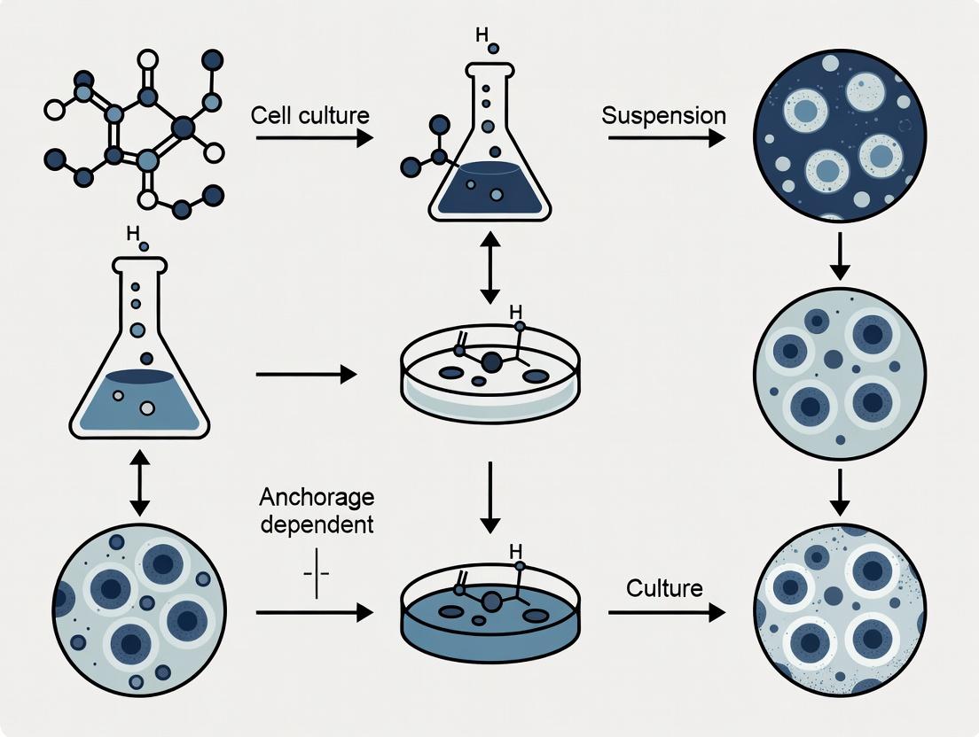
Introduction: Framing the Thesis The central paradigm in mammalian cell culture historically distinguishes anchorage-dependent cells, which require a solid substrate for attachment and proliferation, from suspension-adapted cells, which grow freely in a liquid medium. This article provides an in-depth technical guide to the defining features of suspension-adapted cells, framed within the critical research context of transitioning from adherent to suspension culture for scalable bioprocessing. This adaptation is a cornerstone thesis in modern biologics and cell therapy manufacturing, where suspension systems offer superior scalability, process control, and efficiency over traditional adherent platforms like roller bottles or stacked plates.
Core Hallmarks and Characteristics The adaptation to suspension culture induces and selects for specific phenotypic and genotypic changes. The key hallmarks are summarized below and detailed in the subsequent sections.
Table 1: Quantitative Comparison of Anchorage-Dependent vs. Suspension-Adapted Cell Hallmarks
| Characteristic | Anchorage-Dependent Cells | Suspension-Adapted Cells |
|---|---|---|
| Growth Requirement | Mandatory attachment to solid substrate (e.g., plastic, microcarriers). | No substrate attachment required; growth in free-floating state. |
| Typical Morphology | Spread, flattened, polarized (e.g., fibroblast-like, epithelial-like). | Rounded, spherical, often smaller in diameter (10-20 μm vs. 15-30 μm). |
| Cell Density (viable cells/mL) | Limited by surface area (e.g., ~0.5–2.0 × 10^5 cells/cm²). | High, limited by medium conditions (routinely 5–20 × 10^6 cells/mL, higher for perfusion). |
| Metabolic Profile | Often more glycolytic; metabolism can be influenced by contact signaling. | Highly glycolytic and accelerated; requires careful monitoring of nutrient/ waste levels. |
| Apoptosis Sensitivity | Prone to anoikis (detachment-induced apoptosis). | Anoikis-resistant via altered integrin and survival signaling. |
| Genetic & Proteomic Profile | Expresses full integrin repertoire, focal adhesion kinases (FAK), extracellular matrix (ECM) proteins. | Downregulated integrin signaling; upregulated anti-apoptotic (e.g., Bcl-2), cell cycle, and stress response proteins. |
| Primary Applications | Primary cell expansion, tissue models, some viral vaccine production. | Recombinant protein (mAbs), viral vector (AAV, Lentivirus), vaccine, and cell therapy (CAR-T) production. |
Underlying Molecular Mechanisms and Pathways Suspension adaptation involves a fundamental rewiring of cellular signaling networks, primarily centered on overcoming anoikis and decoupling proliferation from adhesion-based signals.
Diagram 1: Key Signaling Pathways in Suspension Adaptation
Experimental Protocols for Characterization and Adaptation Protocol 1: Serial Adaptation for Generating Suspension Cell Lines Objective: To gradually adapt an adherent cell line (e.g., HEK293, CHO) to growth in suspension and serum-free conditions.
- Seed adherent cells at ~70% confluence in a T-flask.
- Detach cells using standard trypsin/EDTA. Centrifuge and resuspend in Adaptation Medium: a 1:1 mix of the original adherent medium and the target serum-free suspension medium (e.g., CD CHO), supplemented with 1-2% FBS.
- Culture in a low-attachment Erlenmeyer flask placed on an orbital shaker in a humidified CO₂ incubator (37°C, 5% CO₂, 120 rpm). Monitor viability daily.
- Passage cells when viability recovers to >90% and cell density reaches 0.5–1.0 × 10^6 cells/mL. Centrifuge and resuspend in a new medium mix with a higher proportion of target suspension medium (e.g., 30:70 original:target).
- Gradually reduce and eliminate FBS over 5-10 passages, while progressively increasing the target medium to 100%. Incrementally reduce attachment dependence by using poly-lysine coated or finally untreated plastic/shaker flasks.
- Clone by limiting dilution in 96-well plates with conditioned medium to isolate stable, high-viability clones. Expand and cryopreserve the adapted suspension clone.
Protocol 2: Assessing Anoikis Resistance via Soft Agar Colony Formation Objective: To quantitatively evaluate the anchorage-independent growth capacity of adapted cells—a key hallmark of suspension adaptation and transformation.
- Prepare Base Agar Layer: Melt 1.2% agarose in PBS and cool to 45°C. Mix 1:1 with 2X culture medium to create a 0.6% agar solution. Quickly pipette 1.5 mL into each well of a 6-well plate. Allow to solidify at room temperature.
- Prepare Cell Layer: Trypsinize and count suspension and parental adherent cells. Prepare a 0.36% agar solution in 1X medium. Mix the cell suspension with this agar solution to a final density of 5,000–10,000 cells/mL and 0.3% agar. Layer 1.5 mL of this cell-agar mix over the base layer.
- Culture: After the top layer solidifies, carefully add 1 mL of liquid culture medium on top to prevent drying. Incubate at 37°C, 5% CO₂ for 2-3 weeks. Replenish the top medium weekly.
- Stain and Analyze: Add 0.5 mL of 0.005% crystal violet solution for 1+ hours. Count colonies (>50 μm diameter) manually under a microscope or using an automated colony counter. Calculate the colony-forming efficiency (CFE = (colonies counted / cells seeded) × 100%).
Diagram 2: Suspension Cell Line Development Workflow
The Scientist's Toolkit: Essential Research Reagents & Solutions Table 2: Key Reagents for Suspension Cell Research
| Reagent / Material | Function & Application |
|---|---|
| Serum-Free/Suspension Medium (e.g., CD CHO, Freestyle 293) | Chemically defined medium optimized for suspension growth, enhancing reproducibility and downstream purification. |
| Anti-Clumping Agents (e.g., Pluronic F-68) | Non-ionic surfactant added to medium (typically 0.1%) to reduce mechanical shear damage and prevent cell aggregation. |
| Low-Attachment Culture Vessels (Flasks, Plates) | Surface-treated polystyrene that inhibits cell attachment, forcing adaptation to suspension growth. |
| Orbital Shaker & Baffled Flasks | Provides consistent agitation and gas transfer (O₂, CO₂) for suspension cultures in shaker incubators. |
| Automated Cell Counter & Viability Analyzer (e.g., with Trypan Blue) | Essential for daily monitoring of cell density and viability during adaptation and routine culture. |
| Caspase-3/7 Activity Assay Kit | Fluorescence-based assay to quantify apoptosis levels, critical for assessing anoikis during adaptation. |
| Recombinant Growth Factors (e.g., Insulin, Transferrin) | Key supplements in serum-free media to support growth and metabolism in the absence of serum. |
| Cryopreservation Medium (DMSO-based) | For creating stable master cell banks of adapted clones, ensuring long-term genetic stability. |
This technical guide examines the critical choice between primary cells and immortalized cell lines within the context of anchorage-dependent versus suspension culture systems. This decision fundamentally impacts experimental design, data interpretation, and translational relevance in biomedical research and drug development. Anchorage-dependent cells require a solid substrate for growth, while suspension cells grow freely in culture medium, directly influencing scalability and the applicability of findings to in vivo systems, particularly in oncology and immunology.
Core Definitions and Characteristics
Primary Cells
Cells isolated directly from living tissue (human or animal) and cultured for a limited lifespan. They maintain the genotype, phenotype, and functional characteristics of their tissue of origin but have a finite replicative capacity.
Cell Lines
Immortalized populations of cells that can proliferate indefinitely in culture. They are derived from primary cells via spontaneous mutation, genetic manipulation (e.g., introduction of telomerase), or from tumor tissue.
Quantitative Comparison
Table 1: Comparative Analysis of Primary Cells vs. Cell Lines
| Parameter | Primary Cells | Immortalized Cell Lines |
|---|---|---|
| Lifespan/Passages | Finite (usually <10 passages) | Infinite |
| Genetic & Phenotypic Drift | Minimal; high fidelity to tissue of origin | High; accumulate mutations over time |
| Heterogeneity | High; reflects donor tissue diversity | Low; clonal selection leads to uniformity |
| Culture Complexity | High; require optimized, often specialized media | Lower; robust, standardized protocols |
| Cost | High (isolation, characterization, donors) | Low (once established) |
| Experimental Reproducibility | Lower (donor-to-donor variability) | Higher (batch-to-batch consistency) |
| In Vivo Relevance | High - better model for physiology/pathology | Variable - may exhibit artifactual adaptations |
| Typical Culture Format | Often anchorage-dependent; some (e.g., blood cells) are suspension | Both anchorage-dependent and suspension common |
| Scalability for Bioproduction | Challenging and costly | Highly scalable, especially in suspension |
Table 2: Suitability for Culture Types
| Cell Type | Anchorage-Dependent Culture | Suspension Culture | Key Considerations |
|---|---|---|---|
| Primary Epithelial/Fibroblasts | Excellent - natural state | Very Poor | Require coated surfaces (collagen, fibronectin). |
| Primary Hematopoietic Cells | Poor (except adherent subsets) | Excellent - natural state | Media requires cytokines for survival/proliferation. |
| Adherent Cell Lines (e.g., HEK293, HeLa) | Excellent - standard method | Possible with adaptation | Adaptation to suspension alters phenotype. |
| Suspension Cell Lines (e.g., CHO, Jurkat) | Poor | Excellent - standard method | Ideal for high-throughput screening and biomanufacturing. |
Implications for Anchorage-Dependent vs. Suspension Culture Research
The choice between primary cells and cell lines dictates feasible culture methodologies. Anchorage-dependent culture of primary cells is essential for studying tissue architecture, polarity, and cell-matrix interactions but is low-throughput. Suspension culture of adapted cell lines is the cornerstone of industrial bioprocessing (e.g., monoclonal antibody production). A critical research gap lies in developing robust suspension culture systems for primary cells, particularly for immunotherapy (e.g., T-cell, NK cell expansion).
Detailed Experimental Protocols
Protocol 1: Isolation and Anchorage-Dependent Culture of Primary Human Dermal Fibroblasts
Source: Adapted from current tissue dissociation and cell culture guidelines.
- Tissue Acquisition: Obtain de-identified human skin sample in sterile transport medium.
- Dissociation: Mince tissue finely with scalpels. Digest in 5 mL of dissociation solution (see Toolkit) for 2-4 hours at 37°C on a rotator.
- Neutralization & Filtration: Add complete fibroblast growth medium to neutralize enzyme. Filter through a 100µm cell strainer.
- Centrifugation & Seeding: Centrifuge filtrate at 300 x g for 5 min. Resuspend pellet in complete medium. Seed cells at a density of 5,000-10,000 cells/cm² on a T-75 tissue culture flask pre-coated with 1 µg/cm² Fibronectin.
- Culture: Maintain at 37°C, 5% CO₂. Replace medium every 48-72 hours.
- Passaging: At 80% confluence, wash with PBS, dissociate with 0.25% Trypsin-EDTA for 3-5 min, neutralize with serum-containing medium, and re-seed at a 1:3 split ratio.
Protocol 2: Adaptation of an Adherent Cell Line to Suspension Culture
Source: Standard mammalian bioprocess development methodology.
- Baseline Culture: Maintain adherent cells (e.g., HEK293) in standard adherent culture flasks with serum-containing medium.
- Transition to Serum-Free: Gradually adapt cells to a commercial, serum-free suspension medium (see Toolkit) over 3-5 passages by mixing increasing proportions of the new medium with the old.
- Detachment for Suspension: Fully dissociate cells using a gentle, non-enzymatic cell dissociation reagent. Do not use trypsin.
- Initial Suspension Seeding: Seed cells into a low-attachment Erlenmeyer flask at 2-5 x 10⁵ cells/mL in serum-free medium. Place on an orbital shaker in a CO₂ incubator at 37°C, 5% CO₂, 120 rpm.
- Monitoring & Passaging: Monitor cell viability daily via trypan blue exclusion. When cell density reaches 1-2 x 10⁶ cells/mL and viability is >90%, passage by diluting to 3-5 x 10⁵ cells/mL in fresh medium.
- Clone Selection (Optional): Isolate single cells showing robust growth in suspension to select for a clonally adapted population.
Visualizing Culture Decision Pathways
Diagram 1: Cell Source and Culture Type Decision Tree
The Scientist's Toolkit: Key Research Reagent Solutions
Table 3: Essential Materials for Featured Protocols
| Reagent/Material | Function | Example/Catalog |
|---|---|---|
| Collagenase Type IV | Enzyme for gentle tissue dissociation; breaks down collagen in extracellular matrix. | Worthington CLS-4 |
| Fibronectin, Human | Coating protein for culture surfaces to promote attachment and spreading of anchorage-dependent primary cells. | Corning 356008 |
| Serum-Free Suspension Medium | Chemically defined medium supporting growth of specific cell types in suspension without FBS. | Gibco FreeStyle 293 Expression Medium |
| Gentle Cell Dissociation Reagent | Non-enzymatic, EDTA-based solution for detaching cells without damaging surface proteins. | Stemcell Technologies 07174 |
| Low-Attachment Flask | Flask with polymer coating that inhibits cell attachment, forcing growth in suspension. | Corning Ultra-Low Attachment Flask |
| Orbital Shaker for Incubators | Provides consistent agitation to suspension cultures, ensuring proper gas exchange and preventing clumping. | Thermo Scientific MaxQ 6000 |
| Cell Strainer (70µm, 100µm) | Filters out tissue aggregates and debris post-dissociation to obtain a single-cell suspension. | Falcon 352350/352360 |
| Defined FBS Alternative | Animal-free, consistent supplement for primary cell culture, reducing variability vs. FBS. | Gibco KnockOut SR |
The Role of the Extracellular Matrix (ECM) and Integrin Signaling
The fundamental distinction between anchorage-dependent and suspension cell cultures is a cornerstone of cell biology and bioprocessing. Anchorage-dependent cells require attachment to a solid substrate, typically mediated by interactions with the Extracellular Matrix (ECM), to proliferate, differentiate, and avoid anoikis (detachment-induced apoptosis). In contrast, suspension cells, such as hematopoietic lineages or adapted cell lines, grow freely in culture media. This whitepaper delves into the molecular machinery enabling anchorage dependence: the ECM and its primary cellular receptors, the integrins. Their bidirectional signaling is not merely an adhesive event but a master regulatory mechanism controlling cell fate, migration, and survival—critical considerations for scalable culture systems, organoid development, and metastatic cancer research.
The ECM is a dynamic, insoluble network of proteins and polysaccharides. Its composition varies by tissue and dictates mechanical and biochemical signaling.
Table 1: Major ECM Components and Their Functional Properties
| ECM Component | Primary Role/Function | Key Ligands/Receptors | Typical Concentration in Tissues (Range) |
|---|---|---|---|
| Collagen I | Tensile strength, structural scaffold | Integrins α1β1, α2β1, α10β1, α11β1 | 0.5 - 2.0 mg/ml (dermis) |
| Fibronectin | Cell adhesion, migration, assembly of other ECM | Integrin α5β1, αVβ3, αVβ1 | 0.03 - 0.3 mg/ml (plasma) |
| Laminin | Basement membrane foundation, polarity, differentiation | Integrins α3β1, α6β1, α6β4, Dystroglycan | ~0.1 mg/ml (basement membrane) |
| Hyaluronic Acid | Hydration, space-filling, cell migration | CD44, RHAMM | 0.1 - 0.4 mg/ml (synovial fluid) |
| Vitronectin | Serum-borne adhesion, complement regulation | Integrins αVβ3, αVβ5 | 0.2 - 0.5 mg/ml (human plasma) |
Integrin Structure and Activation Mechanism
Integrins are heterodimeric (α and β subunit) transmembrane receptors. In a resting state on suspended or non-adherent cells, integrins often adopt a bent, inactive conformation with low affinity for ligand. Upon intracellular signaling ("inside-out" activation) or ligand binding, they undergo a dramatic conformational shift to an extended, high-affinity state. This allows for the formation of focal adhesions—large, multi-protein complexes that link the ECM to the actin cytoskeleton.
Core Integrin Signaling Pathways: FAK and Beyond
Ligand binding and integrin clustering recruit and activate Focal Adhesion Kinase (FAK), the central signaling node. FAK autophosphorylation at Y397 creates a binding site for Src family kinases, forming an active FAK/Src complex.
Table 2: Major Downstream Pathways of Integrin/FAK Signaling
| Pathway | Key Effectors | Primary Cellular Outcomes | Relevance to Culture Models |
|---|---|---|---|
| Survival (PI3K-Akt) | FAK/Src → PI3K → PDK1 → Akt | Inhibition of pro-apoptotic proteins (e.g., Bad, Caspase-9). | Prevents anoikis in anchorage-dependent culture. |
| Proliferation (MAPK/ERK) | FAK/Src → Grb2/SOS → Ras → Raf → MEK → ERK | Entry into cell cycle, progression through G1/S phase. | Explains lack of proliferation in non-adherent conditions. |
| Cytoskeletal Organization (Rho GTPases) | FAK → p130Cas → DOCK180 → Rac1 (lamellipodia). Integrins → GEFs → RhoA (stress fibers). | Cell spreading, migration, adhesion maturation. | Critical for cell morphology on 2D vs. 3D matrices. |
| Gene Expression | FAK → ERK → Transcription factors (e.g., ETS1). Integrin-linked Kinase (ILK) → β-catenin. | Regulation of differentiation and matrix remodeling genes. | Drives tissue-specific function in 3D organoid cultures. |
Diagram 1: Core Integrin-FAK Signaling Pathway
Experimental Protocols for Key Investigations
Protocol 1: Assessing Anoikis in Suspension Culture Objective: To quantify apoptosis induced by loss of integrin-ECM engagement.
- Cell Preparation: Harvest anchorage-dependent cells (e.g., MCF-10A, primary fibroblasts) using a non-enzymatic cell dissociation buffer to preserve integrin function.
- Suspension Culture: Seed cells (5 x 10^4 cells/mL) onto ultra-low attachment 6-well plates coated with poly(2-hydroxyethyl methacrylate) (poly-HEMA) to prevent adhesion.
- Control: Seed cells on standard tissue culture plates coated with 10 µg/mL human fibronectin.
- Incubation: Culture for 24-72 hours.
- Analysis: Harvest cells.
- Viability: Use flow cytometry with Annexin V-FITC/PI dual staining.
- Caspase Activity: Measure caspase-3/7 activity via luminescent assay.
- Western Blot: Probe for cleaved PARP and cleaved caspase-3.
Protocol 2: Integrin-Mediated Adhesion and Spreading Assay Objective: To characterize specific integrin-ECM interactions.
- Substrate Coating: Coat 96-well plates with ECM proteins (e.g., Collagen I, Fibronectin, BSA control) at 10 µg/mL in PBS overnight at 4°C.
- Blocking: Block with 1% heat-inactivated BSA for 1 hour.
- Cell Treatment: Pre-incubate cells in suspension for 20 min with function-blocking anti-integrin antibodies (e.g., anti-β1, anti-α5) or isotype control.
- Assay: Seed cells (2 x 10^4/well) and allow to adhere for 30-90 min at 37°C.
- Wash & Fix: Gently wash with PBS to remove non-adherent cells, fix with 4% PFA.
- Quantification: Stain actin cytoskeleton (Phalloidin) and nuclei (DAPI). Image with automated microscopy. Analyze cell area and circularity using image analysis software (e.g., ImageJ).
Protocol 3: Analysis of Focal Adhesion Kinase (FAK) Activation Objective: To measure integrin signaling output via FAK phosphorylation.
- Stimulation & Inhibition:
- Re-attachment: Serum-starve cells for 24h, detach, and hold in suspension for 60 min. Replate on fibronectin-coated dishes for 0, 15, 30, 60 min.
- Inhibitor Control: Pre-treat cells with 10 µM PF-573228 (FAK inhibitor) 1h before replating.
- Lysis: Lyse cells in RIPA buffer containing protease and phosphatase inhibitors.
- Immunoblotting: Perform SDS-PAGE and Western blot.
- Primary Antibodies: Anti-phospho-FAK (Y397), anti-total FAK, anti-β-actin (loading control).
- Densitometry: Quantify band intensity; express pFAK signal normalized to total FAK.
The Scientist's Toolkit: Key Research Reagent Solutions
Table 3: Essential Reagents for ECM and Integrin Signaling Research
| Reagent/Material | Function/Application | Example Product/Catalog |
|---|---|---|
| Recombinant Human Fibronectin | Gold-standard substrate for integrin α5β1 and αV family adhesion studies. | Corning, Cat # 356008 |
| Ultra-Low Attachment Plates | Coated with hydrogel to enforce suspension culture for anoikis studies. | Corning Costar, Cat # 3471 |
| Function-Blocking Anti-Integrin Antibodies | To disrupt specific integrin-ligand interactions (e.g., anti-α5β1, anti-αVβ3). | Millipore, MAB1969 (anti-α5) |
| FAK Inhibitor (PF-573228) | Selective, ATP-competitive inhibitor of FAK kinase activity. | Tocris, Cat # 3239 |
| RGD (Arg-Gly-Asp) Peptide | Competitive antagonist for many integrins (e.g., αVβ3, α5β1). Used as soluble inhibitor. | Sigma, Cat # A8052 |
| Cultrex Basement Membrane Extract (BME) | Laminin-rich, natural 3D matrix for organotypic and invasion assays. | R&D Systems, Cat # 3433-005-01 |
| Phospho-FAK (Y397) Antibody | Key reagent for detecting activated FAK via Western blot or immunofluorescence. | Cell Signaling Tech, Cat # 8556 |
| Cell Dissociation Buffer (Enzyme-Free) | For gentle detachment to preserve integrin surface expression for subsequent assays. | Gibco, Cat # 13151014 |
Diagram 2: Experimental Workflow for Integrin Signaling Analysis
Implications for Research and Drug Development
Understanding ECM-integrin signaling bridges fundamental biology with applied research. For regenerative medicine, designing synthetic biomimetic matrices requires precise knowledge of integrin-binding motifs. In cancer therapeutics, targeting integrins (e.g., αVβ3) or FAK is a strategy to inhibit metastasis and overcome therapy resistance. For bioprocessing, transitioning CHO or HEK293 cells to suspension culture in serum-free media often involves adapting integrin signaling networks or providing alternative survival pathways. Thus, dissecting this molecular dialogue is pivotal for advancing both basic science and industrial application across the anchorage-dependence paradigm.
Common Cell Types for Each System (e.g., HEK293, CHO, MSCs, Vero)
The dichotomy between anchorage-dependent (adherent) and suspension cell culture systems is a foundational pillar in bioprocess development. The choice of cell line is intrinsically linked to this paradigm, dictating bioreactor design, scale-up strategy, and ultimately, the economic viability of producing biologics, vaccines, or cell therapies. This guide details the common cell types for each system, framing their characteristics and applications within this core research thesis. Anchorage-dependent lines require a solid substrate (e.g., microcarriers in bioreactors), while suspension-adapted lines grow freely in culture media, offering advantages in scalability and process control.
Core Cell Types: Characteristics and Applications
The following table categorizes prevalent cell lines by their natural or adapted growth phenotype and primary application.
Table 1: Common Cell Types in Bioproduction and Research
| Cell Type | Origin | Growth Phenotype (Natural/Adapted) | Primary Application System | Key Product/Use |
|---|---|---|---|---|
| HEK293 | Human Embryonic Kidney | Adherent / Suspension-adapted variants | Recombinant Protein Production, Viral Vector Production | Transient protein expression, Lentiviral/Adenoviral vectors, Antibodies |
| CHO | Chinese Hamster Ovary | Adherent / Suspension-adapted (industry standard) | Recombinant Protein Production | Monoclonal Antibodies, Fc-fusion proteins, Enzymes |
| MSCs | Mesenchymal Stem/Stromal Cells (Bone Marrow, Adipose) | Strictly Anchorage-Dependent | Cell Therapy, Regenerative Medicine | Allogeneic/autologous cell therapies, Immunomodulation, Tissue repair |
| Vero | African Green Monkey Kidney | Strictly Anchorage-Dependent (on microcarriers at scale) | Viral Vaccine Production | Live-attenuated viral vaccines (Polio, Rabies, COVID-19), Virus isolation |
| NS0/Sp2/0 | Mouse Myeloma | Suspension | Recombinant Protein Production | Monoclonal Antibodies (non-glycosylated or alternative glycosylation) |
| Sf9/Sf21 | Spodoptera frugiperda (Fall Armyworm) Ovary | Suspension (in serum-free media) | Baculovirus-Insect Cell Expression | Recombinant proteins, Virus-like particles (VLPs), Baculovirus insecticides |
Table 2: Quantitative Comparison of Key Bioprocess Parameters
| Parameter | CHO-S (Suspension) | HEK293 Suspension | Vero (on Microcarriers) | MSCs (Adherent, Planar) |
|---|---|---|---|---|
| Typical Peak VCD (cells/mL) | 10–20 x 10⁶ | 5–10 x 10⁶ | 2–4 x 10⁶ | 0.2–0.5 x 10⁶ / cm² |
| Doubling Time (hours) | 14–24 | 20–30 | 24–36 | 24–48 |
| Max. Viable Cell Density in Production Bioreactors (reported) | Up to ~150 x 10⁶ cells/mL (perfusion) | Up to ~20 x 10⁶ cells/mL (batch/fed-batch) | 1–2 x 10⁷ cells/mL (on carriers, perfusion) | Limited by surface area |
| Product Titer Range (Mabs/Proteins) | 3–10+ g/L (fed-batch) | 0.1–1 g/L (transient) | N/A (virus yield: e.g., ~10⁸–10¹⁰ PFU/mL for Polio) | N/A (cellular product) |
| Scalability | High (stirred-tank > 20,000 L) | Moderate-High (stirred-tank to 1,000 L) | Moderate (fixed-bed, stirred-tank with carriers) | Low (multilayer flasks, cell factories) |
Detailed Methodologies: Key Experimental Protocols
Protocol 1: Adaptation of Adherent HEK293 Cells to Serum-Free Suspension Culture
- Objective: Generate a clonal or pooled suspension-adapted HEK293 cell line for bioreactor culture.
- Materials: Adherent HEK293 parent line, serum-containing growth medium, target serum-free suspension medium (e.g., FreeStyle 293, EX-CELL), non-enzymatic cell dissociation reagent, Erlenmeyer shake flasks.
- Procedure:
- Culture adherent cells to ~80% confluence.
- Dissociate using a gentle, non-enzymatic reagent. Centrifuge and resuspend in a 1:1 mix of old adherent medium and new serum-free suspension medium.
- Seed cells into a low-adhesion flask or shake flask at a density of 2-3 x 10⁵ cells/mL. Place on an orbital shaker (110-130 rpm) in a CO₂ incubator.
- Monitor viability daily. When viability drops below 80% or cells double, centrifuge and resuspend in a medium mix with a higher ratio of new suspension medium (e.g., 25:75, then 100%).
- Passage cells every 3-4 days, maintaining viability >90%. Gradually increase shaker speed to the standard 125 rpm.
- After 15-20 passages, assess growth kinetics and product expression. Single-cell cloning may be performed post-adaptation for stable line generation.
Protocol 2: Microcarrier Culture of Vero Cells for Virus Propagation
- Objective: Expand Vero cells on microcarriers in a bioreactor for subsequent viral infection and harvest.
- Materials: Vero cell working bank, GMEM or DMEM medium + 10% FBS, trypsin-EDTA, Cytodex 1 or 3 microcarriers, stirred-tank bioreactor with pH/DO control.
- Procedure:
- Microcarrier Preparation: Hydrate and sterilize microcarriers according to manufacturer's instructions. Wash with PBS and equilibrate in serum-containing growth medium.
- Cell Seeding: Trypsinize adherent Vero cells and resuspend in growth medium. Seed at a ratio of 3-5 cells per microcarrier into the bioreactor vessel containing carriers at 3-5 g/L in a reduced volume.
- Initial Attachment: Run the bioreactor with intermittent stirring (e.g., 30 sec on, 90 sec off) for 4-8 hours to facilitate cell attachment.
- Expansion Phase: After attachment, increase culture volume to working volume and switch to continuous stirring at 40-60 rpm. Maintain at 37°C, pH 7.2, DO >40% air saturation. Perform complete medium exchanges every 2-3 days.
- Infection: Once confluence is reached on carriers (typically 3-5 days), reduce serum concentration. Infect with virus at a predetermined MOI (Multiplicity of Infection) in a small volume.
- Harvest: After appropriate cytopathic effect is observed, lower temperature to halt propagation. Separate virus-containing supernatant from microcarriers via sieving or sedimentation.
Visualizations
Diagram 1: CHO Cell Bioprocess Workflow for mAb Production
Diagram 2: Key Signaling in MSC Expansion vs. Differentiation
The Scientist's Toolkit: Research Reagent Solutions
Table 3: Essential Materials for Featured Cell Culture Systems
| Item | Function | Example Use Case |
|---|---|---|
| Serum-Free/Suspension Medium | Chemically defined medium supporting growth in suspension; eliminates serum variability. | Adaptation and routine culture of CHO-S or HEK293S cells. |
| Microcarriers (e.g., Cytodex) | Solid or porous microspheres providing surface for adherent cell growth in suspension mode. | Scaling up Vero or MSC cultures in stirred-tank bioreactors. |
| Non-Enzymatic Dissociation Reagent | Gentle cell detachment solution that preserves surface proteins and viability. | Passaging sensitive adherent lines (e.g., MSCs) prior to adaptation or bioreactor seeding. |
| Polyethylenimine (PEI) Max | Cationic polymer for transient transfection of suspension cells. | High-yield recombinant protein production in HEK293 or CHO cells. |
| Cell Counters with Viability Staining | Automated or manual systems (with Trypan Blue) for monitoring cell density and health. | Critical for determining infection/passage time in all bioprocesses. |
| Orbital Shaker Flask Systems | Baffled flasks for gas transfer in suspension culture. | Expansion of suspension-adapted lines from shake flask to seed train. |
| Biosafety Cabinet (Class II) | Provides aseptic environment for all open cell culture manipulations. | Essential for maintaining sterility during media changes, passaging, and sampling. |
| Controlled Bioreactor (Benchtop) | System with monitoring/control of pH, DO, temperature, and agitation. | Optimizing fed-batch or perfusion processes for CHO mAb production. |
Practical Protocols: Cultivating and Scaling Adherent & Suspension Systems
The choice between anchorage-dependent and suspension cell culture systems is a foundational decision in bioprocess development, influencing scale-up strategy, productivity, and ultimately, the economic viability of producing biologics, vaccines, and cell therapies. This guide details the core equipment—flasks, microcarriers, and bioreactors—that enable both paradigms, with a focus on transitioning adherent cells to scalable suspension culture via microcarrier technology.
Core Vessels: From Static to Dynamic Culture
Flasks: The Foundation of Adherent Culture
Flasks provide a simple, controlled environment for two-dimensional (2D) monolayer culture of anchorage-dependent cells (e.g., MRC-5, Vero, HEK 293).
Key Types & Quantitative Specifications: Table 1: Common Culture Flask Specifications
| Flask Type (Surface Area) | Typical Working Volume | Seeding Density (cells/cm²) | Material | Primary Use Case |
|---|---|---|---|---|
| T-25 | 5-7 mL | 2.0 - 5.0 x 10⁴ | Polystyrene | Routine maintenance, small-scale experiments |
| T-75 | 15-25 mL | 2.0 - 5.0 x 10⁴ | Polystyrene | Expansion, seed train initiation |
| T-175 | 35-60 mL | 2.0 - 5.0 x 10⁴ | Polystyrene | Large-scale expansion, virus production |
| HyperFlask (1720 cm²) | 300-350 mL | 2.0 - 5.0 x 10⁴ | Polystyrene | High-yield adherent scale-up without microcarriers |
| Roller Bottle (850 cm²) | 100-200 mL | 1.5 - 4.0 x 10⁴ | Polystyrene | Intermediate-scale production |
Experimental Protocol: Seeding and Passaging in T-Flasks
- Trypsinization: Aspirate spent medium. Wash monolayer with 5-10 mL DPBS (Ca²⁺/Mg²⁺ free). Add 1-3 mL of 0.25% Trypsin-EDTA solution (volume varies by flask size). Incubate at 37°C for 2-5 minutes until cells detach.
- Neutralization: Add complete medium containing serum (or a trypsin inhibitor) at a 2:1 ratio to the trypsin volume. Pipette vigorously to create a single-cell suspension.
- Centrifugation: Transfer suspension to a conical tube. Centrifuge at 200-300 x g for 5 minutes. Aspirate supernatant.
- Reseeding: Resuspend pellet in fresh pre-warmed medium. Count cells using a hemocytometer or automated counter. Dilute to required seeding density and dispense into new, pre-incubated flasks.
- Incubation: Place flasks in a humidified incubator at 37°C, 5% CO₂. Check confluence daily.
Microcarriers: Bridging Adherence and Suspension
Microcarriers are solid or porous beads (50-400 µm diameter) that provide a surface for cell attachment, enabling the cultivation of anchorage-dependent cells in stirred-tank bioreactors.
Types and Selection Criteria: Table 2: Microcarrier Types and Characteristics
| Microcarrier Type | Material | Diameter (µm) | Surface Charge/Coating | Key Advantage | Example Cell Lines |
|---|---|---|---|---|---|
| Solid | Dextran, Polystyrene | 150-200 | Cationic (e.g., DEAE) | Simple harvesting, robust | Vero, MRC-5 |
| Porous | Gelatin, Collagen | 100-400 | Natural ECM proteins | High surface area, 3D-like environment | CHO-K1, Mesenchymal Stem Cells |
| Macroporous | Polyethylene, Plastic | 200-400 | Various | Protection from shear, very high cell density | HEK 293, Hepatocytes |
| Dissolvable | Gelatin, Hyaluronic Acid | 100-200 | Customizable | Easy cell recovery without enzymatic treatment | Cell therapy applications |
Experimental Protocol: Scaling Up with Microcarriers in a Spinner Flask
- Microcarrier Preparation: Hydrate and sterilize microcarriers per manufacturer instructions (e.g., autoclave or sterile filtration). For 1g/L final concentration in a 250 mL spinner, hydrate 0.25g in 50 mL PBS, then wash with culture medium.
- Spinner Flask Setup: Transfer carriers and medium to a sterile spinner vessel. Final working volume should be 50-60% of total flask volume to ensure proper gas exchange and mixing.
- Cell Seeding: Trypsinize cells from a near-confluent T-175 flask. Count and resuspend in a small volume of medium. Add cell suspension to the stationary spinner flask at a density of 20-35 cells per microcarrier. For 0.25g of Cytodex 1 (~6000 carriers/g), this requires ~30,000-52,500 cells.
- Attachment Phase: Allow the flask to sit undisturbed in the incubator for 4-8 hours, gently swirling every 30 minutes to ensure uniform distribution.
- Initiation of Agitation: Begin slow agitation (25-40 rpm) to keep carriers suspended. Gradually increase to 50-70 rpm over 24-48 hours.
- Feeding & Monitoring: Perform 50% medium exchanges every 2-3 days. Monitor glucose consumption, cell count via nuclei staining, and carrier confluence microscopically.
Bioreactors: The Apex of Controlled Scale-Up
Bioreactors provide a fully controlled environment (pH, DO, temperature, agitation) for both microcarrier-based adherent culture and suspension culture of non-anchorage-dependent cells (e.g., CHO, Sp2/0, HEK 293 in suspension).
System Comparison: Table 3: Bioreactor Systems for Adherent and Suspension Culture
| Bioreactor Type | Scale Range | Key Control Parameters | Ideal for Cell Type | Shear Stress Management |
|---|---|---|---|---|
| Stirred-Tank (STR) | 1L - 20,000L | pH, DO, Temp, Agitation, Sparging | Suspension, Microcarriers | Impeller design (e.g., pitched blade), controlled agitation |
| Wave-type | 1L - 500L | Rocking rate/angle, Temp, Air flow | Seed train, microcarriers, sensitive cells | Gentle rocking motion, no impeller |
| Fixed-Bed/Packed-Bed | 0.1L - 1000L | Perfusion rate, Temp, pH (in/out) | High-density adherent cells | Cells protected in matrix |
| Hollow Fiber | N/A | Media circulation rate, Harvest rate | Very high-density, secreted products | Minimal shear in extracapillary space |
Experimental Protocol: Inoculating a Bench-Top Bioreactor with Microcarriers
- Bioreactor Preparation: Calibrate pH and DO probes. Add basal medium to the vessel (e.g., 1.5L in a 3L vessel). Set temperature to 37°C, pH to 7.2 (controlled via CO₂ sparging and base addition), and DO to 40% air saturation (controlled via air/O₂ mix and agitation). Allow system to stabilize overnight.
- Microcarrier/Cell Inoculum: Prepare a microcarrier-cell inoculum in a spinner flask as per the protocol above, allowing cells to attach over 24 hours.
- Transfer: Aseptically transfer the entire spinner flask contents to the bioreactor vessel.
- Process Control: Set initial agitation to 50-70 rpm to maintain carrier suspension without damaging cells (tip speed < 1 m/s). Maintain setpoints for pH and DO.
- Perfusion/Fed-Batch: For extended culture, initiate a perfusion system after 72 hours or implement a fed-batch strategy with concentrated nutrient feeds.
- Harvest: For cells: stop agitation, allow carriers to settle, drain medium, and enzymatically digest cells from carriers. For product: continuously harvest from the perfusate.
The Scientist's Toolkit: Essential Research Reagent Solutions
Table 4: Key Reagents for Anchorage-Dependent and Suspension Culture Research
| Reagent/Material | Function | Example Product/Brand |
|---|---|---|
| Trypsin-EDTA (0.05%-0.25%) | Proteolytic enzyme mixture for detaching adherent cells from surfaces. | Gibco Trypsin, Corning |
| Soybean Trypsin Inhibitor | Stops trypsin activity post-detachment, crucial for sensitive cells. | Sigma-Aldrich |
| Defined FBS Alternative | Serum-free supplement for consistent, xeno-free cell growth. | Gibco KnockOut SR, Cytiva HyClone UC |
| Recombinant Attachment Factors | (e.g., Fibronectin, Vitronectin) Enhance cell attachment to surfaces/carriers in defined conditions. | Corning Cell-Tak, Gibco |
| Anti-Clumping Agents | (e.g., Pluronic F-68) Reduce mechanical shear and cell aggregation in suspension/bioreactor cultures. | Gibco |
| Nuclei Counting Dye | (e.g., Crystal Violet, Hoechst) Allows accurate cell counting on microcarriers via lysed nuclei. | Sigma-Aldrich |
| Microcarrier Media | Optimized basal media (low calcium, high buffer capacity) for microcarrier culture. | Gibco DMEM/F-12 for microcarriers |
| pH & DO Probes (Sterilizable) | In-line sensors for real-time monitoring of critical process parameters in bioreactors. | Mettler Toledo, Hamilton |
Visualizing the Workflow and Decision Logic
Title: Cell Culture Scale-Up Decision Workflow
Title: Key Signaling in Anchorage-Dependent Cells
Adherent cell culture is a cornerstone technique for studying anchorage-dependent cells, which require attachment to a solid substrate for proliferation, survival, and function. This dependence contrasts with suspension cultures, where cells grow freely in a medium. The research dichotomy between these two systems is fundamental; adherent cultures often better model in vivo tissue architecture (e.g., epithelial layers, fibroblasts) and are critical for studying cell-matrix interactions, differentiation, and contact inhibition. In drug development, adherent cultures are essential for high-content screening, toxicity assays, and production of certain biologics using engineered cell lines. This protocol provides a standardized, technical guide for establishing, maintaining, and passaging adherent cultures, ensuring reproducibility essential for robust research conclusions in comparative studies.
Key Research Reagent Solutions & Materials
Table 1: Essential Materials for Adherent Cell Culture
| Reagent/Material | Function & Rationale |
|---|---|
| Complete Growth Medium | Typically comprises a basal medium (e.g., DMEM, RPMI-1640) supplemented with fetal bovine serum (FBS, 5-20%), growth factors, and antibiotics. Provides nutrients and adhesion factors. |
| Dulbecco’s Phosphate Buffered Saline (DPBS), Ca²⁺/Mg²⁺-free | Used for rinsing cells without disrupting cell-cell interactions. The absence of divalent cations prevents cadherin-mediated adhesion, aiding cell detachment. |
| Trypsin-EDTA Solution | Proteolytic enzyme (trypsin) digests extracellular matrix and cell-surface proteins; EDTA chelates calcium, further disrupting integrin-mediated adhesion. Standard agent for cell dissociation. |
| Trypsin Neutralizing Solution | Typically serum-containing medium or a defined inhibitor. Halts trypsin activity immediately post-detachment to prevent over-digestion and cell damage. |
| Cell Culture Vessel | Treated polystyrene flask/dish/plate. Surface is often treated with plasma or coated with polymers (e.g., poly-L-lysine, collagen) to enhance hydrophilicity and cell attachment. |
| Cell Counter & Viability Stain | Hemocytometer or automated counter with Trypan Blue dye. Enables accurate quantification of cell number and assessment of viability post-passage. |
| 37°C Incubator with 5% CO₂ | Maintains physiological temperature and pH (via bicarbonate buffer in medium) for optimal cell growth. |
Core Protocol: Establishment and Passaging
Protocol for Thawing and Establishing a New Adherent Culture
Aim: To recover cells from cryopreservation and establish a proliferating monolayer.
- Preparation: Warm complete growth medium and DPBS in a 37°C water bath. Pre-warm the culture vessel.
- Thawing: Rapidly thaw cryovial in a 37°C water bath (~60-90 seconds). Immediately upon ice crystal disappearance, sterilize vial with 70% ethanol.
- Dilution: Transfer cell suspension to a 15 mL conical tube. Slowly add 9 mL of pre-warmed medium drop-wise to dilute the cryoprotectant (DMSO).
- Centrifugation: Centrifuge at 200 x g for 5 minutes to pellet cells. Aspirate supernatant.
- Reseeding: Resuspend cell pellet gently in 1-2 mL of fresh medium. Seed into an appropriately sized culture vessel containing pre-equilibrated medium.
- Incubation: Place vessel in a 37°C, 5% CO₂ incubator. Allow 24-48 hours for attachment before first medium change.
Protocol for Routine Passaging of Adherent Cells
Aim: To subculture a confluent monolayer for maintenance or expansion.
- Assessment: Visually inspect cells under a microscope. Passage when cells are 70-90% confluent (see Table 2).
- Preparation: Warm all solutions (DPBS, trypsin-EDTA, complete medium).
- Rinsing: Aspirate and discard spent medium. Gently rinse cell layer with Ca²⁺/Mg²⁺-free DPBS (e.g., 5 mL for a T75 flask) to remove serum that inhibits trypsin.
- Detachment: Add pre-warmed trypsin-EDTA solution (e.g., 1-2 mL for a T75). Swirl to coat monolayer. Incubate at 37°C for 1-3 minutes.
- Observation & Neutralization: Monitor under microscope until cells round up and detach (tap side of vessel if needed). Immediately add 2-3 volumes of complete medium or trypsin neutralizer to inactivate trypsin.
- Collection: Pipette suspension over the growth surface to collect all cells. Transfer to a conical tube.
- Centrifugation & Counting: Centrifuge at 200 x g for 5 min. Aspirate supernatant. Resuspend in fresh medium. Perform cell count and viability assessment using Trypan Blue exclusion.
- Reseeding: Seed a predetermined number of cells into a new culture vessel containing fresh, pre-warmed medium. Calculate seeding density based on Table 2.
Table 2: Quantitative Guidelines for Common Adherent Cell Lines
| Cell Line | Typical Seeding Density (cells/cm²) | Doubling Time (hours) | Recommended Split Ratio* | Target Confluence for Passage |
|---|---|---|---|---|
| HEK 293 | 2.5 x 10⁴ - 5 x 10⁴ | 20-30 | 1:5 to 1:10 | 80-90% |
| HeLa | 2 x 10⁴ - 4 x 10⁴ | 24 | 1:5 to 1:8 | 80-90% |
| MCF-10A | 3 x 10⁴ - 6 x 10⁴ | 18-24 | 1:3 to 1:6 | 70-80% |
| NIH/3T3 | 1 x 10⁴ - 2 x 10⁴ | 18-24 | 1:5 to 1:10 | 70-80% |
| Primary Human Dermal Fibroblasts (HDFs) | 5 x 10³ - 1 x 10⁴ | 40-60 | 1:2 to 1:4 | 80-100% |
*Split ratio is vessel-dependent (e.g., 1:5 from a T25 means seeding 1/5th of the cell yield into a new T25).
Signaling Pathways in Adhesion and Detachment
Adherent cell survival is governed by integrin-mediated signaling. Upon attachment to extracellular matrix (ECM) components like fibronectin, integrins cluster and activate focal adhesion kinase (FAK), triggering pro-survival (PI3K/Akt) and proliferative (MAPK/ERK) pathways. Trypsinization directly cleaves integrins and ECM proteins, disrupting this survival signaling and inducing a form of anoikis (detachment-induced apoptosis) if not rapidly neutralized by re-plating.
Diagram 1: Adhesion Signaling & Trypsin Disruption
Experimental Workflow: From Culture to Assay
Diagram 2: Adherent Cell Culture & Passage Workflow
This technical guide provides a standardized protocol for suspension cell culture. Within the broader thesis comparing anchorage-dependent (AD) and suspension cell systems, this protocol highlights a key operational advantage: suspension cultures eliminate the need for enzymatic or mechanical detachment required for subculturing adherent lines. This fundamental difference impacts scalability, automation potential, and the physiological relevance of cell-extracellular matrix interactions. Suspension lines, including hematopoietic cells, many cancer lines (e.g., Jurkat, HL-60), and adapted CHO cells for bioproduction, are essential for high-throughput screening, large-scale protein/antibody production, and studies of non-adherent biological systems.
Essential Materials and Reagents (The Scientist's Toolkit)
| Research Reagent / Material | Function / Explanation |
|---|---|
| Suspension-Adapted Cell Line | Cells that proliferate freely in culture media without surface attachment (e.g., Jurkat, THP-1, CHO-S, HEK-293 Suspension). |
| Serum-Free / Defined Medium | Optimized for suspension growth; reduces batch variability, simplifies downstream purification, and enhances reproducibility vs. serum-containing media. |
| Sterile Culture Flasks (Erlenmeyer style with vented cap) | Designed for suspension; baffled base improves aeration and gas exchange during shaking incubation. |
| Orbital Shaker Incubator | Maintains cells in suspension, ensures consistent nutrient and gas distribution, and controls temperature, humidity, and CO₂. |
| Hemocytometer or Automated Cell Counter | For determining cell density and viability via trypan blue exclusion or similar dyes. |
| Sterile Centrifuge Tubes | For pelleting cells during passaging or medium exchange. |
| Cell Strainer (40-70 µm) | To break up small cell clumps and ensure a single-cell suspension for accurate counting. |
| Trypan Blue Solution (0.4%) | Vital dye used to distinguish live (unstained) from dead (blue-stained) cells. |
| Phosphate-Buffered Saline (PBS) | For diluting cells and washing cell pellets if required. |
| Cryopreservation Medium | Typically culture medium with 5-10% DMSO, used for freezing down cell stocks. |
Detailed Maintenance and Subculture Protocol
Routine Monitoring and Key Quantitative Parameters
Monitor cells daily. Key parameters are summarized in the table below.
Table 1: Key Quantitative Parameters for Suspension Cell Culture Maintenance
| Parameter | Typical Target Range | Measurement Method & Notes |
|---|---|---|
| Cell Density | Maintenance: 2.0-5.0 x 10⁵ cells/mL Passaging Trigger: 1.0-2.0 x 10⁶ cells/mL Maximum Density: Varies by line (e.g., 2-4 x 10⁶ cells/mL) | Count using hemocytometer or automated counter. Maintain in exponential growth phase. |
| Viability | ≥ 90% (Routine culture) ≥ 95% (For critical experiments) ≥ 80% (Post-thaw, acceptable) | Trypan Blue exclusion assay. Low viability indicates stress, contamination, or nutrient exhaustion. |
| Doubling Time | 16-48 hours (varies widely by cell line) | Calculated from growth curve data. Critical for experiment planning. |
| Subculture Split Ratio | Commonly 1:3 to 1:10 | Depends on cell line growth rate and target seeding density. |
| Seeding Density | 2.0-5.0 x 10⁵ cells/mL | Optimal density to re-enter log-phase growth post-passage. |
| Shaker Speed | 100-130 rpm (for flasks in standard incubators) | Ensures adequate mixing without causing shear stress or foam formation. |
| Culture Volume | 20-30% of total flask volume (e.g., 20 mL in a 125 mL flask) | Ensures proper aeration (surface area-to-volume ratio). |
Step-by-Step Subculturing (Passaging) Procedure
Materials: Culture flask, pre-warmed complete medium, sterile centrifuge tubes, hemocytometer/slides, trypan blue, pipettes, waste container, incubator.
Protocol:
- Observe: Visually inspect culture for color, turbidity, and any signs of contamination (unusual cloudiness, pH change).
- Count:
- Gently mix the flask to ensure a homogeneous cell suspension.
- Aseptically remove a 0.5-1 mL sample.
- Mix sample 1:1 with 0.4% trypan blue (e.g., 20 µL cells + 20 µL dye).
- Load onto a hemocytometer and count live (clear) and dead (blue) cells. Calculate density and viability.
- Decide: If cell density is at or above the "Passaging Trigger" density (Table 1) and viability is >90%, proceed.
- Subculture (Dilution Method - Most Common):
- Aseptically calculate the volume of old culture needed to seed at the target "Seeding Density" in a new flask with fresh, pre-warmed medium.
- Example: To seed 5.0 x 10⁵ cells/mL in 30 mL of fresh medium, you need 15 x 10⁶ total cells. If your current density is 1.5 x 10⁶ cells/mL, you need 10 mL of the old culture.
- Transfer the required volume of old culture into a new flask.
- Add fresh medium to reach the final working volume (20-30% of flask capacity).
- Label with cell line, passage number, date, and initials.
- Subculture (Centrifugation Method - For Medium Exchange):
- Transfer entire culture to a sterile centrifuge tube.
- Centrifuge at 200-300 x g for 5 minutes.
- Aseptically decant and discard the spent supernatant.
- Gently resuspend the cell pellet in fresh, pre-warmed medium.
- Seed the resuspended cells at the target seeding density in a new flask.
- Incubate: Loosen the flask cap for gas exchange. Place the flask in the orbital shaker incubator (37°C, 5% CO₂, appropriate humidity, 110-130 rpm).
Diagrams of Experimental Workflows and Logical Relationships
Title: Daily Maintenance Decision Workflow
Title: Subculture Method Selection Logic
Media Formulation and Critical Additives (Serum, Growth Factors, Anti-Clumping Agents)
The foundational distinction between anchorage-dependent (AD) and suspension-adapted (SA) cell types dictates every aspect of media formulation. AD cells (e.g., MSCs, HEK293 adherent, many primary cells) require a solid substrate and derive survival signals from integrin-mediated adhesion, necessitating media optimized for attachment and spreading. In contrast, SA cells (e.g., CHO, HEK293 suspension, hybridomas) proliferate freely in liquid medium, prioritizing formulations that prevent aggregation and minimize shear stress. This technical guide details the formulation strategies and critical additives tailored to these divergent paradigms within bioproduction and research.
Serum: Functions, Challenges, and Serum-Free Transitions
Serum (e.g., Fetal Bovine Serum, FBS) is a complex mixture of growth factors, hormones, adhesion factors, lipids, and carriers.
Table 1: Key Serum Components and Their Functions
| Component Class | Example Molecules | Primary Function | Critical for Culture Type |
|---|---|---|---|
| Growth Factors | PDGF, FGF, IGF-1 | Promote proliferation & differentiation | Both (More critical for AD) |
| Adhesion Factors | Fibronectin, Vitronectin | Provide attachment substrate for AD cells | Primarily Anchorage-Dependent |
| Hormones | Insulin, Hydrocortisone | Metabolic regulation & growth promotion | Both |
| Lipids & Carriers | Fatty acids, Albumin | Nutrient source & carrier; detoxifier | Both |
| Protease Inhibitors | α2-Macroglobulin | Protect cells & secreted products | Both |
The drive toward defined, serum-free media (SFM) is motivated by batch variability, pathogen risk, and downstream processing complexity. For AD cells, transition requires supplementation with recombinant adhesion factors (e.g., recombinant human vitronectin). For SA cells, the focus shifts to anti-apoptotic agents and lipid emulsions.
Growth Factors & Cytokines: Targeted Signaling
Growth factors are specific signaling proteins replacing serum's mitogenic activity. Requirements differ markedly.
Table 2: Essential Growth Factors by Cell Culture Paradigm
| Growth Factor | Typical Concentration | Target Receptor | Primary Role | Culture Paradigm Priority |
|---|---|---|---|---|
| Insulin | 1-10 mg/L | Insulin Receptor | Glucose & amino acid uptake | High (Both) |
| EGF | 1-20 ng/mL | EGFR | Proliferation, migration | High (AD), Low (SA) |
| FGF-2 (bFGF) | 5-20 ng/mL | FGFR | Proliferation, stemness maintenance | Very High (AD e.g., MSCs) |
| PDGF | 1-10 ng/mL | PDGFR | Proliferation, migration (wound healing models) | Moderate (AD) |
| Transferrin | 0.5-5 mg/L | Transferrin Receptor | Iron transport | High (Both) |
Experimental Protocol: Titration of FGF-2 for MSC Expansion Objective: Determine optimal FGF-2 concentration for serum-free expansion of human Mesenchymal Stem Cells (hMSCs).
- Cell Seeding: Seed human bone marrow-derived MSCs at P3 in a 96-well plate pre-coated with 0.5 µg/cm² recombinant human vitronectin. Use a base SFM (e.g., STEMPRO MSC SFM minus FGF).
- FGF-2 Dilution: Prepare a 10 ng/µL stock of recombinant human FGF-2 in PBS with 0.1% BSA. Create a 6-point dilution series (0, 1, 2, 5, 10, 20 ng/mL) in the base SFM.
- Feeding: 24h post-seeding, aspirate medium and add 100 µL/well of each FGF-2 concentration condition (n=6 wells per condition).
- Assay & Analysis: Culture for 72h. Perform an ATP-based viability assay (e.g., CellTiter-Glo). Calculate population doubling time over 5 days for the optimal concentration from the viability screen.
Anti-Clumping Agents for Suspension Culture
Preventing cell aggregation is paramount for SA culture viability, accurate cell counting, and consistent productivity.
Table 3: Common Anti-Clumping Agents and Their Applications
| Agent | Typical Working Conc. | Mechanism of Action | Primary Use Case | Key Consideration |
|---|---|---|---|---|
| Polyvinyl Alcohol (PVA) | 0.1 - 1.0 g/L | Synthetic polymer that reduces cell-surface adhesion | CHO, HEK293 suspension | Cost-effective, animal-free |
| Pluronic F-68 | 0.1 - 2.0 g/L | Non-ionic surfactant protecting from shear stress | Most suspension lines, bioreactors | Also mitigates sparging damage |
| Heparin | 1 - 10 U/mL | Binds to cell-surface proteins to inhibit aggregation | Stem cell aggregates, some hybridomas | Can interfere with some assays |
| Methylcellulose | 0.1 - 0.3% w/v | Increases viscosity, reduces collision frequency | Hematopoietic progenitors, difficult lines | Can complicate cell retrieval |
| EDTA / Citrate | 0.1 - 0.5 mM | Chelates divalent cations (Ca2+, Mg2+) needed for adhesion | Transient transfection in suspension | Can be cytotoxic long-term |
Experimental Protocol: Evaluating Anti-Clumping Agent Efficacy Objective: Quantify aggregate reduction in CHO-S cells under different anti-clumping conditions.
- Cell Preparation: Subculture exponentially growing CHO-S cells in standard SFM. Centrifuge and resuspend in fresh medium at 5 x 10^5 cells/mL.
- Condition Preparation: Prepare 50 mL of test media supplemented with: a) 0.2% Pluronic F-68, b) 0.05% PVA, c) 1.0% standard supplement (positive control), d) No additive (negative control).
- Culture & Sampling: Seed 125 mL shake flasks with 25 mL of cell suspension per condition. Culture at 120 rpm, 37°C, 5% CO2. Sample daily for 3 days.
- Analysis: For each sample, take a 20 µL aliquot. Count total cells and number of aggregates (>4 cells) using an automated cell counter with image capture (e.g., Vi-CELL). Calculate % Single Cells = (Total Cells - Cells in Aggregates) / Total Cells * 100.
The Scientist's Toolkit: Research Reagent Solutions
Table 4: Essential Materials for Media Formulation Research
| Item (Example Product) | Function in Research | Specific Application Note |
|---|---|---|
| Recombinant Human Vitronectin (VTN-N) | Defined substrate for AD cell attachment in SFM. | Crucial for transitioning iPSCs or MSCs to SFM. Use 0.5-1 µg/cm² for coating. |
| Lipid Concentrate (Chemically Defined) | Provides cholesterol, fatty acids, and lipids in an albumin-free format. | Essential for long-term SA culture health. Typically added at 1:100 or 1:200 dilution. |
| rBMP-4 (Recombinant Bone Morphogenetic Protein-4) | Growth factor for directed differentiation. | Used in differentiation media for AD stem cells toward mesodermal lineages. |
| Poly-D-Lysine Hydrobromide | Synthetic coating polymer to enhance surface charge and cell adhesion. | For weakly adherent AD lines (e.g., primary neurons). Often used with laminin. |
| Anti-Mycoplasma Agent (e.g., Plasmocin) | Prophylactic or treatment additive to prevent mycoplasma contamination. | Used in both AD and SA culture media, especially with shared incubators or cell lines. |
| Transfection-Grade Heparin Sodium Salt | Inhibits cell aggregation; also used to enhance polyplex-based transfection in SA culture. | For HEK293 suspension transfection, used at 0.1-1 U/mL to boost titers. |
| Chemically Defined Shear Protectant (e.g., Cell Protect) | Specialized poloxamer for high-density bioreactor culture. | Protects SA cells from shear in stirred-tank and perfusion bioreactors. |
Successful media formulation is a balancing act of providing essential signals while mitigating culture-specific challenges.
Table 5: Media Formulation Priorities by Culture Type
| Formulation Aspect | Anchorage-Dependent Culture Priority | Suspension-Adapted Culture Priority |
|---|---|---|
| Attachment Factors | Critical: Recombinant vitronectin, fibronectin, collagen IV. | Unnecessary. |
| Growth Factors | High Complexity: EGF, FGF, TGF-β superfamily often required. | Simpler: Insulin, transferrin, trace elements often sufficient. |
| Anti-Clumping Agents | Low: Used only for dissociation post-trypsinization (e.g., serum). | Critical: Pluronic F-68, PVA essential for single-cell growth. |
| Osmolality | ~300-320 mOsm/kg (standard). | Can be tuned higher (~330-350 mOsm/kg) for specific productivity in SA. |
| pH Buffer System | HCO3-/CO2 dependent. | Often supplemented with HEPES for shake-flask/transient systems. |
| Shear Protection | Low priority (static or low-shear vessels). | High priority: Agents like Pluronic F-68 are mandatory in bioreactors. |
The future of media formulation lies in highly modular, platform-ready systems that allow researchers to selectively add components (e.g., specific growth factor kits, lipid mixes) to a basal medium, precisely tailoring the environment for either AD or SA cell requirements, thereby enhancing reproducibility and performance across research and bioproduction scales.
The progression from lab-scale to industrial-scale mammalian cell culture remains a pivotal challenge in biopharmaceuticals, particularly for anchorage-dependent cells. While suspension-adapted lines (e.g., CHO, HEK293) dominate monoclonal antibody production, many advanced therapies—including viral vectors, vaccines, and cell-based therapies—rely on anchorage-dependent cells (e.g., Vero, MRC-5, HEK293T, mesenchymal stem cells). This guide details the scale-up pathway, framed within the core thesis that the optimal production bioreactor must be selected based on fundamental cell biology (anchorage-dependent vs. suspension) and process economics, not merely volumetric scale.
Foundational Technology: The Roller Bottle
Roller bottles represent the traditional scale-up workhorse for adherent culture, providing a simple, low-shear environment where cells grow on the internal curved surface as the bottle slowly rotates.
Experimental Protocol: Maximizing Yield in Roller Bottles
- Objective: Optimize cell yield per bottle for a representative adherent cell line (e.g., HEK293T).
- Materials: Serum-free medium optimized for adhesion, trypsin/EDTA solution, phosphate-buffered saline (PBS).
- Method:
- Seed cells at a density of 1.5–2.0 x 10^4 cells/cm² in a total volume of 100–150 mL per 850 cm² roller bottle.
- Place bottles in a dedicated roller apparatus inside a 37°C, 5% CO₂ incubator. Set rotation speed to 0.2–0.5 rpm.
- Monitor glucose consumption daily. Perform a complete medium exchange every 48-72 hours.
- At confluence (typically 5-7 days post-seeding), harvest cells: aspirate medium, rinse with PBS, add trypsin/EDTA (10-15 mL), and incubate stationary for 5 minutes. Neutralize with fresh medium.
- Centrifuge, resuspend, and count using an automated cell counter. Calculate total yield and viability.
Scaling Up: Fixed-Bed and Packed-Bed Bioreactors
Fixed-bed bioreactors (FBRs) address the surface area limitation of roller bottles by employing packed beds of porous carriers (e.g., glass, plastic, or fibrous matrices) within a perfused system. Cells grow on the internal surfaces of the carriers, protected from shear, while medium is continuously circulated.
Key Signaling Consideration: Adherent cells in FBRs experience complex mechanotransduction signals from 3D carrier geometry, influencing proliferation, differentiation, and productivity via integrin-mediated pathways (e.g., FAK/PI3K/Akt).
Experimental Protocol: Establishing a Fixed-Bed Bioreactor Run
- Objective: Scale-up production from roller bottles to a bench-scale fixed-bed system.
- Materials: Fixed-bed bioreactor (e.g., 2L bed volume), macroporous carriers, perfusion pump, pH/DO probes, bioreactor control software.
- Method:
- Carrier Preparation & Seeding: Sterilize carriers (e.g., Fibra-Cel disks) in place. Recirculate a high-density cell suspension (e.g., 2 x 10^7 cells/L of bed volume) through the packed bed for 6-8 hours to facilitate attachment.
- Perfusion Initiation: Switch to continuous perfusion mode. Start with a perfusion rate of 0.5-1.0 bed volumes per day, increasing gradually as cell density rises (monitored via glucose consumption rate).
- Process Monitoring: Maintain dissolved oxygen >30% and pH at 7.2 via gas blending. Sample effluent medium daily for metabolites (glucose, lactate, ammonia) and product titer.
- Harvest: For secreted products, continuously harvest from the effluent. For cell-associated products (e.g., viruses), stop perfusion, add lysis or harvest reagent to the bed, and recirculate.
The Industrial Standard: Stirred-Tank Bioreactors
Stirred-tank bioreactors (STRs) are the scalable, homogeneous workhorses of industrial bioprocessing. For suspension cells, scale-up is direct. For adherent cells, microcarriers (MC) are required—small spherical beads (100-300 µm) that suspend in culture, providing growth surface.
Key Workflow: The decision path for implementing adherent cells in STRs.
Experimental Protocol: Microcarrier Culture in a Stirred-Tank Bioreactor
- Objective: Cultivate Vero cells on microcarriers in a 3L STR for vaccine production.
- Materials: STR with pitched-blade or marine impeller, controlled for pH/DO/temperature; Cytodex 1 or similar microcarriers; serum-free medium.
- Method:
- Hydration & Inoculation: Hydrate and sterilize microcarriers (e.g., 3 g/L final concentration). Add to vessel with basal medium. Inoculate with cells at 15-20 cells per microcarrier.
- Initial Phase: Stir intermittently (e.g., 30-50 rpm for 1 min, off for 30 min) for first 4-8 hours to promote attachment without excessive shear.
- Expansion Phase: Switch to continuous, low-shear agitation (60-80 rpm). Begin batch or fed-batch feeding. Control DO via sparging (using anti-foam) and pH via CO₂/Base.
- Monitoring: Sample regularly to count nuclei (via crystal violet lysis) and determine microcarrier confluence microscopically. Monitor metabolite profiles.
- Harvest: For cell harvest, stop agitation, allow microcarriers to settle, drain medium, and treat with trypsin. For product harvest, filter supernatant or lyse cells in situ.
Quantitative Comparison of Scale-Up Platforms
Table 1: Technical and Operational Parameters of Bioreactor Platforms for Adherent Cells
| Parameter | Roller Bottles (850 cm²) | Fixed-Bed Bioreactor (2L bed) | Stirred-Tank with Microcarriers (50L) |
|---|---|---|---|
| Max Cell Yield (per vessel) | ~2.0 x 10^8 cells | ~2.0 x 10^10 cells | ~5.0 x 10^10 cells |
| Volumetric Productivity | Low | Very High | High |
| Surface Area to Volume Ratio (m²/m³) | ~25 | ~500 | ~3,000* |
| Shear Stress | Very Low | Low (on cells) | Medium-High |
| Process Control (pH, DO) | Poor | Good | Excellent |
| Sampling Ease | Low | Medium (effluent only) | High |
| Harvest Complexity | High (manual) | Medium | Medium |
| Scalability Limit | ~100 bottles (cumbersome) | ~500 L bed volume | >10,000 L |
| Capital Cost | Very Low | High | Very High |
| Labor Intensity | Very High | Low-Medium | Low |
*Surface area is a function of microcarrier concentration and type.
Table 2: Application Suitability Based on Cell Type and Product
| Platform | Ideal Cell Types | Typical Applications | Key Limitation |
|---|---|---|---|
| Roller Bottles | Primary cells, finite lines, transient transfections. | Research, vaccine seed train, autologous cell therapy. | Labor, scale, contamination risk. |
| Fixed-Bed Bioreactors | Shear-sensitive adherent lines (e.g., stem cells), high-density perfusion. | Viral vector production (AAV, Lentivirus), continuous secretion. | Gradients (pH, nutrients), carrier cost, harvest inefficiency. |
| Stirred-Tank (MC) | Scalable adherent lines (Vero, MRC-5), suspension-adaptable lines. | Industrial vaccine production, large-scale transient transfection. | Shear damage, microcarrier aggregation, complex scale-up. |
The Scientist's Toolkit: Essential Research Reagent Solutions
Table 3: Key Materials for Anchorage-Dependent Cell Culture Scale-Up
| Item | Function & Rationale |
|---|---|
| Serum-Free, Animal-Component Free Medium | Provides consistent, defined growth conditions essential for regulatory compliance and process consistency at scale. |
| Recombinant Trypsin / TrypLE | Animal-free enzyme for gentle, consistent cell detachment from surfaces or microcarriers, minimizing damage. |
| Microcarriers (e.g., Cytodex, SoloHill) | Provide scalable growth surface in STRs; choice of material (e.g., collagen-coated, plastic) impacts cell attachment and growth. |
| Fixed-Bed Carriers (e.g., Fibra-Cel, CultiSpher G) | Macroporous scaffolds for high-density, perfusion-based culture in FBRs, offering immense internal surface area. |
| Cell Dissociation Reagents (e.g., Accutase) | Gentle enzymes for harvesting sensitive primary or stem cells from complex 3D carrier systems. |
| Anti-Foam Agents (e.g., Antifoam C) | Silicone or organic emulsions to control foam during sparging in STRs, preventing probe fouling and cell damage. |
| Perfusion Medium / Feed Concentrates | High-nutrient concentrates designed for continuous feeding strategies in FBRs and perfusion STRs, maintaining optimal metabolite levels. |
| Viability & Metabolite Assay Kits | Rapid, automated assays (e.g., for glucose/lactate, ATP, LDH) for real-time process monitoring and control. |
The choice between anchorage-dependent (AD) and suspension (S) cell culture systems is a fundamental strategic decision in bioproduction. This decision profoundly impacts process scalability, product quality, cost, and ultimately, the commercial viability of biotherapeutics. This whitepaper explores the application of both paradigms across four critical modalities: monoclonal antibodies (mAbs), vaccines, viral vectors, and cell therapies. The overarching thesis posits that while suspension culture dominates for secreted products like mAbs, anchorage-dependent systems remain indispensable for complex products requiring high cell density, direct cell harvest, or intricate cell-ECM interactions, as seen in many viral vector and cell therapy workflows.
Comparative Analysis: Anchorage-Dependent vs. Suspension Culture
Table 1: Core Characteristics and Applications by Modality
| Modality | Dominant Culture Paradigm | Primary Cell Line/Type | Key Rationale for Paradigm Choice | Scale-Up Technology |
|---|---|---|---|---|
| Monoclonal Antibodies | Suspension | CHO, HEK293, NS0 | High volumetric productivity, ease of scale in stirred-tank bioreactors, well-defined platforms. | Stirred-Tank Bioreactors (STRs) |
| Viral Vaccines | Both (Virus-dependent) | Vero, MDCK, HEK293, Chicken Embryo Fibroblasts | Adenovirus/Vaccinia often in suspension-adapted lines; Measles, Polio, some influenza in AD systems for high infectious titer. | Microcarriers in STRs (AD), STRs (S) |
| Viral Vectors (AAV, Lentivirus) | Shifting to Suspension | HEK293 (Suspension-adapted) | Drive towards higher titers and scalability; transient transfection or stable producer cells. | STRs with perfusion |
| Cell Therapies (CAR-T, etc.) | Primarily Anchorage-Dependent | Primary T cells, MSCs, iPSCs | Requires cell-cell contact, activation, expansion, and harvest of the cell as the product. | Multi-layer flasks, Fixed-bed bioreactors |
Table 2: Quantitative Performance Metrics (Representative Ranges)
| Parameter | Anchorage-Dependent (e.g., on Microcarriers) | Suspension (e.g., CHO for mAb) | Notes |
|---|---|---|---|
| Maximum Cell Density | 5-10 x 10^6 cells/mL (microcarrier surface) | 10-30 x 10^6 cells/mL (bulk volume) | S systems achieve higher volumetric density. |
| Volumetric Productivity (mAb) | Low (if not secretion-optimized) | 1-10 g/L | S culture is industry standard. |
| Viral Titer (AAV) | 1e4 - 1e5 vg/cell (in adherent HEK293) | 1e4 - 1e5 vg/cell (in suspension HEK293) | Titers comparable; S offers scale advantage. |
| CAR-T Expansion Fold | 50-100 fold (over 7-10 days, static culture) | 100-1000 fold (in automated STRs with perfusion) | New S technologies enabling large-scale expansion. |
Detailed Methodologies & Protocols
Protocol 1: Microcarrier-Based Production of Influenza Virus (Adherent Vero Cells)
- Objective: Produce high-titer influenza virus for vaccine purposes using anchorage-dependent Vero cells on microcarriers in a stirred-tank bioreactor.
- Materials: Cytodex 1 microcarriers, Vero cells, serum-free medium, 3L stirred-tank bioreactor with pH/DO control.
- Procedure:
- Microcarrier Preparation: Hydrate and sterilize 5 g/L Cytodex 1 according to manufacturer's protocol.
- Cell Seeding: Seed Vero cells at 20-30 cells per microcarrier in a final concentration of 3 g/L in the bioreactor. Set initial agitation at 30-40 rpm to prevent settling.
- Culture Phase: After 24h, increase agitation to 60 rpm. Maintain at 37°C, pH 7.2, DO at 40% air saturation. Perform medium exchanges as needed.
- Infection: At 80-90% confluence, infect with influenza virus at an MOI of 0.001-0.01 in infection medium (low serum, trypsin).
- Harvest: 72-96 hours post-infection, separate microcarriers from supernatant via sieving. Clarify supernatant by depth filtration for downstream processing.
Protocol 2: Transient Transfection for AAV Production in Suspension HEK293 Cells
- Objective: Generate adeno-associated virus (AAV) vectors via triple transfection in suspension-adapted HEK293 cells in a bioreactor.
- Materials: HEK293 suspension cells, PEIpro transfection reagent, plasmid DNA (Rep/Cap, GOI, Helper), chemically defined medium, 5L STR.
- Procedure:
- Cell Expansion: Grow cells to a target density of 3-4 x 10^6 cells/mL in production medium.
- Transfection Complex Formation: Dilute the three plasmids (1:1:1 mass ratio, total 1 mg/L culture) in medium. In a separate tube, dilute PEIpro (3:1 PEI:DNA ratio). Mix and incubate for 10 minutes at RT.
- Transfection: Add complexes dropwise to the bioreactor. Reduce agitation briefly to ensure mixing.
- Production Phase: Lower temperature to 32-34°C 6h post-transfection to enhance stability. Maintain for 72 hours.
- Harvest: Clarify culture via centrifugation and depth filtration. Process lysate for vector purification.
Signaling Pathway & Workflow Visualizations
Diagram 1: AAV Production in Adherent Cells
Diagram 2: Paradigm Selection Logic by Product
The Scientist's Toolkit: Key Research Reagent Solutions
Table 3: Essential Materials for Bioproduction Research
| Reagent/Material | Function/Application | Example/Notes |
|---|---|---|
| Chemically Defined Medium | Supports consistent growth and production in both S and AD systems, eliminating serum variability. | Gibco Dynamis, EX-CELL, Cellvento |
| Microcarriers | Provides scalable surface for anchorage-dependent cell growth in bioreactors. | Cytodex (DEAE-dextran), Hillex (plastic), Collagen-coated |
| Transfection Reagents | Enables plasmid DNA delivery for viral vector and transient mAb production. | PEIpro, FectoPRO, Lipofectamine 3000 |
| Cell Dissociation Agents | Detaches adherent cells for passaging or harvest (critical in AD workflows). | Trypsin-EDTA, Accutase, TrypLE |
| Growth Factors/Cytokines | Directs stem cell differentiation or primary cell expansion (e.g., T cells, MSCs). | IL-2 (CAR-T), FGF-2 (MSCs), TGF-β (differentiation) |
| Single-Use Bioreactors | Enables flexible, scalable process development for both S and microcarrier-based AD culture. | Ambr, Xcellerex STR, BIOSTAT STR |
| Cell Counting & Viability Assays | Critical for monitoring process kinetics. Distinguishes live/dead and aggregates. | Trypan Blue, NucleoCounter, Vi-CELL |
| Product Titer Assays | Quantifies product concentration for process optimization. | Protein A HPLC (mAbs), ddPCR (viral genomes), ELISA (antigens) |
Advanced Solutions for Common Adherent and Suspension Culture Challenges
Within the paradigm of anchorage-dependent versus suspension cell culture research, the reliable propagation of adherent cell lines is foundational. These cultures, requiring a solid substrate for attachment and growth, are indispensable for modeling tissue biology, cancer research, and drug screening. However, three pervasive pitfalls—Poor Attachment, Detachment Issues, and Contamination—systematically compromise experimental integrity and reproducibility. This technical guide dissects the molecular and procedural origins of these challenges and provides robust experimental protocols for their identification and mitigation.
Quantitative Analysis of Pitfall Impact
Recent data highlight the significant resource and economic costs associated with these common culture failures.
Table 1: Impact Analysis of Adherent Culture Pitfalls
| Pitfall Category | Average Frequency of Occurrence (%) | Typical Project Delay (Days) | Estimated Cost per Incident (USD) | Primary Cell Lines Most Affected |
|---|---|---|---|---|
| Poor Attachment | 15-25% | 3-7 | 500 - 2,500 | Primary epithelial, Neuronal, MSCs |
| Detachment Issues | 10-20% | 2-5 | 300 - 1,500 | Endothelial, Differentiated iPSCs |
| Contamination | 5-10% | 7-14+ | 1,000 - 5,000+ | All, particularly slow-growing lines |
Pitfall 1: Poor Attachment
Mechanisms and Causes
Poor attachment stems from failures in the integrin-mediated focal adhesion pathway. Key factors include:
- Substrate Deficiency: Inadequate or degraded extracellular matrix (ECM) coating (e.g., Collagen I, Fibronectin, Matrigel).
- Serum Quality: Batch variability in Fetal Bovine Serum (FBS), the source of essential attachment factors (vitronectin, fibronectin).
- Cell Health: Passaging errors causing excessive integrin cleavage or cytoskeletal disruption.
Diagnostic Protocol: Quantitative Adhesion Assay
Objective: To measure the percentage of cells successfully attached after a defined period.
Materials:
- 24-well tissue culture plate
- Relevant ECM coating solution
- Cell suspension
- PBS, 4% Paraformaldehyde (PFA), 0.1% Crystal Violet (in 10% ethanol)
- Inverted microscope, spectrophotometer
Procedure:
- Coat wells with appropriate ECM (e.g., 5 µg/cm² Fibronectin in PBS) for 1 hour at 37°C.
- Seed cells at a standardized density (e.g., 2x10⁴ cells/cm²) in triplicate.
- Allow attachment for a defined window (e.g., 2 hours for fast-attaching lines, 6 hours for primary cells).
- Gently wash wells twice with pre-warmed PBS to remove non-adherent cells.
- Fix adherent cells with 4% PFA for 15 minutes, then stain with 0.1% Crystal Violet for 20 minutes.
- Wash thoroughly, solubilize stain with 10% acetic acid, and measure absorbance at 590 nm.
- Calculation: (Absorbance of test well / Absorbance of positive control [optimal conditions]) x 100 = % Attachment.
Signaling Pathway: Integrin-Mediated Cell Adhesion
Pitfall 2: Detachment Issues
Mechanisms and Causes
Over- or under-detachment during passaging impacts cell viability, phenotype, and experimental continuity.
- Enzymatic: Prolonged or harsh trypsin/EDTA exposure damages surface receptors and induces anoikis.
- Mechanical: Aggressive scraping causes physical shear and high rates of apoptosis.
- Inactivation Failure: Residual trypsin in the cell pellet continues to degrade proteins.
Optimization Protocol: Trypsinization Kinetic Assay
Objective: To determine the minimum enzyme exposure time for efficient, gentle detachment.
Materials:
- Pre-confluent (70-80%) adherent cells in a 6-well plate
- PBS, 0.05% Trypsin-EDTA, complete medium (with serum)
- Timer, hemocytometer/automated cell counter
Procedure:
- Wash wells with PBS.
- Add a standardized volume of trypsin-EDTA (e.g., 0.5 mL per well of a 6-well plate).
- Incubate at 37°C and observe under microscope every 30 seconds.
- Record Time Points: a) First sign of rounding; b) ~50% detached; c) >90% detached (target endpoint).
- Immediately add 2x volume of complete medium to inactivate trypsin.
- Gently pipette to collect cells, centrifuge, and resuspend in fresh medium.
- Count cells and assess viability via Trypan Blue exclusion.
- Analysis: Plot viability (%) and cluster integrity vs. trypsinization time. The optimal time is the shortest duration yielding >95% detachment with >95% viability.
Experimental Workflow: Optimal Cell Passaging
Pitfall 3: Contamination
While affecting all culture types, contamination is uniquely detrimental to slow-growing adherent primary cells. Beyond microbes, chemical contamination (e.g., endotoxin, detergent residues) and cross-contamination with other cell lines are critical concerns.
Diagnostic Protocol: Mycoplasma Detection by PCR
Objective: To confirm culture sterility, specifically for mycoplasma, a common occult contaminant.
Materials:
- Cell culture supernatant (conditioned medium, 48 hours post-passage)
- Mycoplasma PCR detection kit (e.g., with universal 16S rRNA primers)
- Thermal cycler, agarose gel electrophoresis equipment
- Positive and negative control DNA
Procedure:
- Collect ~1 mL of supernatant from a test culture. Centrifuge at 12,000xg for 5 min to pellet debris/possible contaminants.
- Extract DNA from the pellet using the kit's protocol.
- Set up a 25 µL PCR reaction per kit instructions. Include provided positive and negative controls.
- Run PCR: Initial denaturation (95°C, 2 min); 35 cycles of [95°C 30s, 55°C 30s, 72°C 45s]; final extension (72°C, 5 min).
- Run products on a 1.5% agarose gel with a DNA ladder.
- Interpretation: Compare test band (~500 bp for universal primers) to controls. A band in the test lane matching the positive control indicates mycoplasma contamination.
The Scientist's Toolkit: Key Research Reagent Solutions
Table 2: Essential Materials for Mitigating Adherent Culture Pitfalls
| Reagent/Material | Primary Function | Application Notes |
|---|---|---|
| Recombinant Human Fibronectin | Defined ECM coating protein promoting integrin binding. | Superior to serum-derived; ensures batch-to-batch consistency for attachment studies. |
| Trypsin Neutralizer Solution | Soybean-based trypsin inhibitor. | Immediate, complete enzyme inactivation post-detachment; more reliable than serum alone. |
| Mycoplasma PCR Detection Kit | Molecular detection of mycoplasma contamination. | Essential for quarterly screening; more sensitive and faster than Hoechst staining. |
| Rho-associated Kinase (ROCK) Inhibitor (Y-27632) | Inhibits ROCK, reduces anoikis. | Add to medium for 24h post-passaging of sensitive cells (e.g., iPSCs, primary epithelial). |
| Polymerase Chain Reaction (PCR) Thermal Cycler | Amplifies specific DNA sequences. | Required for mycoplasma and cross-contamination assays via STR profiling. |
| Defined, Low-Endotoxin FBS | Provides consistent growth and attachment factors. | Minimizes chemical contamination variables; critical for sensitive drug response assays. |
| Automated Cell Counter with Viability Assay | Provides accurate, reproducible cell counts and health metrics. | Removes observer bias in attachment and post-detachment viability calculations. |
The choice between anchorage-dependent (adherent) and suspension culture is foundational in bioprocess development. While adherent cells require a solid substrate for growth, suspension cells proliferate freely in a liquid medium. This whitepaper focuses on suspension culture, a cornerstone for large-scale biomanufacturing of monoclonal antibodies, vaccines, and viral vectors, due to its scalability and homogeneity. However, scaling suspension systems intensifies core challenges: cell aggregation, shear stress, and foaming, which critically impact cell viability, product yield, and process consistency. Understanding and mitigating these challenges is essential for advancing biotherapeutic production.
The Triad of Core Challenges: Mechanisms and Impacts
Cell Aggregation (Clumping)
Aggregation is the unintended adhesion of single cells into clusters, mediated by cell-surface adhesion molecules (e.g., cadherins), extracellular DNA, and secreted proteins.
Primary Impacts:
- Reduced Viability: Hypoxic cores form in large aggregates.
- Sampling & Analysis Error: Inhomogeneous culture leads to inaccurate cell counts and metabolite measurements.
- Downstream Processing Complexity: Clumps clog filtration systems and impede cell separation.
Shear Stress
Shear stress in bioreactors arises from impeller agitation, sparging for oxygenation, and pumping. It can damage cells through direct mechanical force or via energy dissipation in turbulent eddies.
Primary Impacts:
- Direct Cell Damage: Lysis or necrosis from excessive stress.
- Subtle Physiological Effects: Altered metabolism, gene expression, and product quality.
- Increased Aggregation: Shear can paradoxically increase aggregation by enhancing cell-cell collision frequency.
Foaming
Foaming results from the entrainment of sparged gases (air, O2, CO2) into the culture medium, stabilized by proteins and other surfactants secreted by cells.
Primary Impacts:
- Cell Entrainment & Loss: Cells trapped in foam are subjected to dehydration and lethal shear at the gas-liquid interface.
- Contamination Risk: Foam-over can block exhaust filters and lines.
- Reduced Gas Transfer: Impedes efficient oxygen transfer and CO2 stripping.
Table 1: Characteristic Ranges for Shear Stress and Aggregate Size in Bioreactor Systems
| Bioreactor Type | Typical Shear Stress Range (Pa) | Common Aggregate Size (μm) | Key Aggregation Factor |
|---|---|---|---|
| Stirred-Tank Bioreactor | 0.05 - 0.5 | 10 - 500 | Impeller tip speed, cell line |
| Wave Bioreactor | 0.01 - 0.1 | 10 - 200 | Rocking rate/angle |
| Airlift Bioreactor | 0.02 - 0.15 | 10 - 300 | Gas flow rate, riser velocity |
| Microcarrier Culture | 0.1 - 0.6 | Carrier-based (150-300) | Bead-bead collision |
Table 2: Efficacy of Common Anti-Clumping and Anti-Foam Agents
| Agent / Strategy | Typical Working Conc. | Reduction in Aggregation (%) | Impact on Viability | Key Drawback |
|---|---|---|---|---|
| Polyvinyl Alcohol (PVA) | 0.1 - 1.0% (w/v) | 60 - 80 | Neutral to Positive | Potential purification interference |
| Dextran Sulfate | 10 - 100 μg/mL | 50 - 70 | Positive | Cost, batch variability |
| Pluronic F-68 | 0.1 - 0.3% (w/v) | 20 - 40 (indirect) | Strongly Positive | Primarily shear-protectant |
| Enzymatic (DNAse I) | 10 - 100 U/mL | 40 - 60 (DNA-mediated) | Positive | Specific to DNA-based clumping |
| Silicone-based Antifoam | 10 - 100 ppm | N/A (foam control) | Can be Negative at high conc. | Inhibits oxygen transfer, complicates purification |
| Polyol-based Antifoam | 50 - 200 ppm | N/A (foam control) | Minimal | More "clean" but less potent |
Detailed Experimental Protocols
Protocol 4.1: Quantifying Aggregation Dynamics
Title: Static and Dynamic Aggregate Size Distribution Analysis. Objective: To measure the degree and size distribution of cell aggregates under varying culture conditions.
Materials: Bench-top bioreactor or orbital shaker, automated cell counter (e.g., Cedex, Vi-CELL) with image analysis, or flow cytometer with particle size module, 40 μm mesh cell strainer, phosphate-buffered saline (PBS).
Methodology:
- Culture Setup: Inoculate cells at a standard density (e.g., 2 x 10^5 cells/mL) in triplicate under test conditions (varying agitation speed, medium additives).
- Sampling: Aseptically withdraw 2 mL samples at 24-hour intervals.
- Sample Preparation:
- Gently pipet sample 5x to break up weak aggregates.
- Split sample: one aliquot analyzed directly, another filtered through a 40 μm mesh. Cells in the filtrate are counted as "single cells."
- Analysis:
- Use an image-based cell counter. The software's "aggregate detection" feature calculates the percentage of total cells existing in aggregates (typically defined as objects >2x the single-cell diameter).
- Alternatively, use flow cytometry with forward scatter (FSC) pulse-width or height to discriminate singles from doublets/clusters.
- Calculation:
- Aggregation Index (%) = [1 - (Single cell count from filtered sample / Total cell count from unfiltered sample)] x 100.
- Report mean aggregate size (μm) and distribution.
Protocol 4.2: Assessing Shear Sensitivity via Viabliity Loss
Title: Impeller Vortex-Shedding Shear Assay. Objective: To correlate specific power input/impeller speed with acute cell viability loss.
Materials: Small-scale stirred-tank vessels (100-250 mL), marine or pitched-blade impeller, dissolved oxygen (DO) probe, viability stain (e.g., Trypan Blue, propidium iodide), cell counter.
Methodology:
- Baseline Setup: Culture cells to mid-log phase. Determine baseline viability (>95%).
- Shear Exposure: Transfer a known volume of homogeneous culture to the shear vessel. Set impeller to a high, constant speed (e.g., 500-1000 rpm) for a defined exposure period (30-120 minutes). Maintain temperature and DO.
- Control: Run a parallel vessel at standard agitation speed.
- Sampling & Analysis: Take samples at t=0, 30, 60, 120 min. Perform viability counts immediately.
- Data Modeling: Plot viability (%) versus Cumulative Energy Dissipation (ε·t), where ε (W/kg) is estimated from power number (Np) calculations for the impeller. This identifies a critical shear threshold for the cell line.
Protocol 4.3: Foaming Potential and Antifoam Efficacy Test
Title: Sparge-Based Foam Height and Collapse Time Assay. Objective: To evaluate the foam-forming tendency of a culture medium and quantify antifoam agent efficacy.
Materials: Cylindrical glass column (e.g., 2 cm diameter x 30 cm height) with a porous sparger at the bottom, air pump with flow meter, stopwatch, camera.
Methodology:
- Column Preparation: Fill the column with 50 mL of cell-free conditioned medium (or fresh medium with added bovine serum albumin to mimic protein load).
- Baseline Foaming: Initiate air sparging at a fixed rate (e.g., 0.5 L/min). Start timer.
- Measurement:
- Record the maximum foam height (H_max) reached before stabilization.
- Stop sparging. Record the time for foam to collapse to 50% of Hmax (t50).
- Antifoam Testing: Repeat steps 1-3 with medium containing a candidate antifoam agent at varying concentrations.
- Calculation: % Foam Reduction = [(Hmax(control) - Hmax(test)) / Hmax(control)] x 100. Report t50 values.
Visualizations
Diagram Title: Logical Flow of Suspension Culture Challenges and Impacts
Diagram Title: Experimental Workflow for Quantifying Cell Aggregation
The Scientist's Toolkit: Research Reagent Solutions
Table 3: Essential Materials for Addressing Suspension Culture Challenges
| Item / Reagent | Primary Function | Specific Use Case & Notes |
|---|---|---|
| Pluronic F-68 | Non-ionic surfactant, shear protectant. | Forms a protective membrane around cells; standard in many serum-free media to reduce shear damage. Typically used at 0.1-0.3% (w/v). |
| Dextran Sulfate (various MW) | Anti-aggregation agent. | Increases medium viscosity and net negative charge, reducing cell-cell adhesion. Effective for difficult-to-dissociate lines (e.g., some CHO clones). |
| Recombinant Trypsin/Low-Trypsin | Enzymatic dissociation. | Gentle, controlled dissociation of aggregates during passaging or sampling. Superior to animal-sourced trypsin for consistency. |
| DNase I (Recombinant) | Enzyme degrading extracellular DNA. | Breaks down DNA bridges that stabilize large, viscous aggregates. Used as a corrective measure or preventative additive. |
| Polyol-based Antifoam (e.g., Antifoam C) | Foam suppression. | Non-silicone alternative; less inhibitory to downstream purification steps. Often used at 50-200 ppm as needed. |
| Hydrophilic Cell Strainers (40 μm, 70 μm) | Aggregate separation & analysis. | Used to separate single cells from aggregates for quantifying aggregation index. Essential for clumpy cell lines. |
| Image-Based Automated Cell Counter | Viability & aggregate analysis. | Provides objective metrics: total cell density, viability, and mean aggregate size/distribution (e.g., Cedex, NucleoCounter). |
| Single-Use Bioreactors with Tunable Agitation | Scalable process development. | Systems (e.g., Ambr, BIOSTAT) allow high-throughput study of shear and aeration parameters in a controlled environment. |
Optimizing Cell Viability and Growth Kinetics in Both Systems
Introduction This technical guide explores the optimization of cell viability and growth kinetics within the fundamental dichotomy of mammalian cell culture systems: anchorage-dependent (AD) and suspension (S) cultures. Within the broader thesis of comparing these platforms, it is critical to understand that while the core principles of cellular homeostasis are shared, the optimization strategies diverge significantly due to distinct physical and biochemical microenvironments. This document provides a current, in-depth analysis of methodologies and reagents essential for maximizing performance in both systems for research and bioprocessing applications.
Fundamental Growth Kinetics and Optimization Parameters
Cell growth in both systems follows the canonical phases of lag, exponential (log), stationary, and decline. However, the factors limiting each phase differ. Optimization aims to extend the exponential phase and maximize the integral of viable cell density (IVCD).
Table 1: Key Optimization Parameters for AD vs. S Systems
| Parameter | Anchorage-Dependent Culture | Suspension Culture |
|---|---|---|
| Limiting Factor (Growth) | Available surface area & confluency. | Nutrient depletion & metabolite accumulation (e.g., lactate, ammonium). |
| Key Metric | Cell density per cm²; population doubling time (PDT). | Viable Cell Density (VCD, cells/mL); specific growth rate (µ, h⁻¹). |
| Viability Stressors | Trypsinization/Passaging; shear from washing; edge effects in wells. | Shear stress from impeller/sparging; osmotic shock; bubble rupture. |
| Primary Optimization Levers | Substrate coating; medium formulation; passaging protocol. | Bioreactor control (pH, DO, pCO₂); feeding strategies (batch, fed-batch, perfusion); shear reduction. |
| Typical Peak Viability | 95-98% (log phase, pre-confluence) | >95% (mid-log phase in controlled bioreactors). |
Experimental Protocols for Kinetic Analysis
Protocol 2.1: Growth Curve Generation for Both Systems
Objective: To determine population doubling time (PDT), specific growth rate (µ), and maximum viable cell density. Materials: Cell line, appropriate basal medium, fetal bovine serum (FBS) or defined supplement, CO₂ incubator, hemocytometer or automated cell counter (e.g., Vi-Cell), viability dye (Trypan Blue), culture vessels (flasks/multi-well plates for AD; shake flasks/bioreactor for S).
Procedure:
- Seed/Inoculate: For AD cells, seed at a consistent, sub-confluent density (e.g., 5,000 cells/cm²). For S cells, inoculate at a standard low density (e.g., 2.0 x 10⁵ cells/mL).
- Sample Triplicates: At defined intervals (e.g., every 24h for 5-7 days), sample three independent cultures.
- Harvest: For AD cells, aspirate medium, wash with PBS, detach with trypsin-EDTA, and neutralize. For S cells, take a direct aliquot.
- Count & Assess Viability: Mix sample with Trypan Blue (1:1). Count total and viable cells using a hemocytometer or automated counter. Calculate VCD and % viability.
- Analyze: Plot VCD vs. time on a semi-log scale. Calculate µ during exponential phase: µ = (ln(N₂) - ln(N₁)) / (t₂ - t₁). Calculate PDT: PDT = ln(2) / µ.
Protocol 2.2: Fed-Batch Optimization for Suspension Cultures
Objective: To extend exponential growth and increase IVCD by mitigating nutrient limitation. Materials: Basal medium, concentrated nutrient feed (commercial or formulated, high in glucose, glutamine, amino acids), bioreactor or controlled shake flask system, metabolite analyzer (e.g., for glucose/glutamine/lactate).
Procedure:
- Inoculate a batch culture as in Protocol 2.1.
- Monitor Key Metabolites: Daily, measure and record glucose and glutamine levels. Correlate with VCD.
- Initiate Feeding: When glucose drops below a threshold (e.g., 4 g/L), begin feeding. Calculate feed volume to maintain glucose in a target range (e.g., 4-6 g/L).
- Maintain Culture: Continue daily VCD and metabolite monitoring. Adjust feed rate based on consumption rates.
- Harvest & Analyze: Terminate at viability drop <80%. Compare final VCD, IVCD, and product titer (if applicable) to batch control.
Signaling Pathways Governing Viability and Proliferation
The PI3K/Akt and MAPK/ERK pathways are central to transducing growth and survival signals in both AD and S cells. However, in AD cells, integrin-mediated signaling from the extracellular matrix (ECM) is a critical upstream activator.
Diagram Title: Integrin & Growth Factor Signaling in AD vs. S Cells
The Scientist's Toolkit: Essential Research Reagent Solutions
Table 2: Key Reagents for Optimizing Viability and Kinetics
| Reagent / Material | Primary Function | Application Notes |
|---|---|---|
| Recombinant Human Insulin | Activates IGF-1R/PI3K/Akt pathway, promoting anabolic metabolism and survival. | Essential serum-free component. Used at 1-10 µg/mL for both AD and S cells. |
| Fibronectin / Vitronectin | ECM proteins providing integrin-binding sites for cell attachment and spreading. | Critical for AD cell coating (1-5 µg/cm²). Can aid aggregate reduction in some S cells. |
| Rock Inhibitor (Y-27632) | Inhibits ROCK kinase, reducing anoikis (detachment-induced apoptosis) and shear stress response. | Add during passaging of sensitive AD cells (5-10 µM). Can improve single-cell recovery in S cultures. |
| Anti-Apoptic Agents (e.g., Z-VAD-FMK) | Pan-caspase inhibitor, blocks programmed cell death pathways. | Used to troubleshoot acute viability drops (e.g., 20-50 µM). Not for long-term culture. |
| Chemically Defined Lipid Supplement | Provides cholesterol, fatty acids, and lipid precursors for membrane synthesis and signaling. | Crucial for high-density S cultures and serum-free media. Prevents lipid starvation. |
| Pluronic F-68 | Non-ionic surfactant that protects cells from shear and bubble-associated damage. | Standard additive (0.1-1%) for S cultures in bioreactors and shake flasks. |
| GlutaMAX / L-Alanyl-L-Glutamine | Dipeptide form of glutamine, stable and gradually hydrolyzed, providing a steady nitrogen source. | Reduces toxic ammonia accumulation. Standard in fed-batch processes for S cells. |
Diagram Title: Optimization Workflow for Both Culture Systems
Optimizing viability and kinetics requires a system-specific approach rooted in common cell biology principles. For anchorage-dependent cells, the focus is on the cell-substrate interface and passaging trauma. For suspension cells, the battle is against metabolic waste and fluid dynamic stress. Successful optimization in either system is measured by robust, reproducible growth kinetics, and is foundational to reliable experimental data and scalable bioproduction.
Within the broader thesis comparing anchorage-dependent and suspension cell culture systems, monitoring critical bioprocess parameters is paramount for elucidating fundamental biological differences and optimizing production outcomes. While suspension cultures (e.g., CHO, HEK293) dominate biologics manufacturing, anchorage-dependent cells (e.g., MSCs, Vero) remain crucial for vaccines, cell therapy, and specific research applications. The dynamic interplay between cell physiology and culture environment differs significantly between these paradigms, necessitating precise, often distinct, monitoring strategies for pH, dissolved oxygen (DO), metabolites, and cell density to ensure cell health, productivity, and data integrity.
Critical Parameters: Comparative Analysis & Monitoring Rationale
The table below summarizes the core parameters, their significance, and key differences in monitoring context between culture types.
Table 1: Critical Parameter Overview & Culture System Considerations
| Parameter | Optimal Range (General Mammalian) | Primary Impact | Anchorage-Dependent Consideration | Suspension Consideration |
|---|---|---|---|---|
| pH | 7.0 - 7.4 | Enzyme activity, cell growth, protein stability, metabolism. | Gradient risk in dense monolayers; microcarriers require mixing. | Easier homogeneous control; rapid shifts from metabolism. |
| Dissolved Oxygen (DO) | 20-60% air saturation | Oxidative phosphorylation, growth, viability, product quality. | Oxygen diffusion limitations into cell monolayer. | Better bulk phase transfer; potential shear from sparging. |
| Glucose | 4-8 mM (maintained) | Primary carbon & energy source. High levels can cause lactate accumulation. | Consumption may be slower per cell; local depletion zones. | High, rapid consumption; fed-batch control is standard. |
| Lactate | < 20 mM (typically) | Inhibits growth, lowers pH. Metabolic shift to low-lactate phenotypes desired. | Can accumulate in stagnant boundary layers. | Major driver of pH drop and osmolality increase. |
| Cell Density | Varies by cell line | Process scalability, nutrient demand, metabolite accumulation. | Requires detachment for counting; imaging analysis for confluence. | Direct sampling for automated counters (viability via trypan blue). |
Detailed Experimental Protocols for Parameter Measurement
Protocol 1: Off-line Measurement of Metabolites and Cell Density (for Both Systems)
This protocol is for manual sampling and analysis, essential for validating in-line sensors.
- Sample Collection: Aseptically withdraw a representative sample (e.g., 5-15 mL) from the bioreactor or culture vessel.
- Cell Separation: For suspension cells, proceed to step 3. For anchorage-dependent cells on microcarriers or in monolayers:
- Enzymatically detach cells (trypsin/EDTA).
- Inactivate enzyme with complete medium.
- Transfer cell suspension to a tube.
- Cell Density & Viability:
- Mix sample gently but thoroughly.
- Load 10-20 µL onto a hemocytometer or automated cell counter slide.
- Mix with an equal volume of 0.4% Trypan Blue dye.
- Count live (unstained) and dead (blue) cells. Calculate viable cell density (cells/mL) and viability (%).
- Metabolite Analysis:
- Centrifuge the remaining sample at 200 x g for 5 minutes to pellet cells and debris.
- Transfer the clarified supernatant to a new tube.
- Analyze using a biochemistry analyzer (e.g., NOVA, YSI) or validated HPLC/GC-MS method for glucose and lactate concentrations.
Protocol 2: Calibration of In-line pH and DO Electrodes
Essential for ensuring accuracy of real-time monitoring in bioreactors.
- pH Electrode Calibration (2-point):
- Remove electrode from bioreactor and rinse with sterile water.
- Immerse in pH 7.00 standard buffer. Allow reading to stabilize. Calibrate to 7.00.
- Rinse and immerse in pH 4.01 or 10.01 standard buffer. Allow stabilization. Calibrate to second point.
- Re-sterilize per manufacturer's instructions (e.g., autoclave, chemical sterilant) before re-installation.
- DO Electrode Calibration (2-point):
- Zero Point: After sterilization and installation, set bioreactor to 0% DO by sparging with nitrogen gas until the reading is stable (typically <1%). Calibrate to 0%.
- 100% Point: Switch to sparging with air at the standard operating temperature and agitation rate. Allow reading to stabilize (may take 30+ mins). Calibrate to 100% air saturation.
The Scientist's Toolkit: Essential Research Reagent Solutions
Table 2: Key Reagents and Materials for Monitoring
| Item | Function/Application | Example Product/Category |
|---|---|---|
| Biochemistry Analyzer | Quantitative, off-line measurement of glucose, lactate, glutamine, ammonia, etc. | NOVA Bioprofile, YSI 2950 |
| In-line pH Sensor | Sterilizable, real-time monitoring of culture pH within the bioreactor. | Mettler Toledo InPro 3250i, Hamilton Polilyte Plus |
| In-line DO Sensor | Sterilizable, real-time monitoring of dissolved oxygen tension. | Mettler Toledo InPro 6800, Hamilton VisiFerm DO |
| Automated Cell Counter | Rapid, consistent cell density and viability counts from suspension samples. | Bio-Rad TC20, Nexcelom Cellometer |
| Microcarriers | Provide scalable surface for anchorage-dependent cell growth in stirred-tank systems. | Cytodex (Cytiva), SoloHill (Pall) |
| Trypan Blue Solution | Vital dye for distinguishing live (exclude dye) from dead (stained) cells. | 0.4% solution, Gibco |
| Trypsin-EDTA Solution | Detachment enzyme for releasing anchorage-dependent cells for counting or passaging. | 0.05% or 0.25% solutions |
| Calibration Buffers | Certified standards for accurate calibration of pH electrodes. | pH 4.01, 7.00, 10.01 NIST buffers |
| Single-Use Bioreactor | Pre-sterilized, sensor-integrated systems for scalable suspension/microcarrier culture. | Ambr (Sartorius), Xcellerex (Cytiva) |
Integrated Monitoring Workflow & Metabolic Pathways
Integrated Bioprocess Monitoring & Control Workflow
Glucose-Lactate Metabolic Shift Impacting pH and DO
Within the broader research thesis comparing anchorage-dependent versus suspension cell culture systems, the conversion of adherent lines to suspension represents a critical technological bridge. Anchorage dependence, governed by integrin-mediated signaling and cytoskeletal organization, imposes scalability and process limitations. Suspension adaptation liberates cells from substrate attachment, enabling high-density bioreactor culture essential for industrial biologics and vaccine production. This guide details the systematic, multi-pronged strategies required to overcome the inherent apoptotic and proliferative barriers during this transition, focusing on genetic, epigenetic, and microenvironmental manipulations.
The success of conversion relies on manipulating specific cellular pathways. Quantitative outcomes from key studies are summarized below.
Table 1: Efficacy of Primary Adaptation Strategies
| Strategy | Target Cell Line | Success Rate (Suspension Growth) | Key Metric (e.g., Doubling Time) | Duration to Adaptation | Reference Year |
|---|---|---|---|---|---|
| Direct Weaning | HEK 293 | ~60% | DT: 28h → 24h | 8-12 weeks | 2022 |
| Microcarrier Transition | Vero | ~85% | Viability >90% | 4-6 weeks | 2023 |
| Genetic Modification (shRNA p53) | MCF-10A | >95% | DT: 36h → 20h | 2-3 weeks | 2023 |
| Media Engineering (Polymer Additives) | CHO-K1 | ~75% | Aggregation <10% | 6-8 weeks | 2024 |
| CRISPRa (MYC, BCL2) | hMSC | ~70% | Viability maintained at ~88% | 3-4 weeks | 2024 |
Table 2: Comparison of Culture Media Formulations for Adaptation
| Media Component | Function in Adaptation | Typical Concentration Range | Effect on Aggregate Formation |
|---|---|---|---|
| Pluronic F-68 | Surfactant, reduces shear stress | 0.1 - 1.0% | Reduces by up to 60% |
| Hydrocortisone / Dexamethasone | Modulates stress response & apoptosis | 1 - 50 nM | Improves viability by 15-25% |
| Insulin-Transferrin-Selenium (ITS) | Provides defined growth factors | 0.5 - 2X | Supports proliferation in serum-free |
| Y-27632 (ROCK inhibitor) | Inhibits anoikis (detachment-induced apoptosis) | 5 - 20 µM | Critical for first 72h, boosts survival 40% |
| Methylcellulose / Alginate | Viscosity enhancer, mimics ECM | 0.1 - 0.5% | Limits single cell dispersion, can increase aggregation |
Detailed Experimental Protocols
Protocol 1: Sequential Adaptation via Direct Weaning
Objective: Gradually reduce surface dependence by incrementally increasing agitation in low-attachment vessels.
- Seed adherent cells at 90% confluency in standard flasks.
- Detach using a gentle, non-enzymatic solution (e.g., EDTA-based) to preserve surface receptors.
- Seed into low-attachment, U-bottom 6-well plates with standard serum-containing medium. Allow to form transient aggregates.
- Transfer aggregates to a small-volume spinner flask (e.g., 100 mL) with medium supplemented with 10 µM Y-27632. Agitate at 40 rpm.
- Subculture every 3-4 days by gentle sedimentation (100 x g for 5 min). Discard single cells; retain small aggregates.
- Over 4-8 passages, progressively increase agitation speed to 80-100 rpm and begin weaning off serum and ROCK inhibitor.
- Clone isolation: After 15-20 passages, perform single-cell cloning in suspension to select for stable, non-aggregating clones.
Protocol 2: Genetic Adaptation using CRISPR Activation (CRISPRa)
Objective: Stably overexpress anti-apoptotic and proliferation genes to confer suspension competence.
- Design sgRNAs targeting transcriptional start sites of human BCL2 and MYC. Clone into a dCas9-VPR lentiviral vector.
- Produce lentivirus in Lenti-X 293T cells via standard transfection protocols.
- Transduce the target adherent cell line at an MOI of 5-10 in the presence of 8 µg/mL polybrene.
- Select with appropriate antibiotic (e.g., puromycin, 2 µg/mL) for 7 days.
- Detach selected pool and seed directly into suspension culture in medium with Y-27632 for first 72h.
- Monitor growth and viability over 5 passages. Validate overexpression via qPCR and western blot.
Signaling Pathways in Anoikis and Adaptation
Diagram 1: Key Pathways and Interventions in Suspension Adaptation.
Experimental Workflow for Systematic Adaptation
Diagram 2: Systematic Workflow for Cell Line Adaptation.
The Scientist's Toolkit: Key Reagent Solutions
Table 3: Essential Materials for Suspension Adaptation Experiments
| Reagent / Material | Function & Role in Adaptation | Example Product / Component |
|---|---|---|
| Low-Attachment Multiwell Plates | Prevents cell re-attachment, forces aggregate formation. Critical for initial transition. | Corning Ultra-Low Attachment, Nunclon Sphera |
| ROCK Kinase Inhibitor (Y-27632) | Potently inhibits anoikis by blocking ROCK-mediated cytoskeletal collapse and apoptosis. Enhances initial survival post-detachment. | Tocris, Selleckchem Y-27632 dihydrochloride |
| Serum-Free Suspension Medium | Chemically defined formulation eliminates serum variability, supports growth in suspension. Often requires line-specific optimization. | Gibco CD FortiCHO, EX-CELL Advanced |
| Non-Enzymatic Dissociation Agent | Gentle detachment preserving surface receptors (integrins) to reduce initial stress. | Gibco Versene Solution (EDTA), TrypLE Select |
| Small-Scale Agitation Vessels | Provide controlled hydrodynamic environment for aggregate culture and weaning. | 125 mL disposable spinner flasks, 50 mL tube with rotating cap |
| Pluronic F-68 | Non-ionic surfactant that protects cells from shear stress in agitated cultures. | Gibco Pluronic F-68 (100X) |
| Cloning Supplement | Supports single-cell survival and outgrowth during suspension cloning steps. | Gibco CloneDetect Supplement |
| Cell Viability Imaging System | Monitors aggregate size, morphology, and viability in real-time without disturbance. | Incucyte or equivalent live-cell imaging. |
Process Control and the Role of Automated Feeders and Analytics
This whitepaper details the critical role of advanced process control in mammalian cell culture, with a focus on the distinct demands of anchorage-dependent versus suspension culture systems. Precise control over the cellular microenvironment is paramount for consistent, high-yield production of biologics and advanced therapies. Automated feeding systems and integrated analytics form the cornerstone of modern bioreactor control strategies, enabling real-time adjustments that optimize growth, viability, and productivity. The implementation of these technologies differs significantly between adherent and suspension platforms, directly impacting research outcomes and scalability.
The Process Control Imperative in Different Culture Modalities
Cell culture process control aims to maintain key parameters within narrow physiological windows. The control strategies and challenges diverge based on cellular anchorage.
- Anchorage-Dependent Cultures: Grown on microcarriers or in fixed-bed reactors, these systems (e.g., for MSCs, Vero, HEK293 adherent lines) face gradients in nutrients, metabolites, and dissolved gases. Process control must account for the dual-phase system (solid microcarrier/liquid medium) and ensure homogeneity. Automated feeding often involves medium exchange or perfusion with careful consideration of shear stress to prevent detachment.
- Suspension Cultures: The workhorse for monoclonal antibody and viral vector production (e.g., CHO, HEK293 suspension, NK cells), these systems benefit from more homogeneous environments in stirred-tank or wave bioreactors. Process control focuses on high-density growth, often employing concentrated feed strategies or continuous perfusion controlled by automated systems.
Automated feeders transition from simple scheduled bolus additions to advanced, feedback-controlled perfusion or feeding based on real-time analytics.
Automated Feeding Systems: Architectures and Applications
Automated feeders deliver nutrients, supplements, or fresh medium in response to set parameters.
1. Peristaltic Pump-Based Systems:
- Principle: Timed or triggered activation of pumps to deliver feed from external reservoirs.
- Protocol for Fed-Batch Control in Suspension Culture:
- Set-up: Connect sterile feed reservoir(s) containing concentrated nutrients (e.g., Glucose, Amino acids) to bioreactor via sterilized tubing routed through peristaltic pump heads.
- Calibration: Calibrate pump flow rate (mL/min) at the intended tubing size and rotation speed.
- Programming: Program the bioreactor controller to activate feed pumps based on:
- Timer: Fixed intervals (e.g., every 12 hours).
- Demand: Triggered by a drop in metabolite level (e.g., glucose < 4 g/L) as measured by an in-line sensor.
2. Integrated Perfusion Systems with Cell Retention:
- Principle: Continuous medium exchange while retaining cells via an internal filter (acoustic, tangential flow, or spin filter).
- Protocol for Perfusion Process Start-up:
- Retention Device Installation: Install and integrity-test the cell retention device according to manufacturer specifications.
- Harvest Line Calibration: Calibrate the harvest pump to maintain a constant working volume.
- Feedback Control: Set the perfusion rate based on viable cell density (VCD), typically starting at 0.5-1 vessel volumes per day and increasing proportionally to VCD. The fresh medium feed pump is slaved to the harvest pump rate.
The Analytics Feedback Loop
Real-time analytics provide the data necessary for feedback control.
Key Analytical Modalities:
| Analytical Method | Measured Parameter(s) | Typical Frequency | Use in Control Logic |
|---|---|---|---|
| In-line Biolector / Capacitance | Viable Cell Density (VCD) | Continuous | Control perfusion rate or induction timing. |
| In-line pH & Dissolved Oxygen (DO) | pH, pO₂ | Continuous | Control gas mixing (CO₂, O₂, N₂) and base addition. |
| Off-line / At-line Analyzer (e.g., Nova, Cedex) | Metabolites (Glucose, Lactate, Glutamine, Ammonia), Gas (pCO₂), Osmolality | 1-2 times/day | Adjust feed formulation or rate. Trend analysis for process health. |
| In-line Raman or NIR Spectroscopy | Multi-component concentration (Nutrients, Metabolites, Product titer) | Continuous | Predictive, multivariate control of feeding and harvest. |
Experimental Protocol: Implementing a Feedback-Controlled Feed for a Suspension CHO Process
Objective: Maintain glucose concentration within 2-4 g/L in a 5L bioreactor fed-batch run.
Materials:
- Bioreactor with integrated pH & DO probes, automated pump ports, and controller.
- Sterilizable glucose probe (e.g., enzymatic biosensor) or at-line analyzer with automated sampling.
- Peristaltic feed pump.
- Concentrated glucose feed solution.
Method:
- Probe Calibration: Calibrate the glucose probe according to manufacturer protocol prior to sterilization-in-place (SIP).
- Set-point Definition: Program the controller with a glucose set-point of 3 g/L and a control deadband of ±0.5 g/L.
- Control Logic Programming: Implement the following conditional statement in the bioreactor control software:
IF (measured_glucose < 2.5 g/L) THEN (ACTIVATE Feed_Pump_A for X seconds);X = (Volume * (3.0 - measured_glucose)) / Feed_Concentration; - Safety Limits: Program maximum feed volume per day to prevent overfeeding and osmolality spike.
- Verification: Validate control loop performance by taking daily off-line samples for reference glucose analysis.
Signaling Pathways in Cellular Response to Nutrient Dynamics
Nutrient availability, controlled by feeding strategies, directly impacts critical growth and productivity pathways.
Diagram Title: Nutrient Signaling & Cell Fate Pathways
Integrated Process Control Workflow
A modern controlled bioreactor integrates hardware, analytics, and software.
Diagram Title: Automated Bioprocess Control Loop
The Scientist's Toolkit: Key Research Reagent Solutions
| Item | Function in Process Control & Culture |
|---|---|
| Concentrated Nutrient Feeds | Chemically defined supplements for fed-batch processes, enabling high-density culture without dilution. |
| Microcarriers (e.g., Cytodex, SoloHill) | Solid or porous beads for scaling up anchorage-dependent cell cultures in stirred-tank bioreactors. |
| Single-Use Bioreactors | Pre-sterilized, scalable vessels (ambr, Xcellerex) with integrated sensor ports for rapid process development. |
| In-line Viability Probes | Capacitance (dielectric) spectroscopy probes for real-time monitoring of viable cell density and biovolume. |
| Automated Samplers (e.g., FlowCAP) | Aseptically withdraw samples for at-line analysis (blood gas, metabolites) without breaching containment. |
| Process Analytical Technology (PAT) | Suite of tools (Raman, NIR) for real-time monitoring of multiple process variables for quality by design (QbD). |
| Cell Retention Devices | Acoustic settlers, tangential flow filtration (TFF) modules, or spin filters for continuous perfusion culture. |
| Data Historian Software | Centralized platform (e.g., Pi System) for aggregating and time-aligning all process data for multivariate analysis. |
Implementing robust process control through automated feeders and advanced analytics is non-negotiable for reproducible, scalable cell culture. The specific approach is dictated by the fundamental biology of the cells—anchorage-dependent systems require control strategies that manage spatial heterogeneity and shear, while suspension systems leverage homogeneity to achieve ultra-high densities. The integration of real-time sensor data with adaptive control logic creates a dynamic, responsive production environment. This closed-loop control is essential for optimizing yield and quality in both research and cGMP manufacturing, ultimately accelerating the development of novel biologics and cell-based therapies.
Head-to-Head Analysis: Choosing the Right Platform for Your Project
Within the ongoing research thesis comparing anchorage-dependent (AD) and suspension cell culture systems, selecting the optimal platform is critical for bioprocess development. This guide provides a technical comparison of these systems across four key parameters: Cost, Scalability, Yield, and Process Complexity. The choice between AD (e.g., adherent cells on microcarriers) and suspension (e.g., adapted cell lines in stirred-tank reactors) platforms fundamentally impacts research outcomes, process design, and commercial viability in biopharmaceutical production.
Comparative Analysis
Table 1: Direct Comparison of Core Parameters
| Parameter | Anchorage-Dependent Culture | Suspension Culture |
|---|---|---|
| Initial Capital Cost | High. Requires microcarriers, specialized bioreactors (e.g., packed-bed, fixed-bed), or large quantities of 2D vessels (flasks, Cell Factories). | Moderate to Low. Utilizes standard stirred-tank reactors (STRs), a widely available and scalable technology. |
| Operational Cost | Moderate to High. Cost of microcarriers or complex perfusion systems. Higher media consumption per cell in 2D systems. | Generally Lower. Efficient media use in homogeneous systems. No cost for adhesion substrates. |
| Scalability | Challenging & Linear. Scaling requires increased surface area (more flasks, rollers, microcarriers). Physical limitations in mass transfer for high-density microcarrier systems. | Excellent & Exponential. Scalable by simply increasing bioreactor volume. Homogeneous environment supports consistent growth at large scales (10,000L+). |
| Volumetric Yield (Typical) | Low to Moderate. Limited by available surface area. Max cell density for microcarriers: ~5-10 x 10^6 cells/mL. | High. No physical limit from substrate. Common densities: 10-20 x 10^6 cells/mL; high-perfusion can reach >50 x 10^6 cells/mL. |
| Process Complexity | High. Requires cell detachment (trypsinization) for subculture or harvest. Microcarrier systems need careful handling to avoid shear damage. Perfusion systems can be complex. | Low. Cells grow freely in suspension, enabling simple seeding, feeding, and harvesting via direct pumping or centrifugation. Easier to automate. |
| Process Development Time | Longer. Optimization of adhesion surface, detachment enzymes, and specialized equipment. | Shorter. Direct adaptation of microbial fermentation principles. Easier tech transfer. |
| Primary Cell Compatibility | High. Most primary and stem cells require a surface for growth and differentiation. | Very Low. Most non-immortalized cells are inherently anchorage-dependent and undergo anoikis in suspension. |
| Process Monitoring & Control | Difficult. Heterogeneous environment; sampling challenges with microcarriers; pH/O2 gradients in packed beds. | Straightforward. Homogeneous culture allows for real-time, representative sampling and tight control of parameters. |
Table 2: Quantitative Comparison for Representative Bioprocess
| Metric | Anchorage-Dependent (Microcarriers in STR) | Suspension (CHO in STR) |
|---|---|---|
| Maximum Viable Cell Density | 8 x 10^6 cells/mL | 20 x 10^6 cells/mL |
| Specific Productivity (IgG) | 20-50 pg/cell/day | 20-50 pg/cell/day |
| Volumetric Productivity | 0.2-0.4 g/L/day | 0.5-1.0 g/L/day |
| Time to Peak Density | 5-7 days | 3-5 days |
| Scale-Up Limit (Commercial) | ~2,000 L | >20,000 L |
| Media Utilization Efficiency | Lower (supporting non-productive substrate) | Higher |
Experimental Protocols for Critical Comparisons
Protocol 1: Evaluating Scalability in Microcarrier vs. Suspension Systems
Objective: To directly compare the growth kinetics and maximum cell density achievable during scale-up.
- Cell Lines: Use a suspension-adapted CHO cell line and its parental adherent HEK293 cell line.
- Seed Train:
- Suspension: Expand cells in shake flasks (125 mL, 250 mL) to the required inoculum density (0.3 x 10^6 cells/mL) for a 5L stirred-tank bioreactor.
- Microcarrier: Expand adherent cells in T-flasks, then seed onto 5 g/L of collagen-coated microcarriers in a 1L spinner flask. After 3 days, transfer the entire microcarrier culture to a 5L bioreactor.
- Bioreactor Operation:
- Use identical 5L bioreactors with controlled pH (7.2), DO (40%), temperature (37°C).
- For microcarriers, use a low-shear impeller (paddle) and an initial agitation of 40 rpm, increasing to 60 rpm to prevent settling.
- For suspension, use a standard Rushton impeller at 150 rpm.
- Monitoring: Sample daily for viable cell density (trypan blue exclusion), glucose/lactate levels, and metabolite analysis. For microcarrier cultures, use a citrate-based dissociation protocol to release cells for counting.
- Harvest: Terminate the batch at day 10 or when viability drops below 80%. Compare final cell densities and integrated viable cell days.
Protocol 2: Assessing Process Complexity via Metabolic Analysis
Objective: To quantify metabolic differences and monitoring challenges between the two systems.
- Setup: Parallel 1L spinner flask cultures: Suspension CHO cells vs. HEK293 cells on microcarriers.
- Sampling: Take 10 mL samples every 12 hours.
- Sample Processing:
- Suspension: Direct centrifugation (300 x g, 5 min) to separate cells from supernatant.
- Microcarriers: Allow carriers to settle for 2 minutes, aspirate supernatant. Resuspend carriers in PBS, settle, and repeat wash. Use enzymatic detachment to liberate cells for counting before supernatant analysis.
- Analysis: Analyze supernatants via bioanalyzer for glucose, lactate, glutamine, and ammonia. Correlate nutrient consumption/waste production with cell density.
- Complexity Metric: Record the hands-on time and number of processing steps required per sample for each culture type.
Key Signaling Pathways in Cell Adhesion and Anoikis
Diagram: Anoikis Pathway in Suspension vs. Anchorage Signaling
Diagram: Scale-Up Workflow: Adherent vs. Suspension
The Scientist's Toolkit: Research Reagent Solutions
| Item | Function & Application | Key Considerations |
|---|---|---|
| Collagen-Coated Microcarriers (e.g., Cytodex 3) | Provide a biocompatible surface for anchorage-dependent cell growth in 3D bioreactor systems. Used for scaling up adherent cells. | Concentration (1-5 g/L), coating consistency, and shear sensitivity in stirred tanks. |
| Single-Use Stirred-Tank Bioreactor (SUB) | Disposable culture vessel for suspension or microcarrier culture. Eliminates cleaning/sterilization, reduces cross-contamination. | Scalability (1L-2000L), impeller type (paddle for microcarriers), and sensor integration (pH, DO). |
| Trypsin/EDTA Solution | Proteolytic enzyme mixture used to detach adherent cells from culture surfaces for passaging or harvesting from microcarriers. | Exposure time must be minimized to avoid cell damage; requires serum or inhibitor neutralization. |
| Anti-Apoptic Agents (e.g., Y-27632 ROCK inhibitor) | Enhances survival of sensitive cells (e.g., stem cells) during enzymatic detachment and in initial suspension adaptation. | Used transiently to prevent anoikis; can affect differentiation potential. |
| Chemically Defined Media | Serum-free media formulations optimized for either suspension CHO/HEK cultures or for adherent cell types on microcarriers. | Essential for regulatory compliance; formulations differ significantly between cell types and platforms. |
| Perfusion Device (e.g., ATF, TFF) | Enables continuous cell culture by retaining cells/microcarriers while removing spent media and adding fresh. Crucial for high-density suspension and perfusion microcarrier processes. | Choice of pore size (for cells vs. microcarriers), shear stress, and scalability. |
| Metabolite Analyzer (e.g., Bioprofile Analyzer) | Automated analysis of culture supernatant for glucose, lactate, glutamine, ammonia, and other metabolites to monitor cell health and metabolism. | Vital for fed-batch and perfusion optimization in both systems. |
| Cell Counting Solution (with Acridine Orange/Propidium Iodide) | Fluorescence-based viability counting for suspension cells. For microcarriers, requires valid dissociation protocol to liberate cells for accurate counting. | Automated counters (e.g., Vi-Cell) provide consistency over manual hemocytometry. |
The choice between anchorage-dependent (adherent) and suspension cell culture systems is a foundational decision in biopharmaceutical development, directly impacting the Critical Quality Attributes (CQAs) of recombinant proteins, most notably glycosylation and other Post-Translational Modifications (PTMs). Anchorage-dependent cells require a solid substrate for growth, mimicking their natural tissue environment, while suspension cells grow freely in culture media. This fundamental difference influences cellular physiology, metabolic activity, and ultimately, the protein product's heterogeneity, stability, and efficacy. For monoclonal antibodies (mAbs), vaccines, and therapeutic enzymes, precise glycosylation is not merely a product attribute but a key determinant of pharmacokinetics, immunogenicity, and effector function. This guide details the interplay between culture modality and product quality, providing technical insights for process optimization.
Impact of Culture System on Glycosylation and PTMs
Glycosylation, the enzymatic addition of glycan structures to proteins, is highly sensitive to the cellular microenvironment. Key differences between culture systems include:
- Nutrient and Metabolic Gradient: Adherent cultures can experience nutrient and waste gradients, especially in confluent layers, leading to heterogeneous microenvironments that cause glycan heterogeneity. Suspension cultures, when well-mixed, offer a more homogeneous environment, potentially yielding more consistent glycosylation.
- Shear Stress: Suspension cultures, particularly in large bioreactors, are subject to shear stress from impellers and sparging, which can alter cellular metabolism and ER/Golgi function. Adherent cultures are typically protected from such stresses.
- Cell Density and Productivity: High-density perfusion suspension cultures can push protein expression to levels that may overwhelm the PTM machinery, leading to incomplete glycosylation or increased mannose-5/high-mannose types.
- Media and Supplements: Serum-free, chemically defined media, essential for most suspension processes, require precise optimization of substrates like glucose, galactose, uridine, and manganese to support glycan precursor synthesis. Adherent cultures often use serum-containing media, which introduces batch variability.
Recent studies directly comparing systems show measurable differences in glycan profiles, as summarized in Table 1.
Table 1: Comparative Glycosylation Attributes in CHO Cells: Adherent vs. Suspension Culture
| Glycosylation Attribute | Typical Trend in Adherent Culture | Typical Trend in Suspension Culture | Key Influencing Factor |
|---|---|---|---|
| Galactosylation | Often lower | Can be optimized to higher levels | Availability of UDP-galactose, β1,4-galactosyltransferase activity |
| Sialylation | Variable, often lower | Higher with media optimization | CMP-sialic acid availability, sialyltransferase expression, and absence of sialidases |
| Fucosylation | High (~90-95%) | High (~90-95%), but adjustable | GDP-fucose availability and FUT8 activity; less system-dependent |
| High-Mannose | May be elevated at confluence | Can increase at very high cell densities/viability decline | ER stress, overwhelmed processing enzymes, harvest timing |
| Glycan Heterogeneity | Generally higher | Generally lower in fed-batch | Culture homogeneity and process control consistency |
Key Experimental Protocols for Glycosylation Analysis
Protocol: N-Glycan Release, Labeling, and HILIC-UPLC Analysis
This is the standard workflow for detailed glycan profiling of therapeutic proteins.
Materials: Purified protein sample, PNGase F enzyme, 2-AB (2-aminobenzamide) fluorescent label, DMSO, sodium cyanoborohydride, Hydrophilic Interaction Liquid Chromatography (HILIC) column (e.g., Waters BEH Glycan), UPLC system.
Method:
- Denaturation & Release: Denature 50-100 µg of protein with 1% SDS and 50 mM DTT at 60°C for 10 min. Add non-ionic detergent (NP-40) to sequester SDS. Incubate with PNGase F (5 mU) in phosphate buffer (pH 7.5) at 37°C for 18 hours to release N-glycans.
- Clean-up: Pass the digest through a porous graphitized carbon (PGC) or solid-phase extraction (SPE) microplate to separate glycans from protein and salts. Dry eluate under vacuum.
- Fluorescent Labeling: Resuspend dried glycans in 5 µL of a labeling solution containing 0.35 M 2-AB in DMSO/glacial acetic acid (70:30 v/v) and 0.1 M sodium cyanoborohydride. Incubate at 65°C for 2 hours.
- Excess Dye Removal: Dilute the labeling mixture with 100% acetonitrile and purify using a fresh PGC or SPE plate. Elute labeled glycans with water.
- HILIC-UPLC Analysis: Inject sample onto a HILIC BEH Glycan column (1.7 µm, 2.1 x 150 mm) maintained at 60°C. Use a gradient from 75% to 50% buffer B over 30 min at 0.4 mL/min (Buffer A: 50 mM ammonium formate, pH 4.5; Buffer B: 100% acetonitrile). Detect fluorescence (Ex: 330 nm, Em: 420 nm).
- Data Analysis: Identify glycan peaks by comparison with a 2-AB-labeled dextran ladder (GU value calibration) and/or internal standard. Use glycan databases for structural assignment. Quantify by relative peak area percentage.
Protocol: Intact and Middle-Level Mass Analysis for PTM Monitoring
Used for assessing overall glycosylation occupancy, glycoform distribution, and other modifications.
Materials: LC-MS system (Q-TOF or Orbitrap), reversed-phase C4 or C8 column, trifluoroacetic acid (TFA), acetonitrile.
Method:
- Intact Protein Analysis: Dilute 5 µg of protein in 0.1% formic acid. Inject onto a C4 column (300Å, 1.0 x 50 mm) at 60°C. Use a gradient of 20-70% acetonitrile in 0.1% formic acid over 10 minutes at 50 µL/min. Acquire mass spectra under denaturing conditions (ESI positive ion mode). Deconvolute spectra using software (e.g., MaxEnt, BioPharma Finder) to determine intact mass and major glycoforms.
- Middle-Level Analysis (Reduced or IdeS Digested): For mAbs, reduce with DTT or digest with IdeS (FabRICATOR) enzyme to generate Fc/2 and F(ab')2 fragments (approx. 25 kDa each). This simplifies the spectrum, allowing more precise characterization of Fc glycosylation (e.g., G0F, G1F, G2F) and low-abundance modifications like oxidation or deamidation on the Fc fragment.
Signaling and Workflow Visualizations
Diagram 1: Culture System Effects on Cellular PTM Machinery
Diagram 2: Core Analytical Workflow for PTM Characterization
The Scientist's Toolkit: Key Research Reagent Solutions
Table 2: Essential Materials for Glycosylation/PTM Analysis
| Item | Function & Rationale |
|---|---|
| PNGase F (Recombinant) | Enzyme that cleaves N-linked glycans from the protein backbone for downstream analysis. Essential for glycan release. |
| Rapid PNGase F (e.g., FASTdigest) | For quick (1-hour) digestion, useful in high-throughput or rapid release scenarios without compromising efficiency. |
| 2-AB (2-Aminobenzamide) | A common, stable fluorescent label for released glycans, enabling highly sensitive detection in UPLC. |
| PROTASIS Glycan Clean-up Plates | Solid-phase extraction microplates for efficient desalting and purification of labeled or native glycans prior to analysis. |
| Waters ACQUITY UPLC Glycan BEH Amide Column | The industry-standard HILIC column for high-resolution separation of fluorescently labeled N-glycans. |
| 2-AB Labeled Dextran Ladder | Calibration standard for assigning Glucose Unit (GU) values to unknown glycan peaks, enabling structural identification. |
| LudgerTag Sialic Acid Quantification Kit | Fluorometric assay for quantifying total sialic acid content (both NANA and NGNA) on glycoproteins. |
| IdeS (FabRICATOR) Enzyme | Protease that specifically digests IgG below the hinge region, generating Fc and F(ab')2 fragments for simplified middle-level MS analysis. |
| LudgerSure N-Glycan Instant Standards | Pre-labeled, ready-to-inject glycan standards for system suitability testing and method qualification. |
| Tris(2-carboxyethyl)phosphine (TCEP) | A potent, odorless reducing agent for cleaving disulfide bonds prior to peptide mapping or reduced mass analysis. |
The production of monoclonal antibodies (mAbs) has been revolutionized by the adoption of mammalian cell culture systems, with Chinese Hamster Ovary (CHO) cells in suspension emerging as the unequivocal gold standard. This case study examines this dominant platform within the critical thesis context of anchorage-dependent versus suspension cell culture research. The shift from adherent, anchorage-dependent systems to single-cell suspension cultures represents a fundamental technological leap, enabling the scalable, cost-effective, and high-titer production necessary to meet global therapeutic demand. While adherent systems (e.g., using MRC-5, Vero, or HEK293T cells attached to microcarriers) remain vital for certain viral vaccine and cell therapy applications, suspension culture of adapted CHO lines has become the cornerstone of commercial mAb manufacturing.
CHO Suspension: Biological and Engineering Rationale
CHO cells offer a compelling biological foundation for industrial bioprocessing:
- Post-Translational Modification Fidelity: They perform human-compatible glycosylation, particularly adding non-immunogenic sialic acid termini.
- Genetic Plasticity: They are amenable to stable transfection and clonal selection for high-producing cell lines.
- Safety Profile: They have a low risk of propagating human pathogenic viruses.
- Suspension Adaptation: Specific subclones can be selected to thrive in serum-free, chemically defined media in bioreactors.
From an engineering and scalability perspective, suspension culture provides overwhelming advantages:
Table 1: Anchorage-Dependent vs. Suspension Culture for mAb Production
| Feature | Anchorage-Dependent Culture | CHO Suspension Culture |
|---|---|---|
| Scalability | Limited by surface area; requires microcarriers or multi-layer flasks. Complex scale-up. | Directly scalable from shake flask to large-scale bioreactor (≤20,000 L) via volume increase. |
| Process Monitoring & Control | Challenging due to heterogeneity and attachment. | Homogeneous culture enables precise control of pH, DO, temperature, and nutrient feeding. |
| Cell Density | Typically low (≤ 5 x 10^5 cells/cm²). | Very high viable cell densities achievable (10–30 x 10^6 cells/mL in fed-batch). |
| Product Titers | Historically low (0.1-0.5 g/L). | Routinely 3-10 g/L, with state-of-the-art processes exceeding 10 g/L. |
| Automation & Sampling | Difficult. | Straightforward, enabling integration with advanced process analytical technology (PAT). |
| Media/Cost | Often requires serum or complex additives. | Serum-free, chemically defined media reduces cost, variability, and regulatory risk. |
| Footprint | Large for equivalent production capacity. | Compact bioreactor systems. |
Core Experimental Protocol: Establishing a High-Producing CHO Suspension Cell Line
The following detailed methodology outlines the standard industry workflow for generating a clonal cell line for mAb production.
Protocol: Generation of a Recombinant mAb-Producing CHO-S Cell Line
A. Transfection and Selection
- Cell Preparation: Maintain CHO-S cells in serum-free, chemically defined growth medium (e.g., CD CHO). Ensure viability >95%.
- Vector Design: Use a bi-cistronic vector expressing both the mAb heavy and light chains and a selectable marker (e.g., glutamine synthetase (GS) or dihydrofolate reductase (DHFR)).
- Transfection: At a cell density of 0.5-1 x 10^6 cells/mL, transfect using a polyethyleneimine (PEI)-based method.
- For 1 mL transfection in a 24-well plate: Mix 1 µg plasmid DNA with 50 µL OptiPRO SFM. In a separate tube, mix 3 µL PEI (1 mg/mL) with 50 µL OptiPRO. Combine solutions, incubate 10 min, and add dropwise to cells.
- Selection: 48 hours post-transfection, transfer cells to selection medium. For GS systems, use glutamine-free medium. Surviving pools emerge in 2-3 weeks.
B. Single-Cell Cloning and Screening
- Limiting Dilution: Dilute the selected pool to ≤ 5 cells/mL. Dispense 100 µL/well into 96-well plates. Statistically, wells with ≤0.5 cells/well ensure clonality.
- Clonal Expansion: Identify wells with single colonies. Expand sequentially from 96-well to 24-well, 6-well, and shake flasks.
- High-Throughput Screening: Screen clones in 96-deep well plates or micro-bioreactors for:
- Growth Profile (integrated viable cell density).
- Viability (via automated trypan blue exclusion).
- Product Titer (via Protein A HPLC or Octet assay).
- Product Quality Attributes (e.g., glycan profiling via UPLC on a subset).
C. Fed-Batch Process Development
- Basal and Feed Media: Optimize combination of commercial basal and feed media.
- Fed-Batch Cultivation: Inoculate at 0.3 x 10^6 cells/mL in a bench-top bioreactor (e.g., 2 L working volume).
- Process Control: Maintain at 36.5°C, pH 7.0±0.1 (controlled with CO₂ and base), dissolved oxygen (DO) at 40% (via sparging). Agitation at 200 rpm.
- Feeding Strategy: Initiate daily bolus feeds based on glucose consumption or elapsed time, typically starting on day 3.
- Harvest: Terminate when viability drops below 70-80%. Centrifuge and filter (0.22 µm) the supernatant for purification.
Key Signaling Pathways in CHO Cell Culture Performance
CHO cell growth, productivity, and apoptosis are governed by critical intracellular pathways. Understanding these is essential for media design and process optimization.
Diagram Title: Key Signaling Pathways Governing CHO Cell Behavior in Bioprocessing
The Scientist's Toolkit: Research Reagent Solutions for CHO mAb Production
Table 2: Essential Materials and Reagents for CHO Suspension Cell Line Development
| Item Category & Name | Function & Explanation |
|---|---|
| Cell Line | |
| CHO-K1, CHO-DG44, or CHO-S | Different parental hosts with specific gene knockout backgrounds (e.g., DHFR- or GS-) for selection. CHO-S is pre-adapted for suspension. |
| Media & Supplements | |
| Serum-Free, Chemically Defined Basal Medium (e.g., CD CHO, PowerCHO-2) | Provides consistent, animal-component-free nutrients for growth and production, reducing variability. |
| Fed-Batch Nutrient Feeds (e.g., EfficientFeed, Cell Boost) | Concentrated solutions of amino acids, vitamins, and energy sources to extend culture longevity and boost titers. |
| L-Glutamine or GlutaMAX | Crucial energy and nitrogen source. GlutaMAX is a stable dipeptide that reduces toxic ammonia accumulation. |
| Transfection & Selection | |
| Polyethyleneimine (PEI) MAX | Cationic polymer that complexes DNA, facilitating cost-effective, large-scale transfection. |
| Methionine Sulfoximine (MSX) | Inhibitor of glutamine synthetase (GS); used for selection and amplification of GS-system vectors. |
| Methotrexate (MTX) | Inhibitor of dihydrofolate reductase (DHFR); used for selection and amplification of DHFR-system vectors. |
| Analysis & Screening | |
| Automated Cell Counter (e.g., Vi-CELL) | Uses trypan blue dye exclusion to provide rapid, automated viable cell density and viability measurements. |
| Bioanalyzer or Cedex | Advanced systems for real-time monitoring of metabolites (glucose, lactate, glutamine, ammonia) and gases. |
| Protein A Biosensors (for Octet) | Enable high-throughput, label-free quantification of mAb titer directly from culture supernatants. |
| Bioprocess Vessels | |
| Shake Flasks with Baffles (Erlenmeyer) | Provide efficient gas transfer for small-scale suspension culture during cell line development. |
| Ambr or DASGIP Parallel Bioreactor Systems | Automated micro- or mini-bioreactor systems for high-throughput clone screening and process optimization under controlled conditions. |
| Stainless Steel or Single-Use Bioreactor | Production-scale vessels for clinical and commercial manufacturing. Single-use systems eliminate cleaning validation. |
Advanced Workflow: From Clone to Bioreactor
Diagram Title: CHO mAb Cell Line Development and Scale-Up Workflow
Quantitative Performance Metrics and Future Outlook
The success of CHO suspension platforms is quantifiable. Recent industry benchmarks highlight its dominance.
Table 3: Performance Benchmarks for Modern CHO Fed-Batch Processes (2020-2024)
| Metric | Typical Range (Industry Average) | State-of-the-Art/Peak Reported | Measurement Method |
|---|---|---|---|
| Peak Viable Cell Density (PCD) | 15 – 30 x 10^6 cells/mL | > 40 x 10^6 cells/mL | Automated cell counter / in-situ probe |
| Viability at Harvest | 70 – 80% | > 85% (at day 14+) | Trypan blue exclusion |
| Volumetric Titer | 3 – 8 g/L | 10 – 15 g/L | Protein A HPLC |
| Specific Productivity (qP) | 20 – 60 pg/cell/day | > 80 pg/cell/day | Calculated from titer and IVCD* |
| Process Duration | 12 – 16 days | 10 – 14 days | – |
| Lactate Metabolic Shift | Common | Controlled consumption post-shift | Bioanalyzer (enzyme-based) |
*IVCD: Integrated Viable Cell Density.
Future advancements focus on intensifying this gold standard. Continuous and Perfusion Processes are gaining traction, enabling cell densities exceeding 100 x 10^6 cells/mL and more consistent product quality. Cell Engineering targets pathways controlling apoptosis, metabolism, and glycosylation to create next-generation host cells. Process Analytical Technology (PAT) and Machine Learning models are being integrated for real-time monitoring and predictive control, pushing towards fully automated "bioprocessing 4.0."
This case study unequivocally establishes CHO suspension culture as the gold standard for monoclonal antibody production. When framed within the thesis of anchorage-dependent versus suspension research, the advantages of suspension systems—scalability, controllability, and productivity—are decisive for the economic and technical demands of commercial biotherapeutics. The detailed protocols, pathways, and toolkits outlined herein provide a roadmap for researchers. While adherent culture retains its niche, the evolution of CHO suspension platforms, driven by continuous innovation in cell biology, media science, and bioprocess engineering, ensures its dominance for the foreseeable future of mAb manufacturing.
The global demand for viral vaccines, particularly highlighted by influenza and pandemic responses, necessitates robust, scalable, and reliable manufacturing platforms. While suspension culture of cells like PER.C6 or EB66 offers advantages for large-scale production, adherent cell platforms—notably Madin-Darby Canine Kidney (MDCK) and African Green Monkey Kidney (Vero) cells—remain indispensable. This analysis is framed within the broader research thesis contrasting anchorage-dependent and suspension cell culture systems, arguing that adherent platforms offer unique biological and process advantages that ensure their enduring role in vaccine bioprocesses, especially for complex viruses requiring cell-surface interactions for efficient replication.
Core Platform Biology and Advantages
MDCK Cells: A canine kidney epithelial line, highly permissive to influenza virus infection due to abundant surface sialic acids. They are the gold standard for inactivated influenza vaccine (IIV) and live attenuated influenza vaccine (LAIV) production.
Vero Cells: A continuous, aneuploid line derived from monkey kidney epithelium. They are interferon-deficient, making them highly susceptible to infection by a broad range of viruses (e.g., rabies, polio, rotavirus, SARS-CoV-2). They are approved for the production of multiple human vaccines.
Key Adherent Platform Advantages:
- Virus-Host Specificity: Many viruses (e.g., influenza, SARS-CoV-2) have evolved to infect epithelial tissues, and their replication cycle is optimized for interaction with polarized, adherent cell layers.
- Genetic Stability: Adherent lines like Vero and MDCK demonstrate high genomic stability over multiple passages, crucial for regulatory consistency.
- Safety Profile: Decades of safe use have established a well-characterized substrate profile with a low risk of adventitious agent transmission.
- Process Flexibility: Can be scaled via multi-layer vessels (e.g., Cell Factory, HYPERStack), microcarriers in bioreactors, or fixed-bed reactors.
Quantitative Data Comparison
Table 1: Comparative Analysis of Adherent Vaccine Production Platforms
| Parameter | MDCK Platform | Vero Platform | Typical Suspension Platform (e.g., PER.C6) |
|---|---|---|---|
| Primary Use | Influenza (IIV, LAIV) | Rabies, Polio, Rotavirus, COVID-19 (inactivated/viral vector) | Influenza, RSV, Viral Vectors (Adeno, LV) |
| Virus Titer (Example) | 8.0-9.0 log10(TCID50/mL) for influenza H1N1 | 7.5-8.5 log10(FFU/mL) for SARS-CoV-2 (Strain dependent) | 9.0-10.0 log10(vp/mL) for Adenovirus vectors |
| Scalability | ~6000 cm²/L (Microcarriers); 100+ layers (Stacks) | Similar to MDCK | High (>1000 L single-use bioreactor) |
| Regulatory Status | Multiple licensed products (e.g., Flucelvax) | Multiple licensed products (e.g., Rotarix, COVID-19 vaccines) | Licensed products for specific indications (e.g., Ervebo) |
| Key Process Benefit | Supports both attenuated and wild-type strains without adaptation | Interferon deficiency enhances virus yield; supports high-containment pathogens | High volumetric productivity, easier process control |
Table 2: Microcarrier vs. Multi-Layer Vessel Process Metrics
| Scale-Up Method | Volumetric Productivity (Dose/L) | Capital Intensity | Process Complexity | Optimal Virus Type |
|---|---|---|---|---|
| Multi-Layer Stacks | Moderate (10-100 doses/L) | Low | Low | Rabies, Polio (slow-growing, stable) |
| Microcarriers (Stirred Tank) | High (100-1000 doses/L) | High | High (cell-bead attachment, shear) | Influenza, RSV (fast-growing) |
| Fixed-Bed Reactor | High | Medium-High | Medium | Lentivirus, HSV (shear-sensitive) |
Detailed Experimental Protocol: Influenza A Virus Production in MDCK Cells on Microcarriers
Objective: To produce Influenza A/H1N1 virus stock for vaccine antigen using MDCK cells grown on Cytodex 1 microcarriers in a 3L stirred-tank bioreactor.
Materials (Research Reagent Solutions Toolkit): Table 3: Essential Research Reagents & Materials
| Item | Function/Description |
|---|---|
| MDCK-SIAT1 Cells | Engineered MDCK line with enhanced human-type sialic acid receptors for improved human influenza virus growth. |
| Cytodex 1 Microcarriers | Cross-linked dextran beads providing surface for cell adhesion and growth in suspension. |
| VP-SFM (Serum-Free Medium) | Animal-component-free medium optimized for viral production in Vero and MDCK cells. |
| TPCK-Trypsin | Modified trypsin essential for cleavage of influenza HA protein, enabling multi-cycle infection. |
| Infection Medium | VP-SFM supplemented with low concentration of TPCK-trypsin (e.g., 1-5 µg/mL). |
| Bioreactor (3L) | Applikon or equivalent, with controlled pH (7.2), DO (40% saturation), and temperature (35°C). |
| HA Assay Kit | Hemagglutination assay for quantifying viral particles. |
| TCID50 Assay Kit | Tissue Culture Infectious Dose 50 for quantifying infectious virus titer. |
Methodology:
- Microcarrier Preparation: Hydrate 3g/L Cytodex 1 in PBS, autoclave, and equilibrate with VP-SFM in the bioreactor.
- Cell Seeding: Seed MDCK-SIAT1 cells at 10-15 cells/bead into the bioreactor. Set initial agitation at 30-40 rpm intermittently to facilitate attachment.
- Batch Growth Phase: After 4 hours, switch to continuous agitation (50-60 rpm). Maintain pH at 7.2 ± 0.1 via CO₂/NaHCO₃ and DO at 40% via sparging with air/O₂/N₂ mix. Culture to a target cell density of 2-3 × 10⁶ cells/mL (typically 4-5 days).
- Virus Infection: Chill bioreactor to 35°C. Perform a 90% medium exchange to remove growth metabolites and replace with pre-warmed infection medium containing 1-5 µg/mL TPCK-trypsin. Infect with influenza A/H1N1 seed virus at a low Multiplicity of Infection (MOI) of 0.001-0.01.
- Virus Production Phase: Maintain at 35°C with continued agitation. Monitor glucose consumption and lactate production. Harvest the viral supernatant 48-72 hours post-infection (HPI) when cell viability drops below 50% and HA titer plateaus.
- Harvest and Clarification: Transfer broth to a harvest vessel, separate microcarriers and cell debris via depth filtration (e.g., 1.2/0.2 µm filtration train).
- Titration: Quantify yield via HA assay (for total particles) and TCID50 assay on fresh MDCK cells (for infectious titer).
Visualizing Key Pathways and Workflows
Diagram Title: Adherent Cell Vaccine Production Workflow (Upstream & Harvest)
Diagram Title: Influenza Replication in Polarized MDCK Cells
Regulatory and CMC (Chemistry, Manufacturing, and Controls) Implications
The choice between anchorage-dependent (ADC) and suspension cell culture systems is foundational in biopharmaceutical development, with profound Regulatory and CMC implications. ADCs require a solid substrate for growth (e.g., microcarriers, fixed-bed reactors), while suspension cultures grow freely in agitated media. This distinction influences process scalability, product quality attributes, and the entire regulatory filing strategy. This guide details the technical and regulatory consequences of this core decision.
Key Comparative Data: Process and Product Attributes
Table 1: Quantitative Comparison of ADC vs. Suspension Culture Systems
| Attribute | Anchorage-Dependent Culture | Suspension Culture | Primary CMC/Regulatory Impact |
|---|---|---|---|
| Typical Volumetric Productivity | 0.5 - 5 g/L (for mAbs using microcarriers) | 3 - 10+ g/L (for mAbs in CHO) | Batch size definition, facility footprint justification. |
| Maximum Viable Cell Density | 5 - 15 x 10^6 cells/mL (on microcarriers) | 20 - 40 x 10^6 cells/mL | Process efficiency, scale-up model. |
| Scale-Up Limitation | Surface area provision; shear stress on microcarriers. | Oxygen transfer; mixing homogeneity. | Process validation strategy (e.g., mixing studies). |
| Harvest Clarification Complexity | High (requires microcarrier separation). | Low to Moderate. | Validation of cell removal (e.g., for viral safety). |
| Process-Related Impurities | Microcarrier constituents (e.g., gelatin, plastic), serum if used. | Typically fewer unique impurities. | Specification setting, toxicology study design. |
| Glycosylation Pattern Control | More sensitive to local microenvironments (e.g., pH, nutrient gradients). | Generally more consistent and controllable. | Critical Quality Attribute (CQA) definition and control strategy. |
| Regulatory Precedent | Extensive for vaccines, some cell therapies; less for mAbs. | Extensive for mAbs, recombinant proteins. | Comparability burden for novel applications. |
Detailed Experimental Protocol: Microcarrier-Based ADC Process Characterization
Objective: To generate data for a CMC regulatory filing on the characterization of a scalable anchorage-dependent process for a therapeutic protein.
Materials:
- Cell Line: Recombinant CHO-K1 (adherent phenotype).
- Microcarriers: Cytodex 3, at 15 g/L in PBS, hydrated and sterilized.
- Bioreactor: 3L bench-top stirred-tank bioreactor with paddle impeller.
- Media: Serum-free, chemically defined medium.
- Analytics: Metabolite analyzer, cell counter (nuclei staining), ELISA, HPLC for product titer and charge variants.
Methodology:
- Microcarrier Preparation: Hydrate 4.5g Cytodex 3 in 300mL PBS for 3 hours. Wash twice, autoclave at 121°C for 20 minutes.
- Inoculum & Bead Seeding:
- Harvest logarithmically growing cells from T-flasks using a non-enzymatic dissociating reagent.
- Seed cells at a density of 15 cells per microcarrier into the bioreactor containing 1.5L of medium and pre-equilibrated microcarriers.
- Set initial conditions: pH 7.1, DO 40% (air/O2 mix), 37°C, agitation at 40 rpm for the first 4 hours to facilitate attachment.
- Batch Process:
- After attachment, increase agitation to 60 rpm to maintain microcarriers in homogeneous suspension.
- Maintain pH at 7.1 via CO2 sparging or NaHCO3 addition. Maintain DO at 40% via cascade control.
- Daily sampling: Take 15mL sample. Allow microcarriers to settle. Analyze supernatant for metabolites (glucose, lactate, ammonia). For cell count, lyse a known volume of settled microcarriers with 0.1% Crystal Violet in 0.1M Citric Acid; count nuclei.
- Harvest:
- At viability drop to ~70%, stop agitation. Allow microcarriers to settle for 5 minutes.
- Transfer 80% of the supernatant (primary harvest) to a hold vessel.
- Wash remaining microcarriers with 500mL of cold buffer, settle, and combine wash with primary harvest.
- Perform depth filtration (0.5/0.2 µm) on the combined harvest. Filtered harvest is stored at -80°C for purification.
- Analytics for CMC: Perform on purified product from the harvest: SEC-HPLC for aggregates, CE-SDS for purity, peptide map, glycan analysis, and bioassay.
Regulatory Pathway & CMC Control Strategy
Table 2: Key CMC Regulatory Considerations by Culture System
| CMC Section (e.g., CTD) | Anchorage-Dependent Culture Focus | Suspension Culture Focus |
|---|---|---|
| 3.2.S.2.2 (Manufacturing Process Description) | Detailed description of microcarrier handling, cell seeding protocol, and harvest separation steps. | Focus on inoculum train and bioreactor control parameters. |
| 3.2.S.3.1 (Characterization) | Extensive analysis of potential leachables from microcarriers. Demonstration of product consistency across different microcarrier lots. | Characterization of process-related impurities (e.g., host cell proteins, DNA). |
| 3.2.S.4.3 (Process Controls) | In-process controls for cell attachment efficiency (early phase) and microcarrier concentration. | Controls for viable cell density, viability, and metabolite levels. |
| 3.2.S.7 (Stability) | Stability data demonstrating no impact of potential residual microcarrier materials on product shelf-life. | Standard stability protocols apply. |
Signaling Pathways in Cell Adhesion & Proliferation
Diagram Title: Key Adhesion Signaling in Anchorage-Dependent Cells
Experimental Workflow for Process Comparison
Diagram Title: ADC vs Suspension Process Development Workflow
The Scientist's Toolkit: Key Research Reagent Solutions
Table 3: Essential Materials for ADC vs. Suspension Comparative Studies
| Item / Reagent | Function in Research | Specific Application Context |
|---|---|---|
| Functionalized Microcarriers (e.g., collagen-coated, charged) | Provide scalable surface for cell attachment and growth. | Essential for developing a scalable ADC process. Different coatings can impact cell growth and product quality. |
| Single-Use Bioreactors with Agitation Control | Enable parallel, small-scale process development. | Critical for screening ADC (with microcarriers) and suspension conditions side-by-side with high reproducibility. |
| Non-Enzymatic Cell Dissociation Reagents | Detach adherent cells without damaging surface proteins. | Used for passaging anchorage-dependent cells and for seeding microcarriers, preserving cell health. |
| Chemically Defined, Animal-Component Free Media | Support cell growth and production with minimal undefined components. | Vital for both systems to reduce process variability and simplify regulatory approval by eliminating animal-derived risks. |
| Metabolite Analysis Kits (Glucose, Lactate, Glutamine, Ammonia) | Monitor nutrient consumption and waste product accumulation. | Used to optimize feeding strategies and understand metabolic differences between ADC and suspension cultures. |
| Host Cell Protein (HCP) ELISA | Quantify process-related impurity levels. | A key CMC assay. Levels can differ significantly between ADC and suspension harvests, impacting purification validation. |
The production of advanced therapies, including viral vectors, vaccines, and cell-based immunotherapies, is undergoing a pivotal transition. The core of this shift lies in moving from traditional anchorage-dependent to scalable suspension cell culture systems. This whitepaper frames this evolution within the broader thesis of process intensification, where suspension platforms offer a definitive path to achieving the requisite volumetric productivity, cost-effectiveness, and regulatory robustness for commercial-scale manufacturing.
Anchorage-dependent cells require a solid substrate (e.g., microcarriers, fixed-bed reactors) for growth, introducing complexities in scale-up, monitoring, and harvesting. In contrast, suspension-adapted cell lines grow freely in agitated bioreactors, enabling homogeneous culture conditions, precise control of critical process parameters (CPPs), and seamless translation from bench-scale bioreactors to thousand-liter production scales. This guide details the technical rationale, adaptation protocols, and application-specific considerations underpinning this industry trend.
Quantitative Comparison: Adherent vs. Suspension Platforms for Therapy Production
The advantages of suspension culture are quantitatively clear when comparing key performance indicators (KPIs) for a model system like HEK 293 cells used in viral vector production.
Table 1: Performance Comparison of HEK 293 Culture Platforms
| Parameter | Adherent (Microcarriers) | Suspension-Adapted | Advantage Factor |
|---|---|---|---|
| Max Viable Cell Density | 2-4 x 10^6 cells/mL | 10-15 x 10^6 cells/mL | ~3-5x |
| Volumetric Productivity (AAV) | 1-5 x 10^4 VG/mL | 1-5 x 10^5 VG/mL | ~10-100x |
| Bioreactor Footprint | High (surface area-limited) | Low (volume-driven) | Significant |
| Process Automation | Complex (bead handling) | Straightforward | High |
| Seed Train Complexity | Multi-step, enzymatic | Simplified, dilution-based | Reduced by ~70% |
| Cost of Goods (COGs) Impact | High | Lower | 30-50% reduction |
Core Protocol: Adapting an Adherent Cell Line to Serum-Free Suspension
This detailed methodology outlines the stepwise adaptation of adherent HEK 293T cells to a serum-free suspension format suitable for bioreactor culture.
Objective: To generate a clonally derived, suspension-adapted HEK 293T cell pool capable of robust growth in chemically defined, serum-free medium. Materials: Adherent HEK 293T cells, DMEM + 10% FBS (Adaptation Medium A), Hybridoma-SFM + 5% FBS (Adaptation Medium B), Chemically Defined, Serum-Free Medium (CDM; e.g., BalanCD HEK293), 125 mL & 500 mL polycarbonate baffled shake flasks, CO2 shaking incubator (5% CO2, 37°C, 120 rpm). Procedure:
- Pre-adaptation: Subculture adherent 293T cells in Medium A until achieving consistent >90% viability and standard doubling time.
- Stage 1 – Reduced Serum Attachment: Detach cells using a gentle, non-enzymatic reagent (e.g., EDTA-based). Seed cells into a 125 mL shake flask containing Medium B at 3 x 10^5 cells/mL. Incubate with agitation. Cells will initially grow as aggregates.
- Stage 2 – Serum Weaning: Once cells are proliferating consistently in Medium B (typically after 3-5 passages), begin reducing FBS percentage by 1% per passage (e.g., 4% > 3% > 2%) by mixing Medium B with the target CDM.
- Stage 3 – Serum-Free Transition: When cultures are stable in 1% FBS, transition fully to 100% CDM. Passage cells every 3-4 days, maintaining seeding density between 2-5 x 10^5 cells/mL.
- Clonal Selection (Optional but Recommended): After 15-20 passages in CDM, perform single-cell cloning via limiting dilution or FACS into 96-well plates. Screen clones for growth (doubling time <30 hrs), viability (>95%), and specific productivity (e.g., transfection efficiency, AAV yield). Expand the highest-performing clone to create a Master Cell Bank.
- Bioreactor Compatibility Test: Validate the adapted cell line in a controlled benchtop bioreactor (e.g., 2L working volume), assessing growth, pH/DO control, and productivity under fed-batch or perfusion protocols.
Visualizing Key Signaling Pathways and Workflows
The adaptation process involves reprogramming of integrin-mediated survival signaling. The experimental workflow is structured as follows.
Diagram 1: Suspension Adaptation Workflow.
The shift to suspension requires cells to compensate for the loss of integrin-ECM signaling, primarily through PI3K/Akt pathway activation.
Diagram 2: Survival Signaling in Adherent vs. Suspension Cells.
The Scientist's Toolkit: Essential Research Reagents & Materials
Table 2: Key Reagent Solutions for Suspension Cell Line Development
| Item | Function & Rationale |
|---|---|
| Chemically Defined, Serum-Free Medium (CDM) | Provides consistent, animal-component-free nutrition; eliminates lot variability and contamination risks from serum. Essential for regulatory filing. |
| Cell Detachment Reagent (EDTA-based) | Gently dissociates adherent cells without trypsin-mediated surface protein damage, preserving viability for the initial adaptation step. |
| Polycarbonate Baffled Shake Flasks | Provide optimal gas exchange (O2/CO2) and mixing in shaking incubators, preventing cell settling and aggregation. |
| Lipid & Trace Element Supplements | Critical additives in CDM to replace serum-derived growth factors and support membrane synthesis, especially during the serum-weaning phase. |
| Anti-Apoptosis Agents (e.g., Caspase Inhibitors) | Used transiently during early adaptation stages to enhance cell survival by inhibiting anoikis (detachment-induced apoptosis). |
| High-Throughput Cell Counter & Viability Analyzer | For frequent, accurate monitoring of cell density, viability, and aggregate size distribution throughout the adaptation process. |
| Polyethylenimine (PEI) Transfection Reagent | Standard for transient gene expression in HEK 293 suspension systems to rapidly assess viral vector productivity of adapted clones. |
| Cloning Medium (Enriched CDM) | A specialized, nutrient-rich formulation used to support single-cell outgrowth during the clonal selection stage. |
The rise of suspension cell lines is not merely a technical preference but a strategic imperative for the scalable and economically viable manufacture of advanced therapies. By systematically overcoming anchorage dependence through guided adaptation and leveraging the inherent advantages of homogeneous bioreactor culture, developers can achieve the necessary leap in productivity and control. This transition, firmly rooted in the comparative biology of anchorage-dependent versus independent growth, represents the foundational step toward robust, next-generation bioprocesses.
Conclusion
The choice between anchorage-dependent and suspension culture is not merely technical but strategic, impacting everything from research outcomes to commercial manufacturing viability. Adherent cultures remain essential for many primary cells and specific vaccine production platforms, while suspension systems dominate large-scale biopharmaceutical manufacturing due to superior scalability and control. The ongoing development of novel microcarriers, serum-free media, and high-density perfusion bioreactors is blurring the historical limitations of both systems. Future directions point toward greater flexibility, with suspension adaptation of traditionally adherent lines and intensified fed-batch processes pushing volumetric yields higher. For researchers and developers, a deep understanding of both paradigms is crucial for selecting the optimal platform, ensuring robust processes, and accelerating the translation of discoveries into clinical and commercial realities.