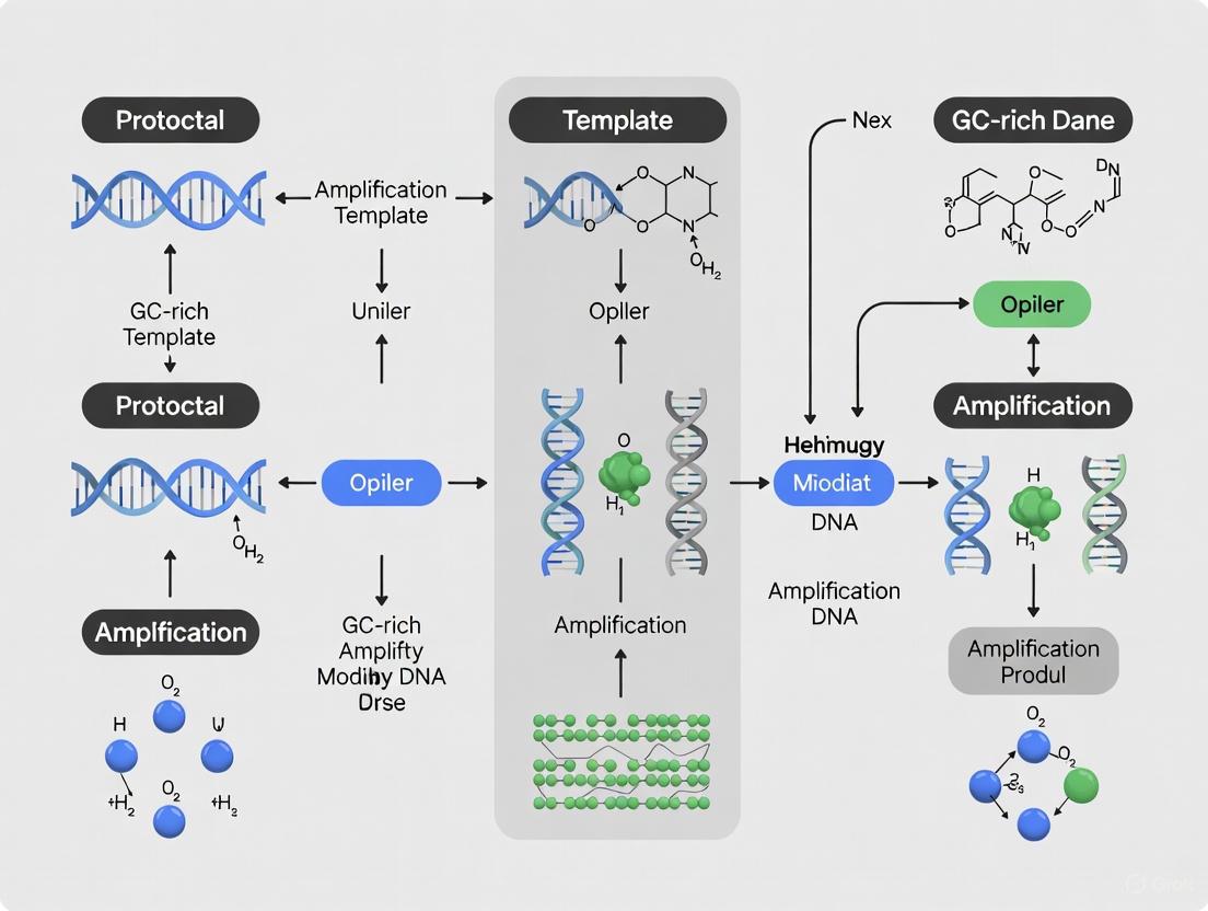
Advanced Strategies for Amplifying GC-Rich DNA Templates: A Comprehensive Protocol for Research and Diagnostics
Amplifying guanine-cytosine (GC)-rich DNA sequences is a common yet formidable challenge in molecular biology, often leading to PCR failure due to stable secondary structures and high thermostability.
Advanced Strategies for Amplifying GC-Rich DNA Templates: A Comprehensive Protocol for Research and Diagnostics
Abstract
Amplifying guanine-cytosine (GC)-rich DNA sequences is a common yet formidable challenge in molecular biology, often leading to PCR failure due to stable secondary structures and high thermostability. This article provides a comprehensive guide for researchers, scientists, and drug development professionals, detailing the foundational principles behind these challenges and presenting a multi-faceted optimization strategy. We cover advanced methodological protocols incorporating specialized polymerases, chemical additives like DMSO and betaine, and refined thermal cycling conditions. The guide further offers a systematic troubleshooting framework and discusses validation techniques to ensure amplification specificity and efficiency, providing an end-to-end solution for successful analysis of GC-rich targets such as gene promoters and key drug targets.
Why GC-Rich DNA is a Challenge: Understanding the Biochemistry and Impact on Research
In molecular biology, a DNA template is defined as GC-rich when 60% or more of its nucleotide bases are guanine (G) or cytosine (C) [1] [2]. While only approximately 3% of the human genome falls into this category, these regions are disproportionately found in crucial regulatory areas, particularly the promoters of housekeeping and tumor suppressor genes [1]. Amplifying these sequences using standard polymerase chain reaction (PCR) protocols presents a significant technical challenge, often resulting in failed amplification, non-specific products, or truncated amplicons. The difficulty primarily arises from two interconnected biophysical properties: exceptional thermal stability and a high propensity to form stable secondary structures [3].
The underlying reason for this stability is often mistakenly attributed solely to the three hydrogen bonds in G-C base pairs versus the two in A-T pairs. However, the primary stabilization mechanism is actually base stacking interactions [3]. This intrinsic stability makes GC-rich DNA more resistant to denaturation, a fundamental step in PCR. Furthermore, these sequences are highly 'bendable,' readily forming complex secondary structures such as hairpins, knots, and tetraplexes [1] [4]. These structures can physically block the progression of the DNA polymerase enzyme and prevent primers from annealing to their target sites, ultimately halting the amplification process [4] [5]. Overcoming these challenges requires a comprehensive understanding of the problem and a systematic optimization of reagents, conditions, and techniques.
The Scientist's Toolkit: Research Reagent Solutions
Successfully amplifying GC-rich templates often requires specialized reagents and additives designed to overcome molecular stability and structural issues. The table below summarizes key solutions and their functions.
Table 1: Key Research Reagent Solutions for GC-Rich PCR
| Reagent Category | Specific Examples | Primary Function & Mechanism of Action |
|---|---|---|
| Specialized Polymerases | OneTaq DNA Polymerase (NEB), Q5 High-Fidelity DNA Polymerase (NEB), PrimeSTAR GXL (Takara), AccuPrime GC-Rich DNA Polymerase (ThermoFisher) [1] [3] [5]. | Engineered for high processivity and efficiency on difficult templates; many are derived from extremophilic organisms and remain stable at high temperatures [3]. |
| GC Enhancers | OneTaq High GC Enhancer, Q5 High GC Enhancer [1] [2]. | Proprietary mixtures that typically contain additives to inhibit secondary structure formation and increase primer stringency. |
| Structure-Disrupting Additives | Dimethyl Sulfoxide (DMSO), Glycerol, Betaine (also known as trimethylglycine) [1] [6] [4]. | Reduce the formation of stable secondary structures (e.g., hairpins) by interfering with hydrogen bonding and base stacking, effectively lowering the melting temperature (Tm) of the DNA [3] [7]. |
| Annealing-Stimulating Additives | Formamide, Tetramethyl ammonium chloride [1] [2]. | Increase primer annealing stringency, which helps reduce non-specific priming and the amplification of off-target products. |
| Nucleotide Analogs | 7-deaza-2′-deoxyguanosine [1] [3]. | A dGTP analog that, when incorporated, improves PCR yield by disrupting normal base pairing and secondary structure stability. Note: it may not stain well with ethidium bromide [3]. |
Optimized Experimental Protocols for GC-Rich Amplification
Standard Three-Step PCR Optimization Protocol
This protocol is adapted from general guidelines for amplifying GC-rich sequences and serves as a strong starting point for optimization [1] [8] [9].
Reagent Setup (50 μL Reaction):
- Polymerase: 1.25 units of a specialized polymerase such as OneTaq or Q5 [8].
- Buffer: Use the manufacturer's recommended buffer, often a dedicated GC Buffer.
- GC Enhancer: If provided, add at the recommended concentration (e.g., 5-10% final volume) [1].
- MgCl₂: Start with 1.5-2.0 mM, but be prepared to optimize using a gradient from 1.0 to 4.0 mM in 0.5 mM increments [1] [8].
- dNTPs: 200 μM of each dNTP [8].
- Primers: 0.1-0.5 μM of each primer [8].
- Template: 1 pg–10 ng of plasmid DNA or 1 ng–1 μg of genomic DNA [8].
- Additives (Optional): If no dedicated GC enhancer is used, test additives like betaine (1 M final) or DMSO (3-10% v/v) individually or in combination [6] [4].
Cycling Conditions:
- Initial Denaturation: 95°C for 2-5 minutes. For extremely GC-rich targets, a longer duration or a temperature of 98°C may be required [9].
- Amplification (25-35 cycles):
- Final Extension: 68-72°C for 5-10 minutes to ensure all products are fully extended [8] [9].
Advanced Two-Step Protocol for Long and Difficult Amplicons
For long targets (>1 kb) with very high GC content (>75%), a two-step protocol that combines annealing and extension has proven superior. The following protocol, optimized for Mycobacterium bovis genes (GC >65%), successfully amplified 51 different GC-rich targets without individual optimization [5] [10].
Reagent Setup (50 μL Reaction):
- Polymerase: PrimeSTAR GXL DNA Polymerase (or equivalent high-fidelity, high-processivity enzyme).
- Enhancer Solution: 1x Final Concentration. This can be a commercial GC enhancer or a mixture of 1 M Betaine and 5% DMSO [5].
- Other Components: As described in Section 3.1, adjusted for the specific polymerase.
Cycling Conditions:
- Initial Denaturation: 98°C for 2-5 minutes.
- Amplification (30-35 cycles):
- Denaturation: 98°C for 10-15 seconds.
- Combined Annealing/Extension: 68°C for 1-2 minutes per 1 kb. Use a slow ramp rate (e.g., 2-3°C/second) between the denaturation and combined steps [5].
- Final Extension: 68°C for 5-10 minutes.
This two-step protocol minimizes the time the reaction spends at non-optimal temperatures, and the higher annealing/extension temperature helps denature secondary structures that persist during cycling [5].
Experimental Workflow and Data Analysis
The following diagram illustrates the logical workflow for troubleshooting and optimizing PCR amplification of a GC-rich DNA template, from initial failure to successful amplification.
Quantitative Data from Protocol Optimization
The effectiveness of a multipronged optimization approach is demonstrated in recent research on amplifying nicotinic acetylcholine receptor subunits from invertebrates. The study quantitatively assessed the impact of different variables on amplifying a 65% GC content target [6] [4].
Table 2: Impact of PCR Additives on Amplification of a 65% GC-Rich Target (Ir-nAChRb1, 1743 bp)
| DNA Polymerase | Additive | Annealing Temperature | Amplification Result |
|---|---|---|---|
| Standard Taq | None | Standard (as per primer Tm) | No Product |
| Standard Taq | DMSO (5%) | Standard | Faint, non-specific bands |
| Standard Taq | Betaine (1 M) | Standard | Weak specific band |
| Standard Taq | DMSO + Betaine | Standard | Strong, specific band |
| High-Fidelity Polymerase (e.g., Q5, PrimeSTAR GXL) | Proprietary GC Enhancer | Elevated (e.g., 5°C higher) | Strong, specific band |
This data underscores that a single adjustment is often insufficient. The combination of a specialized polymerase, structure-disrupting additives, and an optimized annealing temperature was critical for success [6] [4]. Furthermore, a separate study on human genomic DNA demonstrated that for a 78.7% GC-rich template, very short annealing times (3-6 seconds) were optimal, while longer times (>10 seconds) led to smeared products, highlighting the need for precision in cycling parameters [7].
Amplifying GC-rich DNA templates is a common but surmountable challenge in molecular biology. The definition of a "difficult" template—one with >60% GC content—is rooted in the fundamental biophysics of DNA, which confer high thermal stability and promote stable secondary structures. As outlined in this application note, success is not typically achieved by a single universal solution but through a systematic, multipronged strategy. This involves selecting specialized polymerases and reagents, incorporating effective additives like betaine and DMSO, and meticulously optimizing reaction components and thermal cycling conditions. The provided protocols and workflow offer a robust foundation for researchers to reliably amplify even the most challenging GC-rich targets, thereby facilitating the study of critical genes and regulatory elements embedded within these sequences.
The replication and amplification of DNA are fundamental processes in molecular biology, driven by the precise interplay of specific molecular forces. For researchers aiming to amplify genetically rich DNA templates—a common challenge in the study of gene promoters and their role in drug development—understanding these underlying forces is not merely theoretical but a practical necessity. The stability of the DNA double helix is primarily governed by two key interactions: hydrogen bonding between complementary base pairs and base stacking between adjacent nucleotide pairs [11]. While hydrogen bonding is often credited for the specificity of base pairing, it is the base stacking interactions that contribute the majority of the stability to the double-helical structure [3]. GC-rich DNA sequences, defined as those with guanine-cytosine content of 60% or greater, present a formidable challenge in PCR amplification due to their enhanced stability. This application note details the biochemistry of these stabilizing forces and provides optimized, practical protocols for the successful amplification of GC-rich templates, enabling advanced research in genetics and drug discovery.
Quantitative Analysis of Stabilizing Interactions
Hydrogen Bonding
Hydrogen bonds form between complementary bases (G-C and A-T) across the two strands of the DNA double helix. A G-C base pair is stabilized by three hydrogen bonds, while an A-T base pair is stabilized by two [12]. This difference in bond count is a primary reason for the increased thermal stability of GC-rich sequences. Direct mechanical measurements using Atomic Force Microscopy (AFM) have quantified the binding strength of a single dG/dC base pair to be approximately 20.0 ± 0.2 pN, whereas a single dA/dT base pair measures 14.0 ± 0.3 pN [13]. This quantifiable difference underscores the greater energy required to separate GC-rich duplexes.
Base Stacking
Base stacking, or π-π stacking, refers to the hydrophobic interactions between the aromatic rings of adjacent base pairs along the DNA helix. Contrary to common belief, this stacking interaction is a more significant contributor to duplex stability than hydrogen bonding [3]. Base stacking forces have been measured at approximately 2.0 ± 0.1 pN per interaction [13]. The collective effect of these stacking interactions along the helix makes them the dominant force in maintaining the integrity of the DNA molecule, particularly in GC-rich regions where the planar structure of guanine and cytosine favors favorable stacking geometry.
Table 1: Quantitative Forces in DNA Stability
| Interaction Type | Description | Measured Force (pN) | Contribution to Stability |
|---|---|---|---|
| G-C Hydrogen Bonding | Three H-bonds between Guanine and Cytosine | 20.0 ± 0.2 pN [13] | High specificity; major factor in Tm difference |
| A-T Hydrogen Bonding | Two H-bonds between Adenine and Thymine | 14.0 ± 0.3 pN [13] | High specificity; lower thermal stability |
| Base Stacking | Hydrophobic & van der Waals forces between adjacent bases | ~2.0 ± 0.1 pN [13] | Dominant stabilizing force for the double helix |
Experimental Protocols for Investigating DNA Interactions
Protocol: Measuring Base Binding Strength via AFM Unzipping
Principle: This protocol uses Atomic Force Microscopy (AFM) in "unzipping mode" to mechanically disrupt base pairs one by one, directly measuring the hydrogen bond strength of individual base pairs [13].
Methodology:
- Oligonucleotide Design and Immobilization: Synthesize complementary DNA strands of defined length and sequence (e.g., pure dG/dC tracts). Modify the 5'-end of one strand with a thiol (-SH) group and the 3'-end of the complementary strand with an amine (-NH₂) group.
- Surface Functionalization: Covalently immobilize the thiol-modified oligonucleotide onto a gold-coated AFM cantilever tip. Immobilize the amine-modified complementary strand on an aldehyde-functionalized glass slide.
- Duplex Formation and Measurement: Engage the functionalized tip with the surface in TE buffer to allow DNA duplex formation. Retract the cantilever at a controlled speed and loading rate (e.g., 1-4 nN/s). The unzipping process generates a force-extension curve with multiple rupture peaks.
- Data Analysis: Extract rupture forces from the retraction curves. Employ a k-means clustering algorithm to analyze the distribution of rupture events and determine the characteristic unbinding force. Dividing the total rupture force by the number of base pairs yields the average binding strength per base pair.
Workflow: From DNA Stability Analysis to PCR Amplification
The following diagram illustrates the logical relationship between the fundamental forces governing DNA stability and the practical strategies required to overcome challenges in amplifying GC-rich sequences.
Application: Protocol for Amplifying GC-Rich DNA Templates
The inherent stability of GC-rich DNA, governed by the strong forces described above, necessitates specialized PCR protocols. The following optimized procedure is compiled from recent studies.
Reagent Setup
- DNA Polymerase: Utilize polymerases specifically engineered for high GC content, such as Q5 High-Fidelity DNA Polymerase or OneTaq DNA Polymerase [12]. These often demonstrate increased processivity and may be supplied with a proprietary GC Enhancer.
- PCR Buffer with Additives:
- 10X PCR Buffer: 450 mM Tris-HCl (pH 9.0), 110 mM (NH₄)₂SO₄, 45 mM MgCl₂, 67 mM 2-mercaptoethanol, 45 μM EDTA, 1100 μg/mL BSA [14].
- Additives: Supplement the reaction with 5% DMSO, 1.25% formamide, or 1 M betaine [6] [14]. These co-solvents work by reducing secondary structure formation and lowering the melting temperature of GC-rich duplexes.
- Primers: Design primers with a high calculated melting temperature (Tm), ideally between 70°C and 84°C [14]. Software like Primer3 should be used for design.
Thermal Cycling Protocol
A two-step PCR protocol, which combines annealing and extension, has proven superior for long GC-rich amplicons [10]. The following profile is recommended:
- Initial Denaturation: 98°C for 30 seconds.
- High-Stringency Cycling (7 cycles):
- Standard Cycling (30 cycles):
- Denaturation: 98°C for 10 seconds.
- Annealing/Extension: 68°C for 45 seconds per kilobase.
- Final Extension: 72°C for 2 minutes.
Critical Note: Employ a slow ramp rate (e.g., 1-2°C/second) between the annealing/extension and denaturation steps to facilitate efficient primer binding and polymerase loading on difficult templates [10].
The Scientist's Toolkit: Research Reagent Solutions
Table 2: Essential Reagents for GC-Rich PCR and Their Functions
| Reagent / Material | Function / Rationale | Example Use |
|---|---|---|
| High-Processivity DNA Polymerase | Resists stalling at stable secondary structures formed by strong base stacking [12] [3]. | OneTaq or Q5 High-Fidelity DNA Polymerase [12]. |
| GC Enhancer / Betaine | Destabilizes hydrogen bonding and base stacking interactions, effectively lowering the Tm and preventing secondary structure formation [6] [12]. | Used at a concentration of 1 M in the PCR mix [6]. |
| DMSO | Serves as a co-solvent to interfere with hydrogen bonding networks, facilitating strand separation during denaturation [6] [12]. | Typically used at a final concentration of 3-10% [14] [12]. |
| Formamide | Increases primer annealing stringency, reducing non-specific amplification and primer-dimer formation [14] [12]. | Can be used at 1.25% in combination with other additives [14]. |
| Magnesium Chloride (MgCl₂) | Essential cofactor for polymerase activity; optimal concentration is critical for efficiency and specificity [12]. | A gradient from 1.0 mM to 4.0 mM is recommended to find the optimal concentration [12]. |
| 7-deaza-dGTP | Analog of dGTP that incorporates into DNA and reduces base stacking stability, improving polymerase progression [12] [3]. | Used as a partial substitute for dGTP in the dNTP mix [3]. |
The formidable challenge of amplifying GC-rich DNA templates is rooted directly in the fundamental biochemistry of DNA stability. The strong triple hydrogen bonds of G-C base pairs and the profound stabilizing effect of base stacking interactions create a robust structure that resists standard PCR conditions. By understanding these forces—hydrogen bonding providing specificity and base stacking providing the bulk of the stability—researchers can rationally apply specialized polymerases, strategic buffer additives, and tailored thermal cycling profiles. The protocols and data summarized in this application note provide a validated roadmap for overcoming these technical hurdles, thereby facilitating crucial research into GC-rich genomic regions that are of paramount importance in genetics and pharmaceutical development.
The polymerase chain reaction (PCR) is a foundational technique in molecular biology, yet the amplification of DNA templates with high guanine-cytosine (GC) content remains a significant challenge for researchers and drug development professionals. GC-rich regions, defined as sequences exceeding 60% GC content, are of substantial biological importance as they are frequently found in the regulatory regions and first exons of many mammalian genes [14]. The primary obstacle to their amplification lies in the inherent thermodynamic stability of GC base pairs, which facilitates the formation of persistent intra-strand secondary structures—such as hairpin loops and G-quadruplexes—during the PCR cycling process [15]. These structures impede DNA polymerase progression, leading to polymerase stalling, replication fork uncoupling, and ultimately, inefficient or failed amplification [16] [17]. This application note details the mechanisms behind these obstacles and provides a standardized, optimized protocol for the reliable amplification of GC-rich templates, framed within broader research on robust PCR method development.
The Scientific Challenge: Mechanisms of Amplification Failure
Secondary Structure Formation and Polymerase Stalling
The core challenge with GC-rich templates is their propensity to form stable, alternative non-B DNA secondary structures. During the denaturation step of PCR, the separation of DNA strands creates single-stranded templates that are vulnerable to folding into complex conformations. The strong hydrogen bonding between G and C nucleotides allows for the formation of hairpins, G-quadruplexes (G4s), and other secondary structures that are stable at standard PCR annealing temperatures [15]. When the DNA polymerase encounters these structures during the synthesis phase, its progression is physically blocked. Recent studies with reconstituted eukaryotic replisomes have demonstrated that while the CMG helicase can continue unwinding the DNA template ahead of the polymerase, the leading strand synthesis is specifically inhibited, leading to a phenomenon known as helicase-polymerase uncoupling [16]. This stalling is mechanistically similar to that induced by leading-strand DNA lesions, highlighting structured DNA as a significant source of replication stress.
Impact of Specific Repeat Structures
The propensity to form secondary structures and cause stalling varies significantly among different short tandem repeats (STRs). Comprehensive analyses show that structure-prone repeats are a major source of genomic instability and PCR failure [18]. The table below summarizes the impact of common problematic repeats.
Table 1: Impact of Different Short Tandem Repeat (STR) Types on DNA Synthesis
| Repeat Type | Example Motifs | Secondary Structure Formed | Impact on DNA Synthesis & PCR |
|---|---|---|---|
| Mono-nucleotide | (A)~n~, (T)~n~ | Slipped-strand DNA, DNA Unwinding Elements (DUEs) | Correlates strongly with cancer deletion breakpoints; can cause polymerase accumulation [17]. |
| Tri-nucleotide | (CGG)~n~/(CCG)~n~, (CTG)~n~/(CAG)~n~ | Hairpins, G-Quadruplexes (G4s), i-Motifs | Induces significant leading-strand stalling; hairpin stability correlates with expansion and stalling frequency [18] [16]. |
| Tetra-nucleotide | (CCTG•CAGG)~n~ | Hairpin-like structures | Contributes to genetic instability, impeding polymerase progression [18]. |
| Hexa-nucleotide | (GGGGCC)~n~ | G-Quadruplex (G4) | Adopts stable G4 structures that significantly block DNA polymerase [18]. |
Optimized Reagents and Formulations
A multipronged approach using specialized reagent formulations is critical to overcome the challenges of secondary structures. The following combination of PCR additives and enzymes has proven highly effective.
Table 2: Key Research Reagent Solutions for GC-Rich PCR
| Reagent / Material | Function / Explanation | Example Use in Protocol |
|---|---|---|
| Betaine | A chemical chaperone that equalizes the stability of GC and AT base pairs. It disrupts secondary structure by reducing the melting temperature of GC-rich regions without adversely affecting polymerase activity. | Used at a concentration of 1-1.5 M [15] [19]. |
| Dimethyl Sulfoxide (DMSO) | A polar solvent that destabilizes hydrogen bonding, thereby helping to denature DNA secondary structures like hairpins and G-quadruplexes. | Typically used at 5-10% (v/v) [14] [19]. |
| Formamide | A denaturant that further assists in the linearization of structured single-stranded DNA templates, similar to DMSO. | Used at 1.25% (v/v) in combination with DMSO [14]. |
| Bovine Serum Albumin (BSA) | Stabilizes the DNA polymerase enzyme and binds to inhibitors that may be present in the reaction mix, enhancing overall reaction robustness. | Included at 1100 μg/mL in the PCR buffer [14]. |
| 2-Mercaptoethanol | A reducing agent that helps maintain a reducing environment, preventing oxidation of enzyme thiol groups and preserving polymerase activity. | Used at 67 mM in the PCR buffer [14]. |
| High-Performance Taq Polymerase | Specialty polymerases (often engineered or blended) are less prone to stalling at secondary structures compared to standard Taq. | Laboratory-prepared or commercial Taq (e.g., from Fermentas) can be used [14]. |
| dNTPs | Deoxynucleotide triphosphates are the building blocks for DNA synthesis. | Used at standard 200 μM each to ensure sufficient and balanced availability [14]. |
Custom 10X PCR Buffer Formulation: The optimized protocol utilizes a specialized buffer containing 450 mM Tris-HCl (pH 9.0), 110 mM (NH~4~)~2~SO~4~, 45 mM MgCl~2~, 67 mM 2-mercaptoethanol, 45 μM EDTA, and 1100 μg/mL BSA [14]. This formulation provides a high-pH environment and necessary co-factors to support polymerization through difficult templates.
Detailed Experimental Protocol
Primer Design Considerations
- Melting Temperature (T~m~): Design primers with high T~m~ values, ideally between 70°C and 84°C [14].
- Length: Primers of up to 30 nucleotides can be used to achieve the required high T~m~ [14].
- Specificity: Verify primer specificity using software like Primer3 to minimize self- and cross-dimer formation and secondary structure [14].
PCR Master Mix Setup
For a standard 50 μL reaction, combine the following components in order:
- Nuclease-free Water: (Variable volume to reach 50 μL)
- 10X Custom PCR Buffer: 5 μL [14]
- dNTP Mix (10 mM each): 1 μL
- Dimethyl Sulfoxide (DMSO): 2.5 μL (5% final concentration) [14]
- Formamide: 0.625 μL (1.25% final concentration) [14]
- Betaine (5 M): 10 μL (1 M final concentration) [15]
- Forward Primer (10 μM): 1.2 μL
- Reverse Primer (10 μM): 1.2 μL
- Template DNA (50 ng/μL): 1 μL
- Taq DNA Polymerase (5 U/μL): 0.25 μL
Note: A "hot-start" protocol is recommended to prevent non-specific amplification initiated at lower temperatures.
Thermal Cycling Profile
Use the following cycling parameters, which incorporate a touchdown phase to enhance specificity:
- Initial Denaturation: 95°C for 5 minutes.
- 7x Touchdown Cycles:
- Denature: 95°C for 30 seconds.
- Anneal: 72°C for 30 seconds, decreasing by 1°C per cycle.
- Extend: 72°C for 1 minute per kb of amplicon.
- 35x Standard Cycles:
- Denature: 95°C for 30 seconds.
- Anneal: 65°C for 30 seconds.
- Extend: 72°C for 1 minute per kb.
- Final Extension: 72°C for 7 minutes.
- Hold: 4°C indefinitely.
This profile uses a high initial annealing temperature to promote specific primer binding during the critical early cycles, followed by standard cycling for efficient amplification [14].
Workflow and Mechanism Visualization
The following diagram illustrates the core experimental workflow and the molecular mechanisms addressed by this protocol.
Diagram 1: Workflow for Amplifying GC-Rich DNA
Troubleshooting and Validation
Common Issues and Solutions
- No Product or Low Yield: Increase the concentration of Betaine to 1.5 M. Verify the primer T~m~ and ensure the initial annealing temperature is sufficiently high. Check the integrity of the template DNA.
- Non-Specific Bands: Implement or optimize a hot-start protocol. Reduce the number of cycles or lower the primer concentration. Increase the annealing temperature during the standard cycles.
- Smearing on Gel: Reduce the amount of template DNA or Taq polymerase. Optimize the MgCl~2~ concentration in 0.5 mM increments.
Validation of Results
- Gel Electrophoresis: Analyze PCR products on an agarose gel to confirm the expected amplicon size and purity.
- Sequencing: Sanger sequencing of the purified PCR product is essential to confirm the fidelity of amplification and the absence of mutations, especially given the error-prone nature of synthesizing through structured DNA [14].
The amplification of GC-rich DNA templates requires a strategic approach that addresses the fundamental problem of DNA secondary structure-induced polymerase stalling. The combination of a specialized PCR buffer containing betaine, DMSO, formamide, and stabilizing agents, coupled with a tailored thermal cycling profile that includes a high-temperature annealing touchdown phase, provides a robust and reliable method. This optimized protocol enables researchers to consistently amplify challenging targets, including those with GC contents exceeding 80%, thereby facilitating advanced genetic studies, mutation detection, and drug development projects focused on GC-rich genomic regions.
In the human genome and those of other vertebrates, the distribution of guanine (G) and cytosine (C) nucleotides is non-random, forming regions with distinctly high GC-content, particularly around the transcriptional start sites (TSSs) of protein-coding genes [20]. These GC-rich sequences represent fundamental architectural elements that influence multiple levels of gene regulation, from chromatin organization to transcriptional efficiency and RNA processing [20] [21] [22]. A prominent feature of these regions is the presence of CpG islands, defined as DNA segments longer than 200 base pairs with a cytosine/guanine content greater than 55% and a higher observed frequency of CpG dinucleotides than expected [21]. These islands are frequently located proximal to transcription start sites and are associated with the promoters of more than 50% of mammalian genes, including housekeeping genes and tissue-specific genes [21].
The GC-content peaks just downstream of the TSS and slopes down symmetrically into both the upstream intergenic region and downstream into the first exon and first intron, forming a characteristic profile that is conserved across amniotes and likely most vertebrates [20]. This conserved pattern suggests these regions are under significant functional constraint, playing critical roles in gene expression regulation, particularly in neural cells where they influence chromatin organization and genome stability [21]. Understanding the biological significance of these regions is therefore essential for researchers investigating gene regulation, especially those working on challenging templates for PCR amplification in functional studies.
Table 1: Key Genomic Features of GC-Rich Regions in Promoters and First Exons
| Feature | Genomic Location | Characteristics | Functional Significance |
|---|---|---|---|
| CpG Islands | Primarily in promoters, ~5' regulatory regions | >200 bp, >55% GC content, elevated CpG frequency | Epigenetic regulation, transcription factor binding, association with highly expressed genes [21] |
| GC-Content Peak | Transcription Start Site (TSS) | Highest just downstream of TSS, symmetrical decrease upstream and downstream | Promotes efficient nuclear export and translation of mRNAs [20] |
| Nucleosome Positioning | Exonic regions, especially first exons | Higher GC content in exons than introns | Enhanced DNA bendability, influences RNAPII velocity and splicing [22] |
| First Exon & Intron | Beginning of gene structure | First exon has highest GC content; first intron is often longer with distinct composition | Regulatory role in transcription control and splicing [23] |
Biological Significance and Mechanistic Insights
Roles in Transcription and Chromatin Organization
GC-rich sequences in promoters and first exons play multifaceted roles in gene regulation through several interconnected mechanisms. Their high GC content directly influences chromatin architecture by promoting nucleosome positioning, as exonic regions with elevated GC content demonstrate increased DNA bendability, facilitating nucleosome formation [22] [10]. This nucleosome positioning subsequently influences RNA polymerase II velocity, creating a kinetic coupling between transcription and splicing processes [22]. Furthermore, CpG-rich promoters actively recruit specific transcription factors and enable robust, high-level gene expression across diverse tissue types [20] [21]. The methylation status of these GC-rich regions serves as a critical epigenetic mechanism for modulating gene expression in a tissue-specific and developmentally regulated manner, as exemplified by the reelin gene promoter and neuronal nicotinic acetylcholine receptor genes [21].
Implications for mRNA Processing and Stability
The nucleotide composition bias at the 5' ends of genes extends beyond transcriptional regulation to impact subsequent RNA metabolic processes. High GC-content at the 5' end of mRNAs promotes efficient nuclear export, particularly for intron-poor transcripts, by potentially recruiting protein factors like SARNP, SR proteins, and RBM33 that directly interact with nuclear transport receptors [20]. These GC-rich regions also influence splicing regulation, with distinct groups of splicing factors activating either GC-rich exons flanked by small introns or AT-rich exons flanked by large introns [22]. Additionally, the formation of stable G-quadruplex structures in GC-rich regions allows for interactions with RNA-binding proteins such as Fragile X Mental Retardation Protein (FMRP), which modulates translation, particularly of key synaptic proteins involved in neurodevelopment and plasticity [21].
Evolutionary Dynamics and Genomic Distribution
The GC-peak at transcription start sites represents an evolutionarily conserved feature present since the last common ancestor of amniotes, and likely that of vertebrates [20]. However, current evolutionary dynamics reveal this feature is undergoing mutational decay in apes and rodents, where recombination is directed away from TSSs by PRDM9, while GC-content is increasing in canids, which lack PRDM9 and perform recombination at TSSs [20]. This pattern indicates that regional nucleotide composition bias leaves a local footprint at the exon level and establishes a direct link between genome organization and local regulatory processes like alternative splicing [22]. The distribution of these regions also correlates with broader genomic architecture, as the GC content of exons correlates with that of their hosting genes, isochores, and topologically associated domains [22].
Diagram 1: Functional networks of GC-rich regions in gene regulation. GC-rich sequences in promoters and first exons influence gene expression at multiple levels through distinct but interconnected mechanistic pathways.
Technical Challenges in GC-Rich Sequence Amplification
Molecular Basis of Amplification Difficulties
Amplifying GC-rich DNA templates presents significant technical challenges that routinely frustrate molecular biology research. A GC-rich template is formally defined as a DNA sequence where ≥60% of the bases are guanine (G) or cytosine (C) [24] [3]. These regions pose three primary obstacles for successful PCR amplification. First, the enhanced thermodynamic stability of GC-rich sequences arises from base stacking interactions and the presence of three hydrogen bonds in G-C base pairs compared to only two in A-T pairs, resulting in higher melting temperatures that resist DNA denaturation [24] [3]. Second, these sequences readily form stable secondary structures, including hairpin loops and G-quadruplexes, that block polymerase progression and cause stalling, leading to truncated products [24] [3]. Third, primers designed for GC-rich targets frequently form self-dimers, cross-dimers, and stem-loop structures, particularly when GC-rich stretches are present at the 3' end, resulting in mispriming and amplification failure [3].
Impact on Research Applications
These challenges are particularly problematic in neuroscience and biomedical research, as GC-rich sequences are prevalent in promoter regions of housekeeping genes, tumor suppressor genes, and many neuron-specific genes [24] [21]. For instance, studying promoter methylation of neuronal genes like the α4 subunit of neuronal nicotinic acetylcholine receptors requires reliable amplification of their GC-rich promoter sequences [21]. Furthermore, pathogenic expansions of GC-rich hexanucleotide repeats in the C9orf72 gene, associated with amyotrophic lateral sclerosis (ALS) and frontotemporal dementia (FTD), present particular challenges for molecular analysis and therapeutic development [21]. The repetitive and structurally complex nature of these regions complicates experimental analysis and necessitates specialized amplification approaches for accurate genotyping and functional characterization.
Optimized Protocols for GC-Rich Sequence Amplification
Strategic Framework for Protocol Development
Successfully amplifying GC-rich regions requires a systematic, multi-pronged approach that addresses both the biochemical and physical challenges these sequences present. Based on extensive optimization studies, an effective strategy incorporates specialized reaction components, adjusted thermal cycling parameters, and appropriate template handling [24] [3] [10]. The following workflow outlines a comprehensive optimization process that can be adapted for specific GC-rich targets, such as promoter regions or first exons of neuronal genes.
Diagram 2: Systematic optimization workflow for GC-rich PCR. A sequential troubleshooting approach addressing reagent selection first, then cycling conditions, significantly improves success rates with challenging templates.
Reagent Optimization and Selection
The careful selection and optimization of reaction components forms the foundation of successful GC-rich PCR. Different polymerases exhibit varying capabilities in handling complex secondary structures and GC-rich templates, with specialized enzymes consistently outperforming standard Taq polymerase [24] [10] [19].
Table 2: Polymerase Selection for GC-Rich Amplification
| Polymerase Type | Key Features | Recommended Applications | Performance on GC-Rich Templates |
|---|---|---|---|
| Standard Taq | Standard fidelity, common master mixes | Routine PCR on normal templates | Poor, stalls at secondary structures [24] |
| OneTaq DNA Polymerase (NEB) | 2× fidelity of Taq, GC buffer compatible | Routine to GC-rich targets (up to 80% GC with enhancer) | Good to excellent with optimized buffers [24] |
| Q5 High-Fidelity (NEB) | >280× fidelity of Taq, high processivity | Long or difficult amplicons, cloning applications | Excellent with GC enhancer (up to 80% GC) [24] |
| AccuPrime GC-Rich (ThermoFisher) | Derived from Pyrolobus fumarius, extreme thermostability | Extremely GC-rich targets, complex secondary structures | Excellent, maintains activity after 4h at 95°C [3] |
The strategic incorporation of reaction additives significantly improves amplification efficiency by disrupting secondary structures and increasing primer specificity. These additives function through distinct mechanisms, making them appropriate for different specific challenges.
Table 3: PCR Additives for GC-Rich Amplification
| Additive | Recommended Concentration | Mechanism of Action | Considerations |
|---|---|---|---|
| DMSO | 3-10% | Disrupts secondary structures, lowers melting temperature | May inhibit some polymerases at higher concentrations [24] [19] |
| Betaine | 1-1.5 M | Equalizes DNA melting temperatures, disrupts secondary structures | Can be combined with DMSO for synergistic effect [19] |
| Glycerol | 5-10% | Reduces secondary structure formation | Increases enzyme stability but may lower specificity [24] |
| 7-deaza-2'-deoxyguanosine | As dGTP substitute | dGTP analog that reduces secondary structure stability | Does not stain well with ethidium bromide [24] |
| Commercial GC Enhancers | Manufacturer's recommendation | Proprietary blends of multiple additives | Optimized for specific polymerase systems [24] |
Magnesium concentration optimization represents another critical parameter, as Mg²⁺ serves as a essential cofactor for polymerase activity and facilitates primer binding by neutralizing electrostatic repulsion between DNA strands [24]. The standard concentration of 1.5-2.0 mM MgCl₂ may be suboptimal for GC-rich templates, necessitating empirical testing through gradient PCR across a range of 1.0-4.0 mM in 0.5 mM increments to identify the ideal concentration that maximizes yield while minimizing non-specific amplification [24] [3].
Thermal Cycling Parameter Optimization
Adjusting thermal cycling parameters addresses the thermodynamic challenges posed by GC-rich sequences. Implementing a higher denaturation temperature (up to 95-98°C) for the first few cycles helps separate stubborn secondary structures, though this must be balanced against potential polymerase denaturation over extended cycles [3]. Slower temperature ramp rates between denaturation and annealing steps facilitate more complete separation of DNA strands and reorganization of secondary structures [10]. Employing a touchdown PCR approach or higher annealing temperatures increases primer specificity, particularly crucial for preventing mispriming in GC-rich contexts [24] [3]. For extremely challenging templates, a specialized "slow-down PCR" protocol incorporates 7-deaza-2'-deoxyguanosine, uses lowered ramp rates, and extends cycle numbers to dramatically improve amplification efficiency [3].
Case Study: Amplifying Nicotinic Acetylcholine Receptor Subunits
A recent systematic optimization study targeting nicotinic acetylcholine receptor subunits from invertebrates demonstrates the effectiveness of this comprehensive approach [19]. The Ir-nAChRb1 (1743 bp, 65% GC) and Ame-nAChRa1 (1884 bp, 58% GC) subunits presented substantial amplification challenges. The optimized protocol incorporated betaine (1M) and DMSO (5%) as synergistic additives, used Q5 High-Fidelity DNA Polymerase with its corresponding GC enhancer, implemented a 2-step PCR protocol with annealing/extension at 68°C, and applied reduced ramp speeds between temperature steps [19]. This multi-faceted approach successfully amplified these challenging targets where standard protocols failed, providing a template for amplifying other GC-rich neuronal genes.
Successfully working with GC-rich promoter and first exon regions requires access to specialized reagents and computational resources. The following toolkit compiles essential solutions validated for challenging GC-rich templates.
Table 4: Research Reagent Solutions for GC-Rich Genomic Studies
| Category | Specific Product/Resource | Application Notes |
|---|---|---|
| Specialized Polymerases | OneTaq DNA Polymerase with GC Buffer (NEB #M0480) | Ideal for routine GC-rich PCR; compatible with OneTaq High GC Enhancer [24] |
| Q5 High-Fidelity DNA Polymerase (NEB #M0491) | Superior for long or difficult amplicons; high fidelity critical for cloning [24] | |
| AccuPrime GC-Rich DNA Polymerase (ThermoFisher) | Extreme thermostability useful for high denaturation temperatures [3] | |
| Enhancement Reagents | OneTaq High GC Enhancer (NEB) | Proprietary formulation that inhibits secondary structure formation [24] |
| Q5 High GC Enhancer (NEB) | Optimized for use with Q5 polymerase system [24] | |
| DMSO (Molecular Biology Grade) | Versatile additive for reducing secondary structures [24] [19] | |
| Betaine (Molecular Biology Grade) | Effective for equalizing DNA melting temperatures [19] | |
| Computational Tools | NEB Tm Calculator | Web tool for calculating optimal annealing temperatures specific to enzyme/buffer systems [24] |
| NPACT (N-Profile Analysis Computational Tool) | Identifies ORFs with significant periodicities in GC-rich genomes [25] | |
| FramePlot | Visualizes S-profiles and compositional periodicity in coding regions [25] |
GC-rich regions in promoters and first exons represent crucial regulatory elements that present both functional significance and technical challenges for molecular research. Their roles in transcriptional regulation, chromatin organization, and RNA processing make them essential targets for understanding gene expression mechanisms, particularly in neurological contexts. The optimized protocols and systematic approaches outlined in this application note provide researchers with a strategic framework for successfully amplifying these challenging sequences, enabling more reliable investigation of their biological functions. As research continues to uncover the complexities of GC-rich genomic regions, the integration of specialized reagents, optimized conditions, and computational tools will remain essential for advancing our understanding of their significance in genome biology and disease mechanisms.
Amplification failures, particularly with challenging DNA templates, present significant obstacles in molecular biology, impacting fields from basic research to clinical diagnostics. This article details the specific challenges and provides validated protocols to overcome them.
The Core Challenge: Amplification of GC-Rich and Repetitive DNA
The reliable amplification of DNA sequences is a cornerstone of modern genetics. However, certain template characteristics consistently cause PCR failure, leading to incomplete data, erroneous conclusions, and diagnostic inaccuracies.
GC-Rich Sequences: A Problem of Stability and Structure
GC-rich sequences (typically defined as >60% guanine-cytosine content) are difficult to amplify due to their inherent molecular stability [3]. This stability is primarily due to base stacking interactions, not just the three hydrogen bonds of GC pairs, which raise the DNA's melting temperature [3]. Consequently, standard denaturation temperatures (e.g., 94–95°C) may be insufficient to fully separate the DNA strands, preventing primer annealing and polymerase progression.
A critical secondary problem is the formation of stable secondary structures, such as hairpin loops [5] [26] [3]. These structures can block the DNA polymerase, resulting in truncated, non-specific, or absent PCR products [26]. Furthermore, the primers themselves can form dimers or secondary structures, exacerbating the issue [3].
Repetitive DNA: The Problem of Polymerase Slippage
Mononucleotide, dinucleotide, and other repetitive sequences present a different set of challenges. During PCR, the polymerase can slip or "stutter" on these repetitive tracts, leading to the generation of artifacts with varying numbers of repeat units [27] [28]. This phenomenon is a major source of "shadow bands" observed in gel electrophoresis, complicating the analysis of genetic markers and microsatellites [27].
This error rate is not trivial. One study found that while a (T)9 repeat was amplified faithfully, only 33% of clones contained the correct (T)13 repeat length after amplification with Taq polymerase, with most errors being contractions [27]. Proofreading enzymes like Pfu perform better but still show significant error rates with longer repeats [27]. These errors can be mistaken for genuine polymorphisms or mutations, leading to false conclusions in genetic studies [27].
Real-World Impacts on Research and Diagnostics
The failure to robustly amplify these difficult sequences has direct and consequential implications.
Hindrances in Genetic Research
- Gene Cloning and Functional Studies: Research requiring the cloning of GC-rich promoter regions, which are common in housekeeping and tumor suppressor genes, is often hampered [26] [3]. For instance, a study aiming to clone GC-rich open reading frames (ORFs) from Mycobacterium bovis (genome GC content >60%) faced significant obstacles when using conventional PCR protocols and polymerases [5].
- Genome Editing Technologies: The development of technologies based on repetitive DNA-binding domains, such as Transcription-Activator Like Effectors (TALEs), is severely limited because PCR amplification of their repetitive coding sequences consistently fails, generating deleted and hybrid artifacts [28]. This incompatibility with PCR-based cloning methods restricts the full potential of these powerful genome-editing tools.
Consequences for Diagnostic Assay Development
In clinical diagnostics, amplification failures translate directly into reduced sensitivity and accuracy, affecting patient care.
- False Negatives and Inconclusive Results: Failed amplification of a target pathogen sequence can lead to false-negative results. Furthermore, smeared or non-specific products can render a test inconclusive, requiring sample recollection and re-testing, which delays diagnosis [29].
- Misidentification of Mutations and Microsatellite Instability: In oncology and genetic disease testing, errors in amplifying repetitive sequences can lead to misclassification of a patient's status [27]. For example, in acute myeloid leukemia (AML), the detection of residual disease relies on highly sensitive and accurate amplification of specific genetic markers, such as FLT3 mutations [30]. Any amplification infidelity at this stage could compromise the monitoring of treatment response and relapse risk.
Table 1: Quantitative Analysis of PCR Errors at Repetitive Loci
| Locus | Repeat Type & Correct Length | Polymerase | % of Clones with Correct Length | Predominant Error |
|---|---|---|---|---|
| RAC1 | (T)9 | Taq | 100% | None |
| RAC1 | (T)11 | Taq | 90% | Contraction |
| Bat-13 | (T)13 | Taq | 33% | Contraction |
| Bat-26 | (A)26 | Taq | 35% | Contraction |
| Bat-26 | (A)26 | Pfu | 23% | Contraction |
| D15S128 | (CA)18 | Taq | 64% | Contraction/Expansion |
| D15S128 | (CA)18 | Pfu | 33% | Contraction/Expansion |
Data adapted from [27]
Established Protocols and Solutions
To address these challenges, researchers have developed optimized protocols that adjust reaction components and cycling conditions.
Protocol 1: Two-Step PCR for Lengthy GC-Rich Targets
This protocol was developed specifically for amplifying long (>1 kb), GC-rich targets from M. bovis and has been successfully used to amplify 51 different GC-rich targets [5].
Experimental Workflow:
Methodology:
- Reaction Mixture:
- Thermal Cycling Conditions:
- Initial Denaturation: 98°C for 2 minutes.
- 35 Cycles:
- Denaturation: 98°C for 10 seconds.
- Annealing/Extension: 68°C for 1 minute per kilobase of the product.
- Final Extension: 68°C for 5-10 minutes.
- Key Adjustments: This protocol uses a two-step (2St) PCR with a high-temperature (68°C) combined annealing/extension step. This high temperature helps prevent the formation of secondary structures. The use of a slow ramp rate between steps is also critical for success [5].
Protocol 2: Optimized Fast-Cycling for GC-Rich Targets
This protocol, based on fundamental modeling of the annealing process, demonstrates that shorter annealing times are not only sufficient but necessary for efficient amplification of GC-rich templates [29].
Methodology:
- Reaction Mixture:
- Polymerase: KOD Hot Start Polymerase.
- Enhancer: 11% DMSO (v/v) was used for the ARX gene [29].
- Template: 100 ng human genomic DNA.
- Primers: 0.75 µM each for the ARX gene.
- MgSO₄: 4 mM.
- Thermal Cycling Conditions (for a 660 bp, 78.72% GC target):
- Hot Start: 94°C for 30 seconds.
- 35-38 Cycles:
- Denaturation: 94°C for 2 seconds.
- Annealing: 60°C for 3-6 seconds. The study found that annealing times greater than 10 seconds yielded smeared products [29].
- Extension: 72°C for 4 seconds.
- Final Extension: 72°C for 30 seconds.
- Key Principle: The model shows that for GC-rich templates, the window for optimal annealing efficiency is very narrow. Longer annealing times promote competitive binding at incorrect sites (mispriming), leading to smearing and non-specific products [29].
The Scientist's Toolkit: Research Reagent Solutions
A range of specialized reagents and instruments is available to overcome amplification challenges.
Table 2: Essential Reagents and Kits for Difficult Amplicons
| Reagent / Instrument | Supplier / Example | Function and Application |
|---|---|---|
| GC-Rich Optimized Polymerases | OneTaq DNA Polymerase with GC Buffer, Q5 High-Fidelity DNA Polymerase (NEB); AccuPrime GC-Rich DNA Polymerase (ThermoFisher) | Specially formulated enzymes and buffers to polymerize through stable secondary structures and resist denaturation. |
| PCR Enhancers | Betaine, DMSO, Formamide, 7-deaza-dGTP | Destabilize GC-rich DNA secondary structures, increase primer stringency, and improve yield [29] [5] [26]. |
| High-Processivity Polymerase | PrimeSTAR GXL (Takara), KOD Hot-Start (Novagen) | Ideal for long and difficult amplicons due to high processivity and fidelity [29] [5]. |
| Specialized Thermocyclers | PCRJet (Megabase Research Products) | Provides very fast temperature ramping, allowing for the use of extremely short cycling times as required by some optimized protocols [29]. |
Emerging Technologies and Future Directions
While optimized PCR remains fundamental, new technologies are emerging that can circumvent these amplification problems entirely or integrate with PCR to enhance reliability.
- CRISPR-Driven Diagnostics: CRISPR/Cas systems are being harnessed for direct pathogen detection. Platforms like SHERLOCK and DETECTR often couple CRISPR with pre-amplification steps but are evolving towards amplification-free detection, which would eliminate PCR-specific artifacts and errors [31]. These systems use Cas enzymes (e.g., Cas12, Cas13) that, upon binding a target nucleic acid, exhibit collateral activity that cleaves reporter molecules, enabling ultra-sensitive detection down to attomolar (aM) levels [31].
- Advanced Sequencing for Residual Disease: In clinical diagnostics, methods like deep sequencing are being validated for monitoring conditions like AML. These tests can detect mutations (e.g., in FLT3) at extremely low variant allele frequencies (down to 0.0014%), providing a highly quantitative and sensitive alternative to traditional amplification-based assays for residual disease [30].
Logical Workflow for Addressing Amplification Failure:
Amplification failures of GC-rich and repetitive DNA are not merely technical nuisances; they represent a significant source of error that can hinder gene discovery, invalidate experimental results, and compromise diagnostic accuracy. A thorough understanding of the molecular mechanisms behind these failures—thermostability, secondary structure, and polymerase slippage—is the first step toward a solution. By employing strategic reagent selection, such as specialized polymerases and enhancers, and implementing rigorously optimized protocols like the two-step PCR or fast-cycling methods, researchers and diagnosticians can significantly improve the reliability of their assays. As the field advances, leveraging new technologies like CRISPR-based detection and deep sequencing will further mitigate these long-standing challenges, enhancing the fidelity of genetic analysis.
Optimized Protocols for Success: A Step-by-Step Guide to Amplifying GC-Rich Targets
The amplification of guanine-cytosine (GC)-rich DNA templates represents a significant challenge in molecular biology, requiring specialized enzymatic approaches and optimized reaction conditions. GC-rich sequences, typically defined as regions where 60% or more of the bases are guanine or cytosine, pose substantial obstacles to conventional PCR amplification due to their unique biochemical properties [32]. These challenging templates are biologically relevant despite their difficulty—approximately 3% of the human genome consists of GC-rich regions, which are frequently found in the promoters of housekeeping and tumor suppressor genes, making their amplification essential for various research and diagnostic applications [32].
The fundamental challenge in amplifying GC-rich templates stems from the robust nature of GC base pairing, which features three hydrogen bonds compared to the two hydrogen bonds in adenine-thymine (AT) base pairs. This increased bond strength creates exceptionally stable DNA duplexes that require higher denaturation temperatures and are prone to forming complex secondary structures such as hairpins and stem-loops [32]. These structures can block polymerase progression during amplification and interfere with primer annealing, ultimately leading to PCR failure characterized by absent or smeared amplification products on agarose gels. Furthermore, the primers designed for GC-rich regions often form dimers, compounding the challenges of obtaining specific, high-yield amplification [32].
This application note provides a comprehensive framework for selecting appropriate DNA polymerases and optimizing reaction conditions to successfully amplify GC-rich templates. We present detailed protocols and quantitative comparisons to guide researchers in making informed decisions between high-fidelity enzymes and specialized GC-rich polymerases, considering the specific requirements of their experimental applications from basic research to drug development.
Understanding GC-Rich Amplification Challenges
Biochemical Properties of GC-Rich DNA
The amplification difficulties associated with GC-rich templates originate from their distinct molecular characteristics. The additional hydrogen bond in GC base pairs creates significantly greater thermodynamic stability compared to AT-rich regions, requiring more energy input for denaturation. This stability directly translates to higher melting temperatures (Tm), which often exceed standard PCR denaturation conditions [32]. When GC-rich sequences fold back on themselves, they form stable secondary structures that persist even at elevated temperatures, creating physical barriers that impede polymerase progression during the extension phase of PCR.
These structural challenges manifest in several specific ways during amplification attempts. Polymerases frequently stall at the complex secondary structures formed by GC-rich stretches, resulting in truncated amplification products and incomplete synthesis [32]. Additionally, the resistant nature of these regions to complete denaturation prevents primers from accessing their complementary binding sites, while the primers themselves—often designed with high GC content to match their templates—readily form primer-dimers that further reduce amplification efficiency [32]. Understanding these molecular obstacles is essential for selecting appropriate enzymatic solutions and optimization strategies.
Specific Examples of Problematic Templates
The practical implications of these challenges are well-illustrated by the epidermal growth factor receptor (EGFR) promoter region, which features an exceptionally high GC content of up to 88% [33]. This region contains single nucleotide polymorphisms (SNPs) at positions -216G>T and -191C>A that have potential pharmacogenetic significance as biomarkers for predicting efficacy and safety of EGFR tyrosine kinase inhibitor therapies in cancer treatment [33]. However, the extreme GC-rich nature of this template makes it particularly difficult to amplify using standard PCR protocols, necessitating specialized optimization for successful amplification.
Similar challenges occur across various research contexts, including bisulfite-converted DNA used in epigenetic analysis, which contains uracil and often features AT-rich regions flanked by GC-rich areas [34]. The amplification of promoter regions of tumor suppressor genes, complex genomic loci, and templates derived from formalin-fixed paraffin-embedded (FFPE) tissues all present scenarios where conventional polymerases frequently fail, requiring specialized enzymatic formulations and reaction conditions [33].
DNA Polymerase Characteristics for GC-Rich Amplification
Key Enzyme Properties
Successful amplification of GC-rich templates depends on understanding four critical polymerase characteristics that directly impact performance with challenging sequences:
Fidelity: Defined as the accuracy of DNA sequence replication, fidelity is particularly crucial for applications where sequence integrity is paramount, such as cloning and functional analysis. Fidelity is commonly expressed relative to Taq DNA polymerase, with high-fidelity enzymes exhibiting error rates that are 10-300 times lower than standard Taq [35]. The proofreading capability of a DNA polymerase, mediated by its 3'→5' exonuclease activity, defines its fidelity by correcting misincorporated nucleotides [35].
Processivity: This characteristic refers to the number of nucleotides a polymerase can incorporate per single binding event. Highly processive enzymes demonstrate superior performance when amplifying long templates, sequences with substantial secondary structure, and GC-rich regions, as they maintain synthesis through structurally challenging areas that would cause less processive polymerases to dissociate [35]. Enhanced processivity is particularly beneficial for GC-rich amplification where secondary structures frequently impede polymerase progression.
Thermostability: The inherent resistance to thermal denaturation at high temperatures is essential for GC-rich PCR, as these templates require higher denaturation temperatures (often above 95°C) to separate stable GC bonds. Hyperthermostable enzymes isolated from archaeal organisms such as Pyrococcus furiosus (Pfu polymerase) demonstrate approximately 20 times greater stability at 95°C compared to Taq polymerase, enabling them to withstand the stringent denaturation conditions needed for GC-rich templates [35].
Specificity: This property refers to the enzyme's ability to amplify only the intended target while minimizing non-specific products such as primer-dimers and misprimed amplification. Hot-start activation mechanisms, including antibody-based inhibition and chemical modifications, enhance specificity by preventing polymerase activity during reaction setup until high temperatures are reached in the thermal cycler [35]. This feature is particularly valuable for GC-rich amplification where primer-dimer formation is common.
Polymerase Comparison Table
Table 1: Comparison of DNA Polymerases for GC-Rich and High-Fidelity Applications
| Polymerase | 3'→5' Exo | Fidelity (Relative to Taq) | Strand Displacement | dU Tolerance | Resulting Ends | Optimal GC Range | Primary Applications |
|---|---|---|---|---|---|---|---|
| Q5 High-Fidelity | Yes | ~280x | No | No | Blunt | Up to 80% (with GC Enhancer) [32] | High-fidelity PCR, cloning, NGS library prep [36] |
| Phusion High-Fidelity* | Yes | 39-50x | No | No | Blunt | Not specified | High-fidelity PCR, cloning [36] |
| OneTaq DNA Polymerase | Yes | 2x | Limited | Yes | 3'A/Blunt | Up to 80% (with GC Enhancer) [32] | Routine PCR, GC-rich templates, colony PCR [36] |
| Taq DNA Polymerase | No | 1x | Limited | Yes | 3'A Overhang | Moderate (with optimization) | Routine PCR, genotyping [36] |
| LongAmp Taq | Yes | 2x | Yes | Yes | 3'A/Blunt | Not specified | Long range PCR (up to 30 kb) [36] [34] |
| Hemo KlenTaq | No | Not specified | No | Yes | 3'A Overhang | Not specified | Direct PCR from blood [36] |
| Bst Polymerase | No | Lower fidelity | Yes | Yes | 3'A Overhang | Not specified | Isothermal amplification, LAMP [36] |
Note: Phusion DNA Polymerase was developed by Finnzymes Oy, now part of Thermo Fisher Scientific, and is manufactured by New England Biolabs under agreement [36].
Decision Workflow for Polymerase Selection
Table 2: Polymerase Selection Guide Based on Application Requirements
| Application Priority | Recommended Polymerase Type | Key Considerations | Suggested Products |
|---|---|---|---|
| Maximum Fidelity (cloning, sequencing, mutagenesis) | High-fidelity with proofreading | May require GC enhancer for >60% GC content; produces blunt ends requiring different cloning strategies | Q5 High-Fidelity, Phusion High-Fidelity [36] |
| Challenging GC-rich Templates (>70% GC) | Specialist GC-rich enzymes | Often includes proprietary buffers with secondary structure inhibitors; optimized for high annealing temperatures | OneTaq with GC Buffer, PCRBIO HS Taq DNA Polymerase [32] [37] |
| Direct Amplification (from blood, tissue) | Inhibitor-resistant formulations | Tolerant to PCR inhibitors in complex samples; may have lower fidelity but higher robustness | Hemo KlenTaq, Q5 Blood Direct [36] [34] |
| Long Amplicons (>5 kb) | High-processivity blends | Combination of proofreading and non-proofreading enzymes; optimized buffer systems for processivity | LongAmp Taq, specialized long-range mixes [36] [34] |
| Bisulfite-Converted DNA | Uracil-tolerant enzymes | Must amplify uracil-containing templates; often combined with AT-rich sequence capability | EpiMark Hot Start Taq, Q5U Hot Start [36] [34] |
| Multiplex PCR | High-specificity hot-start | Stringent hot-start mechanism critical; balanced buffer system for multiple primers | Multiplex 5X Master Mix, Q5-based multiplex formulations [34] |
The following diagram illustrates the decision-making process for selecting the appropriate polymerase based on template characteristics and application requirements:
Diagram 1: Decision workflow for polymerase selection based on template characteristics and application requirements. The pathway guides users to optimal polymerase choices through a series of key experimental questions.
Experimental Protocols for GC-Rich Amplification
Standardized Optimization Protocol
The following step-by-step protocol provides a systematic approach for optimizing amplification of GC-rich templates, incorporating critical enhancements for challenging sequences:
Initial Template Preparation:
- Use high-quality DNA templates with concentrations of at least 2 μg/ml for optimal results with difficult templates [33].
- For FFPE-derived DNA or other compromised samples, consider additional purification steps to remove inhibitors that may interfere with amplification.
Reaction Setup with Enhanced Components:
- Prepare master mixes on ice to maintain hot-start enzyme inhibition until thermal cycling begins.
- For a 25 μl reaction volume, use 1X reaction buffer, 0.2 μM of each primer, 0.25 mM of each dNTP, and 0.5-1.25 U of selected DNA polymerase.
- Include 5% DMSO as a standard additive for GC-rich amplification to reduce secondary structure formation [33].
- Alternatively, use proprietary GC enhancers such as Q-Solution (QIAGEN) or manufacturer-specific GC enhancers when available [32] [38].
Magnesium Concentration Optimization:
- Test a gradient of MgCl₂ concentrations from 1.0 mM to 4.0 mM in 0.5 mM increments [32].
- Note that excessive Mg²⁺ can lead to non-specific amplification, while insufficient Mg²⁺ reduces polymerase activity [32].
- For the EGFR promoter region (75% GC content), optimal MgCl₂ concentration was determined to be 1.5 mM [33].
Thermal Cycling Parameters:
- Initial denaturation: 94°C for 3 minutes to ensure complete separation of GC-rich strands [33].
- Amplification cycles (30-45 cycles):
- Denaturation: 94°C for 30 seconds (extend to 45-60 seconds for templates >70% GC)
- Annealing: Use a temperature gradient starting 5-7°C above the calculated Tm [33]. For the EGFR promoter, optimal annealing was at 63°C despite a calculated Tm of 56°C [33].
- Extension: 72°C for 60 seconds per kb of amplicon (extend time by 50% for GC-rich templates)
- Final extension: 72°C for 7 minutes to ensure complete product synthesis.
Post-Amplification Analysis:
- Analyze 5-10 μl of PCR product by agarose gel electrophoresis.
- For difficult templates, consider using specialized staining methods with enhanced sensitivity for low-yield amplifications.
Troubleshooting Common Issues
Table 3: Troubleshooting Guide for GC-Rich PCR Amplification
| Problem | Possible Causes | Solutions | Preventive Measures |
|---|---|---|---|
| No Amplification | Excessive secondary structure, insufficient denaturation, inappropriate polymerase | Increase denaturation temperature to 98°C, use specialist GC-rich enzyme, add 5-10% DMSO or betaine | Pre-denature template at 98°C for 5 min before adding polymerase, use touchdown PCR |
| Multiple Bands | Non-specific priming, insufficient annealing stringency, excessive Mg²⁺ | Increase annealing temperature by 2-5°C, optimize Mg²⁺ concentration, use hot-start enzyme | Design primers with uniform Tm, use temperature gradient for annealing optimization |
| Smear of Bands | Primer-dimer formation, excessive cycles, contaminated template | Reduce cycle number to 30-35, increase annealing temperature, use stringent hot-start enzyme | Purify primers, use uracil-DNA glycosylase (UDG) carryover prevention |
| Weak Bands | Polymerase inhibition, insufficient processivity, suboptimal Mg²⁺ | Add GC enhancer, increase template amount, extend extension time | Use high-processivity enzymes, ensure template quality and purity |
| Inconsistent Results | Inhibitors in sample, variable template quality, pipetting errors | Include internal control, standardize template preparation, use master mixes | Implement rigorous quality control for templates, use liquid handling robots |
Research Reagent Solutions
Table 4: Essential Research Reagents for GC-Rich PCR
| Reagent Category | Specific Examples | Function in GC-Rich PCR | Usage Considerations |
|---|---|---|---|
| Specialized Polymerases | Q5 High-Fidelity, OneTaq with GC Buffer, PCRBIO Ultra Polymerase | Enhanced processivity through secondary structures; tolerance to high temperatures | Select based on fidelity requirements; use accompanying proprietary buffers [36] [37] |
| GC Enhancers | DMSO, Betaine, Q-Solution, Proprietary GC Enhancers | Disrupt secondary structures; reduce DNA melting temperature | Typically used at 5-10% concentration; optimize for each template [32] [38] |
| Hot-Start Mechanisms | Antibody-mediated (Platinum Taq), Chemical modification, Aptamer-based | Prevent non-specific amplification during reaction setup; improve yield | Critical for multiplex applications; enables room-temperature setup [35] |
| Optimized Buffer Systems | HF Buffers, GC Buffers, Custom formulations | Provide optimal Mg²⁺ concentration; include stabilizing agents | Use manufacturer-recommended buffers; avoid mixing systems |
| Template Preparation Kits | Blood DNA isolation, FFPE DNA extraction, Inhibitor removal kits | Ensure template quality; remove PCR inhibitors | Essential for direct amplification from complex samples [34] |
| Additives for Specificity | Tetramethylammonium chloride, Formamide, 7-deaza-2'-deoxyguanosine | Increase primer annealing stringency; reduce mispriming | Use when non-specific amplification persists after hot-start implementation |
The successful amplification of GC-rich DNA templates requires a strategic approach to polymerase selection and reaction optimization. Researchers must balance the competing demands of fidelity, processivity, specificity, and thermostability when selecting enzymes for their specific applications. High-fidelity polymerases such as Q5 and Phusion offer superior accuracy for cloning and sequencing applications but may require supplemental enhancers for extremely GC-rich templates. Specialist GC-rich enzymes like OneTaq with GC Buffer provide robust performance for challenging amplifications without the highest fidelity requirements.
The protocols and troubleshooting guides presented in this application note provide a systematic framework for optimizing GC-rich PCR, emphasizing the critical roles of magnesium concentration, annealing temperature, and specialized additives. By following these evidence-based recommendations and utilizing the decision workflow provided, researchers can overcome the historical challenges associated with GC-rich template amplification, enabling more reliable results in gene regulation studies, diagnostic assay development, and pharmaceutical research applications.
The continuing development of engineered DNA polymerases with enhanced characteristics promises further improvements in GC-rich amplification. Future directions include enzymes with combined high fidelity and exceptional processivity, novel buffer formulations that automatically adapt to template characteristics, and integrated systems that minimize optimization requirements for challenging templates.
The amplification of GC-rich DNA templates (typically defined as sequences with >60% GC content) presents a significant challenge in molecular biology, often resulting in poor yield, non-specific amplification, or complete PCR failure [39] [3]. These difficulties arise from the formation of stable secondary structures such as hairpins and the higher thermostability of GC base pairs, which feature three hydrogen bonds compared to the two in AT base pairs [39] [3]. To overcome these challenges, specific chemical additives are employed to modify DNA melting behavior and polymerase activity. This application note details the formulations, concentrations, and experimental protocols for three key additives—DMSO, betaine, and formamide—within the broader context of establishing a robust protocol for amplifying GC-rich DNA templates.
Additive Mechanisms and Formulations
The following table summarizes the primary mechanisms and standard working concentrations for DMSO, betaine, and formamide.
Table 1: Key Additives for Amplifying GC-Rich DNA Templates
| Additive | Primary Mechanism of Action | Common Working Concentration | Key Considerations |
|---|---|---|---|
| DMSO | Disrupts inter- and intrastrand secondary structure formation by reducing DNA melting temperature [40] [41]. | 2–10% (v/v) [41] | Can reduce Taq polymerase activity; requires empirical optimization [41]. |
| Betaine | Equalizes the melting temperature (Tm) disparity between AT and GC base pairs, reducing secondary structures and eliminating base pair composition dependence [41] [42]. | 1.0–1.7 M [41]; Commonly 1.3 M [42] | Use betaine or betaine monohydrate, not betaine HCl [41]. |
| Formamide | Binds to the major and minor grooves of DNA, destabilizing the double helix and lowering the melting temperature [41]. | 1–5% (v/v) [41] | Increases primer annealing stringency, thereby enhancing specificity [41] [39]. |
Synergistic Use of Additives
Research demonstrates that for extremely challenging GC-rich targets, a combination of additives can be essential for successful amplification. One study found that a triple combination of 1.3 M betaine, 5% DMSO, and 50 µM 7-deaza-dGTP was necessary to achieve specific amplification of a 392 bp DNA fragment with 79% GC content, whereas individual additives or pairwise combinations failed [42]. This synergistic effect was also confirmed for other genes with GC contents of 67.8% and 72.7% [42].
The Scientist's Toolkit: Research Reagent Solutions
Table 2: Essential Reagents for GC-Rich PCR Amplification
| Reagent / Solution | Function / Rationale |
|---|---|
| High-Fidelity or Specialty DNA Polymerase | Polymerases like Q5 or OneTaq are often engineered for better performance on difficult templates, including GC-rich regions [39]. |
| PCR-Grade Water | Ensures the reaction is free from nucleases and contaminants that could degrade templates or inhibit the polymerase. |
| Optimized Buffer with MgCl₂ | Provides the optimal ionic environment and pH. Mg2+ is a critical cofactor for polymerase activity [43]. |
| dNTP Mix | The building blocks for new DNA strand synthesis. Typically used at 0.2 mM each for standard PCR [43]. |
| Target-Specific Primers | Oligonucleotides designed to flank the GC-rich region of interest. Must be designed with appropriate Tm and minimal secondary structure [43]. |
| Template DNA | The GC-rich DNA target to be amplified. Quality and quantity should be optimized (e.g., 5-50 ng genomic DNA in a 50 µL reaction) [43]. |
| DMSO, Betaine, Formamide | Additives to disrupt secondary structures and improve amplification efficiency and specificity, as detailed in Table 1. |
| 7-deaza-dGTP | A dGTP analog that can be used to partially replace dGTP in the dNTP mix. It improves the PCR yield of GC-rich regions by disrupting base pairing [39] [42]. |
Experimental Protocol for Optimizing Additives in GC-Rich PCR
Preparation of Additive Stock Solutions
- DMSO (100% Stock): Use molecular biology grade, sterile-filtered DMSO. Aliquot and store at room temperature. Protect from light.
- Betaine (5 M Stock Solution):
- Dissolve betaine (or betaine monohydrate) in PCR-grade water to a final concentration of 5 M.
- Filter sterilize using a 0.22 µm filter.
- Aliquot and store at -20°C.
- Formamide (100% Stock): Use molecular biology grade, high-purity, deionized formamide. Aliquot and store at -20°C.
PCR Setup with Additive Titration
A systematic approach to testing additives is crucial for optimization. The workflow below outlines the key decision points in this process.
For a standard 50 µL PCR reaction, set up master mixes on ice according to the table below. It is critical to include a positive control (a known amplifiable template) and a no-template control.
Table 3: Master Mix Setup for Additive Titration (Volumes for One 50 µL Reaction)
| Component | Control | Test Tubes |
|---|---|---|
| PCR Master Mix (2X) | 25 µL | 25 µL |
| Forward Primer (10 µM) | 2 µL | 2 µL |
| Reverse Primer (10 µM) | 2 µL | 2 µL |
| Template DNA | Variable | Variable |
| PCR-Grade Water | To 50 µL | To 50 µL |
| Additive | None | As below |
| DMSO | - | 1–5 µL (2–10%) |
| 5 M Betaine | - | 10–17 µL (1.0–1.7 M) |
| Formamide | - | 0.5–2.5 µL (1–5%) |
Procedure:
- Prepare a master mix containing all common components (polymerase/buffer, dNTPs, primers, water).
- Aliquot the master mix into individual PCR tubes.
- Add the varying volumes of additive stock solutions and PCR-grade water to each tube to achieve the desired final concentration and volume.
- Add template DNA to all tubes except the no-template control.
- Gently mix and briefly centrifuge the tubes.
- Proceed with the thermal cycling protocol.
Thermal Cycling Conditions
Thermal cycling parameters may require optimization. The following is a suggested starting protocol for GC-rich amplification, which can be performed on a standard thermal cycler [40] [42].
- Initial Denaturation: 94–98°C for 2–5 minutes.
- Amplification (25–40 cycles):
- Denaturation: 94–98°C for 10–30 seconds.
- Annealing: Temperature gradient from 55°C to 65°C for 30 seconds. Use the NEB Tm Calculator for guidance. [39]
- Extension: 68–72°C for 1 minute per kb.
- Final Extension: 68–72°C for 5–10 minutes.
- Hold: 4–10°C.
Note: For the first few cycles, using a higher denaturation temperature (e.g., 95°C) can help separate stubborn secondary structures, but should be used cautiously to avoid excessive polymerase denaturation [3].
Troubleshooting and Additional Considerations
- Magnesium Concentration: Mg2+ is an essential cofactor. If amplification remains poor after testing additives, titrate Mg2+ concentration in 0.5 mM increments between 1.0 and 4.0 mM [39].
- Polymerase Choice: If standard Taq polymerase fails, consider using enzymes specifically engineered for GC-rich templates, such as OneTaq or Q5 DNA Polymerase, which are often supplied with specialized GC buffers and enhancers [39].
- Primer Design: Ensure primers for GC-rich targets have a uniform distribution of G and C bases, and avoid stretches of multiple Gs or Cs at the 3' end to minimize mispriming [43].
By systematically applying the formulations, protocols, and troubleshooting advice outlined in this document, researchers can reliably overcome the significant challenge of amplifying GC-rich DNA templates.
The amplification of GC-rich DNA templates (defined as sequences with ≥60% guanine-cytosine content) presents a significant challenge in molecular biology, particularly in research focused on gene promoters, housekeeping genes, and tumor suppressor genes [44]. The strong hydrogen bonding (three bonds for G-C versus two for A-T) and propensity for forming stable secondary structures, such as hairpins, make these regions resistant to standard denaturation and primer annealing processes in Polymerase Chain Reaction (PCR) [44] [2] [3]. Within this optimization challenge, the composition of the PCR buffer, specifically the concentration and role of magnesium chloride (MgCl₂), emerges as a critical factor influencing enzymatic fidelity, primer annealing specificity, and overall amplification success [44] [45]. This application note details a targeted protocol for determining the optimal MgCl₂ concentration to robustly amplify GC-rich DNA templates.
The Central Role of MgCl₂ in PCR Amplification
Magnesium ions (Mg²⁺) from MgCl₂ serve as an essential cofactor for DNA polymerase activity [45]. They are fundamentally involved in the catalytic mechanism of the enzyme and in facilitating the primer-template interaction. The diagram below illustrates the primary functions of Mg²⁺ in a PCR reaction.
Molecular Mechanisms
- Enhancing Polymerase Activity: The Mg²⁺ ion binds to the alpha-phosphate group of incoming deoxynucleotide triphosphates (dNTPs), facilitating the removal of beta and gamma phosphates and enabling the formation of a phosphodiester bond with the 3' hydroxyl group of the growing DNA strand [44] [45]. Without Mg²⁺, DNA polymerase exhibits minimal to no activity [45].
- Facilitating Primer Binding: Mg²⁺ binds to the negatively charged phosphate backbone of DNA, thereby shielding the charge and reducing electrostatic repulsion between the primer and the template single-stranded DNA [44] [45]. This stabilization lowers the energy required for duplex formation, effectively increasing the primer melting temperature (T
m) and promoting specific annealing [45].
Consequences of Improper MgCl₂ Concentration
The requirement for precise MgCl₂ concentration is heightened with GC-rich templates due to their inherent stability and complex secondary structures.
- Too little MgCl₂ (Typically <1.5 mM): Leads to reduced polymerase processivity and poor primer annealing, resulting in weak amplification or complete PCR failure [44] [46] [45].
- Too much MgCl₂ (Typically >2.5-3.0 mM): Promotes non-specific primer binding, leading to the appearance of multiple erroneous bands on an agarose gel and potential primer-dimer formation [44] [45]. Excessive Mg²⁺ can also stabilize non-productive enzyme-DNA complexes.
Materials and Reagents
Research Reagent Solutions
The following table lists the essential materials required for the optimization protocol.
Table 1: Essential Reagents and Materials for MgCl₂ Optimization
| Item | Function/Description | Example (Non-exhaustive) |
|---|---|---|
| DNA Polymerase | Enzyme for DNA synthesis. Choice impacts fidelity and success with GC-rich templates. | OneTaq DNA Polymerase (NEB #M0480), Q5 High-Fidelity DNA Polymerase (NEB #M0491) [44] |
| 10X Reaction Buffer | Provides baseline ionic strength and pH. Use a buffer without Mg²⁺ for optimization. | Supplied with polymerase (e.g., OneTaq Buffer) |
| MgCl₂ Solution | Adjustable source of Mg²⁺ cofactor. | 25 mM stock solution [46] |
| dNTP Mix | Building blocks for new DNA strand synthesis. | - |
| Template DNA | The GC-rich DNA target to be amplified. | - |
| Primers | Forward and reverse primers specific to the target. | - |
| PCR-Grade Water | Nuclease-free water to bring reaction to volume. | - |
| PCR Additives | Agents to reduce secondary structures in GC-rich DNA. | DMSO, Betaine, Glycerol [44] [6] |
Experimental Protocol: MgCl₂ Concentration Gradient
This protocol outlines a systematic approach to optimizing MgCl₂ concentration for amplifying a specific GC-rich target.
Preliminary Setup
- Reaction Master Mix (MM): Prepare a master mix for
n+1reactions (wherenis the number of MgCl₂ concentrations to be tested) to minimize pipetting error. The components listed below are for a single 50 µL reaction. Combine all components except the MgCl₂ solution and the template DNA.- PCR-Grade H₂O: To a final volume of 50 µL
- 10X Reaction Buffer (without Mg²⁺): 5 µL
- dNTP Mix (e.g., 10 mM each): 1 µL
- Forward Primer (10 µM): 1.25 µL
- Reverse Primer (10 µM): 1.25 µL
- DNA Polymerase (e.g., 1 U/µL): 0.25 µL
- Aliquot MM: Mix the master mix thoroughly by gentle vortexing and brief centrifugation. Aliquot an equal volume (e.g., 43.75 µL if the total volume without MgCl₂ and template is 45 µL) into each PCR tube or well of a PCR plate.
- Add Template DNA: Add the template DNA (e.g., 50-100 ng) to each reaction tube.
- Prepare MgCl₂ Stock Dilutions: Prepare a series of MgCl₂ stock solutions (e.g., 0 mM, 5 mM, 10 mM, 15 mM, 20 mM, 25 mM) from a commercial 25 mM stock [46] to ensure accurate pipetting of small volumes in the next step.
MgCl₂ Gradient Setup
Add a different volume of the MgCl₂ stock solutions to each reaction to create a final concentration gradient. The table below provides a sample setup for a final reaction volume of 50 µL.
Table 2: MgCl₂ Gradient Setup for 50 µL Reactions
| Tube # | Volume of 25 mM MgCl₂ Stock (µL) | Final MgCl₂ Concentration (mM) |
|---|---|---|
| 1 | 0.0 (use water) | 0.0 (negative control) |
| 2 | 1.0 | 1.0 |
| 3 | 1.5 | 1.5 |
| 4 | 2.0 | 2.0 |
| 5 | 2.5 | 2.5 |
| 6 | 3.0 | 3.0 |
| 7 | 4.0 | 4.0 |
PCR Cycling Conditions
Run the reactions using a thermal cycler with the following parameters, adjusting the annealing temperature (Ta) as needed for your specific primer pair [44].
- Initial Denaturation: 98°C for 2-5 minutes (polymerase-dependent).
- Amplification (35-40 cycles):
- Final Extension: 72°C for 5-10 minutes.
- Hold: 4°C.
Analysis and Interpretation
- Agarose Gel Electrophoresis: Analyze 5-10 µL of each PCR product on a standard agarose gel.
- Interpret Results:
- Optimal Concentration: A single, sharp band of the expected size.
- Low [MgCl₂]: Faint or absent target band.
- High [MgCl₂]: Multiple non-specific bands or a DNA smear.
Integrated Optimization Workflow
MgCl₂ optimization is most effective when performed as part of a holistic strategy for amplifying difficult templates. The following workflow integrates MgCl₂ tuning with other key parameters.
Complementary Techniques
- Polymerase and Buffer Selection: Utilize polymerases specifically engineered for high processivity and fidelity on complex templates, such as Q5 or OneTaq, often supplied with specialized GC buffers and enhancers [44] [3].
- Chemical Additives: Incorporate additives like DMSO (1-10%), Betaine (0.5-1.5 M), or commercial GC enhancers. These compounds help destabilize secondary structures by reducing DNA melting temperatures, thereby facilitating polymerase progression [44] [6] [3].
- Thermal Cycling Adjustments: Employ a "touchdown" PCR strategy or a higher initial denaturation temperature (e.g., 95-98°C) for the first few cycles to help melt stubborn secondary structures, then reduce to a standard denaturation temperature [44] [3].
Achieving robust and specific amplification of GC-rich DNA templates hinges on meticulous reaction optimization, with MgCl₂ concentration being a pivotal parameter. The systematic gradient protocol outlined here provides a reliable methodology for identifying the "sweet spot" for Mg²⁺ that balances polymerase activity with primer annealing specificity. When combined with the strategic selection of polymerase systems, judicious use of additives, and optimized thermal cycling profiles, researchers can successfully overcome the challenges posed by GC-rich regions, ensuring reliable results in downstream applications such as cloning, sequencing, and functional genomic analyses.
Within the broader scope of developing a robust protocol for amplifying GC-rich DNA templates, mastering advanced thermal cycling techniques is a critical endeavor. Guanine-cytosine (GC)-rich DNA sequences, typically defined as those with a GC content exceeding 60%, present a formidable challenge in polymerase chain reaction (PCR) due to their propensity to form stable secondary structures and their higher thermostability, which impedes complete denaturation [33] [19]. These obstacles often manifest in failed experiments, characterized by low yield, complete amplification failure, or nonspecific products [47] [33]. This application note details three advanced thermal cycling strategies—Optimized Denaturation, Touchdown PCR, and Slow-Down PCR—that, when integrated into a cohesive workflow, provide a powerful solution for the reliable amplification of difficult GC-rich targets, such as those encountered in promoter regions of pharmacologically relevant genes like the epidermal growth factor receptor (EGFR) [33].
The Challenge of GC-Rich DNA Amplification
The fundamental principles of PCR—denaturation, annealing, and extension—are complicated by the physical chemistry of GC-rich DNA. The three hydrogen bonds of GC base pairs, compared to the two in AT pairs, confer greater stability to the double helix [19]. This often necessitates denaturation at higher temperatures or for longer durations than standard protocols recommend [48]. Furthermore, these sequences are prone to forming intramolecular secondary structures (e.g., hairpins and G-quadruplexes) that block the progression of the DNA polymerase [49] [19]. Consequently, standard PCR protocols frequently result in inefficient or nonexponential amplification, demanding specialized thermal cycling and reagent-based approaches to achieve success.
Advanced Thermal Cycling Strategies
Strategy 1: Optimized Denaturation
The initial and cyclic denaturation steps are paramount for GC-rich templates. Incomplete denaturation is a primary cause of amplification failure.
Protocol:
- Initial Denaturation: Perform a prolonged initial denaturation at 98°C for 2-3 minutes to ensure complete separation of the template DNA strands and to inactivate any contaminating nucleases [48] [50]. For extremely stable templates, this step can be extended to 5 minutes.
- Cyclic Denaturation: In the cycling phase, use a higher denaturation temperature of 98°C for 15-30 seconds, as opposed to the standard 94-95°C [48] [50]. This helps overcome the increased thermal stability of the GC-rich duplex.
Supporting Data: A study targeting the GC-rich EGFR promoter (up to 88% GC content) found that a standard denaturation temperature was insufficient. Implementing a higher denaturation temperature was part of a successful optimization strategy that also included PCR additives [33].
Strategy 2: Touchdown PCR
Touchdown PCR is a powerful technique to enhance amplification specificity by progressively lowering the annealing temperature during the initial cycles [49] [51]. This ensures that the first, most specific amplification products are generated at high stringency and are then preferentially amplified in later cycles.
Protocol:
- Calculate Primer Tm: Determine the melting temperature (
Tm) of your primers using the nearest-neighbor method [48]. - Set Initial Annealing Temperature: Begin cycling with an annealing temperature 10°C above the calculated
Tmof the primers [51]. - Temperature Step-Down: Over the next 10-15 cycles, decrease the annealing temperature by 1°C per cycle until the target
Tm(or 1-2°C below) is reached. For example, start at 72°C and step down to 62°C over 10 cycles [51]. - Final Amplification: Continue for an additional 20-25 cycles at the final, lower annealing temperature.
Table 1: Example Touchdown PCR Protocol for a Primer Set with a Tm of 62°C
| Stage | Cycles | Denaturation | Annealing | Extension |
|---|---|---|---|---|
| Initial Denaturation | 1 | 98°C, 3 min | - | - |
| Touchdown Phase | 10 | 98°C, 30 sec | 72°C → 63°C, 45 sec (-1°C/cycle) | 72°C, 1 min/kb |
| Final Amplification | 25 | 98°C, 30 sec | 62°C, 45 sec | 72°C, 1 min/kb |
| Final Extension | 1 | - | - | 72°C, 5-10 min |
The following diagram illustrates the thermal profile of a Touchdown PCR protocol, showing the stepwise decrease in annealing temperature during the initial cycles:
Strategy 3: Slow-Down PCR
For long or complex GC-rich amplicons, simply extending the extension time is often insufficient. Slow-Down PCR incorporates incremental increases in extension times during the cycling process to accommodate the declining efficiency of the polymerase as reaction components are depleted and by-products accumulate.
Protocol:
- Baseline Extension Time: Calculate the standard extension time based on your polymerase's synthesis rate (e.g., 1 min/kb for Taq polymerase, 15-30 sec/kb for high-processivity enzymes) [48] [50].
- Incremental Increase: Starting at cycle 15-20, automatically increase the extension time by 5-10 seconds per cycle.
- Final Extension: A final, prolonged extension of 10-15 minutes is recommended to ensure all amplicons are fully double-stranded [48].
Table 2: Example Slow-Down PCR Parameters for a 2 kb Amplicon
| Cycle Numbers | Denaturation | Annealing | Extension Time (Rate: 1 min/kb) |
|---|---|---|---|
| 1-15 | 98°C, 30 sec | 62°C, 45 sec | 2 minutes |
| 16-25 | 98°C, 30 sec | 62°C, 45 sec | 2 minutes + 10 sec/cycle |
| 26-35 | 98°C, 30 sec | 62°C, 45 sec | 3.5 - 4.5 minutes (capped) |
Integrated Workflow and Experimental Protocol
For the most challenging GC-rich targets, combining these strategies with optimized reagent solutions yields the highest success rate. The following workflow and detailed protocol provide a template for your experiments.
Detailed Integrated Protocol for GC-Rich Templates:
Reaction Setup (50 µL)
- Assemble components on ice in the following order:
- Sterile Water: Q.S. to 50 µL
- 10X PCR Buffer (with Mg²⁺): 5 µL
- dNTP Mix (10 mM): 1 µL
- MgCl₂ (25 mM): 1.5-2 µL (for a final concentration of 1.5-2.0 mM) [33] [50]
- DMSO: 2.5 µL (5% v/v) [33] OR Betaine (5 M): 5 µL (for a final concentration of 0.5-1.5 M) [47]
- Forward Primer (20 µM): 1 µL
- Reverse Primer (20 µM): 1 µL
- Template DNA: 10-100 ng genomic DNA
- DNA Polymerase: 1-1.5 units of a high-processivity, thermostable enzyme (e.g., Q5, Phusion, or Platinum II Taq)
- Mix gently by pipetting and briefly centrifuge.
- Assemble components on ice in the following order:
Thermal Cycling Profile
- Initial Denaturation: 98°C for 3 minutes [50]
- Touchdown & Slow-Down Cycles (25-30 cycles):
- Denaturation: 98°C for 15-30 seconds
- Annealing: Start at Tm+10°C, decrease by 1°C per cycle for 10 cycles, then hold at the final Tm for the remaining cycles.
- Extension: Begin at 1.5 min/kb and increase by 5 seconds per cycle starting from cycle 15.
- Final Extension: 72°C for 10 minutes [48]
- Hold at 4-10°C.
Post-Amplification Analysis
- Analyze PCR products by agarose gel electrophoresis.
- For cloning or sequencing applications, purify the amplicon using a PCR purification kit to remove primers, dNTPs, and enzymes.
Research Reagent Solutions
The success of advanced thermal cycling is contingent upon the use of appropriate reagents. The following table outlines key solutions for GC-rich PCR.
Table 3: Essential Research Reagents for GC-Rich PCR
| Reagent | Function in GC-Rich PCR | Recommended Usage/Concentration |
|---|---|---|
| High-Processivity DNA Polymerase (e.g., Q5, Phusion, Platinum II Taq) | Binds more tightly to the template, improving read-through of secondary structures and GC stalls [49]. | 0.5 - 2.5 units per 50 µL reaction [47] [50]. |
| Dimethyl Sulfoxide (DMSO) | Disrupts base pairing, lowers DNA melting temperature, and helps denature stable secondary structures [49] [33]. | 1-10% (v/v); commonly 5% is optimal [47] [33]. |
| Betaine | Equalizes the thermodynamic stability of GC and AT base pairs, preventing polymerase pausing and improving yield [47] [19]. | 0.5 M to 2.5 M [47]. |
| MgCl₂ | Cofactor for DNA polymerase; its optimal concentration is critical for enzyme processivity and fidelity [47] [50]. | Typically 1.5 - 2.0 mM; requires titration in 0.2 mM increments [33] [50]. |
| dNTPs | Building blocks for DNA synthesis. Unbalanced or low concentrations can reduce yield and fidelity. | 200 µM of each dNTP [47] [50]. |
Case Study: Amplification of the GC-Rich EGFR Promoter
A study aiming to genotype SNPs in the EGFR promoter region (GC content up to 88%) provides a validated example of this integrated approach [33]. Researchers achieved success by:
- Employing a Hot-Start DNA Polymerase to prevent nonspecific amplification during reaction setup.
- Adding 5% DMSO to the reaction mix, which was found to be essential.
- Optimizing MgCl₂ Concentration, determining 1.5 mM to be optimal.
- Using a Higher Annealing Temperature than calculated (63°C vs. a calculated 56°C) to enhance specificity.
- Ensuring High DNA Template Concentration (at least 2 µg/mL).
This multi-faceted optimization allowed for the specific amplification of a 197 bp fragment from formalin-fixed, paraffin-embedded (FFPE) tissue samples, a notoriously difficult template source [33].
The amplification of GC-rich DNA templates need not be a barrier to scientific progress. The strategic application of Optimized Denaturation, Touchdown PCR, and Slow-Down PCR, supported by a tailored reagent system, creates a robust framework for success. This protocol, validated in challenging contexts like pharmacogenetic research, provides researchers and drug development professionals with a detailed, actionable path to overcome the hurdles of GC-rich amplification, thereby enabling crucial advancements in molecular biology and targeted therapeutics.
Primer Design Considerations for High GC Content and Elevated Melting Temperatures
Within the context of broader research on amplifying GC-rich DNA templates, the efficient amplification of guanine-cytosine (GC)-rich sequences remains a significant challenge in molecular biology. GC-rich regions, defined as DNA sequences where 60% or more of the bases are G or C, are frequently found in biologically critical areas such as gene promoters, including those of housekeeping and tumor suppressor genes [52]. The amplification of these regions is essential for various research and drug development applications, including genotyping, gene expression analysis, and mutational studies in cancer research [33].
The fundamental challenge in amplifying GC-rich templates stems from the thermodynamic properties of GC base pairs, which form three hydrogen bonds compared to the two bonds in AT base pairs. This results in increased thermostability and a higher propensity for forming complex secondary structures, such as hairpins, which can block polymerase progression and lead to inefficient amplification or complete PCR failure [52] [33]. This application note provides detailed methodologies and optimized protocols to overcome these challenges, enabling reliable amplification of even extremely GC-rich targets.
Core Principles and Challenges
Molecular Basis of Amplification Difficulties
The primary challenges associated with GC-rich amplification are directly linked to the physical properties of DNA. The additional hydrogen bond in GC base pairs translates to greater thermodynamic stability, requiring higher denaturation temperatures. This stability facilitates the formation of intramolecular secondary structures when single-stranded DNA templates fold back onto themselves. These structures, particularly stable in GC-rich regions, physically impede polymerase binding and progression [52]. Furthermore, the "bendable" nature of GC-rich sequences further promotes secondary structure formation [52]. Consequently, standard PCR protocols often yield no product, non-specific smearing, or lower-than-expected product yields when applied to these difficult templates.
Key Primer Design Parameters for GC-Rich Templates
Effective primer design is the first critical step towards successful amplification. The following parameters must be carefully considered and optimized for GC-rich targets.
Table 1: Optimal Primer Design Parameters for GC-Rich Templates
| Parameter | Recommended Range | Rationale and Considerations |
|---|---|---|
| Primer Length | 18–30 nucleotides [53] [54] | Shorter primers (18-24 bp) anneal more efficiently, while longer primers (up to 30 bp) can be used to increase specificity for complex samples [55] [54]. |
| GC Content | 40–60% [53] [55] [54] | Maintains a balance; too low reduces Tm and binding strength, too high promotes non-specific binding and secondary structures [55] [54]. |
| Melting Temperature (Tm) | 60–75°C [53] [56] | Forward and reverse primer Tms should be within 5°C of each other for simultaneous binding [53] [54]. |
| GC Clamp | Max 2-3 G/C in last 5 bases at 3' end [53] [55] | Promotes specific binding at the critical point of polymerase extension, but too many can cause non-specific binding [53] [55]. |
| Sequence Repeats | Avoid runs of 4+ identical bases or dinucleotide repeats [53] | Prevents mispriming and reduces the likelihood of secondary structure formation within the primer [53]. |
Experimental Protocols and Optimization Strategies
A Structured Workflow for GC-Rich PCR Amplification
The following workflow outlines a systematic approach to troubleshooting and optimizing PCR for GC-rich templates, integrating both primer design and reaction component adjustments.
Diagram 1: A strategic workflow for systematic optimization of GC-rich PCR.
Detailed Optimization Protocol
This protocol is adapted from proven methods used to amplify challenging targets, such as the GC-rich promoter region of the Epidermal Growth Factor Receptor (EGFR) gene [33].
Reagent Setup and Initial Conditions
- DNA Polymerase Selection: Use a high-fidelity polymerase specifically engineered for GC-rich templates. Q5 High-Fidelity DNA Polymerase (NEB #M0491) or OneTaq DNA Polymerase (NEB #M0480) are excellent choices, as they are supplied with GC Enhancers that help disrupt secondary structures [52].
- Reaction Mix (25 µL):
- Template DNA: 2 µL (concentration ≥ 2 µg/mL for FFPE-derived DNA; 10-40 ng for cDNA) [33] [57].
- Forward & Reverse Primers: 1 µL each (0.2 µM final concentration each) [33].
- dNTP Mix: 1 µL (0.25 mM final concentration each dNTP) [33].
- 10X PCR Buffer: 2.5 µL (as supplied with polymerase).
- MgCl₂: 1.5 µL (1.5 mM final concentration; optimize from 1.0-4.0 mM if needed) [33].
- DMSO: 1.25 µL (5% v/v final concentration) [33].
- DNA Polymerase: 0.625 U.
- PCR-grade water: to 25 µL.
Thermal Cycling Conditions
- Initial Denaturation: 94°C for 3 minutes [33].
- Amplification (45 cycles):
- Final Extension: 72°C for 7 minutes [33].
- Hold: 4°C.
Advanced Primer Design Strategy: Codon Optimization
For exceptionally stubborn targets, a primer-based approach involving codon optimization can be employed. This strategy is particularly useful for amplifying genes from organisms with high genomic GC content, such as Mycobacterium tuberculosis (66% GC) [58].
Method: The degeneracy of the genetic code allows for changes to the primer sequence at the third "wobble" position of certain codons without altering the encoded amino acid sequence. This can disrupt long stretches of GC bases that cause problematic secondary structures.
Example: In a study to amplify the GC-rich Rv0519c gene from M. tuberculosis, researchers replaced a guanine (G) with an adenine (A) at the wobble position of a CGG codon, and a thymine (T) with an adenine (A) in a CGT codon in the forward primer. Similar modifications were made to the reverse primer. This strategy successfully eliminated the inhibitory secondary structures and allowed for specific amplification where standard primers had failed [58].
Key Reagents and Materials
The following toolkit comprises essential reagents for successfully amplifying GC-rich DNA templates.
Table 2: Research Reagent Solutions for GC-Rich PCR
| Reagent / Material | Function / Rationale | Example Products |
|---|---|---|
| Specialized DNA Polymerases | High-fidelity enzymes optimized for difficult amplicons; often include proprietary buffers and enhancers. | Q5 High-Fidelity DNA Polymerase (NEB), OneTaq DNA Polymerase (NEB), Accuprime G-C Rich DNA Polymerase (Thermo Scientific) [52] [57] |
| GC Enhancer / Additives | Chemical additives that help denature stable secondary structures in the template, improving polymerase processivity. | DMSO, Glycerol, Betaine, proprietary GC Enhancer solutions [52] [33] |
| Thermostable Polymerase Buffer | Provides optimal pH, ionic strength, and co-factors (like Mg²⁺) for polymerase activity. | Standard 10X buffer supplied with polymerase, often with optional Mg²⁺ adjustment [57] |
| High-Purity dNTPs | Building blocks for DNA synthesis; quality and concentration affect yield and specificity. | PCR-grade dNTP mix (e.g., 0.25 mM each dNTP) [33] [57] |
| PCR-Grade Water | Nuclease-free water to avoid degradation of reaction components and ensure reaction fidelity. | Various manufacturers |
Visualization of Molecular Challenges and Solutions
The molecular interactions that pose challenges in GC-rich PCR amplification, and the mechanisms by which optimization strategies work, are summarized in the following diagram.
Diagram 2: Molecular challenges in GC-rich PCR and their corresponding solutions.
Amplification of GC-rich DNA templates demands a methodical approach that integrates meticulous primer design with strategic optimization of reaction components and cycling conditions. By adhering to the primer design parameters outlined herein—such as maintaining a GC content of 40-60%, incorporating a judicious GC clamp, and avoiding self-complementary sequences—researchers can establish a solid foundation for successful PCR. When challenges persist, the sequential application of optimized protocols, including the use of specialized polymerases with GC enhancers, adjustment of Mg²⁺ concentration, addition of DMSO, and empirical determination of the optimal annealing temperature via gradient PCR, provides a robust pathway to success. The implementation of these detailed application notes and protocols will equip researchers and drug development professionals with the tools necessary to reliably amplify these critical but challenging genetic sequences.
Formalin-fixed paraffin-embedded (FFPE) tissue samples represent an invaluable resource in biomedical research and clinical diagnostics, particularly in cancer studies and retrospective analyses. These archives constitute an extensive repository of biological material with comprehensive clinical annotations, enabling investigations into disease progression, biomarker discovery, and validation of therapeutic targets. However, the very preservation process that confers stability for long-term storage simultaneously introduces substantial challenges for molecular analyses. The chemical modifications inflicted during FFPE processing significantly compromise DNA integrity, creating formidable obstacles for downstream applications including PCR amplification and next-generation sequencing (NGS).
The fixation process primarily using formalin (a 4% aqueous solution of formaldehyde) initiates DNA-protein cross-linking and induces DNA fragmentation through the formation of methylene bridges between nitrogenous bases [59]. Subsequent paraffin embedding, while stabilizing tissue architecture, subjects nucleic acids to elevated temperatures and dehydration, further exacerbating degradation. These cumulative insults result in fragmented, chemically modified DNA templates that challenge reliable amplification and analysis [59] [60]. The situation is particularly problematic for GC-rich genomic regions, which exhibit inherent structural stability that complicates amplification even from pristine DNA templates. Understanding these challenges and implementing robust quality control measures is therefore prerequisite for successful genetic analysis of FFPE-derived material, especially within the context of amplifying GC-rich DNA templates.
FFPE-Induced DNA Damage: Mechanisms and Consequences
Molecular Pathology of FFPE Tissue Processing
The degradation of DNA in FFPE tissues is not random but follows predictable patterns rooted in the chemistry of fixation. Formalin preservation causes two primary types of damage: chemical modifications and physical fragmentation. The cross-linking between DNA and nuclear proteins creates physical barriers that impede enzyme access during extraction and amplification. Concurrently, hydrolytic processes lead to strand breakage, while chemical modifications such as cytosine deamination to uracil introduce artifactual mutations during amplification [59] [60]. These deamination events manifest as C>T transitions in sequencing data, creating false positive variant calls that can mislead research conclusions and clinical interpretations.
The extent of DNA damage is profoundly influenced by pre-analytical variables, including fixation time, formalin pH, and storage duration. Prolonged fixation beyond 24-48 hours markedly increases DNA damage, while the use of unbuffered formalin (pH <4) accelerates acidic hydrolysis and DNA fragmentation [59]. Tissues fixed in buffered formalin (pH ~7) typically yield DNA fragments of up to ~1 kb, whereas those from unbuffered formalin rarely exceed 100-300 bp [59]. Long-term archival storage introduces additional degradation, with studies indicating substantial quality reduction in samples stored beyond seven years [60].
Quantitative Assessment of DNA Fragmentation
The conventional metric of DNA concentration (ng/μL) provides insufficient information for assessing FFPE DNA quality. A more meaningful approach evaluates the degree of fragmentation and its impact on amplifiable DNA content. Research demonstrates that DNA fragmentation indices strongly correlate with NGS library complexity—a critical quality metric reflecting the number of unique DNA fragments from the original specimen represented in the final sequencing library [61] [62]. Two samples with identical DNA concentrations by fluorometric assessment can yield dramatically different NGS results based on their fragmentation states [62].
Table 1: Impact of FFPE Processing on DNA Quality and Downstream Applications
| Processing Factor | Impact on DNA Quality | Consequence for Downstream Analysis |
|---|---|---|
| Prolonged Formalin Fixation (>48 hours) | Increased DNA-protein cross-linking and fragmentation | Reduced amplification efficiency; increased allele dropout in STR profiling [59] |
| Unbuffered Formalin (pH <4) | Severe fragmentation (100-300 bp fragments) | Limited utility for assays requiring long amplicons; increased C>T artifacts [59] |
| Long-Term Archival Storage (>7 years) | Progressive fragmentation and depurination | Reduced library complexity in NGS; potential assay failure [60] |
| High-Temperature Paraffin Embedding | Additional DNA fragmentation | Further reduction in amplifiable fragment length [59] |
Comprehensive Quality Assessment Framework
Integrated QC Metrics for FFPE DNA
A robust quality control framework for FFPE-derived DNA requires a multi-parametric approach that moves beyond simple spectrophotometric measurements. The nanoscale quality control framework integrates complementary techniques to provide a comprehensive assessment of DNA integrity [60]. This includes gel electrophoresis for visual evaluation of fragment size distribution, fluorometric quantification using dsDNA-specific dyes for accurate concentration measurement, and qPCR amplification of targets of varying lengths to determine amplifiable DNA content.
The qPCR-based integrity assessment typically employs multi-amplicon approaches that target regions of different lengths (e.g., 100 bp, 200 bp, 300 bp). The ratio of amplification efficiency between long and short targets provides a quantitative integrity score, with values below 0.3 indicating severe degradation [60]. This assessment is particularly valuable for predicting NGS performance, as the amount of amplifiable input DNA correlates more strongly with library complexity than does the total DNA mass measured in nanograms [61] [62].
Quantitative Comparison of DNA Extraction Methods
Selection of appropriate DNA extraction methodology significantly influences the quality and utility of FFPE-derived DNA. Comparative studies have systematically evaluated the performance of different commercial kits, revealing substantial variation in both DNA yield and quality.
Table 2: Performance Comparison of FFPE DNA Extraction Methods
| Extraction Method | Average DNA Yield (NanoDrop) | Average DNA Yield (Qubit) | Purity (A260/A280) | Key Advantages |
|---|---|---|---|---|
| Maxwell RSC Xcelerate DNA FFPE (Promega) | 102.72 ng/μl | 31.28 ng/μl | 1.82 | Highest quality DNA; automated workflow; consistently low degradation indices [63] [59] |
| Cobas DNA Sample Preparation (Roche) | 50.60 ng/μl | 9.15 ng/μl | 1.84 | High total yield; compatible with high-throughput processing [63] |
| QIAamp DNA FFPE Tissue (Qiagen) | 18.00 ng/μl | 4.79 ng/μl | 1.78 | Established protocol; widely validated [63] |
The Maxwell system demonstrates particular efficacy for FFPE applications, delivering DNA with consistently low degradation indices that confirm good extraction efficiency [59]. Despite favorable quantitative values, it is important to note that even high-quality extracts from FFPE tissue may still generate incomplete short tandem repeat (STR) profiles characterized by allele dropout and imbalance, underscoring the persistent challenges of FFPE-derived DNA [59].
Optimized Protocol for DNA Extraction from FFPE Tissues
Stepwise Extraction Methodology
The following protocol, adapted from comparative studies and optimized for DNA integrity, provides a robust framework for obtaining high-quality DNA from FFPE tissues [59] [63] [60]:
Sectioning and Deparaffinization
- Sectioning: Condition paraffin blocks at room temperature for at least 30 minutes prior to sectioning. Collect 5-10 μm sections using a microtome, transferring to sterile containers.
- Deparaffinization: Add 1 mL xylene to sections, vortex thoroughly, and incubate at 55°C for 10 minutes. Centrifuge at full speed for 5 minutes and carefully discard supernatant. Repeat this process twice.
- Ethanol Washes: Add 1 mL of absolute ethanol to the pellet, vortex, and incubate at room temperature for 10 minutes. Centrifuge at full speed for 5 minutes and discard supernatant. Repeat twice.
- Drying: Air-dry the pellet for 20-30 minutes until no ethanol residue remains.
Digestion and DNA Extraction
- Proteinase K Digestion: Resuspend the deparaffinized pellet in 200 μL of digestion buffer containing 20 μL proteinase K (20 mg/mL). Incubate at 56°C overnight with gentle agitation until the tissue is completely lysed.
- Cross-link Reversal: Heat samples at 90°C for 60 minutes to reverse formalin-induced cross-links.
- DNA Purification: Continue extraction using the Maxwell RSC Xcelerate DNA FFPE Kit according to manufacturer's instructions, including RNase A treatment step.
- Elution: Elute DNA in 50-100 μL of nuclease-free water or TE buffer.
DNA Integrity Assessment and Repair
Post-Extraction Quality Control
- Quantification: Measure DNA concentration using fluorometric methods (e.g., Qubit dsDNA HS Assay) for accurate assessment of double-stranded DNA content.
- Fragment Analysis: Evaluate DNA integrity via gel electrophoresis (1% agarose) or, preferably, capillary electrophoresis (e.g., TapeStation, Fragment Analyzer).
- PCR Amplification Test: Perform qPCR amplification of both short (100-200 bp) and long (300-500 bp) targets to determine amplifiability and calculate an integrity score.
Enzymatic Repair (Optional)
- DNA Repair: For severely degraded samples, treat with PreCR repair mix (NEB) according to manufacturer's instructions to address base damage and improve amplifiability [60].
Diagram 1: Comprehensive workflow for FFPE DNA extraction and quality assessment. Critical quality control checkpoints ensure DNA integrity before downstream applications.
Specialized Approaches for GC-Rich Template Amplification
Overcoming Amplification Challenges
GC-rich genomic regions (≥60% GC content) present particular challenges for amplification from FFPE-derived DNA due to their inherent structural stability. The strong hydrogen bonding in GC-rich sequences results in elevated melting temperatures and promotes the formation of stable secondary structures that impede polymerase progression [6] [3]. These challenges are exacerbated with fragmented FFPE DNA templates, requiring specialized approaches for successful amplification.
Several strategic modifications can dramatically improve PCR amplification of GC-rich targets from suboptimal templates:
PCR Additives and Enhancers
- DMSO (5-10%): Disrupts base pairing and reduces secondary structure formation
- Betaine (1-1.5 M): Equalizes the contribution of GC and AT base pairs to DNA melting temperature
- GC-Rich Enhancers: Commercial formulations specifically designed to destabilize secondary structures
- 7-deaza-2'-deoxyguanosine: dGTP analog that reduces hydrogen bonding without compromising polymerase recognition [3]
Reaction Optimization
- Increased Denaturation Temperature: Implement higher denaturation temperatures (up to 95°C), particularly in initial cycles
- Slow Ramp Rates: Utilize slower temperature transition rates between denaturation and annealing steps
- Enhanced Polymerase Systems: Employ specialized polymerases with increased processivity (e.g., AccuPrime GC-Rich DNA Polymerase) [3]
- Magnesium Titration: Optimize Mg²⁺ concentration (typically 1.5-4.0 mM) to balance specificity and yield [3]
Research Reagent Solutions for Challenging Templates
Table 3: Essential Reagents for FFPE DNA Analysis and GC-Rich Amplification
| Reagent/Category | Specific Examples | Function and Application |
|---|---|---|
| DNA Extraction Kits | Maxwell RSC Xcelerate DNA FFPE Kit (Promega), QIAamp DNA FFPE Tissue Kit (Qiagen) | Optimized for FFPE tissue; include cross-link reversal and specialized lysis protocols [59] [63] |
| DNA Repair Enzymes | PreCR Repair Mix (NEB) | Enzymatic repair of DNA damage; addresses base modifications and nicks [60] |
| Specialized Polymerases | AccuPrime GC-Rich DNA Polymerase (ThermoFisher), OneTaq GC Buffer (NEB) | Enhanced processivity for GC-rich templates; modified buffer systems to destabilize secondary structures [3] |
| PCR Enhancers | DMSO, Betaine, Commercial GC-rich enhancers | Reduce secondary structure formation; normalize melting temperatures of GC-rich regions [6] [3] |
| Quantification Reagents | Qubit dsDNA HS Assay (ThermoFisher) | Fluorometric quantification of double-stranded DNA; more accurate than spectrophotometry for FFPE samples [63] [60] |
The reliable analysis of genetic material from FFPE tissues demands rigorous quality assessment and specialized protocols tailored to overcome the intrinsic challenges of archived specimens. The integrated quality control framework presented here—encompassing standardized extraction methodologies, multi-parametric DNA integrity assessment, and specialized amplification strategies—provides a robust foundation for generating trustworthy data from these valuable but challenging sources.
Successful application of these principles enables researchers to stratify FFPE samples according to quality, directing high-integrity specimens toward comprehensive analyses like whole-exome sequencing, while reserving severely degraded samples for targeted, short-amplicon assays [60]. This strategic approach maximizes the utility of precious archival specimens while ensuring the reliability of research outcomes, particularly when investigating challenging genomic targets such as GC-rich regions. Through implementation of these comprehensive sample preparation guidelines, the scientific community can continue to unlock the tremendous potential embedded in FFPE tissue archives worldwide.
Diagram 2: Decision framework for directing FFPE DNA samples to appropriate downstream applications based on quality assessment results.
Troubleshooting Failed Reactions and Fine-Tuning Your Protocol for Maximum Yield
Within the specific context of amplifying GC-rich DNA templates (>60% GC content), researchers often encounter a distinct set of challenges that manifest as blank gels, smeared bands, or non-specific amplification. These templates are prone to forming stable secondary structures and require higher denaturation energies, which can lead to PCR failure or aberrant results [4] [5]. This application note provides a structured framework for diagnosing these common failure modes and details optimized protocols to overcome the inherent difficulties of working with GC-rich sequences, such as those found in genes like Mycobacterium bovis and nicotinic acetylcholine receptor subunits [4] [5].
Troubleshooting Common Electrophoresis Anomalies
Blank Gels or Faint Bands
A blank gel indicates a complete absence of amplification, while faint bands suggest insufficient product yield. When working with GC-rich templates, this is often due to failed primer annealing or polymerase stalling.
Table 1: Troubleshooting Blank Gels and Faint Bands
| Possible Cause | Recommended Solution | Specific Application to GC-Rich Templates |
|---|---|---|
| Low DNA template quality/quantity | Check DNA concentration and purity; re-isolate if degraded [64]. | GC-rich DNA is more susceptible to degradation; use fresh, high-quality isolates. |
| Insufficient cycling | Increase cycle number [64]. | Helps overcome low efficiency in early cycles due to difficult templates. |
| Suboptimal primer binding | Increase primer concentration [64]. | Ensures sufficient primer availability despite template secondary structures. |
| Reagent contamination/degradation | Use fresh aliquots of PCR reagents [64]. | -- |
| Incorrect electrophoresis | Load sufficient DNA (0.1–0.2 μg/mm well width); verify correct buffer and voltage [65]. | -- |
| Polymerase incompatibility | Use polymerases designed for high GC content [4] [5]. | Essential for navigating secondary structures and strong hydrogen bonding. |
Smeared Bands
Smearing appears as a diffuse, continuous streak of DNA on the gel and is a common issue when template concentration or extension times are not optimized.
Table 2: Troubleshooting Smeared Bands
| Possible Cause | Recommended Solution | Specific Application to GC-Rich Templates |
|---|---|---|
| Excessive template DNA | Reduce the amount of template DNA in the reaction [64]. | -- |
| Too many PCR cycles | Reduce cycle number to within 20-35 cycles [64]. | Prevents accumulation of non-specific products in later cycles. |
| Nonspecific binding | Raise the annealing temperature; reduce extension times [64]. | Higher annealing temperature increases stringency, reducing mispriming. |
| Gel electrophoresis issues | Change TAE buffer regularly; ensure proper gel thickness (3-4 mm) and well formation [64] [65]. | -- |
| Template degradation | Use fresh reagents and nuclease-free labware [64] [65]. | -- |
| High salt concentration | Dilute or purify the sample to reduce salt content [65]. | -- |
Non-Specific Bands
Non-specific bands are discrete, unwanted amplification products of unexpected sizes, often resulting from primers binding to non-target sequences.
Table 3: Troubleshooting Non-Specific Bands
| Possible Cause | Recommended Solution |
|---|---|
| Low annealing temperature | Increase annealing temperature for greater stringency [64]. |
| Primer dimers | Reduce primer concentration; use a hot-start polymerase; set up reactions on ice [66]. |
| Contaminated reagents | Use fresh aliquots of PCR reagents [64]. |
| High template complexity | For complex samples (e.g., genomic DNA), use touchdown or nested PCR [5]. |
The following workflow provides a systematic approach for diagnosing these common PCR failures, guiding you from the initial gel observation to potential solutions.
Optimized Protocol for Amplifying GC-Rich DNA Templates
Amplifying GC-rich targets requires a multi-pronged approach that addresses the fundamental physical challenges these sequences present, namely their high melting temperatures and propensity to form secondary structures [4] [5].
Research Reagent Solutions
Table 4: Essential Reagents for GC-Rich PCR
| Reagent | Function in GC-Rich PCR | Example Products & Notes |
|---|---|---|
| Specialized Polymerases | High-processivity enzymes that can navigate complex secondary structures. | PrimeSTAR GXL [5], Phusion High-Fidelity, Platinum SuperFi [4]. Often supplied with proprietary GC enhancers. |
| Betaine | A chemical chaperone that destabilizes secondary structures, equalizing the melting temperature of GC- and AT-rich regions [4] [5]. | Typically used at a final concentration of 1–1.5 M. |
| Dimethyl Sulfoxide (DMSO) | Disrupts hydrogen bonding, helping to denature stable DNA duplexes and hairpins [4] [5]. | Commonly used at 5–10% (v/v). Can be combined with betaine. |
| GC-Rich Specific Kits | Pre-optimized master mixes that contain a blend of enhancers and specialized enzymes. | Commercial kits like GC-Rich PCR System (Roche). Save optimization time. |
Detailed Two-Step PCR Protocol
This protocol, adapted from studies on M. bovis and nicotinic acetylcholine receptor subunits, is designed for targets >1 kb with GC content >65% [4] [5].
Step 1: Reaction Setup
- Template DNA: 10–50 ng of high-quality genomic DNA or cDNA.
- Primers: 0.2–0.5 μM each, designed with a Tm of ~65–70°C to accommodate higher annealing temperatures.
- PCR Master Mix:
- 1x PrimeSTAR GXL Buffer
- 200 μM of each dNTP
- 1.5 M Betaine
- 3% DMSO
- 1.25 U PrimeSTAR GXL DNA Polymerase
- Nuclease-free water to a final volume of 50 μL.
Step 2: Thermal Cycling Conditions Use a thermal cycler with adjustable ramp rates. The following two-step protocol combines annealing and extension.
| Cycle Step | Temperature | Time | Notes |
|---|---|---|---|
| Initial Denaturation | 98 °C | 2 min | Ensure complete denaturation of the template. |
| Amplification (35 cycles) | 98 °C | 10 sec | Use a slow ramp rate (e.g., 1–2 °C/sec) to the next step. |
| 68 °C | 1 min/kb | Combined Annealing & Extension. High temperature promotes specificity and prevents secondary structure formation. | |
| Final Extension | 68 °C | 5 min | |
| Hold | 4 °C | ∞ |
The logic of this optimized protocol is summarized in the workflow below, which highlights the key strategic decisions for successful GC-rich amplification.
Advanced Analysis and Future Directions
For precise quantification, traditional densitometry methods that model band intensities as Lorentzian peaks can be used [67]. The field is now being transformed by artificial intelligence. AI-powered tools like GelGenie can automatically and accurately identify and quantify bands from gel images in seconds, surpassing the capabilities of classical software [68]. Furthermore, deep learning models are being developed to predict sequence-specific amplification efficiency directly from DNA sequence data, potentially allowing researchers to pre-emptively identify and re-design primers or templates that are prone to amplification failure [69]. These technologies represent the future of high-fidelity analysis and design in molecular biology.
Systematic Optimization of Annealing Temperature Using Gradient PCR
Within the broader context of research on protocols for amplifying GC-rich DNA templates, the precise optimization of the annealing temperature (Ta) in polymerase chain reaction (PCR) is a critical and often challenging step. This process is paramount for achieving specific amplification of target sequences, particularly for GC-rich genes, such as those encoding nicotinic acetylcholine receptor subunits, where strong hydrogen bonding and secondary structure formation can hinder conventional PCR [19]. Incorrect annealing temperatures can lead to primer binding to unintended targets, resulting in non-specific amplification, or failure to bind, causing low or no yield [48] [70].
Gradient PCR provides a systematic, high-throughput solution to this challenge by enabling the empirical testing of a range of annealing temperatures within a single experiment. This application note details a comprehensive protocol for using gradient PCR to determine the optimal annealing temperature, a method that is indispensable for the accurate amplification of difficult targets like GC-rich DNA, which are frequently encountered by researchers and drug development professionals in gene characterization and therapeutic target validation [19].
Principles of Annealing Temperature Optimization
The annealing step in PCR facilitates the binding of primers to their complementary sequences on the single-stranded DNA template. The temperature of this step must be carefully balanced; a temperature that is too low can permit non-specific primer binding, while a temperature that is too high may prevent binding altogether [48] [71]. The melting temperature (Tm) of a primer, defined as the temperature at which 50% of the primer-DNA duplexes dissociate, serves as the initial guide for selecting an annealing temperature. A common starting point is to set the Ta 3–5°C below the calculated Tm of the primers [48].
However, Tm calculations, which can be based on simple nucleotide count [Tm = 4(G + C) + 2(A + T)] or more sophisticated algorithms that account for salt concentration, are only an estimate [48]. The actual optimal Ta is influenced by multiple factors, including the specific DNA polymerase used, buffer composition, and the presence of PCR additives [48] [72]. Gradient PCR circumvents the limitations of theoretical calculations by allowing up to 12 different annealing temperatures to be tested simultaneously on a single thermal cycler block, thereby empirically determining the Ta that provides the highest yield and specificity for a given primer-template system [73].
Materials and Equipment
Research Reagent Solutions
The following table details the essential reagents and equipment required for successful gradient PCR optimization.
Table 1: Key Research Reagents and Equipment for Gradient PCR
| Item | Function/Description | Example/Note |
|---|---|---|
| DNA Polymerase | Enzyme that synthesizes new DNA strands. | Choice is critical; for GC-rich targets, use enzymes designed for high GC content (e.g., OneTaq, Q5) [72]. |
| PCR Buffer | Provides optimal chemical environment for polymerase activity. | May contain isostabilizing agents for universal annealing or be supplied with GC enhancers [70] [72]. |
| Primers | Short, single-stranded DNA sequences that define the target region. | Typically 20-25 nucleotides; should have similar Tms [71]. |
| dNTPs | Deoxynucleotide triphosphates (dATP, dCTP, dGTP, dTTP); the building blocks for new DNA. | Added to the master mix at a standard concentration (e.g., 200 µM) [73]. |
| Template DNA | The DNA sample containing the target sequence to be amplified. | Can be genomic DNA, plasmid, cDNA, etc. [73]. |
| GC-Rich Enhancers | Additives that destabilize secondary structures and improve amplification. | Betaine, DMSO, or proprietary GC Enhancer solutions [19] [72]. |
| Gradient Thermal Cycler | Instrument that can generate a precise temperature gradient across the sample block. | Instruments with "better-than-gradient" technology (e.g., VeriFlex blocks) provide superior temperature control [74] [75]. |
The following diagram illustrates the systematic workflow for optimizing annealing temperature using gradient PCR, from initial setup to final analysis.
Experimental Protocol
Master Mix Preparation
For an 8-reaction gradient PCR setup (which allows testing of 8 different temperatures, for example), prepare a master mix with one extra volume to account for pipetting error (n+1). The following table provides a detailed breakdown for a standard 25 µL reaction [73].
Table 2: Master Mix Formulation for an 8-Reaction Gradient PCR
| Component | Final Concentration | Volume per Reaction (µL) | Volume for 9 Reactions (µL) |
|---|---|---|---|
| Sterile dH₂O | - | 20.0 | 180.0 |
| 10X PCR Buffer | 1X | 2.5 | 22.5 |
| 10 mM dNTPs | 200 µM | 0.5 | 4.5 |
| 50 µM Forward Primer | 0.5 µM | 0.25 | 2.25 |
| 50 µM Reverse Primer | 0.5 µM | 0.25 | 2.25 |
| DNA Polymerase (5 U/µL) | 0.05 U/µL | 0.25 | 2.25 |
| Subtotal | 23.75 | 213.75 | |
| DNA Template | Variable | 1.0 | 8 x 1.0* |
| Total Volume | 25.0 |
*Note: The DNA template is added individually to each PCR tube after aliquoting the master mix to avoid cross-contamination.
Procedure:
- Thaw all reagents (except the polymerase, which is often stored in a glycerol-containing buffer) on ice [73].
- Prepare the master mix in a sterile microcentrifuge tube by adding the components in the order listed in Table 2, without the template. Mix gently by pipetting up and down. Avoid vortexing after the enzyme has been added.
- Aliquot 23.75 µL of the master mix into each labeled PCR tube.
- Add 1 µL of the DNA template to each corresponding tube and mix gently. Securely close the tube lids.
Gradient PCR Cycling Parameters
Program the thermal cycler with the following universal three-step protocol, setting the annealing step to a gradient that spans the desired temperature range (e.g., from 5°C below to 5°C above the calculated Tm) [48] [73].
Table 3: Standard Gradient PCR Cycling Protocol
| Step | Temperature | Time | Notes |
|---|---|---|---|
| Initial Denaturation | 94–98°C | 1–3 min | Essential for complex (e.g., genomic) or GC-rich templates. Also activates hot-start polymerases [48]. |
| Denaturation | 94–98°C | 15–30 sec | 25–35 cycles. |
| Annealing | X°C (Gradient) | 15–30 sec | This is the optimization variable. Set the gradient across the block. |
| Extension | 68–72°C | Y min | Y = (amplicon length in bp) / (polymerase speed in bp/min). e.g., 1 min/kb for Taq [48]. |
| Final Extension | 68–72°C | 5–10 min | Ensures full-length synthesis of all amplicons. |
| Hold | 4–15°C | ∞ |
Post-Amplification Analysis
- Agarose Gel Electrophoresis: Prepare a 1–2% agarose gel in an appropriate buffer (e.g., 1X TAE or TBE) containing a DNA intercalating dye [73].
- Load 5–10 µL of each PCR product mixed with loading dye into the gel wells. Include an appropriate DNA ladder for size comparison.
- Run the gel at a constant voltage (e.g., 5–10 V/cm distance between electrodes) until adequate separation of DNA bands is achieved.
- Visualize the gel using a UV transilluminator or blue light system.
- Analyze the results: The optimal annealing temperature is identified by the lane(s) that show a single, intense band of the expected size with minimal to no non-specific products or primer-dimers [73].
Troubleshooting and Data Interpretation
Interpretation of Results
The analysis of the agarose gel will reveal the performance of the primers across the temperature gradient. The optimal annealing temperature is typically identified as the highest temperature that still produces a strong, specific band of the expected size. This temperature offers the best balance between yield and specificity [48] [70].
- No amplification at any temperature: Suggests the calculated Tm is too high, primers are problematic, or reaction conditions are suboptimal. Consider lowering the gradient range, checking primer design, or adding enhancers for GC-rich targets [72].
- Smearing or multiple bands at low temperatures: Indicates non-specific binding. The optimal Ta is a higher temperature within the gradient where this smearing disappears and a single clean band remains [48].
- Reduced yield at higher temperatures: While specificity often increases with temperature, an excessive Ta will reduce yield. The optimal Ta is the point just before a significant drop in band intensity occurs.
Advanced Optimization for GC-Rich Templates
Amplifying DNA with high GC content (>60%) often requires modifications beyond Ta optimization. The table below summarizes key strategies to integrate with gradient PCR [19] [72].
Table 4: Additional Strategies for Amplifying GC-Rich Templates
| Strategy | Application | Mechanism |
|---|---|---|
| Specialized Polymerases | Use enzymes designed for GC-rich or long amplicons (e.g., Q5, OneTaq with GC Buffer). | Engineered to overcome stalling at strong secondary structures [72]. |
| Additives | Include DMSO (1-10%), Betaine (0.5-1.5 M), or GC Enhancer in the master mix. | Destabilize GC base pairs, reduce secondary structure formation, and lower the effective Tm [19] [72]. |
| Mg²⁺ Concentration | Test a gradient of MgCl₂ (e.g., 1.0 - 4.0 mM in 0.5 mM increments). | Mg²⁺ is a essential cofactor for polymerase activity; optimal concentration can vary with template [72]. |
| Touchdown PCR | Start with an annealing temperature above the estimated Tm and decrease it incrementally over subsequent cycles. | Favors amplification of specific targets in the initial cycles, which are then preferentially amplified [72]. |
Systematic optimization of annealing temperature via gradient PCR is a fundamental and robust method for ensuring successful DNA amplification, a cornerstone of molecular biology research. This protocol provides a detailed framework for researchers to efficiently determine the optimal Ta, thereby enhancing the specificity and yield of their PCR assays. When dealing with challenging templates, such as GC-rich sequences prevalent in promoter regions of genes like tumor suppressors, combining gradient PCR with specialized reagents and additives is not just beneficial but often necessary [19] [72]. This comprehensive approach ensures reliability and reproducibility in experiments, from basic gene characterization to the development of molecular diagnostics.
Adjusting Magnesium Ion Concentration to Balance Specificity and Yield
Within the broader research on protocols for amplifying GC-rich DNA templates, the optimization of magnesium ion (Mg²⁺) concentration emerges as a critical, non-negotiable parameter for achieving robust polymerase chain reaction (PCR) results. Magnesium chloride (MgCl₂) serves as an essential cofactor for DNA polymerase activity, directly influencing the enzyme's fidelity and efficiency [76] [77]. Furthermore, Mg²⁺ concentration profoundly affects reaction thermodynamics by stabilizing the primer-template duplex and influencing DNA melting temperature (Tm) [76] [78]. For GC-rich templates, which are prone to forming stable secondary structures and exhibit high melting temperatures, the precise modulation of MgCl₂ is paramount to balancing the competing demands of amplification yield and product specificity. This application note provides a detailed, evidence-based framework for optimizing Mg²⁺ concentration, integrating quantitative models and practical protocols to overcome the significant challenges associated with amplifying complex genomic sequences.
The Role of Magnesium Ions in PCR
Magnesium ions are fundamental to the PCR process, fulfilling two primary biochemical roles. First, they act as an essential cofactor for thermostable DNA polymerases. The enzyme requires Mg²⁺ to form a catalytically active complex with the DNA template and the incoming deoxynucleoside triphosphates (dNTPs) [77]. Second, Mg²⁺ cations stabilize the double-stranded DNA structure by electrostatically shielding the negative charges on the sugar-phosphate backbone, thereby influencing the kinetics of primer annealing and the overall stringency of the reaction [76] [79].
The concentration of MgCl₂ has a direct and quantifiable impact on DNA melting temperature. A comprehensive meta-analysis established a logarithmic relationship between MgCl₂ concentration and Tm, demonstrating that within the optimal range of 1.5 to 3.0 mM, every 0.5 mM increase in MgCl₂ raises the DNA melting temperature by approximately 1.2 °C [76]. This relationship is crucial for GC-rich templates, where the inherent stability of the DNA duplex is already high. An imbalance in Mg²⁺ concentration can lead to two common failure modes: low yield resulting from insufficient enzyme activity and primer annealing, or low specificity caused by non-specific primer binding and spurious amplification [79] [77].
Quantitative Data and Optimization Models
Empirical and modeling studies provide clear quantitative guidelines for Mg²⁺ optimization. The following table summarizes the key quantitative relationships and optimal ranges identified in the literature.
Table 1: Quantitative Effects of MgCl₂ Concentration on PCR Parameters
| Parameter | Effect or Optimal Range | Notes and Context |
|---|---|---|
| General Optimal Range | 1.5 - 3.0 mM [76] | Foundational range for standard PCR; a starting point for optimization. |
| Impact on Melting Temp. | +1.2 °C per 0.5 mM MgCl₂ [76] | Observed within the 1.5-3.0 mM range; critical for Tm calculations. |
| Template-Specific Needs | Higher concentrations for genomic DNA [76] | Complex templates like genomic DNA require more Mg²⁺ than simple plasmids. |
| Fidelity Consideration | Higher fidelity at lower (0.5 mM) Mg²⁺ [80] | Demonstrated for HIV-1 RT; physiological free Mg²⁺ is ~0.5 mM. |
| Standard Starting Point | 2.0 mM [77] | Common initial concentration in commercial master mixes. |
For GC-rich templates, which often require deviation from standard conditions, recent mathematical modeling offers a powerful tool for prediction. A 2025 study developed a predictive framework using thermodynamic integration and multivariate Taylor series expansion, achieving a high coefficient of determination (R² = 0.9942) for forecasting optimal MgCl₂ concentration [78]. The model highlights the significant influence of interaction terms, particularly between dNTP and primer concentrations. The resulting predictive equation is:
(MgCl₂) ≈ 1.5625 + (-0.0073 × Tm) + (-0.0629 × GC) + (0.0273 × L) + (0.0013 × dNTP) + (-0.0120 × Primers) + (0.0007 × Polymerase) + ... [78]
Table 2: Variable Importance in Predictive MgCl₂ Optimization Model [78]
| Model Variable | Relative Importance (%) |
|---|---|
| dNTP_Primers Interaction | 28.5% |
| GC Content | 22.1% |
| Amplicon Length (L) | 15.7% |
| Primer Melting Temp. (Tm) | 12.3% |
| Primer Concentration | 8.9% |
| pH_Polymerase Interaction | 5.6% |
| Tm_GC Interaction | 3.2% |
The following diagram illustrates the logical relationship between Mg²⁺ concentration and its opposing effects on PCR outcomes, which forms the core challenge of optimization.
Diagram 1: Balancing Act of Mg²⁺ Concentration. Low Mg²⁺ levels reduce enzyme activity and annealing, leading to low yield. High Mg²⁺ levels promote non-specific binding and reduce fidelity, leading to low specificity.
Experimental Protocols for Optimization
Protocol 1: Empirical MgCl₂ Titration for GC-Rich Templates
This protocol is adapted from studies on amplifying GC-rich nicotinic acetylcholine receptor subunits and is designed to empirically determine the optimal MgCl₂ concentration [76] [6].
Research Reagent Solutions
- Template DNA: GC-rich target (e.g., genomic DNA, plasmid with >60% GC content).
- Primers: Specifically designed for the GC-rich target, with matched Tm.
- MgCl₂ Stock Solution: Typically 25 mM or 50 mM, provided with the polymerase.
- 10X Reaction Buffer: Supplied with the DNA polymerase, without MgCl₂.
- DNA Polymerase: A high-fidelity enzyme (e.g., Pfu, KOD) is recommended for complex templates [77].
- dNTP Mix: 10 mM each.
- PCR Enhancers: DMSO (100%) and/or Betaine (5M) [6] [77].
Procedure
- Prepare a Master Mix: For N reactions, combine the following components on ice, calculating for one extra reaction to account for pipetting error:
- N+1 µL of 10X Reaction Buffer (without MgCl₂)
- N+1 µL of dNTP Mix (10 mM each)
- N+1 µL of Forward Primer (10 µM)
- N+1 µL of Reverse Primer (10 µM)
- N+1 µL of DNA Polymerase (e.g., 1.25 U/µL)
- N+1 µL of DMSO (optional, but recommended at 5-10% final concentration for GC-rich templates)
- N+1 µL of Betaine (optional, 1.0-1.5 M final)
- PCR-grade water to a volume of (N+1) * (18 - X) µL, where X is the volume of MgCl₂ and template to be added.
- Aliquot the Master Mix: Dispense 18 µL of the master mix into each of N PCR tubes/strips.
- Titrate MgCl₂: Add MgCl₂ stock solution to each tube to achieve a final concentration gradient. A recommended range is 1.0 mM to 4.0 mM in 0.5 mM increments.
- Example: To a 25 µL final reaction, add 1.0 µL of 25 mM MgCl₂ for 1.0 mM final, 1.5 µL for 1.5 mM, and so on.
- Add Template DNA: Add a constant, optimal amount of template DNA (e.g., 50 ng genomic DNA or 10 pg plasmid) to each tube.
- Run PCR: Place tubes in a thermal cycler and use the following cycling parameters, adjusting the annealing temperature (Ta) as needed for your primers:
- Initial Denaturation: 98 °C for 2 min.
- 35 Cycles:
- Denaturation: 98 °C for 20 sec.
- Annealing: Gradient from 55°C to 70°C or primer-specific Ta for 30 sec.
- Extension: 72 °C for 1 min/kb.
- Final Extension: 72 °C for 5 min.
- Hold: 4 °C.
- Analyze Results: Analyze amplification products by agarose gel electrophoresis. The condition that produces a single, intense band of the expected size with minimal background smearing or non-specific bands indicates the optimal MgCl₂ concentration.
Protocol 2: A Combined Optimization Workflow
For the most challenging templates, a multi-parameter approach is necessary. The following workflow integrates Mg²⁺ optimization with other key factors, as demonstrated in successful amplifications of GC-rich sequences [6] [81].
Diagram 2: Integrated PCR Optimization Workflow. A sequential, iterative protocol for challenging templates like GC-rich DNA, combining primer design, reagent selection, and parameter tuning.
Detailed Methodological Steps
Primer Design and In-silico Quality Control: Design primers 18-24 bases long with a Tm between 55-65°C and closely matched (within 1-2°C). Aim for a GC content of 40-60%. Use software tools to avoid secondary structures like hairpins and primer-dimers [77]. For GC-rich targets, this step is critical to ensure specific binding.
Strategic Selection of Polymerase and Additives:
- Polymerase: Choose a polymerase suited to the application. Standard Taq is sufficient for routine screening, but high-fidelity enzymes (e.g., Pfu, KOD) are preferred for cloning and sequencing due to their proofreading activity, which lowers error rates [77].
- Additives: Include PCR enhancers in the initial optimization.
Annealing Temperature Optimization via Gradient PCR: Using a mid-range MgCl₂ concentration (e.g., 2.0 mM), perform a PCR with a gradient of annealing temperatures spanning at least 10°C above and below the calculated primer Tm. This identifies the most stringent temperature that still allows for specific product yield [77].
MgCl₂ Titration: Once the optimal annealing temperature and additive cocktail are established, perform the MgCl₂ titration as described in Protocol 1. This sequential approach prevents confounding effects between temperature and Mg²⁺ concentration.
Validation and Scale-up: Validate the optimized protocol by running replicate reactions. If the product is for cloning or sequencing, confirm its sequence fidelity. The established conditions can then be scaled up for larger volume preparations.
The Scientist's Toolkit
Table 3: Essential Research Reagents for Optimizing GC-Rich PCR
| Reagent / Material | Function / Rationale |
|---|---|
| High-Fidelity DNA Polymerase | Engineered enzymes (e.g., Pfu, KOD) with 3'→5' exonuclease (proofreading) activity for superior accuracy, crucial for downstream applications like cloning [77]. |
| Molecular Biology Grade MgCl₂ | A pure, standardized stock solution is essential for reproducible optimization and avoiding contaminants that can inhibit PCR [79]. |
| PCR Enhancers (DMSO, Betaine) | Additives that destabilize secondary structures and homogenize base-pair stability, directly addressing the challenges of high GC content [6] [77]. |
| Hot-Start Taq Polymerase | A modified enzyme that remains inactive until the initial denaturation step, preventing non-specific amplification and primer-dimer formation during reaction setup [77]. |
| Gradient Thermal Cycler | Instrumentation that allows for the simultaneous testing of multiple annealing temperatures in a single run, drastically accelerating the optimization process [77]. |
Incorporating GC Enhancers and Co-Solvents in Your Reaction Mix
The amplification of GC-rich DNA templates presents a significant challenge in molecular biology, often leading to PCR failure due to the formation of stable secondary structures and incomplete denaturation. GC-rich regions, typically defined as having a GC content exceeding 60%, are common in genomic areas of great biological importance, including gene promoters and first exons [82] [19]. The fundamental issue stems from the three hydrogen bonds formed between guanine and cytosine bases, creating DNA fragments that are much more prone to form strong secondary structures after the denaturation stage, which DNA polymerase cannot easily overcome [82]. This application note provides detailed protocols and data-driven recommendations for incorporating GC enhancers and co-solvents to successfully amplify these challenging templates, framed within broader thesis research on optimizing GC-rich DNA amplification protocols.
Understanding GC-Rich Amplification Challenges
Amplifying GC-rich DNA templates is problematic due to several interconnected factors. The strong hydrogen bonding between G and C bases results in higher melting temperatures (Tm), making complete denaturation difficult under standard PCR conditions [82]. Furthermore, these templates readily form stable secondary structures such as hairpins and quadruplexes, which can cause DNA polymerase to stall during elongation, resulting in incomplete or non-specific amplification products [82] [83]. The issues are compounded by the fact that the percentage of GC pairs alone provides only a rough description of template difficulty; the entire nucleotide sequence contributes significantly to amplification efficiency, and local regions with extremely high GC composition can create problems even when the overall GC content is moderate [82].
GC enhancers work through diverse biochemical mechanisms to facilitate the amplification of difficult templates. The following table summarizes the primary categories of enhancers and their modes of action.
Table 1: Categories and Mechanisms of Common GC-Rich PCR Enhancers
| Category | Representative Agents | Primary Mechanism of Action | Typical Working Concentration |
|---|---|---|---|
| Organic Solvents | DMSO, Glycerol, Formamide | Reduce DNA melting temperature, prevent formation of secondary structures [82] [83] | DMSO: 1-10%; Glycerol: 5-10%; Formamide: 1-5% [83] [14] |
| Compatible Solutes | Betaine, Trehalose | Destabilize GC base pairs, homogenize DNA melting temperatures, stabilize enzymes [82] [84] | Betaine: 0.5-3 M; Trehalose: 0.1-0.4 M [84] |
| Protein Additives | BSA, SSB Protein | Bind inhibitors, stabilize polymerase, bind single-stranded DNA to prevent secondary structure formation [14] [84] | BSA: 10-1000 µg/mL; SSB: 5-10 µg/mL [84] |
| Modified Nucleotides | 7-deaza-dGTP, N4-methyl-2'-deoxycytidine | Incorporate into nascent DNA, reduce hydrogen bonding, destabilize secondary structures [82] | Partial substitution for dGTP or dCTP |
| Ionic Additives | TMAC, Tetramethylammonium chloride | Alter DNA Tm, reduce base composition dependence of melting temperature, improve specificity [84] | 10-40 mM [84] |
| Nanomaterials | Bismuth-based materials | Surface interaction with PCR components, modulate polymerase activity, facilitate DNA dissociation [83] | Varies by material |
The enhancement effects can be synergistic. For instance, a combination of DMSO and betaine has proven particularly powerful for amplifying GC-rich sequences, as DMSO reduces secondary structure formation while betaine counteracts the inhibitory effects of organic solvents on DNA polymerase activity [82] [19]. Similarly, bismuth-based materials dispersed in DMSO and glycerol have demonstrated significant enhancement effects on GC-rich PCR amplification [83].
Quantitative Analysis of Enhancement Effects
The effectiveness of enhancers must be quantitatively evaluated to establish optimal protocols. The following table synthesizes experimental data from multiple studies on the performance of various enhancers with different GC-rich templates.
Table 2: Quantitative Performance of Enhancers on GC-Rich Templates
| Enhancer | Template (GC %, Size) | DNA Polymerase | Key Performance Metrics | Reference |
|---|---|---|---|---|
| Betaine + DMSO | nAChR subunits (65%, 1743 bp) | Not specified | Successful amplification where standard PCR failed; required optimized annealing temperature [19] | [19] |
| Ammonium Bismuth Citrate + 3% DMSO + 5% Glycerol | GNAS1 promoter (~84%, unspecified) | Ex Taq | Enabled specific amplification of extremely GC-rich target; optimal with 1.5 mM Mg2+ [83] | [83] |
| 5% DMSO + 1.25% Formamide + BSA | Multiple first exons (65-85%, up to 870 bp) | Standard Taq | Consistently amplified large fragments with very high GC content; suitable for high-throughput applications [14] | [14] |
| Trehalose (0.1-0.4 M) | EXPAR template (isothermal) | Bst DNA polymerase | Increased product yield (ssDNA) by approximately 2-fold; higher concentrations slowed reaction rate [84] | [84] |
| TMAC (40 mM) | EXPAR template (isothermal) | Bst DNA polymerase | Improved specificity, reduced non-specific amplification by 50% (band intensity) [84] | [84] |
| 7-deaza-dGTP | GC-rich templates | Various | Successful amplification of GC-rich templates by reducing secondary structure stability [82] | [82] |
Detailed Experimental Protocols
Protocol 1: Standard PCR with Additives for GC-Rich Templates
This protocol is adapted from methods successfully used to amplify nicotinic acetylcholine receptor subunits and other GC-rich targets [19] [14].
Reaction Setup:
- Prepare a master mix on ice with the following components for a 50 µL reaction:
- 1X PCR buffer (supplied with polymerase)
- 200 µM of each dNTP (consider partial substitution with 7-deaza-dGTP for extremely GC-rich targets) [82]
- 0.2-0.5 µM of each primer (with Tm of 70°C or higher for better specificity) [14]
- 1-3% DMSO (v/v) [19] [83]
- 0.5-1.5 M Betaine [19]
- 50-100 ng genomic DNA template
- 1.25-2.5 U DNA polymerase (choice of polymerase is critical; see section 5.2)
- Nuclease-free water to 50 µL
- Gently mix and briefly centrifuge to collect contents at the bottom of the tube.
- If using a hot-start polymerase, activate according to manufacturer's instructions.
Thermal Cycling Conditions:
- Initial Denaturation: 95°C for 2-5 minutes
- Amplification (30-40 cycles):
- Denaturation: 95°C for 20-30 seconds
- Annealing: Use a temperature 3-5°C above the calculated primer Tm for the first 7 cycles, then reduce to the calculated Tm for the remaining cycles [14]. For primers designed with high Tm (70-84°C), annealing can be performed at 68-72°C [14].
- Extension: 72°C for 1 minute per kb (consider longer extensions for targets >1 kb)
- Final Extension: 72°C for 5-10 minutes
- Hold: 4°C
Protocol 2: Enhanced Protocol for Extremely GC-Rich Targets (>80% GC)
This protocol incorporates a "slowdown" approach and specialized enhancer mixtures for the most challenging templates, such as the GNAS1 promoter region [82] [83].
Reaction Setup:
- Prepare a master mix on ice with the following components for a 50 µL reaction:
- 1X PCR buffer
- 200 µM of each dNTP
- 0.2-0.4 µM of each primer
- 3% DMSO (v/v) and 5% Glycerol (v/v) [83]
- Add one of the following enhanced enhancer systems:
- 50 ng genomic DNA template
- 2.5 U of a high-performance DNA polymerase (e.g., Ex Taq or other proprietary blends formulated for GC-rich templates)
- Nuclease-free water to 50 µL
- Gently mix and briefly centrifuge.
Thermal Cycling Conditions with "Slowdown" or "Subcycling":
- Initial Denaturation: 95°C for 5 minutes
- Amplification (35-40 cycles):
- Denaturation: 95°C for 30 seconds
- Annealing: Use a touchdown approach starting 5°C above calculated Tm and decreasing by 0.5°C per cycle for the first 10 cycles, then remaining at the final temperature for subsequent cycles [82].
- Extension: 72°C for 1 minute per kb. For extremely difficult templates, implement a "subcycling" extension with multiple short heat pulses (e.g., 10 seconds at 72°C, 10 seconds at 80°C, repeated) to help polymerase through secondary structures [82].
- Final Extension: 72°C for 10 minutes
- Hold: 4°C
Diagram 1: A systematic workflow for developing a robust PCR protocol for GC-rich DNA templates.
The Scientist's Toolkit: Essential Research Reagent Solutions
Successful amplification of GC-rich templates requires careful selection of reagents and materials. The following table details essential components for your research toolkit.
Table 3: Essential Research Reagent Solutions for GC-Rich PCR
| Reagent/Material | Function & Importance | Selection Criteria & Notes |
|---|---|---|
| High-Performance DNA Polymerase | Critical for processivity through secondary structures; some are specifically engineered for GC-rich and difficult templates [19]. | Choose polymerases with high displacement activity or proprietary blends. Test several brands as performance varies significantly [19]. |
| Chemical Enhancers | Fundamental for reducing secondary structures and lowering effective Tm of GC-rich templates [82]. | Maintain stock solutions at high purity. DMSO is hygroscopic; betaine solutions should be prepared fresh or stored at -20°C. |
| Modified Nucleotides (7-deaza-dGTP) | Directly reduces stability of GC pairs by altering hydrogen bonding capacity [82]. | Can be used to partially or completely replace dGTP in reactions. Note: may require adjustment of polymerase type and Mg2+ concentration. |
| Primers with High Tm | Enables use of higher annealing temperatures, improving specificity in GC-rich regions [14]. | Design primers with Tm of 70-84°C. Use software to check for secondary structures. GC content of 40-60% is generally recommended [43]. |
| Optimized Buffer Systems | Provides optimal ionic environment for polymerase activity and DNA denaturation/annealing. | May contain proprietary enhancers. Consider buffers with (NH4)2SO4 rather than KCl, as they can provide better specificity for some templates [14]. |
| Mg2+ Solution | Cofactor for DNA polymerase; concentration critically affects specificity and yield [43]. | Optimize concentration (1.5-2.5 mM) for each template/enhancer combination. Free Mg2+ is reduced by dNTPs and enhancers [83]. |
Troubleshooting and Optimization Guidance
Despite using enhancers, amplification may fail without careful optimization. Common issues and solutions include:
- No Amplification or Weak Yield: Increase betaine concentration to 1.5-2 M; extend denaturation time; incorporate "subcycling" at the extension stage; try a different high-performance DNA polymerase; test 7-deaza-dGTP substitution [82].
- Non-Specific Bands or Primer-Dimers: Increase annealing temperature; reduce primer concentration to 0.1-0.2 µM; reduce polymerase amount; add TMAC (20-40 mM) or BSA (40 µg/mL) to improve specificity; optimize Mg2+ concentration [14] [84].
- Inconsistent Results Between Samples: Include BSA (100-1000 µg/mL) to bind inhibitors; ensure uniform template quality; prepare master mixes to minimize tube-to-tube variation; consider the use of SSB protein (5-10 µg/mL) to stabilize single-stranded DNA [14] [84].
There is no universal recipe for PCR with any GC-rich template. Achieving desired results typically requires empirical testing of various enhancers and their combinations, different DNA polymerases, and thermal cycling parameters [82].
Evaluating and Switching to Specialized Buffers and Polymerase Blends
Within the broader context of establishing robust protocols for amplifying GC-rich DNA templates, the selection of appropriate biochemical reagents is paramount. GC-rich DNA sequences, typically defined as those comprising 60% or greater guanine (G) and cytosine (C) bases, present a formidable challenge in polymerase chain reaction (PCR) applications [85] [3]. These regions, while making up only approximately 3% of the human genome, are frequently found in the promoter regions of critical genes, including housekeeping and tumor suppressor genes, making their amplification essential for many research and diagnostic applications [85].
The primary challenges in amplifying these sequences stem from the intrinsic physicochemical properties of GC-rich DNA. The three hydrogen bonds in a G-C base pair confer greater thermostability compared to the two bonds in an A-T pair, requiring more energy to denature the double strand [85]. Furthermore, GC-rich sequences are highly prone to forming stable secondary structures, such as hairpins and stem-loops, which can cause DNA polymerases to stall during amplification [85] [3]. This often results in failed PCRs, characterized by blank gels, non-specific smears, or significantly reduced yield [85]. This application note details the evaluation and adoption of specialized buffers and polymerase blends to overcome these obstacles, providing a reliable framework for researchers tackling similar challenges.
The Scientific Basis for Specialized Reagents
Mechanistic Challenges of GC-Rich Amplification
The difficulties encountered when amplifying GC-rich regions are not merely practical but are rooted in the fundamental biophysics of nucleic acids. The enhanced stability of GC-rich DNA is often incorrectly attributed solely to hydrogen bonding; however, base stacking interactions play a predominant role in this stabilization [3]. This increased stability elevates the melting temperature ((T_m)) of the template, making complete denaturation difficult under standard PCR conditions.
When GC-rich regions fold into secondary structures, they create physical barriers that block the progression of the DNA polymerase, leading to truncated, incomplete products [85]. Additionally, the primers themselves can form stable self-dimers or hairpins, especially if their sequences are also GC-rich, which further reduces amplification efficiency and can promote mispriming [3]. These phenomena collectively underscore the necessity of moving beyond standard PCR formulations to specialized reagent systems designed to disrupt secondary structures and enhance enzyme processivity.
How Specialized Polymerases and Buffers Mitigate These Challenges
Specialized polymerases and buffer systems are engineered to address the specific fail points in GC-rich amplification. Key advancements include:
- Enhanced Processivity and Fidelity: Polymerases like Q5 High-Fidelity DNA Polymerase are chimeric enzymes fused to a DNA-binding domain. This fusion significantly increases processivity—the number of nucleotides incorporated per enzyme binding event—allowing the polymerase to traverse through complex secondary structures more effectively [86]. Furthermore, Q5 offers a fidelity that is 280 times greater than that of Taq polymerase, minimizing errors during amplification [85] [86].
- Integrated Proofreading: Some high-fidelity polymerases possess 3'→5' exonuclease (proofreading) activity. This allows the enzyme to detect and remove misincorporated nucleotides, which is crucial for accurately amplifying long or difficult templates [86].
- Buffer Chemistry and GC Enhancers: Specialized buffers often include proprietary GC Enhancers which contain a mix of additives like betaine, DMSO, and other compounds. These additives work by destabilizing secondary structures and increasing primer annealing stringency, thereby facilitating the denaturation of the GC-rich template and preventing the formation of hairpins [85] [4].
Research Reagent Solutions
The following table summarizes key reagent solutions essential for successful amplification of GC-rich templates.
Table 1: Key Research Reagent Solutions for GC-Rich PCR
| Reagent Category | Specific Examples | Function & Mechanism of Action |
|---|---|---|
| Specialized Polymerases | OneTaq DNA Polymerase (NEB #M0480), Q5 High-Fidelity DNA Polymerase (NEB #M0491) | Optimized for high GC content; Q5 offers >280X Taq fidelity and robust performance on long or difficult amplicons [85]. |
| Specialized Master Mixes | OneTaq Hot Start 2X Master Mix with GC Buffer, Q5 High-Fidelity 2X Master Mix (NEB #M0492) | Provide convenience and include optimized buffer compositions, often with supplemental enhancers, for robust amplification [85]. |
| GC Enhancer Additives | OneTaq High GC Enhancer, Q5 High GC Enhancer | Proprietary mixtures that help inhibit secondary structure formation and increase primer stringency, enabling amplification of up to 80% GC content [85]. |
| Magnesium Chloride (MgCl₂) | Supplied with polymerase buffers | A critical co-factor for polymerase activity and primer binding; concentration must be optimized (typically 1.0-4.0 mM) to balance yield and specificity [85]. |
| Organic Additives | DMSO, Betaine, Glycerol, Formamide | Reduce secondary structures (DMSO, Glycerol, Betaine) or increase primer annealing stringency (Formamide) to improve specificity and yield of GC-rich targets [85] [29] [4]. |
Quantitative Comparison of Specialized Polymerase Systems
Selecting the appropriate polymerase is a critical first step. The table below provides a structured comparison of several specialized polymerases to guide this decision.
Table 2: Quantitative Comparison of Polymerases for GC-Rich PCR
| Polymerase | Fidelity (Relative to Taq) | Proofreading | Recommended GC Content | Key Features & optimal format for GC-rich targets |
|---|---|---|---|---|
| Q5 High-Fidelity DNA Polymerase | ~280X [85] [86] | Yes [86] | Up to 80% (with GC Enhancer) [85] | Ideal for long or difficult amplicons, high fidelity. Use standalone polymerase with GC Enhancer for maximum flexibility [85]. |
| OneTaq DNA Polymerase | 2X [85] | No | Up to 80% (with GC Enhancer) [85] | Ideal for routine or GC-rich PCR. Supplied with standard and GC buffers [85]. |
| Platinum SuperFi DNA Polymerase | >100X (Inferred) | Yes (Inferred) | High (Not specified) | Proofreading activity, designed for amplification of GC-rich targets [4]. |
| KOD Hot-Start Polymerase | High (Not specified) | Yes | High (e.g., 78.7% demonstrated) | Used with DMSO for successful amplification of very high GC content templates (e.g., 78.7% ARX gene) [29]. |
Experimental Protocols and Methodologies
Protocol 1: Standardized Workflow for Evaluating GC-Rich PCR Reagents
The following diagram outlines a logical workflow for evaluating and optimizing your switch to specialized buffers and polymerases.
Diagram 1: GC-rich PCR optimization workflow.
Detailed Methodology:
Initial Polymerase and Buffer Setup:
- For a target with >60% GC content, begin with a standalone high-fidelity polymerase (e.g., Q5) rather than a pre-mixed master mix, as this offers greater flexibility for component adjustment [85].
- Prepare a 50 µL reaction using the manufacturer's recommended concentrations of dNTPs, primers, and template DNA.
- Use the accompanying GC Buffer and supplement it with the recommended percentage of the proprietary GC Enhancer (e.g., 10% final concentration) as a starting point [85].
Thermal Cycling with Temperature Gradient:
- Use the following cycling conditions for initial screening:
- Initial Denaturation: 98°C for 30 seconds.
- Amplification (35 cycles):
- Denaturation: 98°C for 5-10 seconds.
- Annealing: Test a temperature gradient from 60°C to 72°C for 10-30 seconds. The NEB Tm Calculator is recommended for determining the theoretical annealing temperature based on the specific enzyme and buffer [85].
- Extension: 72°C for 20-30 seconds per kb.
- Final Extension: 72°C for 2 minutes.
- Use the following cycling conditions for initial screening:
Analysis and Iterative Optimization:
- Analyze the PCR products by agarose gel electrophoresis.
- Based on the outcome (as per Diagram 1), proceed with further optimization of Mg²⁺ concentration, additives, or annealing time as detailed in the subsequent protocols.
Protocol 2: Optimization of Magnesium and Additive Concentrations
This protocol is invoked when the initial PCR yields weak, non-specific, or no product.
- Magnesium Titration: Prepare a series of reactions with MgCl₂ concentrations varying from 1.0 mM to 4.0 mM in 0.5 mM increments [85]. Magnesium is a crucial cofactor, and its optimal concentration is template-specific. Too little reduces polymerase activity, while too much promotes non-specific priming.
- Additive Screening: Prepare separate reaction tubes containing various additives at different concentrations. A typical screening matrix includes:
- DMSO: 3%, 5%, 10% (v/v)
- Betaine: 0.5 M, 1.0 M
- A combination of DMSO (5%) and Betaine (1.0 M) [4]
- Note: Some additives, like high concentrations of DMSO, can inhibit certain polymerases, so it is crucial to consult the manufacturer's guidelines.
Protocol 3: Annealing Time Optimization for GC-Rich Templates
A fundamental study demonstrated that shorter annealing times are not only sufficient but necessary for efficient PCR amplification of GC-rich templates [29]. This counters the conventional wisdom of using longer annealing times to ensure primer binding.
Detailed Methodology:
- Setup: Using the optimized polymerase, buffer, and additive conditions from previous protocols, set up a series of reactions.
- Annealing Time Gradient: Program the thermocycler to use a range of very short annealing times. For a GC-rich target like the human ARX gene (78.7% GC), successful amplification was achieved with annealing times between 3 and 6 seconds, while longer times (>10s) produced smeared products [29].
- Analysis: The goal is to find the shortest annealing time that produces a strong, specific band. Shorter times minimize the opportunity for primers to bind to incorrect, non-specific sites, which is a significant problem with GC-rich sequences due to their high stability [29].
Data Presentation and Analysis
The success of the optimization process is quantitatively assessed by analyzing PCR yield and specificity. The following experimental data from the literature illustrates the impact of key variables.
Table 3: Impact of Annealing Time on Specificity of a GC-Rich (78.7%) ARX Gene Amplicon [29]
| Annealing Temperature | Annealing Time | PCR Outcome |
|---|---|---|
| 58°C | 5s | Faint smear begins to appear |
| 60°C | 3-4s | Specific band, increased yield |
| 60°C | 6s | Distinguishable smear appears |
| 62°C | 9s | Specific band, fewer incorrect products |
The reliable amplification of GC-rich DNA templates necessitates a strategic shift from standard PCR reagents to specialized systems. As detailed in these application notes, a multipronged approach is most effective. This involves the selection of a high-fidelity, processive polymerase with matched specialized buffers and GC enhancers, followed by systematic optimization of Mg²⁺ concentration, organic additives, and critically, annealing temperature and time. The experimental protocols and quantitative data provided herein offer researchers a clear roadmap for evaluating and switching to these specialized buffers and polymerase blends, thereby overcoming one of the most persistent challenges in molecular biology and advancing research in genomics, gene regulation, and drug development.
Within the broader scope of establishing robust protocols for amplifying GC-rich DNA templates, the amplification of the epidermal growth factor receptor (EGFR) promoter sequence presents a particularly formidable challenge [33]. This region is of significant clinical and research importance as it contains single nucleotide polymorphisms (SNPs), specifically -216G>T and -191C>A, which have been investigated as potential biomarkers for predicting the efficacy and safety of EGFR tyrosine kinase inhibitor therapy in cancers such as lung carcinoma [33]. However, the extremely high GC content of this promoter region, which can reach up to 88%, often results in failed amplification using standard polymerase chain reaction (PCR) protocols [33]. This case study details a systematic optimization approach to successfully amplify this difficult target, providing a validated framework that can be adapted for other GC-rich sequences.
The primary challenge in amplifying GC-rich DNA stems from the inherent physicochemical properties of these sequences. Guanine-cytosine (G-C) base pairs form three hydrogen bonds, compared to the two bonds in adenine-thymine (A-T) pairs, leading to significantly higher thermostability [87]. This results in incomplete denaturation of the DNA template during standard PCR cycling [19]. Furthermore, GC-rich regions are highly prone to forming stable secondary structures, such as hairpin loops and stem-loop structures, which can physically block polymerase progression and lead to truncated or non-specific amplification products [87] [3].
Experimental Design and Optimization Strategy
Template DNA and Primer Design
Genomic DNA was extracted from formalin-fixed paraffin-embedded (FFPE) lung tumor tissue using the PureLink Genomic DNA Kits, presenting an additional challenge due to the cross-linking and fragmentation often associated with FFPE samples [33]. DNA concentration was accurately quantified using a Qubit Fluorometer.
Bioinformatic analysis of the target EGFR promoter region (GenBank reference: M11234.1) revealed an extremely high GC content of 75.45% across a 660 bp sequence, with a CpG island region spanning 558 bp [33]. Primer melting temperature (Tm) was calculated using the formula: Tm = 4 × (G + C) + 2 × (A + T). The annealing temperature (Ta) was subsequently determined using the specialized formula: Ta = 0.3 × (Tm of primer) + 0.7 × (Tm of product) − 25 [33].
Optimization Parameters and Experimental Approach
A systematic approach was employed to optimize critical PCR parameters. The following components were identified as key variables requiring optimization:
- PCR Additives: Type and concentration of additives including dimethyl sulfoxide (DMSO)
- Magnesium Ion Concentration: Testing MgCl₂ concentrations across a defined range
- Thermal Cycling Conditions: Particularly annealing temperature optimization
- Template DNA Concentration: Determining the minimum effective concentration
The optimization process utilized gradient PCR to efficiently identify optimal conditions for each parameter.
Results and Discussion
Optimization of Critical PCR Components
Effect of DMSO Concentration
DMSO functions as a secondary structure destabilizer, helping to unwind the stable hairpin structures formed by GC-rich templates and facilitating primer access [87]. The optimization experiments demonstrated that a final concentration of 5% DMSO was necessary and optimal for successful amplification of the EGFR promoter region, providing sufficient amplicon yield without non-specific amplification [33]. Lower concentrations (1% and 3%) proved ineffective in consistently amplifying this challenging target.
Magnesium Chloride Concentration Optimization
Magnesium ions (Mg²⁺) serve as an essential cofactor for DNA polymerase activity and facilitate primer binding by neutralizing the negative charges on DNA strands [87]. Testing MgCl₂ concentrations from 0.5 to 2.5 mM revealed an optimal range of 1.5 to 2.0 mM for the EGFR promoter amplification [33]. This concentration supported robust polymerase activity while maintaining primer specificity.
Annealing Temperature Optimization
Despite a calculated annealing temperature of 56°C based on standard formulas, empirical testing using a gradient from 61°C to 69°C established that an optimal annealing temperature of 63°C—7°C higher than calculated—was required for specific amplification [33]. This higher temperature increased primer stringency, preventing non-specific binding in the challenging GC-rich context [87].
Template DNA Concentration Requirements
The study established a minimum DNA concentration threshold of 1.86 μg/mL for reliable amplification [33]. Samples with DNA concentrations below this threshold failed to amplify even under otherwise optimized conditions, highlighting the importance of adequate template quantity when working with difficult targets like the EGFR promoter.
Table 1: Summary of Optimized Reaction Conditions for EGFR Promoter Amplification
| Component | Optimized Condition | Function in GC-Rich PCR |
|---|---|---|
| DMSO | 5% | Destabilizes secondary structures [87] |
| MgCl₂ | 1.5 - 2.0 mM | Polymerase cofactor; facilitates primer binding [33] [87] |
| Annealing Temperature | 63°C (7°C > calculated) | Increases primer stringency [33] |
| DNA Concentration | ≥ 1.86 μg/mL | Ensures sufficient template quantity [33] |
| Cycling Conditions | 45 cycles | Compensates for potentially lower efficiency [33] |
Additional Considerations for GC-Rich PCR Amplification
Beyond the parameters optimized in this specific case study, the broader literature suggests several additional strategies that can be employed for particularly recalcitrant GC-rich targets:
Alternative and Specialized Reagents
- Polymerase Selection: Specialized polymerases such as Q5 High-Fidelity DNA Polymerase or OneTaq DNA Polymerase, often supplied with GC Enhancers, can significantly improve amplification of GC-rich templates [87].
- Additional Additives: Betaine (also known as trimethylglycine) can be used as an alternative or supplement to DMSO. It functions by equalizing the contribution of GC and AT base pairs to DNA thermostability, effectively reducing the overall melting temperature of GC-rich regions [19]. Formamide can also increase primer annealing stringency [87].
Modified Thermal Cycling Profiles
- Two-Step PCR: For some extremely GC-rich targets, a 2-step PCR protocol (combining annealing and extension) performed at higher temperatures with adjusted ramp speeds has demonstrated superiority over standard 3-step protocols [10].
- Touchdown PCR: This technique can be beneficial, using a high initial annealing temperature that is gradually decreased in subsequent cycles to favor specific amplification early in the reaction [14].
- Slow Ramp Rates: Reducing the temperature transition rate between annealing and extension steps can provide more time for the polymerase to navigate through complex secondary structures [10].
Recommended Optimized Protocol
Reagent Setup
Table 2: Research Reagent Solutions for GC-Rich PCR
| Reagent | Final Concentration/Amount | Purpose/Notes |
|---|---|---|
| Template DNA | 50 ng (≥1.86 μg/mL) | High-quality, accurately quantified DNA [33] |
| Primers (Forward & Reverse) | 0.2 μM each | Designed per Section 2.1 [33] |
| dNTPs | 0.25 mM each | Standard dNTP mixture [33] |
| Taq DNA Polymerase | 0.625 U | Can substitute with specialized polymerases [33] [87] |
| PCR Buffer (10X) | 1X | Provided with polymerase [33] |
| MgCl₂ | 1.5 mM | Optimized cofactor concentration [33] |
| DMSO | 5% | Critical additive for secondary structure destabilization [33] [87] |
Thermal Cycling Conditions
The optimized thermal cycling protocol is as follows:
- Initial Denaturation: 94°C for 3 minutes
- Amplification (45 cycles):
- Denaturation: 94°C for 30 seconds
- Annealing: 63°C for 20 seconds
- Extension: 72°C for 60 seconds
- Final Extension: 72°C for 7 minutes
- Hold: 4°C
This workflow can be visualized as follows:
Analysis and Validation
PCR products were detected by gel electrophoresis on a 2% agarose gel stained with SYBR Safe DNA Gel Stain [33]. The specificity of the 197 bp amplicon was confirmed by direct sequencing using the same amplification primers and comparison with the reference sequence (M11234.1) [33].
This case study demonstrates that successful amplification of the extremely GC-rich EGFR promoter region requires a systematic, multi-parameter optimization approach. The critical factors identified were: the mandatory inclusion of 5% DMSO, optimization of MgCl₂ concentration to 1.5-2.0 mM, utilization of an annealing temperature 7°C higher than calculated, and ensuring adequate template DNA concentration. The optimized protocol provides a reliable method for genotyping the clinically relevant -216G>T and -191C>A SNPs and serves as a valuable template for amplifying other challenging GC-rich targets in both research and clinical settings. This work underscores that while GC-rich regions present significant technical challenges, they can be consistently amplified through careful optimization of reaction components and cycling parameters.
Ensuring Specificity and Exploring Alternatives: Validation and Next-Generation Methods
The successful amplification of DNA, particularly from challenging GC-rich templates, is only the first step in a genetic analysis workflow. Post-amplification validation is crucial to confirm the identity, specificity, and fidelity of the generated amplicons. This application note provides detailed methodologies for three fundamental validation techniques—gel electrophoresis, Sanger sequencing, and restriction fragment length polymorphism (RFLP) analysis—within the specific context of verifying amplification products from GC-rich templates. These techniques form an essential verification pipeline for researchers, scientists, and drug development professionals working with genetically complex regions, including gene promoters and first exons often characterized by high GC content [88] [14].
The Scientist's Toolkit: Research Reagent Solutions
The following reagents are essential for overcoming challenges in amplifying and validating GC-rich DNA regions.
Table 1: Key Research Reagents for GC-rich PCR and Validation
| Reagent Category | Specific Examples | Function in GC-rich PCR & Validation |
|---|---|---|
| Specialized Polymerases | OneTaq Hot Start DNA Polymerase, Q5 High-Fidelity DNA Polymerase [88] | Engineered to stall less at strong secondary structures; often supplied with proprietary GC enhancers. |
| PCR Additives | DMSO, Betaine, Formamide [19] [88] [14] | Disrupt secondary structures by reducing hydrogen bonding and lowering DNA melting temperature. |
| Enhanced Staining Dyes | Silver Nitrate [89] | Provides superior sensitivity for visualizing low-yield amplicons on gels after challenging GC-rich PCR. |
| Restriction Enzymes | Various (e.g., from Thermo Fisher Scientific) [90] | Used in RFLP analysis to digest amplified products and generate sequence-specific fragment patterns. |
| Sanger Sequencing Kits | BigDye Terminator Kit [91] | Enables definitive nucleotide-level confirmation of the identity of GC-rich amplicons. |
Gel Electrophoresis: Primary Verification of Amplification Success
Gel electrophoresis serves as the first-line, qualitative method for confirming the presence and size of a PCR product.
Detailed Protocol
- Gel Preparation: Prepare a 1-2% agarose gel by dissolving agarose in TAE or TBE buffer. For higher resolution of smaller fragments, use a higher percentage agarose. Add a fluorescent DNA intercalating dye, such as ethidium bromide or a safer alternative, to the molten gel before casting.
- Sample Loading: Mix 5-10 µL of the PCR amplification product with a DNA loading dye. Load this mixture into a well of the agarose gel. In an adjacent well, load a DNA ladder of appropriate size range for molecular weight comparison.
- Electrophoresis: Run the gel at 5-10 V/cm for 30-60 minutes, or until the dye front has migrated sufficiently.
- Visualization: Image the gel using a UV or blue light transilluminator. A single, sharp band at the expected size indicates specific amplification.
Application Notes for GC-Rich Amplicons
- Challenge: GC-rich templates often form secondary structures (e.g., hairpins) that can cause polymerase stalling, resulting in incomplete products or a "smear" on the gel [88].
- Solution: If a smear or no product is observed, consider optimizing the PCR using reagents from Table 1. Furthermore, using silver nitrate staining, as demonstrated in detection of genetically modified components, can significantly enhance detection sensitivity for faint bands [89].
Sanger Sequencing: Definitive Confirmation of Amplicon Identity
Sanger sequencing provides the ultimate verification of a PCR product's nucleotide sequence, ensuring the amplified region matches the intended target.
Detailed Protocol
- PCR Product Purification: Clean the amplification product to remove excess primers, dNTPs, and polymerase. This can be done using enzymatic cleanup (e.g., Exonuclease I and FastAP) [91] or column-based purification kits.
- Sequencing Reaction: Set up a cycle sequencing reaction using:
- 1-10 ng of purified PCR product
- 3.2 pmol of a single sequencing primer
- Terminator ready reaction mix (e.g., BigDye Terminator v1.1)
- Thermal Cycling: Run the reaction in a thermal cycler with a rapid denaturation-annealing-extension cycle profile (e.g., 25 cycles of 96°C for 10s, 50°C for 5s, 60°C for 4 min).
- Post-Reaction Purification: Remove unincorporated dye terminators using column-based cleanup or ethanol precipitation.
- Capillary Electrophoresis: Load the purified product onto a genetic analyzer (e.g., ABI 3500Dx). The instrument will separate the DNA fragments by size and detect the fluorescent dye at each terminating nucleotide.
- Data Analysis: Use sequence analysis software to compare the resulting chromatogram to the reference sequence.
Application Notes for GC-Rich Amplicons
- Primer Design: When designing primers for sequencing GC-rich regions, check for the presence of SNPs in the primer-binding site to prevent allelic dropout, which can lead to false homozygous calls [91].
- Validation Role: While Next-Generation Sequencing (NGS) is powerful, Sanger sequencing remains the "gold standard" for validating variants detected by NGS, especially those with borderline quality scores or in difficult-to-sequence regions [91].
RFLP Analysis: Detecting Sequence Variations
RFLP analysis is a powerful technique for identifying DNA polymorphisms based on variations in restriction enzyme recognition sites.
Detailed Protocol
- Restriction Digest: Digest 5-20 µL of the purified PCR product in a reaction containing:
- 1X restriction enzyme buffer
- 10-20 units of the appropriate restriction endonuclease
- Nuclease-free water to a final volume of 20-50 µL
- Incubate at the optimal temperature for the enzyme (usually 37°C) for 1 hour or more.
- Electrophoresis: Separate the digested fragments on a 2-3% agarose gel, as described in Section 3.1. A higher percentage gel provides better resolution for smaller fragments.
- Analysis: Visualize the gel and document the fragment pattern. Compare the observed fragment sizes against the expected sizes for the wild-type and variant sequences.
Application Notes for GC-Rich Amplicons
- Utility: PCR-RFLP has gained popularity for its accuracy in identifying transgenic varieties and other genetic polymorphisms [89]. It can be used to confirm the integrity of specific sites within a GC-rich amplicon.
- Modern Context: While RFLP can be slower than newer techniques like fragment analysis by capillary electrophoresis, its variations, such as terminal-RFLP (T-RFLP), remain useful for characterizing microbial communities [90].
The following table summarizes key quantitative findings from the literature regarding the application of these validation techniques.
Table 2: Summary of Quantitative Data from Application Studies
| Technique | Application Context | Key Quantitative Result | Reference |
|---|---|---|---|
| PCR-RFLP | Detection of GM components in 384 puffed cereal samples | 24.3% contained CaMV P35S; 27% had NPT II; 38.8% were positive for MON 810. | [89] |
| Sanger Sequencing | Validation of NGS-detected variants in a 218-patient cohort | 945 rare variants validated; 3 discrepancies with NGS resolved in favor of NGS after investigation (99.7% concordance). | [91] |
| qPCR with Machine Learning | Automated analysis of 165,214 qPCR amplification curves | Machine learning model achieved 0.996 accuracy and 0.611 mean absolute error in Ct value prediction. | [92] |
| GC-rich PCR Protocol | Amplification of 20 targets with 65-85% GC content | Successful amplification of fragments up to 870 bp using a optimized buffer and cycling profile. | [14] |
Integrated Workflow for GC-Rich Amplicon Validation
A robust validation pipeline for GC-rich amplicons should leverage the complementary strengths of all three techniques. Gel electrophoresis provides a quick check for amplification success and product purity. RFLP analysis can then be employed as an intermediate validation step to confirm the presence or absence of a specific genetic feature without the need for full sequencing. Finally, Sanger sequencing delivers definitive, nucleotide-level confirmation of the amplicon's identity, which is especially critical for sequences prone to secondary structures that might induce polymerase errors [19] [88]. This multi-layered approach ensures the reliability of results in downstream applications, from functional genetics to diagnostic assay development.
The amplification of GC-rich DNA templates presents a significant challenge in molecular biology, impacting applications from gene regulation studies to the characterization of inherited disorders. Researchers often face a choice between utilizing convenient, proprietary commercial kits or developing customizable, cost-effective in-house buffer formulations. This application note provides a structured comparison and detailed protocols to guide researchers in selecting and optimizing the most effective approach for their specific needs, particularly when working with recalcitrant GC-rich sequences. The performance of PCR amplification is critically dependent on the reaction components, which must overcome the strong secondary structures and high thermodynamic stability associated with GC-rich regions [14].
Performance Comparison: Commercial Kits vs. In-House Formulations
The table below summarizes the key characteristics and performance metrics of selected commercial kits and in-house formulations for GC-rich DNA amplification.
Table 1: Performance Comparison of Commercial Kits and In-House Formulations for GC-Rich DNA Amplification
| Method | Maximum GC% | Amplicon Size | Key Components | Advantages | Limitations |
|---|---|---|---|---|---|
| Advantage GC 2 PCR Kit (Commercial) [93] | Up to 90% | Up to 6 kb | Titanium Taq DNA Polymerase, proofreading polymerase, GC-Melt Reagent, DMSO-containing buffer | Optimized for complex templates; hot-start capability; minimal optimization required | Higher cost per reaction; proprietary composition |
| In-House GC-Rich Protocol [14] | 65-85% | Up to 870 bp | Laboratory-prepared Taq, DMSO, formamide, 2-mercaptoethanol, BSA, specific salt buffer | Cost-effective; highly customizable; scalable for large projects | Requires manual optimization; preparation time needed |
| PrimeSTAR GXL (Commercial) [94] | Not specified | Up to 13.6 kb (genomic DNA) | PrimeSTAR GXL DNA Polymerase, proprietary buffer system | Superior performance for long-range PCR; high fidelity | Not specifically optimized for GC-rich content |
| KOD One PCR Master Mix (Commercial) [95] | Not specified | <10 kb | KOD DNA Polymerase (UKOD variant), novel extension accelerators | Ultra-fast PCR (5 sec/kb); ultra-high fidelity | Premium pricing; not GC-specific |
Cost-Benefit Analysis
Commercial kits such as the Advantage GC 2 PCR Kit provide a ready-to-use solution with minimal optimization requirements, making them ideal for standardized applications and laboratories with limited method development resources [93]. These kits incorporate specialized components like GC-Melt Reagent and optimized DMSO concentrations specifically formulated to denature stable secondary structures in GC-rich templates.
In contrast, in-house formulations offer significant cost savings and greater flexibility for customization. Researchers can adjust component concentrations based on specific template challenges and scale up for high-throughput applications without substantial budget increases [14]. The ability to modify individual components allows for systematic optimization of parameters such as denaturation time and temperature, which is crucial for difficult templates.
Detailed Experimental Protocols
Commercial Kit Protocol: Advantage GC 2 PCR Kit
Table 2: Reaction Setup for Advantage GC 2 PCR Kit
| Component | Volume for 50 μL Reaction | Final Concentration |
|---|---|---|
| Sterile distilled water | Variable | - |
| 5× GC 2 PCR Buffer | 10 μL | 1× |
| dNTP Mix (10 mM each) | 1 μL | 200 μM each |
| Advantage GC 2 Polymerase Mix | 1 μL | - |
| GC-Melt Solution (25×) | 2 μL | 1× |
| Template DNA | Variable | 50-100 ng |
| Forward Primer (10 μM) | 2 μL | 0.4 μM |
| Reverse Primer (10 μM) | 2 μL | 0.4 μM |
Thermal Cycling Conditions:
- Initial Denaturation: 94°C for 1 minute
- Amplification (30-35 cycles):
- Denaturation: 94°C for 30 seconds
- Annealing: 68°C for 30 seconds
- Extension: 68°C for 1 minute per kb
- Final Extension: 68°C for 2 minutes
- Hold: 4°C
This protocol is designed specifically for GC-rich cDNA and genomic DNA templates, with the proprietary GC-Melt reagent and DMSO-containing buffer working synergistically to destabilize secondary structures that inhibit amplification [93].
In-House Formulation Protocol for GC-Rich DNA Amplification
Table 3: Reaction Setup for In-House GC-Rich PCR Formulation
| Component | Volume for 50 μL Reaction | Final Concentration |
|---|---|---|
| 10× Custom PCR Buffer | 5 μL | 1× |
| dNTP Mix (10 mM each) | 1 μL | 200 μM each |
| DMSO | 2.5 μL | 5% |
| Formamide | 0.625 μL | 1.25% |
| Forward Primer (10 μM) | 1.2 μL | 0.24 μM |
| Reverse Primer (10 μM) | 1.2 μL | 0.24 μM |
| Taq DNA Polymerase (5 U/μL) | 0.24 μL | 1.2 U |
| Template DNA | Variable | 50 ng |
| Sterile distilled water | To 50 μL | - |
10× Custom PCR Buffer Composition [14]:
- 450 mM Tris-HCl (pH 9.0)
- 110 mM (NH₄)₂SO₄
- 67 mM 2-mercaptoethanol
- 45 μM EDTA
- 1100 μg/mL BSA
- 45 mM MgCl₂
Optimized Thermal Cycling Profile [14]:
- Initial Denaturation: 95°C for 5 minutes
- High-Stringency Phase (7 cycles):
- Denaturation: 95°C for 45 seconds
- Annealing: 70°C for 45 seconds
- Extension: 72°C for 1 minute per kb
- Reduced-Stringency Phase (28 cycles):
- Denaturation: 95°C for 45 seconds
- Annealing: 65°C for 45 seconds
- Extension: 72°C for 1 minute per kb
- Final Extension: 72°C for 7 minutes
- Hold: 4°C
The protocol employs a two-phase cycling approach with an initial high annealing temperature to enhance specificity, followed by standard cycling conditions. Key additives include DMSO and formamide as destabilizing agents, BSA to stabilize the polymerase, and 2-mercaptoethanol to reduce oxidative damage [14].
The Scientist's Toolkit: Essential Reagents for GC-Rich PCR
Table 4: Key Research Reagent Solutions for GC-Rich DNA Amplification
| Reagent Category | Specific Examples | Function in GC-Rich PCR |
|---|---|---|
| Specialized Polymerases | Advantage GC 2 Polymerase Mix [93], PrimeSTAR GXL [94], KOD One Polymerase [95] | Enhanced processivity through secondary structures; proofreading activity for accuracy |
| Buffer Additives | GC-Melt Reagent [93], DMSO [14], formamide [14] | Reduce DNA secondary structure stability; lower melting temperature of GC-rich regions |
| Stabilizing Agents | BSA [14], 2-mercaptoethanol [14] | Protect enzyme activity; prevent oxidative damage during extended cycling |
| Enhanced Salt Solutions | Tetramethylammonium chloride (TMAC), betaine | Equalize AT and GC bond melting temperatures; improve primer specificity |
| Optimized Primer Design | High-Tm primers (70-84°C) [14] | Increase binding specificity to GC-rich templates; reduce spurious amplification |
Workflow Visualization
Diagram 1: Decision workflow for selecting between commercial kits and in-house formulations for GC-rich DNA amplification, highlighting key differentiators including challenge level, components, and expected outcomes.
The selection between commercial kits and in-house formulations for GC-rich DNA amplification involves careful consideration of performance requirements, resource constraints, and application specificity. Commercial kits provide a reliable, optimized solution for challenging applications and extreme GC content, while in-house formulations offer unparalleled flexibility and cost-effectiveness for high-throughput applications. The protocols and comparative data presented herein enable researchers to make informed decisions and implement successful amplification strategies for their GC-rich template challenges. As demonstrated in the performance comparisons, both approaches can successfully amplify templates with GC content exceeding 80%, though they achieve this through different mechanistic and practical pathways [93] [14].
Within the broader research on protocols for amplifying GC-rich DNA templates, benchmarking the performance of different methods is paramount. GC-rich sequences (defined as those with 60% or greater guanine-cytosine content) present a formidable challenge in polymerase chain reaction (PCR) due to their propensity to form stable secondary structures and their high thermostability, which can lead to poor yield, low fidelity, and unsatisfactory performance in downstream applications [96]. This document provides detailed application notes and protocols for systematically evaluating PCR methods to overcome these challenges, ensuring that amplified products meet the rigorous demands of subsequent research and drug development processes.
The necessity for robust benchmarking is underscored by the importance of GC-rich regions in the genome; they are often found in the promoters of genes, including those for housekeeping and tumor suppressor genes [96]. Consequently, failure to reliably amplify these regions can impede critical research avenues. This protocol establishes a framework for comparing key performance indicators—yield, fidelity, and utility—across different experimental conditions, reagents, and templates, enabling researchers to identify the optimal amplification strategy for their specific needs.
Experimental Protocols for Benchmarking GC-Rich Amplification
Core Reagent Setup and Reaction Assembly
The foundational step for benchmarking is the consistent assembly of PCR reactions. The following protocol is adapted from established methods for amplifying GC-rich templates and is designed to be modular, allowing for the systematic testing of variables such as polymerase choice and additive concentration [14].
- Primer Design and Resuspension: Design primers with calculated melting temperatures (Tm) between 70°C and 84°C using software such as Primer3. For especially difficult templates, manually designing primers with very high Tms (80–90°C) can be beneficial [14]. Resuspend primers in nuclease-free water or TE buffer to a stock concentration of 100 µM. Prepare a working aliquot of 10 µM for use in PCR setups.
- Master Mix Preparation (50 µL Reaction): Assemble reactions on ice in thin-walled PCR tubes or plates. For a single 50 µL reaction, combine the components in the order listed below to ensure homogeneity and prevent precipitation. A master mix excluding the template and polymerase is recommended for multiple reactions to minimize pipetting error.
- Nuclease-free water: to 50 µL final volume
- 10X PCR Buffer (with additives): 5 µL (Final 1X) [Note: Buffer composition is critical; see Section 2.2]
- dNTP Mix (10 mM each): 1 µL (Final 200 µM each)
- Forward Primer (10 µM): 1.2 µL (Final 0.24 µM)
- Reverse Primer (10 µM): 1.2 µL (Final 0.24 µM)
- DNA Template (e.g., human genomic DNA): 1–100 ng (typically 1–2 µL of a 50 ng/µL stock)
- DMSO: 2.5 µL (Final 5%)
- Formamide: 0.625 µL (Final 1.25%) [Note: Additives can be optimized; see Section 3.2]
- Taq DNA Polymerase (e.g., 5 U/µL): 0.24 µL (Final 1.2 U)
- Thermal Cycling: Use the following thermal cycling profile, which incorporates a touchdown approach to enhance specificity for GC-rich targets [14]:
- Initial Denaturation: 95°C for 5 minutes.
- 7 "Touchdown" Cycles:
- Denature: 95°C for 30 seconds.
- Anneal: 70°C for 30 seconds. [Note: The annealing temperature in these initial cycles is high for increased stringency. The temperature is reduced in subsequent cycles.]
- Extend: 72°C for 1 minute per kilobase of amplicon.
- 30–35 Standard Cycles:
- Denature: 95°C for 30 seconds.
- Anneal: 65°C for 30 seconds. [Note: This is the final annealing temperature. A gradient PCR block can be used to optimize this temperature for each primer-template system.]
- Extend: 72°C for 1 minute per kilobase.
- Final Extension: 72°C for 7 minutes.
- Hold: 4°C ∞.
Specialized Buffer Formulation
A critical factor for success is the use of a specialized PCR buffer containing co-solvents and enhancers. The following buffer formulation has been shown to effectively amplify GC-rich fragments up to 870 base pairs with GC content ranging from 65% to 85% [14].
- 10X GC-Rich PCR Buffer:
- 450 mM Tris-HCl (pH 9.0)
- 110 mM (NH₄)₂SO₄
- 45 mM MgCl₂ [Note: Mg²⁺ concentration is elevated and can be optimized; see Section 3.2]
- 67 mM 2-mercaptoethanol
- 45 µM EDTA
- 1.1 mg/mL Bovine Serum Albumin (BSA)
- This buffer can be prepared as a 10X stock, aliquoted, and stored at -20°C.
Post-Amplification Analysis Workflow
A consistent workflow for analyzing PCR products is essential for reliable benchmarking.
- Gel Electrophoresis: Separate 5–10 µL of the PCR product on a 1–2% agarose gel stained with an intercalating dye. Include a DNA ladder suitable for the expected amplicon size.
- Yield Quantification: Quantify the amplified DNA yield using a fluorescence-based method (e.g., Qubit) or by comparing band intensity on the gel to a quantitative ladder. Record yields for comparison.
- Product Purification: Purify the remaining PCR product using a PCR clean-up kit according to the manufacturer's instructions.
- Downstream Application Testing: Use the purified amplicon in the intended downstream application (e.g., Sanger sequencing, cloning, restriction digestion) following standard protocols. The success of these applications is a key utility metric.
The following workflow diagram summarizes the key experimental and benchmarking processes.
Key Performance Metrics and Data Presentation
Benchmarking requires the quantification of specific, relevant metrics to facilitate comparison across different protocols and conditions.
Defining Performance Metrics
- Yield: The total mass of DNA amplicon produced, typically measured in nanograms. This is a primary indicator of amplification efficiency.
- Fidelity: The accuracy of the DNA replication process by the polymerase. It is often expressed as error frequency (errors per base pair duplicated). High-fidelity polymerases possess proofreading (3'→5' exonuclease) activity [96].
- Utility/Suitability for Downstream Applications: A qualitative or semi-quantitative measure of how well the amplified product performs in subsequent experiments, such as successful sequencing (high-quality chromatograms without drops), efficient cloning (high colony count with correct inserts), or accurate restriction digestion.
Quantitative Benchmarking Data
The following tables summarize key quantitative data for benchmarking, derived from the cited literature and manufacturer specifications.
Table 1: Benchmarking Polymerase Performance on GC-Rich Templates [96]
| Polymerase | Relative Fidelity (vs. Taq) | Effective GC Range | Key Features for GC-Rich PCR |
|---|---|---|---|
| Standard Taq | 1x | Up to ~60% | Baseline; often requires extensive optimization |
| OneTaq DNA Polymerase | 2x | Up to 80% (with GC Enhancer) | Supplied with standard and GC buffers; ideal for routine or GC-rich PCR |
| Q5 High-Fidelity DNA Polymerase | >280x | Up to 80% (with GC Enhancer) | Ideal for long or difficult amplicons; high specificity and yield |
Table 2: Benchmarking the Impact of Reaction Additives and Components [96] [14]
| Component | Typical Concentration Range | Effect on GC-Rich PCR | Mechanism of Action |
|---|---|---|---|
| MgCl₂ | 1.0 - 4.0 mM (test in 0.5 mM steps) | Critical cofactor; optimal concentration is template-specific | Enzyme cofactor; facilitates primer binding by neutralizing charge |
| DMSO | 1 - 10% (common: 5%) | Reduces secondary structure formation | Disrupts base pairing, lowering DNA melting temperature |
| Formamide | 1 - 5% (common: 1.25%) | Reduces secondary structure formation | Denaturant that helps keep DNA strands separated |
| Betaine | 0.5 - 2.0 M | Reduces secondary structure formation | Equalizes the contribution of GC and AT base pairs to stability |
| 7-deaza-dGTP | Substitute for 50-100% of dGTP | Can improve yield of some GC-rich targets | dGTP analog that disrupts Hoogsteen base pairing |
| GC Enhancer | As per manufacturer (e.g., 10-20%) | Proprietary blend to inhibit secondary structures | Often a combination of multiple additives like DMSO, betaine, and glycerol |
The Scientist's Toolkit: Research Reagent Solutions
Selecting the appropriate reagents is a prerequisite for successful amplification. The following table details essential materials and their functions.
Table 3: Essential Research Reagents for GC-Rich PCR
| Reagent / Solution | Function / Rationale | Example Products / Formulations |
|---|---|---|
| High-Fidelity Polymerase with Proofreading | Ensures accurate replication of the template DNA, critical for sequencing and cloning. Higher fidelity is often correlated with better performance on complex templates. | Q5 High-Fidelity DNA Polymerase (NEB #M0491), OneTaq Hot Start 2X Master Mix with GC Buffer (NEB) [96] |
| Specialized PCR Buffers | Provides optimal pH, salt conditions, and often includes additives to destabilize secondary structures and increase primer stringency. | OneTaq GC Buffer, Q5 High GC Enhancer, laboratory-formulated 10X GC-Rich PCR Buffer [96] [14] |
| Chemical Additives | Co-solvents that help denature stable GC-rich duplexes and prevent the formation of secondary structures like hairpins, allowing the polymerase to proceed. | DMSO, Formamide, Betaine [96] |
| Magnesium Chloride (MgCl₂) | An essential cofactor for DNA polymerase activity. Fine-tuning its concentration is one of the most effective ways to optimize specificity and yield. | Supplied with polymerase; may require titration from a separate stock solution (e.g., 50 mM) [96] |
| dNTP Mix | The building blocks for DNA synthesis. A balanced, high-quality mix is crucial for efficient amplification and high fidelity. | Various commercial suppliers (e.g., 10 mM each dNTP, PCR Grade) |
Troubleshooting and Optimization Workflow
Systematic troubleshooting is required when amplification fails. The following decision tree guides this process, helping to diagnose the most likely cause of failure and suggesting targeted interventions.
Within molecular biology research, particularly in studies involving GC-rich DNA templates, the amplification of nucleic acids is a foundational step. While the polymerase chain reaction (PCR) is the most widely known method, isothermal amplification techniques present powerful alternatives that do not require thermal cycling. These methods rely on enzymatic strand displacement to unwind DNA and amplify target sequences at a single, constant temperature [97] [98]. This application note details the core principles, provides structured comparisons, and outlines specific protocols for key isothermal methods, framing them within the context of overcoming challenges associated with amplifying GC-rich regions, which are prone to forming stable secondary structures that can hinder conventional PCR.
Various isothermal techniques have been developed, each with unique mechanisms, optimal temperatures, and application strengths. The selection of a method depends on the target (DNA or RNA), desired amplicon size, available reaction time, and required sensitivity [97] [99].
Table 1: Comparison of Key Isothermal Amplification Methods
| Method | Acronym | Typical Reaction Temperature | Key Enzymes Used | Primary Template | Key Characteristics |
|---|---|---|---|---|---|
| Loop-Mediated Isothermal Amplification [97] | LAMP | 60–65°C | Bst DNA Polymerase | DNA | Uses 4-6 primers for high specificity; suitable for colorimetric detection [100] |
| Recombinase Polymerase Amplification [97] | RPA | 37–42°C | Recombinase (e.g., T4 UvsX), Strand-displacing Polymerase | DNA/RNA | Fast amplification (<20 min); operates at low temperatures [101] |
| Helicase-Dependent Amplification [97] | HDA | 37–65°C | DNA Helicase, DNA Polymerase | DNA | Mimics in vivo DNA replication; uses only two primers [100] |
| Nucleic Acid Sequence-Based Amplification [101] | NASBA | ~41°C | AMV-RT, RNase H, T7 RNA Polymerase | RNA | Specifically designed for RNA amplification; produces RNA amplicons [99] |
| Multiple Displacement Amplification [97] | MDA | 30–42°C | phi29 DNA Polymerase | DNA | Used for whole genome amplification (WGA); generates long, branched products [100] |
| Rolling Circle Amplification [97] | RCA | 37–65°C | phi29 or Bst DNA Polymerase | Circular DNA | Amplifies circular templates; produces long concatemers of the target [100] |
Experimental Protocols
The following protocols are adapted for robustness, which is particularly beneficial when working with difficult templates like GC-rich sequences.
Protocol: Loop-Mediated Isothermal Amplification (LAMP)
LAMP is renowned for its high specificity and efficiency, making it suitable for detecting low-abundance targets, even in the presence of inhibitors commonly found in field samples [97] [100].
1. Primer Design
- Design two inner primers (FIP, BIP) and two outer primers (F3, B3) targeting 6 distinct regions of the DNA sequence.
- Optional loop primers (LF, LB) can be added to accelerate the reaction [97].
- For GC-rich targets, ensure primers are designed to avoid stable secondary structures. Software tools, such as the NEB LAMP Primer Design Tool, are strongly recommended [97].
- Primers are typically 20-35 nucleotides in length [99].
2. Reaction Setup
- Prepare a master mix on ice with the following components per 25 µL reaction:
- 12.5 µL 2X LAMP Master Mix (containing buffer, dNTPs, and Bst DNA polymerase)
- 1.6 µM each of FIP and BIP primers
- 0.2 µM each of F3 and B3 primers
- 0.8 µM each of LF and LB primers (if using)
- 1 µL Target DNA (0.02 ng to 100 ng, depending on source)
- Nuclease-free water to 25 µL
- For colorimetric detection, ensure the master mix contains a pH-sensitive dye (e.g., phenol red) [97].
3. Amplification
- Incubate the reaction tube at 60–65°C for 15–60 minutes.
- Use a heated lid or mineral oil to prevent evaporation if using a water bath or heat block.
4. Detection & Analysis
- Real-time monitoring: Use a fluorescence reader or a smartphone-based setup to track amplification [102].
- Endpoint detection: Visually inspect for a color change from pink to yellow (pH-sensitive dye) or from violet to blue (metal indicator dye) [97] [102]. Turbidity due to magnesium pyrophosphate precipitation can also be observed.
Protocol: Multiple Displacement Amplification (MDA) for Whole Genome Amplification
MDA is ideal for amplifying entire genomes from limited or precious samples, such as single cells, and is highly effective for GC-rich genomes due to the high processivity and strand-displacement power of phi29 DNA polymerase [97].
1. DNA Denaturation
- Dilute genomic DNA in nuclease-free water. For GC-rich templates, avoid excessive fragmentation.
- Denature the DNA by heating to 95°C for 3 minutes.
- Immediately transfer to ice for at least 2 minutes.
2. Reaction Setup
- Prepare a master mix on ice with the following components per 50 µL reaction:
- 20 µL Reaction Buffer (supplied with kit)
- 2.5 µL Exonuclease-resistant Random Hexamer Primers (50 µM)
- 1 µL dNTP Mix (10 mM each)
- 0.5 µL phi29-XT DNA Polymerase
- Denatured DNA template (up to 10 µL volume)
- Nuclease-free water to 50 µL
3. Amplification
- Incubate the reaction at 30–35°C for 2–6 hours. Longer incubation times may yield higher molecular weight products.
- Terminate the reaction by heating to 65°C for 10 minutes to inactivate the enzyme.
4. Purification & Analysis
- Purify the amplified DNA using a standard DNA clean-up kit to remove enzymes and salts.
- Analyze the yield and fragment size by spectrophotometry/fluorometry and gel electrophoresis. Products are typically >10 kb in length [97].
The Scientist's Toolkit: Essential Research Reagents
The successful implementation of isothermal amplification relies on a specific set of enzymatic and chemical reagents.
Table 2: Key Research Reagent Solutions for Isothermal Amplification
| Reagent | Function | Example Enzymes & Notes |
|---|---|---|
| Strand-Displacing DNA Polymerase | Synthesizes new DNA strands while simultaneously unwinding the downstream double-stranded DNA. Essential for all isothermal methods. | Bst DNA Polymerase (Large Fragment): Optimal at 65°C; common in LAMP, SDA [97] [100]. phi29 DNA Polymerase: Highly processive with proofreading activity; used in MDA and RCA [97]. |
| Reverse Transcriptase | Converts RNA templates into complementary DNA (cDNA) for amplification in RNA-targeting assays. | Compatible with the reaction temperature of the chosen isothermal method (e.g., used in RT-LAMP, RT-RPA) [97]. |
| Recombinase Enzyme | Facilitates primer invasion into double-stranded DNA by forming D-loop structures, eliminating the need for heat denaturation. | T4 UvsX recombinase: A core component of the RPA system [97]. |
| DNA Helicase | Unwinds double-stranded DNA enzymatically to provide single-stranded templates for primer binding. | Used in Helicase-Dependent Amplification (HDA) to replace thermal denaturation [97] [100]. |
| Nicking Enzyme | Creates a single-strand break ("nick") in the DNA, which serves as a priming site for the polymerase. | Used in Strand Displacement Amplification (SDA) and the Nicking Enzyme Amplification Reaction (NEAR) [97]. |
| Specialized Primers | Designed to facilitate specific amplification mechanisms under isothermal conditions. | LAMP uses 4-6 primers for high specificity. MDA uses random hexamers for whole genome amplification [97]. |
| Detection Reagents | Enable visual or instrumental readout of the amplification reaction. | pH-sensitive dyes (phenol red) or metal indicator dyes (Eriochrome Black T, HNB) for colorimetric LAMP [97] [102]. |
Workflow and Signaling Pathways
The diagrams below illustrate the logical workflow for selecting an isothermal method and the core mechanism of LAMP amplification.
LAMP Amplification Mechanism
Isothermal Method Selection Workflow
The amplification and sequencing of GC-rich DNA templates—defined as sequences exceeding 60% guanine-cytosine content—presents a significant challenge in molecular biology and genomics [103] [104]. These regions are biologically crucial, as they are often concentrated in gene regulatory elements such as promoters, enhancers, and cis-regulatory modules, particularly of housekeeping and tumor suppressor genes [103]. Successful interrogation of these regions is therefore essential for comprehensive genetic analysis.
The primary difficulties in amplifying GC-rich sequences stem from their biochemical properties. The three hydrogen bonds between G-C base pairs confer higher thermostability compared to A-T pairs with only two bonds [103]. This enhanced stability leads to two main technical issues: incomplete denaturation of DNA strands during PCR thermal cycling, and the formation of stable secondary structures such as hairpins and stem-loops that hinder polymerase progression [19] [103]. These challenges typically manifest experimentally as poor amplification yields, complete amplification failure, or non-specific products that complicate downstream sequencing applications.
This application note details optimized protocols to overcome these challenges, enabling reliable amplification and sequencing of GC-rich templates through integrated adjustments to reaction components, cycling conditions, and enzyme selection.
Technical Challenges and Underlying Principles
Molecular Obstacles in GC-Rich Amplification
The fundamental challenge in GC-rich template amplification lies in the strong hydrogen bonding between guanine and cytosine bases. This bonding creates DNA duplexes with significantly higher melting temperatures than AT-rich regions [103]. During standard PCR denaturation steps, these regions may not fully separate, preventing primer access and annealing.
Additionally, GC-rich sequences readily form intramolecular secondary structures through inverted repeats. These hairpin structures cause polymerase stalling, premature termination, and truncated amplicons [103] [104]. The templates also demonstrate "bendable" characteristics that further complicate polymerase processivity [103].
The following diagram illustrates the molecular challenges and strategic solutions for GC-rich amplicons:
Impact on Sequencing Technologies
GC-content bias significantly affects downstream sequencing applications. In metagenomic sequencing, GC-dependent biases can lead to inaccurate species abundance estimates, particularly affecting pathogens with extreme GC content like Fusobacterium nucleatum (28% GC) [105]. Single-cell whole-genome amplification methods also exhibit performance variability dependent on GC content, affecting genome breadth and amplification uniformity [106].
For long-read sequencing technologies, GC-rich regions traditionally presented mapping and assembly challenges, though newer approaches like Illumina's mapped read technology and PacBio's HiFi sequencing have improved resolution in these difficult areas [107] [108].
Optimized Reagents and Composition
Polymerase Selection
The choice of DNA polymerase is critical for successful GC-rich amplicon generation. Standard Taq polymerase often stalls at complex secondary structures, while specialized polymerases have been engineered to overcome these challenges:
Table 1: Polymerase Options for GC-Rich Amplification
| Polymerase | Fidelity Relative to Taq | Recommended GC Content | Special Features |
|---|---|---|---|
| OneTaq DNA Polymerase | 2x higher | Up to 80% (with GC Enhancer) | Supplied with standard and GC buffers; ideal for routine or GC-rich PCR [103] |
| Q5 High-Fidelity DNA Polymerase | 280x higher | Up to 80% (with GC Enhancer) | Ideal for long or difficult amplicons including GC-rich DNA [103] |
| PrimeSTAR GXL DNA Polymerase | Not specified | High GC content | Contains proprietary elongation factor; allows high-speed reactions [104] |
| LA Taq Polymerase | Not specified | High GC content | Optimized for long-range PCR with GC buffer available [104] |
Chemical Additives and Buffers
Chemical additives improve GC-rich amplification through two primary mechanisms: reduction of secondary structures and increased primer annealing stringency:
Table 2: Chemical Additives for GC-Rich PCR
| Additive | Recommended Concentration | Mechanism of Action | Effect on Amplification |
|---|---|---|---|
| DMSO | 2.5-5% [104] | Reduces secondary structures | Increases target amplification by destabilizing hairpins |
| Betaine | Not specified | Reduces secondary structures | Improves yield by equalizing GC and AT base pairing stability [19] |
| Formamide | Not specified | Increases primer stringency | Reduces non-specific amplification |
| 7-deaza-2'-deoxyguanosine | Not specified | dGTP analog that incorporates into DNA | Improves PCR yield of GC-rich regions (note: stains poorly with ethidium bromide) [103] |
| GC Enhancer | 10-20% (target specific) [103] | Proprietary mixture of additives | Combines multiple beneficial effects; concentration must be optimized per target |
Magnesium and Salt Optimization
Magnesium ion concentration significantly influences PCR success. Mg2+ serves as an essential cofactor for polymerase activity and facilitates primer binding by neutralizing electrostatic repulsion between DNA strands [103]. Standard concentrations of 1.5-2 mM may require adjustment for GC-rich templates:
- Optimization range: 1.0-4.0 mM in 0.5 mM increments [103]
- Low Mg2+ (≤1.0 mM): Reduced polymerase activity leading to weak or no amplification
- High Mg2+ (≥3.0 mM): Non-specific primer binding and multiple DNA bands
Potassium chloride concentration also affects amplification efficiency. Higher concentrations (70-100 mM) improve short fragment amplification (100-1000 bp), while lower concentrations benefit longer products [104].
Experimental Protocols
Standardized Workflow for GC-Rich Amplification
The following experimental workflow integrates the key optimization strategies for reliable amplification of GC-rich templates:
Step-by-Step Protocol
Template Preparation and Quality Control
- DNA Extraction: Use high-quality template DNA with minimal contaminating proteins, carbohydrates, or phenolic compounds that inhibit polymerase activity [109].
- Quality Assessment:
- Concentration Adjustment: Dilute template to appropriate working concentration (typically 10-100 ng for complex genomes) [104].
Reaction Setup
Table 3: Recommended Reaction Components for GC-Rich PCR
| Component | Volume | Final Concentration |
|---|---|---|
| GC-optimized Polymerase | 0.5-1.0 µL | As manufacturer specifies |
| 5X or 10X GC Buffer | 2-5 µL | 1X |
| GC Enhancer | 2-5 µL | 10-20% (v/v) |
| dNTP Mix | 0.5-1.0 µL | 200 µM each |
| Template DNA | 1-5 µL | 10-100 ng |
| Forward Primer | 0.5-1.0 µL | 0.2-0.5 µM |
| Reverse Primer | 0.5-1.0 µL | 0.2-0.5 µM |
| MgCl2 (if separate) | Variable | 1.0-4.0 mM (optimized) |
| Nuclease-free Water | To final volume | - |
Procedure:
- Prepare master mix on ice, adding components in the order listed
- Include appropriate controls (no-template, positive control if available)
- For difficult templates, test a Mg2+ gradient (1.0, 1.5, 2.0, 2.5, 3.0, 3.5, 4.0 mM) in initial optimization
- Mix thoroughly by pipetting, avoid vortexing after polymerase addition
- Centrifuge briefly to collect reaction at tube bottom
Thermal Cycling Parameters
Initial Denaturation:
Amplification Cycles (25-35 cycles):
- Denaturation: 98°C for 10-30 seconds [104]
- Annealing: Temperature gradient recommended (see section 4.3)
- Extension: 68°C for 1 min/kb for products >4 kb; 72°C for shorter fragments [104]
Final Extension:
- 68-72°C for 5-10 minutes
- Hold at 4-10°C
Primer Design and Annealing Optimization
Primer Design Specifications
For GC-rich targets, primer design requires special considerations:
- Higher Tm: Design primers with melting temperatures >68°C [104]
- Exon-Exon Junctions: When working with cDNA, design primers spanning exon-exon boundaries to avoid genomic DNA amplification [109]
- Dimer Formation: Analyze potential homodimers, heterodimers, and hairpins using tools like OligoAnalyzer [109]
- Length: Typically 20-25 nucleotides with balanced GC content (40-60%)
Annealing Temperature Optimization
The annealing step is critical for specificity and yield in GC-rich PCR:
- Initial Estimate: Calculate primer Tm using specialized tools (e.g., NEB Tm Calculator) that account for enzyme and buffer composition [103]
- Temperature Gradient: Test a range of ±5-7°C around the calculated Tm
- Two-Step PCR: For primers with Tm close to extension temperature (72°C), consider two-step PCR with combined annealing/extension at 68°C [104]
- Touchdown Approach: Start 2-3°C above calculated Tm and decrease by 0.5°C per cycle for increased specificity [104]
The Scientist's Toolkit
Essential Research Reagent Solutions
Table 4: Key Reagents for GC-Rich Template Amplification
| Reagent Category | Specific Examples | Function/Application |
|---|---|---|
| Specialized Polymerases | OneTaq DNA Polymerase (NEB #M0480), Q5 High-Fidelity DNA Polymerase (NEB #M0491), PrimeSTAR GXL DNA Polymerase | Engineered for processivity through difficult secondary structures; often supplied with GC-specific buffers [103] [104] |
| GC Enhancer Buffers | OneTaq GC Buffer, Q5 High GC Enhancer | Proprietary formulations containing additives that destabilize secondary structures and improve yield [103] |
| Chemical Additives | DMSO, Betaine, Formamide | Destabilize secondary structures (DMSO, Betaine) or increase primer stringency (Formamide) [19] [103] [104] |
| MgCl2 Solutions | 25 mM MgCl2 (supplied separately with some polymerases) | Enables fine-tuning of magnesium concentration to optimize polymerase activity and primer binding [103] [104] |
| High-Quality Template Prep Kits | Various commercial DNA/RNA extraction kits | Ensure template free of contaminants (proteins, carbohydrates, phenols) that inhibit polymerase activity [109] |
Troubleshooting and Quality Assessment
Common Issues and Solutions
- No Amplification: Increase template amount; reduce annealing temperature; add DMSO or betaine; increase Mg2+ concentration; use specialized GC polymerase [103] [104]
- Multiple Bands: Increase annealing temperature; reduce Mg2+ concentration; use touchdown PCR; optimize primer design [103]
- Smear Formation: Reduce cycle number; decrease extension time; increase annealing temperature; use hot-start polymerase [103]
- Weak Bands: Increase Mg2+ concentration; add GC enhancers; increase template concentration; increase cycle number [103] [104]
Quantitative Assessment of Success
For qPCR applications, additional validation is essential:
- Efficiency Calculation: Generate standard curve with serial dilutions; efficiency = (10^(-1/slope) - 1) × 100% [109]
- Specificity Verification: Analyze melting curves with single peaks >78°C to confirm specific amplification and absence of primer dimers [109]
- Reference Genes: Use stable reference genes validated for specific experimental conditions with tools like geNorm or BestKeeper [109]
Successful sequencing of GC-rich templates requires a systematic approach addressing the unique biochemical challenges of these difficult regions. Through integrated optimization of polymerase selection, buffer composition, cycling parameters, and primer design, researchers can achieve robust amplification of even the most challenging GC-rich targets. The protocols detailed in this application note provide a validated foundation for investigating these biologically significant genomic regions, enabling more comprehensive genetic analysis in research and diagnostic applications.
The amplification of GC-rich DNA templates presents a significant bottleneck in large-scale genetic studies, such as those investigating gene regulatory regions or first exons, which are often characterized by high GC content [14]. Conventional polymerase chain reaction (PCR) protocols frequently fail to consistently amplify these challenging regions, especially when dealing with fragments exceeding 300 base pairs and GC content above 80% [14]. For research laboratories and drug development professionals engaged in high-throughput sequencing, mutation screening in large pedigrees, or population-scale studies, this limitation translates into incomplete data, reduced throughput, and increased costs. This application note details a scalable and efficient protocol for amplifying GC-rich DNA from large numbers of samples, enabling robust integration into large-scale genomic studies.
Scalable Method for GC-Rich DNA Amplification
Core Challenge and Solution Principle
The primary challenge in amplifying GC-rich DNA is the formation of stable secondary structures and intramolecular base pairing within the template DNA, which impede polymerase progression and lead to reaction failure or poor yield [14]. While numerous methods exist to address this, many are cost-prohibitive for large-scale studies or require labor-intensive steps (such as multiple enzyme additions) that preclude automation and scalability [14].
The protocol described here overcomes these limitations through a optimized PCR buffer system combined with a specialized thermal cycling profile. This approach uses common laboratory reagents, making it economical, and is designed for a single addition of polymerase, facilitating its use in 96-well or 384-well plate formats for processing hundreds to thousands of samples [14].
Detailed Experimental Protocol
Reagent Setup
Prepare a 50 µL PCR reaction mixture with the following components:
- 5 µL of 10X PCR Buffer: 450 mM Tris-HCl (pH 9.0), 110 mM (NH₄)₂SO₄, 67 mM 2-mercaptoethanol, 45 µM EDTA, 1100 µg/mL Bovine Serum Albumin (BSA), 45 mM MgCl₂ [14].
- 200 µM of each dNTP.
- 0.24 µM of each forward and reverse primer.
- 50 ng of human genomic DNA template.
- 5% DMSO.
- 1.25% formamide.
- 1.2 U of Taq DNA polymerase.
- Nuclease-free water to 50 µL.
Thermal Cycling Conditions
Perform amplification using the following cycling parameters [14]:
- Initial Denaturation: 95°C for 5 minutes.
- 7 "Touchdown" Cycles:
- Denature at 95°C for 30 seconds.
- Anneal at 70°C for 30 seconds. (This high initial annealing temperature enhances specificity).
- Extend at 72°C for 1 minute (adjust extension time based on amplicon size, typically 1 minute per 1 kbp).
- 30 Standard Cycles:
- Denature at 95°C for 30 seconds.
- Anneal at 65°C for 30 seconds.
- Extend at 72°C for 1 minute.
- Final Extension: 72°C for 7 minutes.
- Hold: 4°C.
The following diagram illustrates the streamlined workflow of this scalable protocol:
Key Research Reagent Solutions
The successful amplification of GC-rich DNA relies on a specific combination of reagents. The table below catalogs the essential components and their functions in the protocol.
Table 1: Essential Reagents for Scalable GC-Rich DNA Amplification
| Reagent | Function in Protocol | Specific Recommendation |
|---|---|---|
| Specialized PCR Buffer | Provides an optimal chemical environment, stabilizes the polymerase, and helps denature secondary structures. | 450 mM Tris-HCl (pH 9), 110 mM (NH₄)₂SO₄, 45 mM MgCl₂, 1100 µg/mL BSA, 67 mM 2-mercaptoethanol [14]. |
| Co-solvents (DMSO & Formamide) | Disrupt hydrogen bonding and lower the melting temperature (Tm) of GC-rich templates, preventing secondary structure formation. | 5% DMSO and 1.25% formamide in the final reaction volume [14]. |
| High-Quality Taq Polymerase | Catalyzes the DNA synthesis. A robust, standard polymerase is sufficient, avoiding the need for expensive specialized enzymes. | 1.2 U per 50 µL reaction [14]. |
| Primers with High Tm | Ensures specific binding during the initial high-temperature annealing phase, reducing non-specific amplification. | Designed with calculated melting temperatures of 70°C–84°C using software like Primer3 [14]. |
Performance Data and Validation
This protocol has been rigorously tested for scalability and efficiency. The method successfully amplified 20 different GC-rich targets from human genomic DNA, with product sizes ranging from 260 to 865 base pairs and GC content from 65% to 85% [14]. The key performance metrics are summarized below.
Table 2: Performance Metrics of the Scalable GC-Rich PCR Protocol
| Parameter | Performance Result | Implication for High-Throughput Studies |
|---|---|---|
| Sample Throughput | Designed for simultaneous processing of a large number of DNA samples in a single experiment [14]. | Eliminates the need for multiple enzyme additions, making it suitable for automated liquid handling systems and multi-well plates. |
| Amplicon Size Range | Up to 870 base pairs successfully amplified [14]. | Enables analysis of larger genomic regions, including full first exons and promoter areas, which are often GC-rich. |
| GC Content Range | 65% to 85% [14]. | Covers the majority of challenging GC-rich regulatory regions in the human genome. |
| Reaction Consistency | Robust amplification across all 20 primer sets tested without protocol modification [14]. | Reduces the need for extensive primer-specific optimization, saving time and resources in large-scale projects. |
Application in Large-Scale Genomic Studies
The scalability of this protocol makes it directly applicable to several key areas in modern genomics and drug development:
- High-Throughput Mutation Screening: The ability to reliably amplify specific GC-rich exons from hundreds of patient and control DNA samples is crucial for establishing the segregation of mutations with disease phenotypes in large pedigrees and for determining allele frequencies in population controls [14].
- Sequencing of Regulatory Regions: The protocol enables the targeted sequencing of promoter regions and first exons of genes, which are frequently GC-rich, for large-scale association studies aiming to link regulatory variants to complex traits or drug responses [14].
- Integration with Next-Generation Sequencing (NGS): The high-quality, specific amplicons generated are suitable for various downstream molecular biology applications, including sequencing [14]. This complements the massive scalability of modern NGS platforms from Illumina and MGI, which can process billions of reads in a single run [110].
The following diagram contrasts the streamlined workflow of this protocol with the limitations of conventional methods, highlighting its suitability for high-throughput applications.
Conclusion
Successfully amplifying GC-rich DNA templates requires a holistic and often iterative approach that integrates specialized reagents, tailored cycling conditions, and systematic troubleshooting. As summarized in this guide, there is no universal solution; however, by understanding the foundational challenges and applying a structured optimization strategy targeting polymerase choice, buffer additives, and thermal profile refinement, researchers can reliably overcome these hurdles. The ability to consistently work with these difficult regions is paramount for advancing biomedical research, particularly in the study of gene regulation and the development of targeted therapies. Future directions will likely see the continued evolution of novel polymerases, more robust commercial kits, and the increased integration of amplification-free sequencing technologies, further empowering discoveries in genetics and personalized medicine.
References
- 1. Four tips for PCR amplification of GC-rich sequences [https://www.neb.com/en-us/nebinspired-blog/four-tips-for-pcr-amplification-of-gc-rich-sequences?srsltid=AfmBOoojF9_MKlRmcwM1_dCvho7MplRTOYZpzy0vNNM3ZKrl7ZhAinjl]
- 2. Having trouble when amplifying GC-rich sequences? [https://bionordika.no/science-hub/blog-articles/having-trouble-when-amplifying-gc-rich-sequences-here-are-four-tips-on-how-to-troubleshoot]
- 3. 5 easy tips to address problems amplifying GC-rich regions [https://bitesizebio.com/24002/problems-amplifying-gc-rich-regions-problem-solved/]
- 4. Optimizing PCR amplification of GC-rich nicotinic ... [https://pmc.ncbi.nlm.nih.gov/articles/PMC12145843/]
- 5. PCR procedures to amplify GC-rich DNA sequences of ... [https://www.sciencedirect.com/science/article/abs/pii/S016770122030837X]
- 6. Optimizing PCR amplification of GC-rich nicotinic ... [https://pubmed.ncbi.nlm.nih.gov/40486497/]
- 7. A fundamental study of the PCR amplification of GC-rich ... [https://www.sciencedirect.com/science/article/abs/pii/S1476927108000881]
- 8. Guidelines for PCR Optimization with Taq DNA Polymerase [https://www.neb.com/en/tools-and-resources/usage-guidelines/guidelines-for-pcr-optimization-with-taq-dna-polymerase?srsltid=AfmBOorefvQEiRPaV_9zXCwh0QJ9VPbAggvAy8P2E9IPmpSYH-_lEl8r]
- 9. PCR Cycling Parameters—Six Key Considerations for ... [https://www.thermofisher.com/br/en/home/life-science/cloning/cloning-learning-center/invitrogen-school-of-molecular-biology/pcr-education/pcr-reagents-enzymes/pcr-cycling-considerations.html]
- 10. PCR procedures to amplify GC-rich DNA sequences of ... [https://pubmed.ncbi.nlm.nih.gov/33316290/]
- 11. Hydrogen bonding, base stacking, and steric effects in dna ... [https://pubmed.ncbi.nlm.nih.gov/11340050/]
- 12. Four tips for PCR amplification of GC-rich sequences [https://www.neb.com/en-us/nebinspired-blog/four-tips-for-pcr-amplification-of-gc-rich-sequences?srsltid=AfmBOoran0zcAnJkG3QjBtUqPXoHe4YOJnPavShQIqETBSS0vLv3UUiN]
- 13. Determination of Base Binding Strength and Base Stacking ... [https://www.nature.com/articles/srep09143]
- 14. Amplification of GC-rich DNA for high-throughput family ... [https://pmc.ncbi.nlm.nih.gov/articles/PMC3581729/]
- 15. Polymerase Chain Reaction (PCR) Amplification of GC-Rich Templates - PubMed [https://pubmed.ncbi.nlm.nih.gov/30710022/]
- 16. The mechanism of replication stalling and recovery within ... [https://www.nature.com/articles/s41467-022-31657-x]
- 17. The role of fork stalling and DNA structures in causing ... [https://pmc.ncbi.nlm.nih.gov/articles/PMC7083089/]
- 18. DNA polymerase stalling at structured DNA constrains ... - PMC [https://pmc.ncbi.nlm.nih.gov/articles/PMC7441554/]
- 19. Optimizing PCR amplification of GC-rich nicotinic acetylcholine receptor subunits from invertebrates - ScienceDirect [https://www.sciencedirect.com/science/article/pii/S2405580825001396]
- 20. The GC-content at the 5′ ends of human protein-coding ... [https://pmc.ncbi.nlm.nih.gov/articles/PMC11323403/]
- 21. GC Rich Sequence - an overview [https://www.sciencedirect.com/topics/neuroscience/gc-rich-sequence]
- 22. Characterizing the interplay between gene nucleotide ... [https://genomebiology.biomedcentral.com/articles/10.1186/s13059-019-1869-y]
- 23. [PDF] First Exons and Introns – A Survey of GC Content ... [https://www.semanticscholar.org/paper/First-Exons-and-Introns-%E2%80%93-A-Survey-of-GC-Content-in-Kalari-Casavant/6a3c2fb739b328c74bc4563f85654864c825053e]
- 24. Four tips for PCR amplification of GC-rich sequences [https://www.neb.com/en-us/nebinspired-blog/four-tips-for-pcr-amplification-of-gc-rich-sequences?srsltid=AfmBOorfjjfIQATzjcsiF5FoH6Pycfx5vJh0fYnmSu_2WwwauI9wNUh-]
- 25. Quantitative frame analysis and the annotation of GC-rich ... [https://pmc.ncbi.nlm.nih.gov/articles/PMC4595893/]
- 26. Four tips for PCR amplification of GC-rich sequences [https://www.neb.com/en-us/nebinspired-blog/four-tips-for-pcr-amplification-of-gc-rich-sequences?srsltid=AfmBOorA2C4CRexvVRUuz7vdXwObfNAL2X24dSrucyuyqTk_3-5uON3k]
- 27. PCR amplification introduces errors into mononucleotide ... [https://pmc.ncbi.nlm.nih.gov/articles/PMC1187094/]
- 28. PCR amplification of repetitive DNA: a limitation to genome ... [https://www.nature.com/articles/srep05052]
- 29. A Fundamental Study of the PCR Amplification of GC-Rich ... [https://pmc.ncbi.nlm.nih.gov/articles/PMC2727727/]
- 30. New Research in Blood Cancer Diagnostics to Be ... [https://www.clinicallab.com/new-research-in-blood-cancer-diagnostics-to-be-featured-at-amp-2025-28476]
- 31. CRISPR‐driven diagnostics: Molecular mechanisms, clinical ... [https://pmc.ncbi.nlm.nih.gov/articles/PMC12464570/]
- 32. Four tips for PCR amplification of GC-rich sequences [https://www.neb.com/en-us/nebinspired-blog/four-tips-for-pcr-amplification-of-gc-rich-sequences?srsltid=AfmBOopSfMZwKpptg_hfpHCh0KQ8o3oMIfT__Z-twSgS04Pij5R5oa0A]
- 33. Optimization of PCR Conditions for Amplification of GC‐ ... [https://pmc.ncbi.nlm.nih.gov/articles/PMC6807403/]
- 34. Specialty PCR [https://www.neb.com/en-us/applications/dna-amplification-pcr-and-qpcr/specialty-pcr?srsltid=AfmBOorImWG-NiVh_XifNT9VAPC5GAY-EMbuDf2OlVDnd_15EvvNywbz]
- 35. DNA Polymerase—Four Key Characteristics for PCR [https://www.thermofisher.com/us/en/home/life-science/cloning/cloning-learning-center/invitrogen-school-of-molecular-biology/pcr-education/pcr-reagents-enzymes/dna-polymerase-characteristics.html]
- 36. DNA Polymerase Selection Chart [https://www.neb.com/en-us/tools-and-resources/selection-charts/dna-polymerase-selection-chart?srsltid=AfmBOoox-zgLee_5NVvcUYSpvPRCAVVPza37JFUYQ_xDy90CKLSQS4wA]
- 37. Polymerases for GC-Rich PCR [https://pcrbio.com/applications/pcr/gc-rich-pcr/]
- 38. Taq DNA Polymerase [https://www.qiagen.com/us/products/discovery-and-translational-research/enzymes-for-molecular-biology/taq-dna-polymerase]
- 39. Four tips for PCR amplification of GC-rich sequences [https://www.neb.com/en-us/nebinspired-blog/four-tips-for-pcr-amplification-of-gc-rich-sequences?srsltid=AfmBOoqKpbpJHWeyz_l9hUEwgcWBBGPvYExzTduU_KkX2gqtktbTtDTW]
- 40. DMSO and Betaine Greatly Improve Amplification of GC- ... [https://pmc.ncbi.nlm.nih.gov/articles/PMC2883997/]
- 41. Just What Do All These Additives Do? [https://bitesizebio.com/19420/just-what-do-all-these-additives-do/]
- 42. Betaine, Dimethyl Sulfoxide, and 7-Deaza-dGTP, a Powerful ... [https://pmc.ncbi.nlm.nih.gov/articles/PMC1876170/]
- 43. PCR Setup—Six Critical Components to Consider [https://www.thermofisher.com/us/en/home/life-science/cloning/cloning-learning-center/invitrogen-school-of-molecular-biology/pcr-education/pcr-reagents-enzymes/pcr-component-considerations.html]
- 44. Four tips for PCR amplification of GC-rich sequences [https://www.neb.com/en/nebinspired-blog/four-tips-for-pcr-amplification-of-gc-rich-sequences?srsltid=AfmBOoop7-1KnkZa4roZECaMWUdDVG5u2ty4MzkpRL89EAGHqL8lAgjw]
- 45. What Is the Role of MgCl2 in PCR Amplification Reactions? [https://www.excedr.com/resources/what-is-the-role-of-mgcl2-in-pcr]
- 46. Magnesium Chloride (25 mM MgCl2) [https://bento.bio/product/magnesium-chloride/]
- 47. Polymerase Chain Reaction: Basic Protocol Plus ... [https://pmc.ncbi.nlm.nih.gov/articles/PMC4846334/]
- 48. PCR Cycling Parameters—Six Key Considerations for ... [https://www.thermofisher.com/us/en/home/life-science/cloning/cloning-learning-center/invitrogen-school-of-molecular-biology/pcr-education/pcr-reagents-enzymes/pcr-cycling-considerations.html]
- 49. PCR Methods - Top Ten Strategies [https://www.thermofisher.com/us/en/home/life-science/cloning/cloning-learning-center/invitrogen-school-of-molecular-biology/pcr-education/pcr-reagents-enzymes/pcr-methods.html]
- 50. Guidelines for PCR Optimization with Thermophilic DNA ... [https://www.neb.com/en-us/tools-and-resources/usage-guidelines/guidelines-for-pcr-optimization-with-thermophilic-dna-polymerases?srsltid=AfmBOoqfLJtiBjjlKjJRVQ9klQ3rnnI8f4dY-LTnPZNoK9jKkSG9l-i7]
- 51. Touchdown PCR: Easy Intro Plus 5 Expert Tips [https://bitesizebio.com/2203/touchdown-pcr-a-primer-and-some-tips/]
- 52. Four tips for PCR amplification of GC-rich sequences [https://www.neb.com/en-us/nebinspired-blog/four-tips-for-pcr-amplification-of-gc-rich-sequences?srsltid=AfmBOorlQiIxehaAcSRS6odCPkXrwnRZiAT82KmFOS5bs1xBX4i-D5eB]
- 53. PCR Primer Design Tips - Behind the Bench [https://www.thermofisher.com/blog/behindthebench/pcr-primer-design-tips/]
- 54. Proven tips for PCR primer design [https://www.neb.com/en-ca/nebinspired-blog/proven-tips-for-pcr-primer-design?srsltid=AfmBOorVsPm1MlUW2JGTTOWFwTSlnf4s8naoBrZNQAh9Bg8JEWpkChJu]
- 55. Primer design guide - 5 tips for best PCR results [https://the-dna-universe.com/2022/09/05/primer-design-guide-the-top-5-factors-to-consider-for-optimum-performance/]
- 56. Rules and Tips for PCR & qPCR Primer Design | IDT [https://www.idtdna.com/page/support-and-education/decoded-plus/how-to-design-primers-and-probes-for-pcr-and-qpcr/]
- 57. Faster PCR Optimization [https://bitesizebio.com/31937/optimizing-your-pcr-faster/]
- 58. Primer Based Approach for PCR Amplification of High GC ... [https://pmc.ncbi.nlm.nih.gov/articles/PMC3982478/]
- 59. Forensic DNA Recovery from FFPE Tissue Using the ... [https://pmc.ncbi.nlm.nih.gov/articles/PMC12469798/]
- 60. A nanoscale quality control framework for assessing FFPE ... [https://pubs.rsc.org/en/content/articlehtml/2025/nh/d5nh00176e]
- 61. Fragmentation assessment of FFPE DNA helps in ... [https://pubmed.ncbi.nlm.nih.gov/35427578/]
- 62. Fragmentation assessment of FFPE DNA helps in ... [https://www.sciencedirect.com/science/article/pii/S0014480022000314]
- 63. DNA extraction from FFPE tissue samples – a comparison ... [https://pmc.ncbi.nlm.nih.gov/articles/PMC6500389/]
- 64. Weak PCR Band Results or Smearing - What To Do [https://www.goldbio.com/blogs/articles/troubleshooting-pcr-part-three-solutions-for-weak-bands-and-smearing?srsltid=AfmBOopChKxp_OtHXPCp-urd4vYh1YZAm53sqdyJyDfjTuC6aozoku9G]
- 65. Nucleic Acid Gel Electrophoresis Troubleshooting [https://www.thermofisher.com/us/en/home/life-science/cloning/cloning-learning-center/invitrogen-school-of-molecular-biology/na-electrophoresis-education/na-electrophoresis-troubleshooting.html]
- 66. Troubleshooting Non-Specific Amplification [https://bento.bio/resources/bento-lab-advice/troubleshooting-non-specific-amplification-with-bento-lab/]
- 67. Quantitative analysis of electrophoresis data: novel curve ... [https://pmc.ncbi.nlm.nih.gov/articles/PMC146501/]
- 68. GelGenie: an AI-powered framework for gel ... [https://www.nature.com/articles/s41467-025-59189-0]
- 69. Predicting sequence-specific amplification efficiency in ... [https://www.nature.com/articles/s41467-025-64221-4]
- 70. How to Simplify PCR Optimization Steps for Primer Annealing [https://www.thermofisher.com/us/en/home/brands/thermo-scientific/molecular-biology/molecular-biology-learning-center/molecular-biology-resource-library/spotlight-articles/pcr-annealing-optimization-universal-annealing.html]
- 71. Polymerase Chain Reaction (PCR) - StatPearls - NCBI - NIH [https://www.ncbi.nlm.nih.gov/books/NBK589663/]
- 72. Four tips for PCR amplification of GC-rich sequences [https://www.neb.com/en-us/nebinspired-blog/four-tips-for-pcr-amplification-of-gc-rich-sequences?srsltid=AfmBOoo5pbHWuTejt7uGSRitPG-qhM0ZyZWF0VbkFtYdQaMLoc8mRVgi]
- 73. Gradient Polymerase Chain Reaction (PCR) [https://www.neosynbio.com/gradient-pcr]
- 74. PCR Thermal Cyclers Education [https://www.thermofisher.com/bi/en/home/life-science/cloning/cloning-learning-center/invitrogen-school-of-molecular-biology/pcr-education/pcr-thermal-cyclers.html]
- 75. More efficient PCR? Challenge accepted [https://www.nature.com/articles/d42473-022-00358-w]
- 76. Investigating concentration effects on reaction efficiency ... [https://www.sciencedirect.com/science/article/abs/pii/S0003269725001472]
- 77. Optimizing PCR Reaction Conditions for High Fidelity and ... [https://www.labx.com/resources/optimizing-pcr-reaction-conditions-for-high-fidelity-and-yield/5579]
- 78. Mathematical Modeling and Thermodynamic Integration for ... [https://pmc.ncbi.nlm.nih.gov/articles/PMC12620865/]
- 79. PCR Optimization: Factors Affecting Reaction Specificity ... [https://www.mybiosource.com/learn/pcr-optimization-factors-affecting-reaction-specificity-and-efficien/]
- 80. Physiological magnesium concentrations increase fidelity ... [https://pmc.ncbi.nlm.nih.gov/articles/PMC10019084/]
- 81. Optimization of loop-mediated isothermal amplification ... [https://www.nature.com/articles/s41598-024-72228-y]
- 82. PCR enhancers. IV. Amplification of GC-rich templates - Biomics [https://biomicsj.ru/en/archive-of-issues/2025/volume-17,-no-2/articles/932.html]
- 83. Enhancement Effects and Mechanism Studies of Two ... [https://www.mdpi.com/1420-3049/28/11/4515]
- 84. Comprehensive evaluation of molecular enhancers of the ... [https://www.nature.com/articles/srep37837]
- 85. Four tips for PCR amplification of GC-rich sequences [https://www.neb.com/en/nebinspired-blog/four-tips-for-pcr-amplification-of-gc-rich-sequences?srsltid=AfmBOoo4tIgBZenMkZbtfa4unH0ehCt5_33uj_aySSegOyEvoX26iV-m]
- 86. Anatomy of a Polymerase - How Function and Structure are ... [https://www.neb.com/en-us/tools-and-resources/feature-articles/anatomy-of-a-polymerase-how-structure-effects-function?srsltid=AfmBOoo2QsA1r-8VFLGjL4kv2lkpsyvJlBuospPfI2_x7J50leRiBGyW]
- 87. Four tips for PCR amplification of GC-rich sequences [https://www.neb.com/en-us/nebinspired-blog/four-tips-for-pcr-amplification-of-gc-rich-sequences?srsltid=AfmBOoptdPAuINjd_NYqv6_5zTEHXDUIC4plYRyRhlJxxI1IAAaxMdKP]
- 88. Four tips for PCR amplification of GC-rich sequences [https://www.neb.com/en-us/nebinspired-blog/four-tips-for-pcr-amplification-of-gc-rich-sequences?srsltid=AfmBOoqU5dVDdmYu62c_MCmHsEQy0ZsHua670432hJ7fYVfcQ9izdYWM]
- 89. Discovering Genetic Insights by PCR-RFLP, Real-Time ... [https://www.sciencedirect.com/science/article/pii/S0023643825013210]
- 90. Restriction Fragment Length Polymorphisms (RFLP) Analysis [https://www.thermofisher.com/us/en/home/life-science/sequencing/fragment-analysis/restriction-fragment-length-polymorphism-rflp-analysis.html]
- 91. Sanger Validation of High-Throughput Sequencing in ... [https://pmc.ncbi.nlm.nih.gov/articles/PMC7738558/]
- 92. Automated post-run analysis of arrayed quantitative PCR ... [https://gatesopenresearch.org/articles/9-1]
- 93. Advantage GC 2 polymerase mix & PCR kit [https://www.takarabio.com/products/pcr/gc-rich-pcr/advantage-gc-2-polymerase?srsltid=AfmBOoo-x12SHYeqvpwQK5I_aY6V1mkkzfAr96JcvUSiTwRMew5H35Ln]
- 94. Long-range PCR in next-generation sequencing [https://www.nature.com/articles/srep05737]
- 95. PCR Reagents & Kits [https://www.sigmaaldrich.com/US/en/products/molecular-biology-and-functional-genomics/pcr-and-amplification/pcr-reagents-and-kits?srsltid=AfmBOooY6wR6D801fiR6HYfiq4Ghk0SW9NGEW58XPG1tHX14K8sgbQaB]
- 96. Four tips for PCR amplification of GC-rich sequences [https://www.neb.com/en-us/nebinspired-blog/four-tips-for-pcr-amplification-of-gc-rich-sequences?srsltid=AfmBOopXFGwMIg1mP44B7fSneM9RKkUOTDECDPUXWnZQhYQhmubOiAsW]
- 97. Isothermal Amplification [https://www.neb.com/en-us/applications/dna-amplification-pcr-and-qpcr/isothermal-amplification?srsltid=AfmBOopTPKtQ28F7Ocv_zYYXjZoXAkY2dN6Y4QGbeS9iuFvYSr3dx2-f]
- 98. What are the differences between isothermal amplification ... [https://www.aatbio.com/resources/faq-frequently-asked-questions/what-are-the-differences-between-isothermal-amplification-and-pcr]
- 99. Critical Review of Methods for Isothermal Amplification ... - PMC [https://pmc.ncbi.nlm.nih.gov/articles/PMC7726081/]
- 100. Isothermal Nucleic Acid Amplification Techniques (INAAT) [https://www.thermofisher.com/us/en/home/brands/thermo-scientific/molecular-biology/molecular-biology-learning-center/molecular-biology-resource-library/spotlight-articles/isothermal-nucleic-acid-amplification.html]
- 101. Progress in the application of isothermal amplification ... [https://www.frontiersin.org/journals/microbiology/articles/10.3389/fmicb.2025.1601644/full]
- 102. Quantification of colorimetric isothermal amplification on ... [https://www.nature.com/articles/s41598-020-72095-3]
- 103. Four tips for PCR amplification of GC-rich sequences [https://www.neb.com/en-us/nebinspired-blog/four-tips-for-pcr-amplification-of-gc-rich-sequences?srsltid=AfmBOopLXJsGxUnSVBJt4lOpoRbS4JbCacwa86tW68TjSXwYJSokS-RP]
- 104. Optimizing your PCR [https://www.takarabio.com/learning-centers/pcr/faq/optimization?srsltid=AfmBOoq25N-fl64xon4ZogcI-EeA2UYUJJnfzeWjAdq8oX8D8QNgke-l]
- 105. Genomic GC bias correction improves species abundance ... [https://www.nature.com/articles/s41467-025-65530-4]
- 106. Differential performance of strategies for single-cell whole ... [https://pmc.ncbi.nlm.nih.gov/articles/PMC12256942/]
- 107. Long-Read Sequencing Technology | For challenging ... [https://www.illumina.com/science/technology/next-generation-sequencing/long-read-sequencing.html]
- 108. Gaining deeper insight with targeted sequencing [https://www.pacb.com/blog/gaining-deeper-insight-with-targeted-sequencing/]
- 109. Guidelines for Successful Quantitative Gene Expression in ... [https://www.intechopen.com/chapters/53176]
- 110. Scalability of Illumina and MGI Sequencing Technologies [https://alitheagenomics.com/blog/scalability-of-illumina-and-mgi-sequencing-technologies]