Advanced PCR Optimization: Strategies to Maximize Specificity, Yield, and Reproducibility in Biomedical Research
This article provides a comprehensive guide for researchers and drug development professionals on optimizing Polymerase Chain Reaction (PCR) to achieve high specificity and yield.
Advanced PCR Optimization: Strategies to Maximize Specificity, Yield, and Reproducibility in Biomedical Research
Abstract
This article provides a comprehensive guide for researchers and drug development professionals on optimizing Polymerase Chain Reaction (PCR) to achieve high specificity and yield. Covering foundational principles to advanced methodologies, it details strategic primer design, reaction component optimization, and systematic troubleshooting. The content explores the application of advanced techniques like quantitative, digital, and multiplex PCR in diagnostic and research settings. It also provides frameworks for method validation and comparative analysis of PCR technologies, supported by the latest research and ISO standards to ensure reliable, reproducible results in clinical and pharmaceutical applications.
Mastering the Core Principles of PCR for Robust Assay Design
The Critical Role of Primer Design in Amplification Specificity
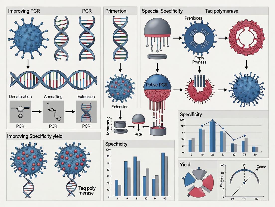
In molecular biology, the polymerase chain reaction (PCR) serves as a fundamental technique for gene amplification, with its success critically dependent on the specificity of the amplification process. Primer design is the cornerstone of achieving this specificity; well-designed primers ensure efficient and accurate amplification of the intended target sequence, while poor design leads to nonspecific products, reduced yield, and compromised data integrity. This application note details the principles of specific primer design and provides validated protocols to equip researchers with the tools to overcome common PCR challenges, thereby enhancing the reliability of their results in research and diagnostic applications.
The Fundamental Principles of Specific Primer Design
The specificity of a PCR amplification is governed by several interconnected physicochemical properties of the primers. These parameters determine how exclusively the primers anneal to their intended target sequence during the critical annealing step of the PCR cycle.
Primer Length: The specificity of a primer is directly influenced by its length. For optimal amplification, PCR primers should be between 18 and 24 nucleotides long [1] [2]. Primers shorter than this range risk binding to multiple non-target sites, producing nonspecific amplification. Conversely, primers longer than 30 base pairs exhibit slower hybridization rates and can be less efficient during the annealing phase, leading to reduced amplicon yield [1].
Melting Temperature (
T_m): The melting temperature is the temperature at which 50% of the DNA duplex dissociates into single strands. For primers, it defines the annealing conditions. The optimalT_mfor maintaining primer specificity is 54°C or higher, with an ideal range of 54°C to 65°C [1]. Crucially, the two primers in a pair should haveT_mvalues within 5°C of each other to ensure synchronized binding to the template [1] [2]. The annealing temperature (T_a) of the PCR cycle is typically set 2-5°C above theT_mof the primers for maximum specificity [1].GC Content: The GC content, which is the percentage of guanine (G) and cytosine (C) bases in the primer, should be maintained between 40% and 60% [1] [2]. Since G and C bases form three hydrogen bonds—compared to two for A and T—a higher GC content stabilizes the primer-template duplex. However, a GC content that is too high can promote non-specific binding and primer-dimer formation [1]. A useful design feature is the GC clamp, where the last five nucleotides at the 3' end contain one or two G or C bases. This promotes strong binding at the site of elongation, but more than three G or C bases at the 3' end should be avoided as it can cause non-specific binding [1].
Table 1: Optimal Design Parameters for PCR Primers
| Parameter | Optimal Range | Rationale |
|---|---|---|
| Length | 18 - 24 nucleotides [1] [2] | Balances specificity (longer) with hybridization efficiency and yield (shorter). |
Melting Temperature (T_m) |
54°C - 65°C [1] | Ensures stable and specific annealing. Primer pairs should be within 5°C [2]. |
| GC Content | 40% - 60% [2] | Provides sufficient duplex stability without risking non-specific binding. |
| GC Clamp | 1-2 G/C bases in last 5 bases at 3' end [1] | Strengthens binding at the critical point of polymerase elongation. |
Advanced Considerations for Avoiding Amplification Artifacts
Beyond the core principles, successful primer design must account for and prevent secondary structures and homologous sequences that severely compromise amplification specificity.
Secondary Structures: Primers must be screened for self-complementarity and 3'-self-complementarity to avoid intramolecular structures like hairpins [1]. Hairpins form when regions within a single primer are complementary, causing the primer to fold onto itself and preventing it from binding to the template. Similarly, primer-dimers—both self-dimers (between two identical primers) and cross-dimers (between forward and reverse primers)—occur due to inter-primer complementarity [1]. These dimers act as efficient templates for amplification, consuming reagents and outcompeting the desired target, which results in little to no yield of the intended amplicon [1].
Specificity and Exon Spanning: For amplifying cDNA (from mRNA), primers should be designed to span an exon-exon junction whenever possible [3]. This technique ensures that amplification is specific to mRNA and not contaminating genomic DNA, as the primer binding site would be disrupted by an intron in the genomic sequence. Tools like NCBI's Primer-BLAST can enforce this rule and are essential for verifying that primer pairs are unique to the intended template and will not produce amplicons from unrelated sequences in the database [3].
The following workflow diagrams the logical process for designing specific primers and the cascade of problems that arise from poor design.
Experimental Protocol for Primer Design and Validation
This section provides a detailed, step-by-step protocol for designing, testing, and utilizing primers in a PCR assay to ensure high specificity and yield.
Protocol: A Step-by-Step Guide to Specific Primer Design
Principle: To systematically create and validate primer pairs that exclusively amplify the target DNA sequence.
Materials:
- Template DNA Sequence (FASTA format).
- Primer Design Software (e.g., NCBI Primer-BLAST, Eurofins Genomics tools).
- Oligonucleotide Synthesis Service.
- PCR Reagents: Thermostable DNA polymerase (e.g., Pfu or Taq), corresponding buffer, MgCl₂, dNTPs, nuclease-free water [4] [5].
- Thermal Cycler.
- Agarose Gel Electrophoresis system.
Procedure:
Define the Target:
- Obtain the complete nucleotide sequence of your target gene or region.
- Identify the precise start and end points of the sequence you wish to amplify.
Generate Candidate Primers:
- Input your target sequence into a reliable primer design tool like NCBI Primer-BLAST [3].
- Set the following parameters in the tool:
- Product Size: 100-500 bp for standard PCR [5].
- Primer Length: Min=18, Opt=20, Max=24.
- Tm: Min=54°C, Opt=60°C, Max=65°C.
- GC Content: Min=40%, Opt=50%, Max=60%.
- For mRNA/cDNA work, select the option "Primer must span an exon-exon junction" to avoid genomic DNA amplification [3].
- Run the tool to generate a list of candidate primer pairs.
Manually Check and Select Primers:
- From the candidate list, select a pair where the
T_mvalues are within 1-2°C of each other. - Inspect the sequences for a GC clamp (1-2 G/C bases at the 3' end).
- Check the reported parameters for "self-complementarity" and "self 3'-complementarity" and select the pair with the lowest scores to minimize secondary structures [1].
- If adding restriction enzyme sites for cloning, add the recognition sequence plus a 3-6 bp "clamp" at the 5' end of the primer [2].
- From the candidate list, select a pair where the
Validate Primers Experimentally:
- Reconstitute the synthesized primers to a stock concentration (e.g., 100 µM).
- Set up a 25 µL PCR reaction as follows:
- Use Touchdown PCR for initial testing: Start with an annealing temperature 5-10°C above the calculated
T_m, and decrease it by 1°C per cycle for the first 10-15 cycles, then continue for another 15-20 cycles at the final, lowerT_m[6]. - Include a negative control (no template) to detect contamination or primer-dimer formation.
Analyze the Results:
- Separate the PCR products on an agarose gel.
- A successful, specific reaction will show a single, sharp band of the expected size.
- If multiple bands, smearing, or primer-dimers are present, proceed to optimization (Section 5.1).
Integrating Primer Design with Broader PCR Optimization
Protocol: Troubleshooting and Enhancing Specificity with Additives
Even well-designed primers may require reaction optimization. This protocol outlines steps to resolve issues of nonspecific amplification.
Materials:
- Hot-Start DNA Polymerase: An enzyme modified to be inactive at room temperature, preventing mispriming during reaction setup [6].
- PCR Additives: DMSO, Betaine, BSA, or specialized commercial enhancers.
- Novel Additives: Zwitterionic polymer-modified Graphene Oxide (GO-pSB) [4].
Procedure:
- Optimize Annealing Temperature:
- Perform a gradient PCR with the annealing temperature varying across a 10-15°C range centered on the calculated
T_a. - Analyze the gel to identify the temperature that produces the strongest target band with the least background.
- Perform a gradient PCR with the annealing temperature varying across a 10-15°C range centered on the calculated
Titrate Magnesium Concentration:
- Magnesium (
Mg²⁺) is a crucial cofactor for DNA polymerase. Test a series ofMgCl₂concentrations (e.g., 1.0, 1.5, 2.0, 2.5, 3.0 mM) while keeping other components constant [5].
- Magnesium (
Incorporate Specificity-Enhancing Additives:
- DMSO: Often effective for GC-rich templates. Test at 1-5% (v/v) [6].
- Graphene Oxide (GO) Derivatives: Based on recent research, consider adding zwitterionic polymer-modified GO (GO-pSB) to the PCR mix. Studies show it can significantly enhance specificity by interacting with the DNA polymerase [4]. A recommended starting point is to add 1-5 µL of a prepared GO-pSB dispersion to a 25 µL reaction [4].
Research Reagent Solutions for PCR Specificity
Table 2: Key Reagents for Optimizing PCR Specificity and Yield
| Reagent / Solution | Function / Application | Example Use Case |
|---|---|---|
| Hot-Start DNA Polymerase | Inhibits polymerase activity at low temperatures, preventing nonspecific amplification and primer-dimer formation during reaction setup [6]. | Essential for multiplex PCR and high-throughput setups where reactions are assembled at room temperature. |
| Pfu or Vent Polymerase | High-fidelity, thermostable DNA polymerases with 3'→5' proofreading exonuclease activity, resulting in lower error rates than Taq polymerase [5]. | Critical for cloning, sequencing, and any application where sequence accuracy is paramount. |
| MgCl₂ Solution | Essential cofactor for DNA polymerase activity. Its concentration dramatically affects primer annealing, duplex stability, and enzyme fidelity [5]. | Requires titration for each new primer-template system; a standard starting point is 1.5-2.0 mM. |
| DMSO | Additive that disrupts base pairing, helping to denature GC-rich secondary structures in the template DNA [6]. | Amplification of GC-rich targets (>65% GC). Note: DMSO lowers the effective primer Tm. |
| Zwitterionic GO (GO-pSB) | Novel nanomaterial additive that interacts with DNA polymerase, significantly improving PCR specificity, particularly for complex templates like genomic DNA [4]. | Challenging amplifications from clinical samples (e.g., blood genomic DNA) or when traditional additives fail. |
| Platinum II Taq Buffer | A commercially available, pre-optimized buffer system often containing proprietary stabilizers and enhancers. | Simplifies optimization, especially for fast PCR, GC-rich PCR, or direct PCR from crude samples [6]. |
The meticulous design of primers is an indispensable first step in achieving specific and efficient DNA amplification. By adhering to the established principles of length, melting temperature, and GC content, and by rigorously avoiding secondary structures, researchers can lay a solid foundation for successful PCR. When this careful design is coupled with robust experimental protocols and strategic use of modern reagents—from hot-start polymerases to novel nanomaterials like graphene oxide—the challenges of nonspecific amplification and low yield can be effectively overcome. This integrated approach ensures the generation of reliable, reproducible data that is critical for advancing research and development in the life sciences.
The polymerase chain reaction (PCR) is a foundational technique in molecular biology, enabling the rapid in vitro amplification of specific DNA sequences. Since its introduction by Kary Mullis in 1985, PCR has become an indispensable tool for researchers and clinicians in diverse fields, including diagnostics, genomics, and drug development [7]. The efficiency and specificity of PCR are not inherent but are profoundly influenced by the careful selection and optimization of core reaction components. Within the context of a broader thesis on methods to improve PCR specificity and yield, this application note provides a detailed examination of these critical elements—DNA polymerases, buffers, and co-factors. A thorough understanding of their properties, interactions, and mechanisms is essential for designing robust and reliable PCR protocols, particularly when dealing with challenging samples or complex templates such as those encountered in pharmaceutical research [8] [9].
Critical Reaction Components and Their Functions
A standard PCR requires a basic set of components, each fulfilling a specific role in the enzymatic amplification of DNA. The precise concentration and quality of each component are crucial for successful amplification [10] [11].
DNA Polymerases
The DNA polymerase is the core enzyme of the PCR, responsible for synthesizing new DNA strands. Its characteristics directly determine the success of the amplification.
- Taq DNA Polymerase: Derived from Thermus aquaticus, this was the first thermostable polymerase used in PCR and remains widely used. It has a half-life of approximately 40 minutes at 95°C and lacks 3'→5' proofreading exonuclease activity, resulting in a relatively low fidelity [8] [10].
- Proofreading DNA Polymerases: Enzymes such as Pfu (from Pyrococcus furiosus) possess 3'→5' exonuclease activity, which allows them to excise misincorporated nucleotides during synthesis. This proofreading capability increases fidelity by 10-fold or more compared to Taq polymerase. However, they often exhibit slower synthesis rates and cannot amplify uracil-containing templates [8] [9].
- Engineered DNA Polymerases: Modern PCR often utilizes engineered enzymes optimized for specific applications. These include "hot-start" polymerases (e.g., antibody-inactivated) that remain inactive at room temperature to prevent non-specific amplification, and chimeric polymerases with enhanced processivity and fidelity, sometimes exceeding 300 times that of standard Taq [8].
Table 1: Key Characteristics of DNA Polymerases
| Characteristic | Description | Impact on PCR | Example Enzymes |
|---|---|---|---|
| Thermostability | Ability to withstand high denaturation temperatures. | Essential for PCR cycling; hyperthermostable enzymes (e.g., from archaea) have longer half-lives at >95°C. | Taq, Pfu, KOD |
| Fidelity | Accuracy of DNA synthesis. | Critical for cloning, sequencing, and mutagenesis; proofreading enzymes have higher fidelity. | Pfu (High), Taq (Standard) |
| Processivity | Number of nucleotides added per enzyme-binding event. | Important for amplifying long templates, GC-rich regions, and in the presence of inhibitors. | Engineered polymerases |
| Specificity | Ability to amplify only the intended target. | Enhanced by hot-start mechanisms that inhibit activity until high temperatures are reached. | Hot-start Taq |
Buffers and Co-factors
The reaction buffer provides the optimal chemical environment for the DNA polymerase to function.
- Magnesium Ions (Mg²⁺): This divalent cation is an essential co-factor for all DNA polymerases. It catalyzes phosphodiester bond formation and facilitates the formation of primer-template complexes by stabilizing the negative charges on the phosphate backbones. The concentration of Mg²⁺ is a critical optimization parameter, typically used in a range of 0.5 to 5.0 mM. It directly affects enzyme activity, fidelity, and primer annealing [10] [11].
- Monovalent Cations: The buffer typically contains potassium ions (K⁺), usually at a concentration of 35 to 100 mM. K⁺ promotes primer annealing and increases the specificity of the reaction by destabilizing imperfect primer-template hybrids [9] [11].
- Tris-HCl: This buffering agent, usually at pH 8.0-8.8, maintains a stable pH throughout the thermal cycling process [11].
Other Essential Components
- Primers: These short, single-stranded DNA oligonucleotides (typically 15-30 nucleotides) define the start and end of the target sequence to be amplified. Careful design is paramount. Primers should have a melting temperature (Tm) of 55-70°C, with the two primers within 5°C of each other, and a GC content of 40-60%. The 3' end should avoid complementarity to prevent primer-dimer formation [10] [11].
- Deoxynucleoside Triphosphates (dNTPs): These are the building blocks (dATP, dCTP, dGTP, dTTP) for new DNA strands. They are typically used at equimolar concentrations of 0.2 mM each. Higher concentrations can inhibit PCR, while concentrations below the Km (0.010–0.015 mM) can lead to premature termination [10].
- Template DNA: The quality and quantity of the input DNA are crucial. Recommended amounts are 0.1–1 ng of plasmid DNA, 5–50 ng of genomic DNA, or 1-1000 ng of PCR product for re-amplification in a 50 µL reaction [10] [11].
(caption: PCR Component Optimization Workflow)
Advanced Optimization: PCR Enhancers and Additives
For challenging PCR applications, such as amplifying GC-rich sequences, long fragments, or templates with secondary structures, the inclusion of enhancing additives can be decisive. These compounds work through various mechanisms to improve yield and specificity [9].
- Betaine: Used at a concentration of 0.5 M to 2.5 M, betaine (trimethylglycine) can help amplify GC-rich templates by reducing the strand separation temperature. It acts as a stabilizing osmolyte, neutralizing the base-pairing energy differences between GC and AT pairs, which helps polymerases traverse through regions of high secondary structure [9] [11].
- Dimethyl Sulfoxide (DMSO): Typically used at 1-10%, DMSO is a polar solvent that interferes with the formation of secondary DNA structures by reducing DNA melting temperature. This facilitates the denaturation of complex templates and improves primer annealing specificity [9] [11].
- Other Additives:
- Formamide (1.25-10%): Similar to DMSO, it lowers the melting temperature of DNA, aiding in denaturation.
- Bovine Serum Albumin (BSA) (10-100 µg/mL): BSA can bind to inhibitors commonly found in biological samples (e.g., phenols, humic acid), thereby neutralizing their negative effects on the DNA polymerase.
- Tetramethylammonium oxalate (TMAO): A novel enhancer shown at 2 mM to significantly increase specificity and yield by suppressing non-specific amplification and primer-dimer formation [12].
Table 2: Common PCR Enhancers and Their Applications
| Additive | Common Concentrations | Proposed Mechanism of Action | Typical Application |
|---|---|---|---|
| Betaine | 0.5 M - 2.5 M | Equalizes DNA melting temperatures; reduces secondary structure. | GC-rich templates, long amplicons. |
| DMSO | 1% - 10% | Lowers DNA Tm; disrupts secondary structures. | GC-rich templates, complex genomes. |
| Formamide | 1.25% - 10% | Denaturant that lowers DNA Tm. | Difficult templates. |
| BSA | 10 - 100 µg/mL | Binds and neutralizes PCR inhibitors. | Crude samples (e.g., blood, plant). |
| TMA Oxalate | ~2 mM | Increases specificity and efficiency; reduces non-specific bands. | General specificity enhancement. |
Detailed Protocol for a Standard PCR Setup
The following protocol is adapted from established molecular biology methods and is designed for a 50 µL reaction volume [11]. This serves as a starting point from which optimizations can be made.
Materials and Reagents
- The Scientist's Toolkit:
- Thermostable DNA Polymerase (e.g., Taq, Pfu, or an engineered high-fidelity enzyme).
- 10X PCR Buffer (often supplied with the enzyme; may contain MgCl₂).
- 25 mM MgCl₂ Solution (if not included in the buffer or if optimization is required).
- 10 mM dNTP Mix (2.5 mM of each dNTP).
- Forward and Reverse Primers (20 µM stock solutions).
- Template DNA (e.g., genomic DNA, plasmid, cDNA).
- Nuclease-Free Water.
- PCR Additives (e.g., DMSO, Betaine) as needed.
- Equipment:
- Thermal cycler, microcentrifuge, pipettes and sterile aerosol-resistant tips, sterile 0.2 mL PCR tubes or plates, ice bucket.
Procedure
- Prepare the Reaction Mixture:
- Thaw all reagents on ice and briefly centrifuge to collect the contents at the bottom of the tubes.
- It is highly recommended to prepare a Master Mix when setting up multiple reactions to minimize pipetting error and ensure consistency. For a single 50 µL reaction, combine the components in a sterile 0.2 mL tube in the following order:
| Component | Volume (µL) | Final Concentration/Amount |
|---|---|---|
| Nuclease-Free Water | Q.S. to 50 µL | - |
| 10X PCR Buffer | 5.0 | 1X |
| 10 mM dNTP Mix | 1.0 | 200 µM each |
| 25 mM MgCl₂ (if needed) | Variable (e.g., 3.0) | 1.5 mM |
| Forward Primer (20 µM) | 1.0 | 0.4 µM |
| Reverse Primer (20 µM) | 1.0 | 0.4 µM |
| Template DNA | Variable (e.g., 1.0) | e.g., 50 ng genomic DNA |
| DNA Polymerase | 0.5 | 1.25 Units |
Thermal Cycling:
- Place the tubes in a thermal cycler and run the following standard program:
- Initial Denaturation: 94–95 °C for 2–5 minutes.
- Amplification (25–40 cycles):
- Denature: 94–95 °C for 15–30 seconds.
- Anneal: 45–65 °C for 15–30 seconds. The temperature must be optimized based on the primer Tm.
- Extend: 72 °C for 1 minute per kb of amplicon.
- Final Extension: 72 °C for 5–10 minutes.
- Hold: 4–10 °C.
- Place the tubes in a thermal cycler and run the following standard program:
Analysis of PCR Products:
- Analyze the amplified DNA by agarose gel electrophoresis. Use an appropriate DNA ladder to confirm the size and assess the specificity and yield of the amplification.
The path to achieving optimal PCR results is a systematic process of understanding and controlling reaction components. The choice of DNA polymerase—with its unique profile of thermostability, fidelity, processivity, and specificity—sets the foundation for the experiment. This foundation is supported by a carefully optimized buffer system, particularly the concentration of Mg²⁺, and the quality of primers and template. For the most challenging applications, a strategic selection of PCR enhancers can overcome significant obstacles. By applying the principles and detailed protocols outlined in this note, researchers can rationally troubleshoot and refine their PCR conditions, thereby enhancing the specificity and yield critical for advancing research and development in the life sciences.
The Impact of Thermal Cycling Parameters on Reaction Efficiency
The polymerase chain reaction (PCR) is a foundational technique in molecular biology, yet its efficiency is profoundly influenced by the specific parameters of the thermal cycling process. Reaction efficiency directly impacts specificity, yield, and the reliability of downstream results. Within the broader context of improving PCR specificity and yield, the optimization of thermal cycling parameters emerges as a critical, yet often overlooked, factor. The pursuit of faster, more specific, and more robust amplification protocols requires a deep understanding of the interplay between temperature, time, and enzyme kinetics [13]. This application note provides a detailed examination of how thermal cycling parameters—including denaturation, annealing, and extension temperatures and durations—affect reaction efficiency. We present structured quantitative data, optimized experimental protocols, and visual workflows to guide researchers and drug development professionals in systematically enhancing their PCR methodologies.
Key Thermal Cycling Parameters and Their Impact on Efficiency
The fundamental PCR process consists of three temperature-dependent steps that are repeated for 25-40 cycles: denaturation, annealing, and extension. The precise execution of each step is governed by the thermal cycler and reaction components [13].
Denaturation
The initial and cycle denaturation steps separate double-stranded DNA into single strands, enabling primer binding. Incomplete denaturation leads to poor yield and efficiency.
- Temperature and Time: Denaturation is typically performed at 94–98°C. The required time depends on template complexity; mammalian genomic DNA or GC-rich regions may require longer incubation or higher temperatures than plasmids and PCR products [14]. For routine templates, denaturation times can be as short as 1-5 seconds per cycle without sacrificing efficiency [15].
- GC-Rich Templates: DNA with high GC content (>65%) often necessitates longer denaturation or the use of additives like DMSO, formamide, or betaine to facilitate strand separation [14].
Annealing
The annealing step is the primary determinant of reaction specificity. Here, primers bind to their complementary sequences on the template DNA.
- Temperature Calculation: The annealing temperature is determined by the primer melting temperature (Tm), defined as the temperature at which 50% of the oligonucleotide molecules are in a duplex state [16]. Tm can be calculated using several formulas, with the Nearest Neighbor method being among the most accurate [14].
- Optimization Strategy: A starting annealing temperature of 3–5°C below the lowest primer Tm is recommended [14] [16]. If non-specific amplification occurs, the temperature should be increased in increments of 2–3°C. Conversely, low yield may require a temperature decrease [14]. The use of a thermal cycler with a precise gradient function is invaluable for this empirical optimization.
Extension
During extension, the DNA polymerase synthesizes a new DNA strand complementary to the template.
- Temperature and Duration: The extension temperature is set to the optimal temperature for the DNA polymerase, typically 70–75°C. The extension time is proportional to the length of the amplicon and the synthesis rate of the enzyme. While traditional guidelines suggest 1 minute per kilobase for Taq polymerase, recent studies show that extension times can be significantly shortened. For a 1.5 kb fragment, durations of 25 seconds can be sufficient with robust master mixes [15].
- Two-Step PCR: For primers with annealing temperatures within 3°C of the extension temperature, a two-step PCR (combining annealing and extension) can be used to shorten cycle times [14].
Quantitative Comparison of Thermal Cycling Performance
The performance of a PCR assay is heavily dependent on the instrument's ability to accurately and uniformly control temperature. The following table compares the advertised performance of different thermal cycler technologies, which directly impacts the efficiency and reproducibility of the reactions they run.
Table 1: Performance Comparison of qPCR Thermal Cycler Technologies
| qPCR Platform | Thermal System | Fastest Ramp Rate (°C/sec) | Thermal Uniformity (°C) |
|---|---|---|---|
| ABI Prism 7900HT | Block/Peltier | 1.5 | ±0.5 |
| Bio-Rad CFX96 | Block/Peltier | 3.3 (average) | ±0.4 |
| Qiagen Rotor-Gene Q | Air | 15 (peak) | ±0.02 |
| BJS Biotechnologies xxpress | Resistive Heating | 10 | ±0.3 |
Data adapted from a comparative study of qPCR instrumentation [17]
The relationship between instrument performance and assay outcome is clear. In a study comparing these systems, the time to complete 40 cycles varied dramatically from 12 to 58 minutes [17]. Despite these differences in speed, the amplification efficiency across platforms was comparable, with Ct values for the same target ranging from 13.6 to 16.8 [17]. This demonstrates that novel heating technologies can provide speed without necessarily compromising efficiency.
Advanced Protocols for Enhanced Efficiency
Protocol: Rapid PCR with Shortened Cycling Parameters
This protocol demonstrates that significantly shortened cycling times are feasible without loss of yield or specificity, enabling higher throughput and reduced energy consumption [15].
Application: Amplification of a 1466 bp fragment from the 16S rRNA gene from bacterial templates with low-, mid-, and high-GC content. Objective: To achieve efficient amplification with a 46% reduction in program duration and 50% less electricity consumption compared to typical protocols.
Materials:
- Template DNA: Bacterial genomic DNA (e.g., 10-100 ng per reaction).
- Primers: Specific to the 16S rRNA gene target.
- Master Mix: PCRBIO Ultra Mix or equivalent robust, fast-cycling polymerase mix.
- Thermal Cycler: Instrument capable of fast ramping and precise control of short hold times.
Method:
- Reaction Setup: Prepare a 25 µL reaction mix according to the master mix manufacturer's instructions, including primers and template DNA.
- Initial Denaturation: 95°C for 1 minute.
- Cycling (30 cycles):
- Denaturation: 95°C for 5 seconds.
- Annealing: Primer-specific Tm for 25 seconds.
- Extension: 72°C for 25 seconds.
- Final Extension: 72°C for 1 minute.
- Hold: 4°C forever.
Validation: Analyze 5 µL of the PCR product by agarose gel electrophoresis. A single, sharp band of the expected size (1466 bp) should be visible. The amplicon yield should be sufficient for downstream applications such as sequencing [15].
Protocol: "V" Shape PCR (VPCR)
VPCR is a radical approach that eliminates hold times entirely, performing the three PCR steps during the heating and cooling transitions of the thermal cycler. This can reduce amplification time by two-thirds [18].
Application: Ultra-fast amplification of short to medium-length DNA fragments (e.g., 98-500 bp). Objective: To complete PCR amplification in minutes using a conventional thermal cycler.
Materials:
- Template DNA: λ-DNA or target DNA (e.g., 0.1-1 ng/µL).
- Primers: Designed for the target amplicon; longer primers with higher Tm may perform better.
- Polymerase System: KAPA2G Robust HotStart DNA Polymerase or a similar robust, fast enzyme.
- Thermal Cycler: Standard block-based instrument.
Method:
- Reaction Setup: Prepare a 10 µL reaction mix. For a 500 bp λ DNA fragment:
- 1X Reaction Buffer
- Additional 1 mM MgCl₂ (if required)
- 0.2 mM dNTPs
- 0.5 U/µL KAPA2G Robust DNA Polymerase
- 0.2-0.5 µM of each primer
- Template DNA
- Cycling (30 cycles):
- Denaturation: 94°C for 0 seconds.
- Annealing/Extension: 60°C for 0 seconds.
- Note: The instrument will ramp between these two temperatures. The critical parameters are the ramp rate and the upper/lower temperature setpoints.
Validation: For a 500 bp amplicon, VPCR can produce a specific band identical to that from a conventional 66-minute protocol in under 17 minutes [18]. Specificity should be confirmed by gel electrophoresis.
Experimental Workflow for Systematic Optimization
The following diagram illustrates a logical pathway for researchers to systematically optimize their thermal cycling parameters to maximize reaction efficiency.
Figure 1: A decision workflow for troubleshooting and optimizing PCR thermal cycling parameters to improve reaction efficiency. The path is determined by the initial symptom observed (e.g., no product, low yield, or non-specific bands).
The Scientist's Toolkit: Essential Research Reagent Solutions
The following table outlines key reagents and their critical functions in optimizing thermal cycling and ensuring high PCR efficiency.
Table 2: Essential Reagents for Optimizing PCR Efficiency
| Reagent / Solution | Function in PCR | Considerations for Optimization |
|---|---|---|
| Robust Hot-Start DNA Polymerase | Catalyzes DNA synthesis; "Hot-Start" reduces non-specific amplification during reaction setup. | Select enzymes with high processivity and speed for fast cycling. Verify thermostability for prolonged or high-temperature denaturation [14] [18]. |
| Optimized Buffer Systems | Provides optimal pH, ionic strength, and co-factors (e.g., Mg²⁺) for polymerase activity. | Mg²⁺ concentration is critical; it must be optimized as it affects primer annealing and enzyme fidelity. Some buffers offer isostabilizing properties for universal annealing temperatures [14]. |
| PCR Additives (DMSO, Betaine) | Reduces secondary structure in GC-rich templates; lowers the effective Tm of the primer-template duplex. | Use at appropriate concentrations (e.g., 1-10% DMSO). Requires adjustment of annealing temperature downward [14]. |
| High-Purity dNTPs | Building blocks for new DNA strand synthesis. | Ensure quality and balance of all four dNTPs to prevent misincorporation and early reaction plateau. |
| Well-Designed Primers | Provides specificity and initiation points for DNA synthesis. | Both primers should have similar Tm. Use tools like IDT OligoAnalyzer for accurate Tm prediction based on reaction conditions [16]. |
Thermal cycling parameters are not fixed constants but variables that can be strategically manipulated to significantly enhance PCR efficiency. As demonstrated, moving beyond standard, lengthy protocols to optimized, rapid cycling is readily achievable. This optimization requires a holistic approach, considering the interplay between the thermal cycler's performance, the biochemical properties of the reagents, and the sequence of the target and primers. By applying the structured data, protocols, and troubleshooting workflows provided in this application note, researchers can systematically refine their PCR methods. This leads to more specific amplification, higher yields, faster turnaround times, and more sustainable laboratory operations, thereby directly supporting advanced research and robust drug development processes.
Polymerase Chain Reaction (PCR) is a foundational technique in molecular biology, yet the amplification of templates with high GC content (>60%) and stable secondary structures presents a significant challenge for researchers and drug development professionals [19]. GC-rich DNA sequences exhibit greater thermal stability primarily due to base stacking interactions, not just hydrogen bonding, which raises the melting point of the DNA and hinders complete denaturation [20]. Furthermore, these regions readily form stable intramolecular secondary structures, such as hairpin loops, which can cause polymerase stalling, premature termination, and ultimately lead to PCR failure or the production of truncated amplicons [21] [20]. This application note, framed within a broader thesis on enhancing PCR specificity and yield, details optimized protocols and reagent solutions to overcome these obstacles, enabling reliable analysis of challenging targets like promoter regions, nicotinic acetylcholine receptor subunits, and viral inverted terminal repeats (ITRs) [19] [21].
Underlying Mechanisms and Strategic Workflow
The core challenge with complex templates lies in the interference of two natural DNA properties with the PCR process. The strong triple hydrogen bonding between guanine and cytosine creates regions of high thermal stability that resist standard denaturation temperatures. Concurrently, the complementary nature of these sequences promotes the formation of stable secondary structures within the single-stranded template itself before primers can anneal. These structures physically block polymerase progression and can lead to enzymatic errors, such as endonucleolytic cleavage by Taq polymerase, further reducing yield and specificity [21].
A successful strategy requires a multi-pronged approach that addresses both thermodynamic stability and structural conformation, as outlined in the workflow below.
Research Reagent Solutions
A curated selection of reagents is essential for tackling difficult templates. The following table summarizes key solutions and their functions.
Table 1: Research Reagent Solutions for Complex PCRs
| Reagent Category | Specific Example | Function & Mechanism |
|---|---|---|
| Specialized Polymerases | Proofreading Blends (e.g., Pfu, KOD) [22] | High-fidelity enzymes with 3'→5' exonuclease activity reduce error rates for cloning and sequencing. |
| Inhibitor-Tolerant (e.g., Phusion Flash) [23] | Engineered for robust activity in the presence of common inhibitors from direct PCR protocols. | |
| Highly Processive (e.g., AccuPrime) [20] | Derived from extremophiles, these polymerases remain stable at high temperatures, aiding denaturation. | |
| Organic Additives | DMSO (2-10%) [22] [20] | Disrupts secondary structures by lowering DNA melting temperature and interfering with base pairing. |
| Betaine (1-2 M) [19] [22] | Homogenizes the thermodynamic stability of DNA, equivalentizing GC- and AT-rich regions. | |
| 7-deaza-dGTP [21] [20] | A dGTP analog that reduces hydrogen bonding, facilitating the denaturation of GC-rich structures. | |
| Novel Oligonucleotides | Disruptors [21] | Sequence-specific oligonucleotides that bind and unwind stable intramolecular secondary structures in the template. |
| Enhanced Buffer Systems | OneTaq GC Buffer / GC Enhancer [20] | Commercially optimized buffers specifically formulated to improve amplification efficiency of GC-rich targets. |
Optimized Experimental Protocols
Core PCR Protocol with Additives
This protocol is designed for amplifying GC-rich targets, such as nicotinic acetylcholine receptor subunits, and incorporates a multi-factorial optimization strategy proven to amplify targets with GC contents up to 65% [19].
Table 2: Reaction Setup for GC-Rich PCR
| Component | Final Concentration/Amount | Notes & Rationale |
|---|---|---|
| Template DNA | 5-50 ng (genomic DNA) | High purity is critical; dilute if inhibitors are suspected [10] [5]. |
| Forward/Reverse Primer | 0.4 - 0.5 µM each | Designed with Tm within 55-70°C; avoid GC clamps at 3' end [10] [24]. |
| High-Fidelity Polymerase | 1-2 units | Use a proofreading enzyme or blend for targets >5kb or for cloning [22]. |
| dNTPs | 0.2 mM each | Balanced equimolar mixture; avoid excess to maintain fidelity [10]. |
| Mg²⁺ | 1.5 - 4.0 mM (start at 2.0 mM) | Requires titration; essential cofactor whose concentration critically affects specificity [22] [5]. |
| PCR Buffer | 1X | Use the manufacturer's supplied buffer. |
| DMSO | 2 - 10% (v/v) | Critical additive. Start with 5% to help denature secondary structures [19] [22]. |
| Betaine | 1 - 1.5 M | Critical additive. Can be used alone or with DMSO to destabilize GC-rich structures [19] [22]. |
| Water | To volume | Nuclease-free. |
| Total Reaction Volume | 50 µL |
Thermal Cycling Conditions [19] [20]:
- Initial Denaturation: 98°C for 2 minutes (or per polymerase guidelines).
- Cycling (30-40 cycles):
- Denaturation: 98°C for 10-30 seconds. For extreme GC content, a higher denaturation temperature (e.g., 99-100°C) can be tested for the first 5 cycles, but be mindful of polymerase half-life [20].
- Annealing: Temperature gradient from 55°C to 72°C for 15-60 seconds. The optimal temperature (Ta) must be determined empirically and is often 3-5°C below the primer Tm [22] [5].
- Extension: 72°C for 15-60 seconds per kilobase.
- Final Extension: 72°C for 5-10 minutes.
- Hold: 4°C.
Protocol for Using Disruptor Oligonucleotides
For templates with ultra-stable secondary structures, such as the inverted terminal repeats (ITRs) of adeno-associated virus (AAV) vectors, conventional additives may fail. Disruptors offer a novel, sequence-specific solution [21].
Mechanism of Action: A disruptor oligonucleotide contains three functional domains: an "anchor" for initial template binding, an "effector" that is reverse-complementary to part of the structured region to mediate strand invasion, and a 3' blocker (e.g., C3-Spacer) to prevent its extension by the polymerase [21]. The diagram below illustrates this mechanism.
Procedure:
- Disruptor Design: Design disruptors to be reverse-complementary to the template sequence, partially overlapping the duplex region of the intramolecular secondary structure. The anchor domain is the most critical for initial binding and function [21].
- Reaction Setup: Incorporate the disruptor oligonucleotide into the standard PCR mix described in Protocol 4.1 at a final concentration of 0.1 - 0.5 µM.
- Thermal Cycling: Use a standard thermal cycling profile. The disruptor functions during the annealing step, obviating the need for specialized cycling conditions [21].
Data Presentation and Optimization Guidelines
Systematic optimization of reaction components is non-negotiable for success with challenging templates. The following tables consolidate quantitative data to guide this process.
Table 3: Optimization of Critical PCR Components [22] [10] [5]
| Component | Suboptimal Condition | Effect | Recommended Optimization |
|---|---|---|---|
| Annealing Temp. (Ta) | Too Low | High nonspecific amplification, smearing. | Perform gradient PCR. Set Ta 3-5°C below the average primer Tm. |
| Too High | Low or no yield due to inefficient primer binding. | ||
| Mg²⁺ Concentration | < 1.5 mM | Greatly reduced polymerase activity, low yield. | Perform titration (e.g., 1.5, 2.0, 2.5, 3.0, 3.5, 4.0 mM). |
| > 4.0 mM | Increased nonspecific products, reduced fidelity. | ||
| Primer Concentration | < 0.2 µM | Low reaction yield. | Use 0.4 - 0.5 µM as a standard; titrate if issues persist [24]. |
| > 1.0 µM | Primer-dimer formation, nonspecific binding. | ||
| Cycle Number | Too Few | Insufficient product from low-copy templates. | Use 30-40 cycles; >35 cycles can increase background [24]. |
| Too Many | Plateau phase, accumulation of nonspecific products. |
Table 4: Troubleshooting Common Scenarios
| Problem | Possible Cause | Suggested Remedies |
|---|---|---|
| No Product | Overly stable template secondary structure. | Implement Protocol 4.1 with 5% DMSO + 1M Betaine. Use a polymerase blend. Increase denaturation temperature for first 5 cycles [20]. |
| Annealing temperature too high. | Run a gradient PCR to determine the optimal Ta [22]. | |
| Inhibitors co-purified with template. | Dilute template, re-purify, or use an inhibitor-tolerant polymerase [23]. | |
| Smearing / Non-specific Bands | Annealing temperature too low. | Increase Ta stringency using a gradient [22] [5]. |
| Mg²⁺ concentration too high. | Titrate Mg²⁺ down in 0.5 mM increments [22]. | |
| Primer concentration too high. | Reduce primer concentration to 0.2-0.4 µM [10]. | |
| Primer-Dimer | Low annealing stringency. | Increase Ta. Use a hot-start polymerase to prevent activity at room temperature [25]. |
| Primer 3'-end complementarity. | Redesign primers to avoid 3' self-complementarity [10]. |
Implementing Advanced PCR Techniques for Complex Applications
Real-Time PCR (qPCR) for Sensitive Quantification and Cosmetic Product Quality Control
The preservation of microbial safety in cosmetic products is paramount for consumer health, necessitating rapid and accurate detection strategies [26]. Traditional microbiological quality control methods, such as plate counts, are often time-consuming, labor-intensive, and fail to detect viable but non-cultivable (VBNC) cells, a common physiological state for many microorganisms [26]. Molecular techniques, particularly real-time PCR (qPCR), have revolutionized this field by offering enhanced detection sensitivity, specificity, and speed [26]. This document details the application of qPCR for the quantification of specific pathogens and quality control in cosmetics, providing validated protocols and frameworks that align with international standards to ensure product safety and regulatory compliance.
Experimental Design and Workflow
The application of qPCR for cosmetic quality control involves a multi-stage process, from sample preparation to final data analysis. The workflow below outlines the key steps to ensure accurate and reliable pathogen detection.
Sample Preparation and Inoculation
Cosmetic products with varying physical characteristics (e.g., creamy, oily, milky, solid) are selected [26]. For the detection of major cosmetic pathogens—Escherichia coli, Staphylacus aureus, Pseudomonas aeruginosa, and Candida albicans—samples are spiked with low levels (3–5 colony-forming units, CFU) of the target organisms [26]. Spiked samples are then enriched in an appropriate broth, such as Eugon broth, and incubated at 32.5°C for 20–24 hours to allow for microbial growth, thereby increasing the target DNA load for subsequent detection [26]. Complex matrices, such as certain soaps, may require a longer enrichment time (e.g., 36 hours) and dilution to mitigate the effects of antimicrobial ingredients [26].
Genomic DNA Extraction
Post-enrichment, genomic DNA is isolated from the samples. This can be performed using commercial DNA extraction kits, such as the PowerSoil Pro kit (Qiagen), following the manufacturer's instructions and processed with automated systems like the QIAcube Connect [26]. The extraction process should include several controls: a medium control, a zero control (no template), and an extraction control to monitor for potential contamination and verify the integrity of the extraction process [26].
qPCR Assay Design and Validation
Primer and Probe Design
The success of a qPCR assay is fundamentally dependent on the careful design of primers and probes. The table below summarizes the key design criteria for achieving high specificity and efficiency.
Table 1: Golden Rules for PCR Primer and Probe Design
| Design Parameter | Optimal Specification | Rationale |
|---|---|---|
| Length | 18–30 nucleotides (primers); 15–30 nucleotides (probes) [27] [1] | Balances specificity with efficient hybridization and amplification [1]. |
| GC Content | 40%–60% [27] [1] | Ensures stable primer-template binding; a GC clamp (G or C bases at the 3' end) promotes specific binding [1]. |
| Melting Temperature (Tm) | 54°C–65°C; forward and reverse primers should have similar Tm (difference ≤ 2°C) [1] | Allows for synchronized primer annealing during the PCR cycle, which is essential for efficiency [1]. |
| 3' End Specificity | Avoid A; prefer T; avoid runs of 3+ G/C bases [27] [1] | Prevents mispriming and non-specific amplification due to weaker base pairing at the initiation site [27]. |
| Sequence Composition | Avoid self-complementarity, cross-dimers, and secondary structures [27] | Prevents the formation of primer-dimers and hairpins, which compete with target amplification and reduce yield [27] [1]. |
| Specificity Verification | Perform in silico BLAST search [27] | Confirms that primers will bind uniquely to the intended target sequence, minimizing off-target amplification [27]. |
The following diagram illustrates the logical workflow for designing and validating these critical oligonucleotides.
qPCR Assay Validation
Before implementation in a quality control setting, the developed qPCR assay must undergo rigorous validation to confirm its performance characteristics. Key validation parameters, as demonstrated in vaccine and environmental surveillance studies, are summarized below [28] [29].
Table 2: Key Validation Parameters for a qPCR Assay
| Validation Parameter | Target Performance | Experimental Approach |
|---|---|---|
| Linearity and Range | Coefficient of determination (R²) ≥ 0.990 [29] | Analysis of a standard curve from a 10-fold dilution series of target DNA [29]. |
| Efficiency | 90–110% [28] | Calculated from the slope of the standard curve [28]. |
| Limit of Detection (LOD) | Varies by target (e.g., 0.003 pg/reaction) [29] | Determination of the lowest concentration detectable in 95% of replicates [29]. |
| Limit of Quantification (LOQ) | Varies by target (e.g., 0.03 pg/reaction) [29] | Determination of the lowest concentration quantifiable with acceptable precision and accuracy (e.g., RSD < 25%, recovery rate 80–120%) [29]. |
| Specificity | No cross-reactivity with non-target strains [28] [29] | Testing against DNA from a panel of related and common contaminating organisms [28] [29]. |
| Precision (Repeatability) | Relative Standard Deviation (RSD) ≤ 25% [29] | Multiple replicates of samples at different concentrations within the same run [29]. |
The Scientist's Toolkit: Essential Research Reagents
The following table catalogs the key reagents and materials required for implementing qPCR for cosmetic quality control.
Table 3: Essential Research Reagent Solutions for qPCR-based Quality Control
| Item | Function / Description | Example / Specification |
|---|---|---|
| DNA Extraction Kit | Isolates high-purity genomic DNA from complex cosmetic matrices, minimizing PCR inhibitors. | PowerSoil Pro Kit (Qiagen) [26]. |
| qPCR Master Mix | Provides the necessary buffer, enzymes (Taq polymerase), dNTPs, and MgCl₂ for the amplification reaction. | Commercial kits (e.g., R-Biopharm SureFast PLUS) [26]. |
| Primers & Probes | Oligonucleotides that specifically bind and detect the target pathogen DNA. | Designed per parameters in Table 1; can be unlabeled or dual-labeled (e.g., TaqMan) probes [28] [1]. |
| DNA Standards | Used to generate a standard curve for absolute quantification of target DNA in samples. | Synthetic gBlocks [28] or genomic DNA from reference strains [29]. |
| Internal Control | Distinguishes between true target negatives and PCR inhibition. | A non-competitive synthetic DNA sequence or a gene from a different species spiked into the reaction [26]. |
| Reference Strains | Provide positive control DNA for assay development and validation. | ATCC strains (e.g., E. coli ATCC 8739, S. aureus ATCC 6538) [26]. |
Detailed Experimental Protocol
qPCR Reaction Setup and Thermal Cycling
This protocol is adapted from methods used for pathogen detection in complex matrices [28] [26].
Reaction Mix Preparation: Prepare a master mix for the number of reactions needed, including controls (no-template control, positive control, extraction controls). A typical 30 µL reaction volume may contain:
Thermal Cycling Conditions: Program the real-time PCR instrument with the following standard protocol:
Data Analysis and Interpretation
- Threshold and Cq Determination: Set the fluorescence threshold in the exponential phase of the amplification plot across all standards and samples. The cycle quantification (Cq) value for each reaction is the cycle number at which the fluorescence crosses this threshold.
- Standard Curve Generation: Plot the Cq values of the DNA standards against the logarithm of their known concentrations. The slope, y-intercept, and R² value are used to determine amplification efficiency and for absolute quantification of unknown samples [29].
- Result Interpretation: A sample is considered positive if amplification occurs and the Cq value is at or below the validated limit of quantification (LOQ). The result is negative if no amplification occurs or if the Cq is above the LOQ, provided the internal and extraction controls are valid.
Real-time PCR represents a significant advancement in microbial safety for the cosmetics industry. Its superior sensitivity, specificity, and speed compared to traditional culture-based methods make it an ideal tool for rapid and reliable quality control [26]. By adhering to standardized protocols, conducting thorough validation as outlined in this document, and aligning with international guidelines, manufacturers can effectively integrate qPCR into their production workflows. This integration reinforces product safety, ensures regulatory compliance, and ultimately protects consumer health.
Digital PCR (dPCR) for Absolute Quantification and Rare Mutation Detection
Digital PCR (dPCR) represents the third generation of Polymerase Chain Reaction technology, following conventional PCR and real-time quantitative PCR (qPCR). This technology is founded on the partitioning of a PCR mixture containing the sample into thousands to millions of separate reactions, so that each partition contains either zero, one, or a few nucleic acid targets according to a Poisson distribution. Following PCR amplification, the fraction of positive partitions is counted via end-point measurement, enabling absolute quantification of the target concentration through Poisson statistics without the need for a standard curve [30].
The fundamental difference between dPCR and qPCR lies in their quantification methods. While qPCR relies on comparing amplification curves to standards for relative quantification, dPCR provides direct, absolute quantification by counting individual molecules, conferring significant advantages for applications requiring high sensitivity and precision, such as rare mutation detection [31] [30]. The first clinically relevant applications of dPCR leveraged its ability to detect rare genetic mutations within a background of wild-type genes, paving the way for tumor heterogeneity analysis and liquid biopsy applications in oncology [30].
Principles and Advantages of dPCR
Core Technological Principles
The dPCR workflow consists of four key steps: (1) partitioning the PCR mixture containing the sample into thousands of individual compartments; (2) amplifying the target sequences within each partition through thermal cycling; (3) performing end-point fluorescence analysis of all partitions; and (4) calculating the target concentration using Poisson statistics based on the fraction of positive and negative partitions [30].
Two major partitioning methodologies have emerged: water-in-oil droplet emulsification (droplet digital PCR or ddPCR) and microchamber-based systems (chip-based dPCR). Droplet systems utilize microfluidics to create monodisperse droplets at high speed (typically 1-100 kHz), while microchamber systems employ fixed arrays of microscopic wells embedded in a solid chip [30]. Each approach offers distinct advantages, with ddPCR providing greater scalability and cost-effectiveness, while microchamber dPCR typically offers higher reproducibility and ease of automation [32].
Comparative Advantages Over qPCR
dPCR offers several distinct advantages for absolute quantification and rare mutation detection:
- Absolute Quantification Without Standard Curves: By providing direct molecular counting, dPCR eliminates the need for standard curves, improving accuracy and reproducibility [31] [33].
- Enhanced Sensitivity for Rare Targets: dPCR can detect rare mutations with allele frequencies as low as 0.1% by effectively enriching low-level targets through sample partitioning [33].
- Superior Tolerance to PCR Inhibitors: The partitioning process dilutes inhibitors present in the sample across multiple reaction chambers, reducing their impact on amplification efficiency [30].
- Precise Quantification of Small Fold Changes: The technology's precision makes it particularly suitable for detecting minor variations in copy number and gene expression [32].
Table 1: Comparison of dPCR and qPCR Characteristics
| Parameter | Digital PCR (dPCR) | Quantitative PCR (qPCR) |
|---|---|---|
| Quantification Method | Absolute counting of molecules | Relative to standard curve |
| Standard Curve Requirement | Not required | Required |
| Sensitivity | Can detect rare targets (0.1% MAF) | Limited sensitivity for rare targets |
| Precision | High precision for small fold-changes | Moderate precision |
| Inhibitor Tolerance | High (inhibitors diluted) | Low (inhibitors affect reaction) |
| Data Output | Copies/μL | Cq values |
| Best Applications | Rare mutation detection, absolute quantification, liquid biopsy | Gene expression, pathogen detection |
dPCR for Rare Mutation Detection
Technical Considerations for Rare Allele Detection
The exceptional sensitivity of dPCR for rare mutation detection stems from its ability to physically separate mutant sequences from abundant wild-type DNA through partitioning. When the sample is partitioned, mutant sequences become concentrated in specific chambers, effectively enriching them and enabling detection even at very low frequencies [34]. This partitioning, combined with the binary nature of the endpoint detection (positive vs. negative), allows dPCR to overcome the limitations of traditional PCR when discriminating single-nucleotide variations [31].
For optimal rare mutation detection, several factors must be considered. The number of partitions directly impacts sensitivity, with higher partition counts increasing the probability of capturing and detecting rare targets. Sample input and quality are also critical, as degraded DNA or insufficient template can compromise results. Additionally, careful assay design is essential, typically employing two differently labeled hydrolysis probes (e.g., FAM for mutant sequences and VIC/HEX for wild-type) with the same primer set to ensure specific discrimination [34].
Application in Liquid Biopsies and Oncology
dPCR has emerged as a transformative technology for liquid biopsy analysis, enabling non-invasive detection and monitoring of cancer through circulating tumor DNA (ctDNA). ctDNA fragments released from apoptotic and necrotic cancer cells are typically short and exist in very low concentrations relative to total cell-free DNA, making the exceptional sensitivity of dPCR particularly valuable for their quantification [33].
In clinical oncology, dPCR applications include early cancer detection, measurement of therapeutic response, quantification of residual tumor burden, and monitoring of emerging treatment resistance. The technology's ability to detect mutant allele frequencies as low as 0.1% makes it suitable for tracking minimal residual disease and tumor evolution [33]. For example, in patients with advanced EGFR-mutant non-small cell lung cancer (NSCLC), dPCR enables early detection of the EGFR T790M mutation, which confers resistance to first and second-generation tyrosine kinase inhibitors, allowing for timely adjustment of treatment strategies [34].
Table 2: dPCR Performance Characteristics for Rare Mutation Detection
| Parameter | Performance Characteristics | Impact on Detection |
|---|---|---|
| Theoretical Detection Sensitivity | As low as 0.1% mutant allele frequency (MAF) | Enables identification of rare variants |
| DNA Input Requirements | 10-100ng human genomic DNA | Higher input increases sensitivity |
| Partition Numbers | 20,000-1,000,000+ | More partitions enhance rare target detection |
| Limit of Detection (LOD) | Theoretical LOD of 0.2 copies/μL with 95% confidence | Determines lowest detectable concentration |
| Practical Sensitivity | Down to 0.15% with 10ng DNA input | Function of system LOD and DNA input |
Experimental Protocol: dPCR for EGFR T790M Mutation Detection
Assay Design and Workflow
This protocol details the detection of the EGFR T790M mutation, a clinically relevant resistance mutation in non-small cell lung cancer, using a duplex dPCR approach with hydrolysis probes [34].
Assay Design Strategy:
- Use a single primer set amplifying the EGFR T790 locus
- Employ two TaqMan hydrolysis probes with different fluorophores:
- FAM-labeled probe targeting the EGFR T790M mutant sequence
- Cy3/VIC/HEX-labeled probe targeting the wild-type EGFR sequence
- Validate probe specificity and ensure fluorophore compatibility with the dPCR system
Required Materials and Reagents:
- dPCR system and appropriate consumables
- PCR master mix (2X or 5X concentration)
- Reference dye (if required by manufacturer)
- EGFR T790 forward and reverse primers (500 nM final concentration)
- EGFR T790M mutant probe (250 nM final concentration)
- EGFR wild-type probe (250 nM final concentration)
- Human genomic DNA samples (10-100ng input recommended)
- Nuclease-free water
Step-by-Step Procedure
A. PCR Mix Preparation
- Calculate the required number of reactions including:
- Test samples
- Non-template control (NTC; all components except DNA)
- Monocolor controls for each probe (for fluorescence compensation)
- Prepare master mix for n+1 samples to account for pipetting error
- Prepare PCR master mix according to Table 3:
Table 3: PCR Master Mix Formulation
| Reagent | Final Concentration | Volume per 25μL Reaction |
|---|---|---|
| PCR Master Mix (2X) | 1X | 12.5 μL |
| Reference Dye | As per manufacturer's instructions | Variable |
| EGFR T790 Forward Primer | 500 nM | Variable |
| EGFR T790 Reverse Primer | 500 nM | Variable |
| EGFR T790WT Probe | 250 nM | Variable |
| EGFR T790M Probe | 250 nM | Variable |
| Human Genomic DNA | 10-100 ng total | Variable |
| Nuclease-Free Water | - | To 25 μL total volume |
- Mix components thoroughly by vortexing and brief centrifugation
B. DNA Input Calculation and Sensitivity Determination
- Calculate DNA copy number using the formula:
- Number of copies = mass of DNA in reaction volume (ng) / 0.003
- The 0.003 factor represents the approximate mass in ng of a single haploid human genome
- Determine theoretical detection sensitivity:
- With 10ng DNA input: 10/0.003 = 3,333 copies
- Final concentration = 3,333 copies / 25μL = 133 copies/μL
- Sensitivity = Theoretical LOD / Concentration = 0.2 / 133 = 0.15%
C. Partitioning and Thermal Cycling
- Load PCR mix into appropriate dPCR consumable
- Perform partitioning according to manufacturer's instructions
- Run thermal cycling using optimized conditions:
Table 4: EGFR T790M Assay Cycling Conditions
| Cycles | Temperature | Time | Purpose |
|---|---|---|---|
| 1 | 95°C | 10 minutes | Initial denaturation, enzyme activation |
| 45 | 95°C | 30 seconds | Denaturation |
| 45 | 62°C | 15 seconds | Primer annealing/extension |
D. Data Acquisition and Analysis
- Perform endpoint fluorescence measurement according to system requirements
- Apply fluorescence compensation if necessary using monocolor controls
- Analyze data using 2D scatter plots to distinguish populations:
- Double-negative partitions (no target)
- FAM-positive only (mutant)
- Cy3/VIC/HEX-positive only (wild-type)
- Double-positive partitions (potential hybrid or errors)
- Calculate mutant allele frequency using Poisson correction:
- Mutant allele frequency = (Mutant concentration / Total DNA concentration) × 100
dPCR Platform Comparison and Selection
Technology Platforms and Characteristics
The dPCR landscape includes multiple commercial platforms employing different partitioning and detection technologies. Key systems include Applied Biosystems' QuantStudio Absolute Q, Bio-Rad's QX200/QX600 ddPCR systems, Qiagen's QIAcuity, and Roche's Digital LightCycler [30] [32].
Each platform offers distinct advantages depending on application requirements. Chip-based systems like the QuantStudio Absolute Q and QIAcuity provide fixed arrays of microchambers with simplified workflows, while droplet-based systems like Bio-Rad's ddPCR platforms create water-in-oil emulsions for partitioning [32]. The choice between systems depends on factors including throughput requirements, multiplexing capabilities, sample volume, and workflow preferences.
Table 5: Comparison of dPCR Platform Technologies
| Parameter | Chip-Based dPCR | Droplet Digital PCR (ddPCR) |
|---|---|---|
| Partitioning Mechanism | Fixed microchambers/wells | Water-in-oil emulsion droplets |
| Typical Partition Numbers | 20,000-30,000+ | 20,000-1,000,000+ |
| Partition Volume | Nanoliter range | Picoliter to nanoliter range |
| Workflow Integration | Integrated, automated systems | Multiple instruments/steps |
| Multiplexing Capability | Available for 4-12 targets | Limited in earlier systems, improved in newer models |
| Throughput Time | <90 minutes for some systems | 6-8 hours for complete workflow |
| Ideal Application Context | QC environments, routine testing | Research labs, development work |
Platform Selection Considerations
For regulated environments and quality control applications, integrated dPCR systems with automated workflows offer significant advantages. These systems typically provide "sample-in, results-out" functionality that reduces hands-on time, minimizes contamination risk, and includes features supporting 21 CFR Part 11 compliance [32]. The reduced manual intervention also decreases the potential for operator error, improving reproducibility.
For research applications requiring maximum flexibility and partition numbers, droplet digital PCR systems may be preferable. These platforms typically generate higher numbers of partitions, potentially increasing sensitivity for very rare targets, and offer established protocols for a wide range of applications [30] [32]. However, they generally require more manual steps and longer processing times.
Research Reagent Solutions
Table 6: Essential Reagents for dPCR Experiments
| Reagent/Category | Function | Examples & Considerations |
|---|---|---|
| dPCR Master Mix | Provides DNA polymerase, dNTPs, buffer, MgCl₂ for amplification | Manufacturer-specific formulations; choose based on compatibility with dPCR system and application requirements [34] [35] |
| Hydrolysis Probes | Sequence-specific detection with fluorescent reporters | TaqMan-style probes; FAM, VIC, HEX, Cy3, Cy5 fluorophores; requires compensation for spectral overlap [34] |
| Primers | Sequence-specific amplification of target region | Standard DNA primers; 500nM final concentration typical; thiol-modified primers may enhance sensitivity in some applications [36] |
| Reference Dye | Passive reference for normalization | Required for some systems; follow manufacturer recommendations [34] |
| Partitioning Oil/Stabilizer | Creates stable emulsion for ddPCR | Critical for droplet stability during thermal cycling; system-specific formulations [30] |
| DNA Standards/Controls | Assay validation and quality control | Synthetic oligonucleotides, certified reference materials for absolute quantification [35] |
| PCR Additives | Enhance specificity and yield | DMSO, betaine, glycerol; graphene oxide derivatives may improve specificity [4] |
Workflow Visualization
Diagram 1: Digital PCR Workflow - This diagram illustrates the five key steps in digital PCR analysis, from sample preparation through final quantification, highlighting the partitioning and binary detection principles that enable absolute quantification.
Digital PCR represents a significant advancement in nucleic acid quantification technology, offering unparalleled sensitivity and precision for absolute quantification and rare mutation detection. Its ability to provide calibration-free quantification and detect rare variants at frequencies as low as 0.1% makes it particularly valuable for applications in oncology, liquid biopsy analysis, and disease monitoring. As the technology continues to evolve with improvements in multiplexing, automation, and throughput, dPCR is poised to play an increasingly important role in both research and clinical diagnostics, enabling researchers and clinicians to address biological questions that were previously beyond the reach of conventional PCR technologies.
Multiplex Polymerase Chain Reaction (PCR) represents a significant advancement in molecular biology, enabling the simultaneous amplification of multiple target DNA sequences in a single reaction. This technique has transformed diagnostic laboratories by allowing researchers to conserve precious time, reagents, and samples while obtaining comprehensive results from minimal starting material. As a cornerstone of modern biotechnology, multiplex PCR has established itself as an essential method for identifying genetic markers with high accuracy and sensitivity across various fields, including infectious disease diagnostics, genetic mutation analysis, and blood group genotyping [37]. The integration of multiplex PCR into routine diagnostic workflows has been particularly valuable for improving PCR specificity and yield, addressing key challenges in molecular detection methods. By incorporating multiple primer sets optimized to work under identical cycling conditions, this approach provides a built-in control system that enhances reliability while reducing the potential for contamination through minimized sample handling. These characteristics make multiplex PCR an indispensable tool for researchers and clinicians seeking efficient, cost-effective molecular solutions.
Applications of Multiplex PCR
The versatility of multiplex PCR has led to its adoption across diverse scientific disciplines. Table 1 summarizes several key applications documented in recent research, highlighting the technique's breadth and impact.
Table 1: Diverse Applications of Multiplex PCR Across Research Fields
| Application Area | Specific Targets | Key Findings/Performance | Reference |
|---|---|---|---|
| Antimicrobial Resistance (AMR) Surveillance | tet(X) variants in Acinetobacter species (A. indicus, A. amyesii, A. towneri) | High accuracy (≥97.8% for DNA, ≥93.5% for bacterial suspensions); detection limit of 0.3 ng/μL; results within 2 hours. | [38] |
| Respiratory Pathogen Detection | SARS-CoV-2, Influenza A/B, RSV, hADV, M. pneumoniae | 98.81% agreement with RT-qPCR; detected 51.54% positives (6.07% co-infections); LOD: 4.94-14.03 copies/μL. | [39] |
| Blood Group Genotyping | GYPAM, GYPAN, GYPBS, GYPBs alleles | Results aligned with serological phenotyping and DNA sequencing with maximum accuracy and reliability. | [40] |
| Febrile Disease Panel | 19 pathogens including Plasmodium spp., Dengue, Ebola virus | 85.71% positive percentage agreement versus conventional diagnostics; results in <1 hour. | [41] |
The data in Table 1 demonstrates how multiplex PCR addresses the critical need for rapid, specific, and simultaneous detection of multiple targets. In clinical diagnostics, this technology has significantly reduced time-to-diagnosis for conditions like bloodstream and respiratory infections, enabling faster adjustments to targeted antimicrobial therapy and improving patient outcomes [42]. The technique's ability to detect co-infections is particularly valuable during seasonal outbreaks when overlapping clinical symptoms complicate diagnosis.
Detailed Experimental Protocol
This protocol outlines the development and optimization of a multiplex PCR assay for detecting tigecycline resistance genes in Acinetobacter species, based on a recently published study [38]. The process can be adapted for other targets with appropriate primer modifications.
Primer Design and Pan-Genome Analysis
- Pan-Genome Analysis: Conduct pan-genome analysis of the target organisms using appropriate bioinformatics software (e.g., IPGA version 1.09) to identify species-specific signature genes [38].
- Primer Design: Design primers targeting the specific genes of interest using tools like Primer-BLAST.
- Melting Temperature (Tm): Aim for primers with similar Tm, ideally between 48°C to 60°C [38].
- Amplicon Size: Design primers to generate products of significantly different sizes (e.g., 267 bp, 424 bp, 690 bp, and 990 bp) to facilitate clear distinction via gel electrophoresis [38].
- Specificity Check: Verify primer specificity against nucleotide databases to minimize non-specific binding.
Optimal Reaction Setup and Thermal Cycling
The following reaction conditions and thermal cycling profile were optimized for the simultaneous detection of four targets [38].
Table 2: Multiplex PCR Reaction Setup
| Component | Final Concentration/Amount |
|---|---|
| 2× Taq Master Mix | 1× |
| Forward and Reverse Primers (each) | Optimized ratio (e.g., 1:1:1:1) |
| Genomic DNA Template | 0.3 ng/μL (or determined optimal concentration) |
| Nuclease-Free Water | To volume |
| Total Reaction Volume | 20-50 μL |
Table 3: Thermal Cycling Protocol
| Step | Temperature | Time | Cycles |
|---|---|---|---|
| Initial Denaturation | 95°C | 5 min | 1 |
| Denaturation | 95°C | 30 sec | |
| Annealing | 52.8°C (gradient 47.0-61.5°C for optimization) | 30 sec | 35-45 |
| Extension | 72°C | 1 min/kb | |
| Final Extension | 72°C | 5 min | 1 |
| Hold | 4°C | ∞ |
Analysis and Validation
- Electrophoresis: Separate PCR products using agarose gel electrophoresis (e.g., 1.5-2% gel) to visualize distinct bands corresponding to the different amplicon sizes.
- Sequencing: Confirm the identity of the amplicons by Sanger sequencing and subsequent sequence alignment [38].
- Sensitivity and Specificity: Determine the limit of detection (LOD) using serial dilutions of the target DNA. Test against a panel of non-target organisms to validate specificity [39].
- Stability Assessment: Evaluate the stability of the assay reagents and results under different storage conditions (e.g., -20°C, 4°C, 20°C) [38].
Diagram 1: A workflow for developing and optimizing a multiplex PCR assay, from initial bioinformatic analysis to final result interpretation.
The Scientist's Toolkit: Research Reagent Solutions
Successful implementation of multiplex PCR relies on a suite of optimized reagents and materials. The table below lists essential components and their critical functions in the reaction.
Table 4: Essential Reagents for Multiplex PCR
| Reagent/Material | Function | Key Considerations |
|---|---|---|
| Thermostable DNA Polymerase | Enzymatically amplifies target DNA sequences. | Use a high-fidelity, salt-tolerant polymerase capable of amplifying GC-rich targets with high yield. |
| dNTP Mix | Provides the essential nucleotides (A, T, G, C) for DNA synthesis. | Use a balanced mixture to prevent misincorporation errors. |
| Primer Sets | Sequence-specific oligonucleotides that define the amplification targets. | Must be designed for compatibility (similar Tm, minimal hairpins/dimers); ratio may require optimization [38]. |
| Reaction Buffer | Provides optimal chemical environment (pH, salts) for polymerase activity. | MgCl₂ concentration is critical; may require optimization (e.g., 1.5-3.0 mM). |
| Template DNA | Contains the target sequences to be amplified. | Quality and quantity are vital; common sources are genomic DNA, cDNA, or plasmid DNA. |
| Nuclease-Free Water | Solvent for the reaction mixture. | Must be free of nucleases to prevent degradation of primers and templates. |
Diagram 2: Essential reagents required to assemble a multiplex PCR reaction. The compatibility of primer sets is a fundamental requirement for success.
Multiplex PCR stands as a powerful, efficient, and specific method that directly addresses common challenges in PCR specificity and yield research. Its ability to co-amplify multiple targets in a single tube not only streamlines laboratory workflows but also provides built-in controls that enhance the reliability of results. As demonstrated by its diverse applications in antimicrobial resistance tracking, respiratory pathogen detection, and blood group genotyping, this technique offers a robust solution for complex diagnostic and research scenarios. Ongoing innovations in primer design, enzyme engineering, and detection chemistries, including digital PCR and point-of-care devices, continue to expand the potential of multiplex PCR [42] [39] [37]. By following optimized protocols and understanding the critical reagents involved, researchers can effectively leverage this technology to advance scientific discovery and improve diagnostic outcomes across numerous fields.
The relentless pursuit of faster, more specific, and higher-yield nucleic acid amplification has driven the evolution of the polymerase chain reaction (PCR) beyond conventional benchtop thermal cyclers. This evolution is critical for applications ranging from point-of-care (POC) diagnostics to high-throughput clinical screening, where time, portability, and accuracy are paramount [43] [44]. The limitations of traditional PCR systems, which often rely on bulky Peltier elements for thermal cycling, include prolonged amplification times, high power consumption, and constraints on miniaturization and integration [45] [44]. In response, innovative platforms have emerged, leveraging principles from microfluidics, photonics, and advanced engineering to overcome these hurdles. This document details the application notes and experimental protocols for three transformative platforms: a Fully Automated Rotary Microfluidic system, a Plasmonic Photonic PCR system, and a High-Resolution Melting (HRM) Analysis platform. Developed within the context of a broader thesis on enhancing PCR specificity and yield, these protocols provide researchers and drug development professionals with the methodologies to implement these cutting-edge technologies in their own work, thereby pushing the boundaries of molecular diagnostics and genetic analysis.
The following table summarizes the key performance characteristics of the three featured novel PCR platforms, providing a direct comparison of their throughput, speed, and detection capabilities.
Table 1: Quantitative Performance Comparison of Novel PCR Platforms
| Platform Name | Core Technology | Throughput | Time to Result | Limit of Detection (LoD) | Key Quantitative Performance |
|---|---|---|---|---|---|
| Fully Automated Rotary Microfluidic Platform (FA-RMP) [43] | Centrifugal Microfluidics, RT-LAMP | 4 samples simultaneously, 16 reactions per run | ~30 minutes | 50 copies/μL (for M. pneumoniae) | Log-linear correlation between threshold time and template load (R² = 0.9528) |
| Plasmonic Photonic PCR System [45] [46] | Plasmonic Photothermal Heating (Gold Nanofilm) | Single sample per device | 5 - 7.5 minutes for 30 cycles | Not Specified | Heating/Cooling rates: ~13.2 °C/s and ~7.9 °C/s; Max temperature variations < 1°C |
| High-Resolution Melting (HRM) Analysis [47] | Real-time PCR with High-Resolution Melting | Standard real-time PCR throughput | Standard PCR time + melting analysis | N/A (for species differentiation) | Significant Tm differentiation of 2.73°C between P. falciparum and P. vivax |
Application Notes & Experimental Protocols
Fully Automated Rotary Microfluidic Platform (FA-RMP) for Multiplex Respiratory Pathogen Detection
Application Notes
The FA-RMP is designed for high-throughput, multiplex detection of pathogens at the point of care, effectively delivering laboratory-grade performance in a benchtop system. It integrates all steps from sample lysis to result readout into a single, automated "sample-in, result-out" workflow [43]. This platform is particularly suited for scenarios requiring rapid, simultaneous testing for multiple targets, such as routine screening of respiratory tract infections (e.g., Influenza A, Influenza B, and Mycoplasma pneumoniae) in clinical settings or during epidemic outbreaks. Its disposable cartridge design minimizes the risk of cross-contamination, and the use of isothermal (LAMP) amplification reduces the complexity of thermal control compared to conventional PCR [43].
Detailed Experimental Protocol
A. Materials and Reagents
- FA-RMP Benchtop Reader: A 4-channel, 9 kg instrument [43].
- Disposable Microfluidic Cartridge: Pre-loaded with lyophilized RT-LAMP reagents [43].
- LAMP Reagents: Lyo-Ready Direct RNA/DNA LAMP 4× reaction mixture, Eva Green fluorescent dye, primer mixtures for target pathogens (e.g., MP, Flu A, Flu B) [43].
- Sample Lysis Reagent: Commercially available nucleic acid release reagent [43].
- Clinical Samples: Nasopharyngeal swab specimens, preferably heat-inactivated for biosafety [43].
B. Primer Design
- Target Selection: Identify a unique genomic region of the target pathogen (e.g., the P1 gene for Mycoplasma pneumoniae).
- Software Design: Use PrimerExplorer V5 software to design a set of six LAMP primers (F3, B3, FIP, BIP, LF, LB) targeting the selected region [43].
- Validation: Confirm primer specificity in silico against databases of non-target pathogens.
C. Workflow Procedure The following diagram illustrates the fully automated "sample-in, result-out" workflow within the microfluidic cartridge.
- Sample Introduction: Load the raw clinical sample (e.g., nasopharyngeal swab in lysis buffer) directly into the dedicated inlet on the microfluidic cartridge [43].
- Automated Processing: Seal the cartridge and insert it into the FA-RMP reader. The run is initiated, and the platform automatically executes:
- Sample Lysis: The rotary module spins, moving the sample through the lysis chamber.
- Reagent Partitioning and Mixing: The lysate is partitioned and mixed with the pre-stored, lyophilized LAMP reagents in the reaction chambers.
- Amplification: The cartridge is heated to 65°C for 30 minutes for isothermal nucleic acid amplification. The real-time fluorescence is monitored in each chamber via a moving detection head [43].
- Data Analysis: The embedded software records fluorescence signals and calculates amplification rates. A positive result is determined based on the threshold time, providing quantitative or qualitative output [43].
Ultrafast Photonic PCR Based on Plasmonic Heating
Application Notes
This photonic PCR system utilizes the plasmonic photothermal effect of gold nanofilms to achieve ultrafast thermal cycling, completing 30 cycles in as little as 5-7.5 minutes [45] [46]. Its key advantages are exceptionally high heating and cooling rates (over 12 °C/s and 6 °C/s, respectively), low power consumption (~3.5 W), and a simple, compact configuration ideal for developing portable POC diagnostic devices [45] [46]. The system ensures uniform temperature distribution across the sample, which is critical for reliable and efficient amplification.
Detailed Experimental Protocol
A. Materials and Reagents
- Photonic Thermocycler: Custom-built system containing:
- High-Power LED Chip: Royal blue LED (peak wavelength ~447-450 nm) as the light source [45] [46].
- PCR Well Fabrication: PMMA well with a thin gold (Au) nanofilm (~120 nm thick) deposited on its bottom surface via electron beam evaporation. The film is passivated with poly(dimethylsiloxane) to prevent PCR inhibition [45].
- Temperature Monitoring: Type-K thermocouple for real-time temperature feedback.
- Cooling Fan: For active cooling between heating phases.
- Control System: Microcontroller (e.g., using LabVIEW) to regulate the LED and fan based on thermocouple input [45].
- PCR Reagents: DNA polymerase (e.g., Z-Taq), corresponding buffer, dNTPs, forward and reverse primers, and DNA template [45].
- Mineral Oil: To prevent sample evaporation during rapid thermal cycling.
B. Device Fabrication and Principle The core mechanism of the photonic PCR system is based on plasmonic photothermal conversion.
- Gold Nanofilm Preparation: Deposit a ~120 nm thin gold film onto a laser-cut PMMA substrate using electron beam evaporation under high vacuum [45].
- Photothermal Principle: When the royal blue LED illuminates the gold nanofilm, it excites surface plasmon polaritons (SPPs). The energy from this resonance is rapidly converted to heat via photon-electron-phonon coupling, providing a highly efficient and localized heat source for the PCR mixture directly above it [45] [44].
C. Workflow Procedure
- Reaction Setup:
- Prepare a standard PCR mixture (e.g., 10-20 μL volume) containing all necessary reagents and template DNA [45].
- Pipette the mixture into the Au-nanofilm-coated PMMA well.
- Overlay the reaction with 30 μL of mineral oil to prevent evaporation.
- Ultrafast Thermal Cycling:
- Place the reaction well into the photonic thermocycler, ensuring the thermocouple is properly positioned.
- Run the cycling protocol controlled by the LabVIEW program. A typical 30-cycle protocol may be:
- The controller uses feedback from the thermocouple to pulse the LED on and off, maintaining precise temperature setpoints with minimal overshoot.
- Product Analysis: After amplification (typically 5-7.5 minutes), retrieve the PCR product and analyze it using standard methods like agarose gel electrophoresis [45].
High-Resolution Melting (HRM) Analysis for Pathogen Genotyping
Application Notes
HRM analysis is a powerful, closed-tube post-PCR method used for genotyping, mutation scanning, and species identification based on the dissociation behavior of double-stranded PCR products. It is highly sensitive and specific, capable of discriminating sequences that differ by even a single base pair [47]. This protocol outlines its application for differentiating Plasmodium species (P. falciparum and P. vivax) in malaria diagnostics, demonstrating its utility as an optimal method for precise species identification where morphological similarity or co-infections pose challenges [47].
Detailed Experimental Protocol
A. Materials and Reagents
- Real-time PCR Instrument: Capable of high-resolution melting data acquisition (e.g., Light Cycler 96 Instrument, Roche) [47].
- HRM Master Mix: A commercial mix containing a saturating DNA dye that fluoresces specifically in the presence of double-stranded DNA (e.g., EvaGreen, SYTO 9).
- Primers: Specifically designed primers targeting a conserved yet variable region (e.g., 18S SSU rRNA for Plasmodium) to generate a short amplicon (e.g., <300 bp) for optimal HRM discrimination [47].
- Genomic DNA: Extracted from clinical samples (e.g., peripheral blood of suspected malaria patients).
B. Primer and Amplicon Design
- Target Selection: Choose a genetic region with sufficient sequence variation between the species of interest to yield a difference in melting temperature (Tm).
- Amplicon Length: Design primers to generate a short amplicon (ideally 100-300 bp). Shorter amplicons are more sensitive to sequence variations, enhancing HRM discrimination power [5].
- Specificity Check: Ensure primers are specific and do not form primer-dimers, which can interfere with the melting curve.
C. Workflow Procedure The HRM process follows real-time PCR amplification to enable precise genotyping.
PCR Amplification:
- Set up a real-time PCR reaction in a 96-well plate according to the following table.
- Run a standard real-time PCR cycling protocol optimized for the primer set and amplicon.
Table 2: Example PCR Reaction Setup for HRM
Component Final Volume/Concentration HRM Master Mix (2X) 1X Forward Primer (10 μM) 0.2 - 0.5 μM Reverse Primer (10 μM) 0.2 - 0.5 μM Template DNA 1 - 50 ng Nuclease-free Water To final volume Total Volume 10 - 20 μL High-Resolution Melting:
- After the final PCR amplification cycle, the instrument automatically proceeds to the HRM step.
- Denaturation: Heat the product to 95°C for a short time to completely denature all DNA.
- Renaturation: Rapidly cool to a temperature below the expected Tm (e.g., 40°C) to allow the DNA strands to reanneal.
- Data Acquisition: Gradually increase the temperature (e.g., from 65°C to 95°C with increments of 0.1-0.2°C per second) while continuously monitoring the fluorescence. The intercalating dye is released as the DNA melts, causing a decrease in fluorescence [47].
- Data Analysis:
- The instrument software plots the fluorescence against temperature to generate melting curves.
- Normalize and shift these curves to clearly visualize differences.
- Species are identified based on the characteristic Tm of their amplicon. For example, the 18S SSU rRNA amplicon for P. falciparum and P. vivax showed a significant Tm difference of 2.73°C, allowing for clear discrimination [47].
The Scientist's Toolkit: Essential Research Reagent Solutions
The successful implementation of the protocols above relies on carefully selected reagents and materials. The following table catalogs key solutions and their functions.
Table 3: Essential Reagents and Materials for Novel PCR Platforms
| Item Name | Function/Application | Example/Citation |
|---|---|---|
| Lyophilized LAMP Beads | Pre-mixed, stable reagents for isothermal amplification in microfluidic devices; enable room-temperature storage and rapid rehydration. | Pre-loaded in FA-RMP cartridges [43] |
| IsoFast Hot Start Bst Polymerase | Engineered for high specificity in LAMP; Hot Start technology minimizes non-specific amplification during reaction setup. | PCR Biosystems [48] |
| Gold Nanofilm Substrate | Serves as a highly efficient photothermal converter in photonic PCR; plasmonic effect rapidly heats the PCR sample upon LED illumination. | ~120 nm Au film on PMMA [45] [46] |
| Saturating dsDNA Dye (e.g., EvaGreen) | Fluorescent dye for real-time monitoring and HRM; binds reversibly to dsDNA without inhibiting PCR, enabling high-resolution melting analysis. | Used in HRM and FA-RMP [43] [47] |
| High-Fidelity Polymerase (e.g., Pfu, Vent) | Possesses 3'→5' exonuclease (proofreading) activity for high-fidelity amplification, crucial for cloning and sequencing applications. | Recommended for high-fidelity needs [22] |
| Rapid PCR Barcoding Primers | Unique oligonucleotide sequences for multiplexing samples in next-generation sequencing workflows, such as on Oxford Nanopore platforms. | Rapid PCR Barcoding Kit 24 V14 [49] |
| Buffer Additives (e.g., DMSO, Betaine) | Enhances amplification efficiency of complex templates (e.g., high GC-content) by lowering DNA melting temperature and resolving secondary structures. | Used in PCR optimization [22] |
Systematic Troubleshooting for Common PCR Challenges
Diagnosing and Resolving Low Yield and No Amplification
Within the broader scope of research aimed at improving polymerase chain reaction (PCR) specificity and yield, the failure of a reaction to produce a sufficient amount of the desired amplicon represents a significant and common obstacle. "No amplification" refers to the complete absence of a detectable product, while "low yield" describes a situation where the product is present but in quantities insufficient for downstream applications [50]. Successful amplification hinges on a delicate balance between numerous reaction components and cycling conditions. This application note provides a systematic framework for diagnosing the root causes of these issues and presents detailed, actionable protocols to resolve them, thereby enhancing the reliability and efficiency of PCR in research and development settings.
Initial Diagnosis and Systematic Troubleshooting
A logical, step-by-step approach is critical for efficiently identifying the cause of PCR failure. The following workflow provides a recommended diagnostic pathway.
Diagnostic Workflow
The diagram below outlines a systematic procedure for diagnosing the causes of low yield or no amplification in PCR experiments.
Key Diagnostic Checks
- Confirm Template DNA Quality and Quantity: The first critical step is to verify the integrity, purity, and amount of the input DNA template. Degraded DNA or the presence of inhibitors (e.g., phenol, EDTA, or salts) can prevent amplification [50] [51]. Assess integrity by gel electrophoresis and quantify using spectrophotometry (ensuring an A260/A280 ratio of ~1.8) or fluorometry [50]. For standard PCR, 30–100 ng of human genomic DNA is typically sufficient, while high-copy targets may require only 10 ng [52] [53].
- Verify Primer Design and Integrity: Primers must be specific to the target and well-designed. Confirm that primers are not degraded and are resuspended at the correct concentration. Optimal primer length is generally 15–30 nucleotides, with a GC content of 40–60%, and melting temperatures (Tm) for forward and reverse primers within 5°C of each other [52] [5]. The 3' ends should avoid complementarity to prevent primer-dimer formation [50] [51].
- Inspect Basic Reaction Components: The absence or inaccuracy of any core reagent will lead to failure. Systematically check that all components have been added, including Mg²⁺, dNTPs, and polymerase. Prepare a fresh master mix from stock solutions to rule out reagent degradation [50].
Optimization of Reaction Components
Once a preliminary diagnosis is made, targeted optimization of specific reaction components is necessary. The following table summarizes key parameters and their optimal ranges.
Table 1: Optimization of PCR Reaction Components for Improved Yield
| Component | Optimal Range/Value | Optimization Protocol | Effect of Suboptimal Concentration |
|---|---|---|---|
| Template DNA | 10–100 ng (genomic DNA); 10 pg–1 ng (plasmid/cDNA) [52] [53] | Perform a concentration gradient (e.g., 1 pg–1 µg). | Low: No amplification. High: Non-specific amplification. |
| Primers | 0.1–1.0 µM each [52] [51] | Test a concentration series (e.g., 0.05–1.5 µM). | Low: Low yield. High: Primer-dimer/non-specific bands. |
| Mg²⁺ | 1.5–2.5 mM (starting point) [52] [53] | Titrate MgCl₂ in 0.5 mM increments (0.5–5.0 mM). | Low: No/low yield. High: Non-specific products, reduced fidelity. |
| dNTPs | 200 µM each [52] | Ensure equimolar concentrations of all four dNTPs. | Low: Low yield. Unbalanced: Increased error rate. |
| DNA Polymerase | 0.5–2.5 U/50 µL reaction [52] | Follow manufacturer's recommendation; increase for difficult templates. | Low: No/low yield. High: Non-specific amplification. |
Detailed Protocol: Magnesium Titration
Magnesium ion (Mg²⁺) concentration is a critical cofactor for DNA polymerase activity and profoundly influences primer annealing and strand dissociation [50] [5] [53].
- Prepare a Master Mix containing all standard PCR components except MgCl₂. Use a template and primer set known to be problematic.
- Aliquot equal volumes of the master mix into 8 PCR tubes.
- Add MgCl₂ to each tube to create a final concentration series: 0.5, 1.0, 1.5, 2.0, 2.5, 3.0, 4.0, and 5.0 mM.
- Run the PCR using standard cycling conditions.
- Analyze the results by agarose gel electrophoresis. Identify the concentration that yields the brightest, most specific band with the least background.
Optimization of Thermal Cycling Conditions
Suboptimal thermal cycling is a frequent cause of poor amplification. The parameters below should be systematically investigated.
Table 2: Optimization of Thermal Cycling Parameters
| Parameter | Standard Conditions | Optimization Strategy | Impact on Yield/Specificity |
|---|---|---|---|
| Initial Denaturation | 94–98°C for 1–2 min [52] [53] | Increase to 2–5 min for complex templates (e.g., genomic DNA). | Ensures complete strand separation. |
| Denaturation | 94–98°C for 10–60 sec [52] | Increase temperature (to 98°C) or time for GC-rich templates. | Prevents reannealing of complex templates. |
| Annealing Temperature | 5°C below primer Tm [52] | Use a gradient thermal cycler; test ±10°C range. | Low: Non-specific binding. High: No primer binding. |
| Annealing Time | 15–60 sec [53] | Shorten to 15–30 sec to reduce mispriming. | Long times can promote non-specific binding. |
| Extension Time | 1 min/kb [52] [53] | Increase for long targets (>3 kb) or complex templates. | Short: Incomplete products. Long: May promote artifacts. |
| Cycle Number | 25–35 | Increase to 40 for low-copy templates [51]. | High: Accumulation of non-specific products. |
Detailed Protocol: Annealing Temperature Optimization via Gradient PCR
The annealing temperature is one of the most critical parameters for specificity and yield [5] [51].
- Calculate the Tm of both forward and reverse primers using the formula or software provided by the primer manufacturer.
- Prepare a single, large-volume PCR master mix containing all components and aliquot it equally across the gradient wells of the thermal cycler.
- Set the thermal cycler's gradient function to span a temperature range, for example, from 5°C below the lowest primer Tm to 5°C above the highest primer Tm.
- Execute the PCR run.
- Analyze the products by gel electrophoresis. The optimal annealing temperature produces a single, intense band of the expected size. Select the highest temperature that gives high yield for maximum specificity.
Advanced Strategies for Challenging Templates
For templates that remain recalcitrant after standard optimization, advanced techniques and specialized reagents are required.
Research Reagent Solutions
Table 3: Key Reagents for Solving Complex PCR Problems
| Reagent / Solution | Function | Application Example |
|---|---|---|
| Hot-Start DNA Polymerase | Enzyme is inactive at room temperature, preventing non-specific priming and primer-dimer formation during reaction setup [6] [52]. | Essential for high-specificity PCR, multiplex PCR, and when setting up reactions at room temperature. |
| PCR Additives (DMSO, BSA, Betaine) | DMSO and betaine help denature GC-rich secondary structures; BSA binds inhibitors present in the sample [50] [52] [51]. | Amplification of GC-rich templates (>65% GC) or templates from complex biological samples (e.g., blood, soil). |
| High-Processivity DNA Polymerase | Polymerase remains bound to the template for longer, incorporating more nucleotides per binding event [6] [52]. | Amplification of long targets (>5 kb), difficult templates, or direct PCR from crude samples. |
| GC Enhancer / Specialized Buffers | Proprietary buffer systems formulated to lower melting temperatures and stabilize DNA polymerases [6] [51]. | Included with some enzyme systems for reliable amplification of GC-rich sequences. |
Protocol for GC-Rich Amplification
GC-rich sequences (>65%) form stable secondary structures that impede polymerase progression [6] [51] [53].
- Select a specialized polymerase: Choose a polymerase blend or a highly processive enzyme designed for GC-rich templates.
- Modify the reaction mix:
- Optimize thermal cycling conditions:
Protocol for Touchdown PCR
Touchdown PCR is a powerful method to increase specificity by progressively increasing stringency in the initial cycles [6].
- Set the initial annealing temperature 5–10°C above the calculated Tm of the primers.
- Program the thermal cycler to decrease the annealing temperature by 1°C every cycle for the first 10–15 cycles.
- Continue with the final, lowered annealing temperature (typically 3–5°C below the Tm) for the remaining 20–25 cycles.
- This method ensures that the first products amplified are the most specific ones, which are then preferentially amplified in later cycles.
The following diagram illustrates the logic and workflow for selecting the appropriate advanced strategy based on the nature of the PCR problem.
Diagnosing and resolving PCR issues related to low yield and no amplification requires a structured and systematic approach. Researchers should begin by verifying the most fundamental components—template DNA and primers—before proceeding to fine-tune reaction buffers and thermal cycling parameters. For persistently challenging applications, the implementation of advanced strategies such as hot-start enzymes, touchdown protocols, and specialized reagent systems provides a robust path to success. By adhering to these detailed protocols, scientists can significantly improve the reliability and specificity of their PCR assays, thereby accelerating progress in drug development and molecular biology research.
Eliminating Non-Specific Amplification and Primer-Dimers
Non-specific amplification and primer-dimer formation represent two of the most prevalent challenges in polymerase chain reaction (PCR), significantly compromising assay sensitivity, specificity, and reproducibility in molecular biology research and diagnostic applications. Non-specific amplification occurs when primers anneal to non-target DNA sequences, while primer-dimers form through self-annealing of primers, creating short, amplifiable artifacts [54] [55]. These phenomena competitively consume reaction components, reduce target amplification efficiency, and complicate result interpretation [54] [56]. Within the broader context of improving PCR specificity and yield, understanding and addressing these artifacts is fundamental to advancing reliable genetic analysis, particularly in low-template applications and quantitative studies where precision is paramount.
Understanding the Problem
Types and Identification of Non-Specific Products
Non-specific amplification manifests in several distinct forms, each with characteristic features on electrophoretic analysis. Primer-dimers are short, double-stranded artifacts (typically 20-60 bp) resulting from primer self-annealing, appearing as bright bands at the bottom of agarose gels [54]. These can further polymerize into primer multimers, creating ladder-like patterns of 100 bp or more [54]. Non-target amplicons are longer amplification products from off-target binding, visible as discrete bands at unexpected sizes, while PCR smears indicate random DNA amplification across a wide size range, often obscuring target bands [54].
Differentiating these artifacts from specific amplification requires careful analysis. As illustrated in Figure 1, gel electrophoresis reveals distinct banding patterns: target amplicons appear as clean, discrete bands at expected molecular weights, while non-specific products show diffuse smearing, multiple bands, or low-molecular-weight bands characteristic of primer artifacts [54].
Figure 1. Decision workflow for identifying PCR artifacts through gel electrophoresis analysis.
Underlying Mechanisms
Non-specific amplification initiates during reaction setup when DNA polymerase exhibits low-temperature activity, enabling extension of misprimed templates and primer self-structures [57] [58]. This premature polymerization creates amplifiable byproducts that compete with target sequences throughout thermal cycling [54]. The thermodynamic stability of these artifacts, particularly primer-dimers with complementary 3' ends, allows efficient amplification that can overwhelm target products, especially in later PCR cycles [54] [55]. Template complexity, primer characteristics, and reaction conditions collectively influence artifact prevalence, with factors like high GC content, excessive primer concentration, and suboptimal magnesium levels exacerbating the problem [59] [52].
Optimization Strategies
Primer Design Optimization
Strategic primer design represents the most effective approach for minimizing non-specific amplification. Optimal primers should demonstrate minimal self-complementarity and 3'-end complementarity to prevent hairpin formation and primer-dimer artifacts [1].
Table 1: Optimal Primer Design Parameters for Specific Amplification
| Parameter | Optimal Range | Rationale | Design Tool Considerations |
|---|---|---|---|
| Length | 18-24 nucleotides [1] | Balances specificity with efficient hybridization | Longer primers (≥30 bp) reduce hybridization efficiency |
| GC Content | 40-60% [1] [52] | Prevents overly stable or unstable priming | 3 G/Cs in last 5 bases at 3' end (GC clamp) improves binding but >3 causes non-specificity [1] |
| Melting Temperature (T~m~) | 54-65°C [1] | Enables specific annealing | Both primers should have T~m~ within 2-5°C [1] [52] |
| Self-Complementarity | Minimal (low scores in design tools) [1] | Preforms primer-dimers and hairpins | Check parameters "self-complementarity" and "self 3'-complementarity" [1] |
| 3' End Specificity | Avoid complementary ends between forward/reverse primers [52] | Prevents cross-dimer formation | Especially avoid 3+ complementary bases at 3' ends |
Reaction Component Optimization
Fine-tuning reaction components dramatically reduces non-specific amplification by creating conditions that favor specific primer-template interactions.
Table 2: Reaction Component Optimization Guidelines
| Component | Optimal Concentration | Effect on Specificity | Optimization Approach |
|---|---|---|---|
| MgCl~2~ | 1.5-3.0 mM [59] | Critical cofactor; every 0.5 mM increase raises DNA T~m~ by ~1.2°C [59] | Titrate in 0.5 mM increments; genomic DNA may require higher concentrations [59] |
| Primers | 0.1-1.0 μM each [52] | High concentrations promote primer-dimer formation | Reduce concentration if dimers persist [54] |
| DNA Template | 10^4^ copies for standard PCR [52] | Excessive template increases mispriming probability | Dilute template 10-100x if smearing occurs [54] |
| DNA Polymerase | Follow manufacturer recommendations (e.g., 2.5 U/50 μL for Taq) [52] | Excess enzyme amplifies non-specific products | Titrate down to 0.5 U in problematic cases [58] |
| dNTPs | 20-200 μM each [52] | Imbalances can reduce fidelity | Maintain equimolar concentrations [52] |
Magnesium concentration requires particular attention as it profoundly influences PCR thermodynamics. A recent meta-analysis established that MgCl~2~ concentration exhibits a logarithmic relationship with DNA melting temperature, directly impacting amplification specificity [59]. Template characteristics significantly affect optimal magnesium requirements, with complex genomic DNA typically needing higher concentrations than simpler templates [59].
Specialized PCR Methods
Several modified PCR approaches specifically address non-specific amplification by controlling enzyme activity and annealing specificity:
Hot-Start PCR employs inhibited DNA polymerases that activate only at high temperatures, preventing pre-PCR mispriming [57] [6]. Three primary methods achieve this effect:
- Antibody-mediated: Taq-specific antibodies block active sites until denaturation [57]
- Aptamer-mediated: Oligonucleotide inhibitors dissociate at lower temperatures, enabling shorter protocols [57]
- Chemical modification: Thermolabile groups block enzyme activity until initial heating [57]
Touchdown PCR begins with annealing temperatures 3-5°C above primer T~m~, progressively decreasing to optimal temperatures in subsequent cycles [6]. This approach preferentially enriches specific products formed during high-stringency early cycles [6].
Nested PCR utilizes two sequential primer sets, where outer primers amplify the target region followed by internal primers that bind within the first amplicon [6]. This double selection mechanism virtually eliminates non-specific products [6].
Figure 2. Specialized PCR methods for reducing non-specific amplification.
Thermal Cycling Parameters
Optimizing thermal cycling conditions enhances amplification specificity by controlling hybridization stringency and enzyme fidelity:
- Annealing Temperature: Set 2-5°C below primer T~m~ for specific binding [1]. Increase temperature incrementally if non-specific products persist [52]
- Two-Step PCR: Combine annealing and extension at 68-72°C for short amplicons (<500 bp) to reduce non-specific binding [6]
- Ramped Annealing: Gradually decrease temperature from higher stringency during initial cycles (Touchdown approach) [6]
- Cycle Number: Limit to 25-35 cycles; excessive cycling amplifies late-forming artifacts [54]
Advanced Approaches and Additives
Chemical Enhancers and Additives
Specific additives significantly improve amplification specificity, particularly for challenging templates:
- DMSO (1-10%): Disrupts secondary structure in GC-rich templates (>60% GC) by reducing DNA melting temperature [52]
- Formamide (1.25-10%): Weakens base pairing strength, increasing primer annealing specificity [52]
- BSA (400 ng/μL): Binds inhibitors present in biological samples, preventing polymerase interference [52]
- Non-ionic Detergents (Tween 20, Triton X-100; 0.1-1%): Stabilize polymerase enzymes and prevent secondary structure formation [52]
Nanoparticle-Assisted PCR (nanoPCR)
Nanomaterials increasingly facilitate PCR optimization through multiple mechanisms. Gold nanoparticles (13-100 nm) improve specificity by adsorbing primers and increasing melting temperature differences between matched and mismatched templates [60]. Carbon-based nanomaterials (carbon nanotubes, graphene) and quantum dots enhance thermal conductivity and reaction kinetics through superior heat transfer properties [60]. These materials typically function at optimal concentrations between 0.1-500 nM, depending on nanoparticle type and size [60].
Experimental Protocol: Systematic Optimization for Problematic Targets
Objective: Establish specific amplification for targets with persistent non-specific products or primer-dimer formation.
Materials:
- Hot-Start DNA polymerase (aptamer- or antibody-mediated)
- MgCl~2~ stock solution (25 mM)
- Optimized primers (HPLC purified)
- Template DNA (10-100 ng genomic DNA)
- DMSO, BSA, and other additives
- Thermal cycler with gradient functionality
Procedure:
- Initial Hot-Start Setup:
- Prepare master mix on ice: 1X PCR buffer, 200 μM dNTPs, 0.2-0.5 μM each primer, 1.5-2.0 mM MgCl~2~, 0.5-2.5 U hot-start polymerase, template DNA
- Include negative controls without template
- Use thin-walled PCR tubes for efficient heat transfer
Magnesium Titration:
- Set up reactions with MgCl~2~ concentrations: 1.0, 1.5, 2.0, 2.5, 3.0, 3.5 mM
- Use touchdown cycling: initial denaturation 95°C/2 min; 10 cycles of 95°C/30s, 65-55°C/30s (decreasing 1°C/cycle), 72°C/1 kb/min; 25 cycles of 95°C/30s, 55°C/30s, 72°C/1 kb/min; final extension 72°C/5 min
Annealing Temperature Optimization:
- Using optimal Mg^2+^ concentration, perform gradient PCR across annealing temperature range (T~m~ ± 7°C)
- Analyze products by agarose gel electrophoresis for specific single bands
Additive Screening:
- Test DMSO (3%, 5%, 7%), formamide (1.5%, 3%), and BSA (0.2, 0.4 μg/μL) in separate reactions
- Compare band intensity and specificity against no-additive controls
Validation:
- Sequence specific products to confirm target amplification
- Optimize cycle number to minimum required for detectable amplification
The Scientist's Toolkit
Table 3: Essential Reagents for Preventing Non-Specific Amplification
| Reagent/Tool | Function | Application Notes |
|---|---|---|
| Hot-Start DNA Polymerase | Inhibits enzyme activity at room temperature | Choose antibody-, aptamer-, or chemically modified versions; aptamer-mediated allows lower activation temperatures [57] |
| Gradient Thermal Cycler | Enables empirical annealing temperature optimization | Essential for testing multiple temperatures simultaneously [52] |
| Primer Design Software | Identifies self-complementarity and optimal T~m~ | Tools like Primer-Blast, OligoAnalyzer check dimer potential (ΔG ≤ -9 kcal/mol) [55] [1] |
| HPLC-Purified Primers | Reduces truncated primers that contribute to artifacts | Particularly important for long primers and low-template applications [55] |
| MgCl~2~ Stock Solutions | Precisely controls magnesium concentration | Prepare fresh solutions; concentration significantly affects specificity [59] |
| PCR Additives | Modifies DNA melting behavior | DMSO for GC-rich templates; BSA for inhibitor-containing samples [52] |
| Nanoparticles | Enhances thermal conductivity and specificity | Gold nanoparticles (13-50 nm) at 0.1-10 nM improve yield and specificity [60] |
Eliminating non-specific amplification and primer-dimers requires a systematic approach addressing primer design, reaction components, cycling parameters, and specialized methodologies. The integration of hot-start enzymes, magnesium optimization, and strategic primer design establishes a foundation for specific amplification. For persistently challenging targets, advanced approaches including touchdown protocols, nanoparticle addition, and additive incorporation provide effective solutions. These optimization strategies collectively enhance PCR reliability, supporting accurate genetic analysis across research and diagnostic applications. Through methodical implementation of these techniques, researchers can achieve the high specificity essential for advancing molecular biology research and drug development programs.
Optimization of Mg2+ Concentration and PCR Additives (DMSO, Betaine)
Within the broader scope of research aimed at improving polymerase chain reaction (PCR) specificity and yield, the precise optimization of reaction components stands as a cornerstone of methodological rigor. The critical role of magnesium ions (Mg2+) as an essential cofactor for DNA polymerase is well-established, yet its concentration requires empirical tuning for each experimental setup [59] [22]. Furthermore, challenging templates, particularly those with high GC-content, often necessitate the use of specialized additives such as dimethyl sulfoxide (DMSO) and betaine to overcome amplification bottlenecks [19] [61]. This application note synthesizes current evidence and provides detailed protocols for optimizing these key parameters, thereby enhancing the reliability and efficiency of PCR in research and diagnostic applications.
The Critical Role of Mg2+ in PCR and Optimization Strategies
Magnesium chloride (MgCl2) is a fundamental PCR component, serving primarily as a cofactor for thermostable DNA polymerases. Its concentration directly influences enzyme activity, fidelity, and the stability of primer-template hybrids [59] [22]. A recent comprehensive meta-analysis of 61 studies established a clear quantitative relationship between MgCl2 concentration and PCR performance, identifying an optimal range of 1.5 to 3.0 mM for efficient amplification [59]. The same analysis quantified the effect of Mg2+ on DNA melting temperature (Tm), finding that every 0.5 mM increase in MgCl2 raises the DNA Tm by approximately 1.2°C [59]. This underscores the profound influence of Mg2+ on reaction stringency.
The optimal Mg2+ concentration is not universal but is significantly affected by template complexity. Genomic DNA templates, with their inherent complexity, generally require higher MgCl2 concentrations compared to simpler plasmid or synthetic DNA templates [59]. Furthermore, the concentration of deoxynucleotide triphosphates (dNTPs) is a key interacting variable, as Mg2+ ions bind to dNTPs in the reaction mix. An imbalance can lead to suboptimal performance [22].
Table 1: Effects of MgCl2 Concentration on PCR Performance
| MgCl2 Concentration | Impact on DNA Melting Temperature (Tm) | Impact on Polymerase Fidelity | Impact on Specificity |
|---|---|---|---|
| Too Low (< 1.5 mM) | Increased Tm, hindering denaturation | Reduced enzyme activity; low yield | Increased stringency; may cause failure |
| Optimal (1.5 - 3.0 mM) | Maintains appropriate Tm for cycling | High fidelity and efficient extension | High specificity and yield [59] |
| Too High (> 3.0 - 5.0 mM) | Decreased Tm, lowering annealing stringency | Reduced fidelity; increased error rate | Increased non-specific amplification [62] [22] |
Advanced Predictive Modeling for Mg2+ Optimization
Moving beyond empirical titration, recent advances employ predictive modeling to determine optimal MgCl2 concentrations. One study developed a multivariate Taylor series expansion model that achieved a high coefficient of determination (R² = 0.9942) for predicting MgCl2 concentration based on multiple reaction parameters [63]. The resulting predictive equation incorporates variables such as primer melting temperature (Tm), GC content, amplicon length (L), and concentrations of dNTPs and primers [63]. Variable importance analysis revealed that the interaction between dNTP and primer concentrations was the most significant factor (28.5% relative importance), followed by GC content (22.1%) and amplicon length (15.7%) [63]. This model provides a robust, theoretical framework for initial parameter estimation, reducing the need for extensive trial-and-error.
PCR Additives for Challenging Templates: DMSO and Betaine
GC-rich DNA sequences (GC content >60%) present a major challenge in PCR due to the formation of stable secondary structures and strong hydrogen bonding, which impede DNA polymerase progression and primer annealing [19]. The use of PCR enhancers is a established strategy to mitigate these issues.
Betaine (also known as trimethylglycine) is a highly effective additive for GC-rich templates. It functions by homogenizing the base pair stability of DNA, effectively reducing the difference in melting temperature between GC-rich and AT-rich regions [61]. This prevents the localized "breathing" or partial denaturation of AT-rich clusters within an otherwise stable GC-rich template, allowing for more uniform and efficient amplification. Studies show that betaine outperforms other enhancers in amplifying GC-rich fragments and also improves Taq polymerase thermostability and tolerance to PCR inhibitors [61].
DMSO (Dimethyl sulfoxide) is a polar aprotic solvent that aids in the amplification of GC-rich templates by lowering the overall melting temperature of DNA, thereby facilitating the denaturation of strong secondary structures [22] [64]. Biophysical studies using magnetic tweezers and atomic force microscopy have demonstrated that DMSO concentrations up to 20% moderately reduce DNA's bending persistence length and compact its conformations, which can help overcome structural barriers to amplification [64].
Table 2: Optimization of PCR Additives for GC-Rich Templates
| Additive | Mechanism of Action | Recommended Concentration | Key Considerations |
|---|---|---|---|
| Betaine | Homogenizes DNA duplex stability; reduces Tm differences between GC/AT regions [61]. | 0.5 M - 1.0 M | Superior for very high GC content; has enzyme-stabilizing properties [61]. |
| DMSO | Lowers DNA melting temperature; disrupts secondary structures; reduces DNA rigidity [22] [64]. | 2% - 10% (v/v) | Can inhibit PCR at high concentrations (>10%); destabilizing to enzymes [22] [61]. |
| Betaine + Sucrose | Combined effect of betaine with the thermostabilizing effect of sucrose on Taq polymerase [61]. | 0.5 M Betaine + 0.2 M Sucrose | Effective for long, GC-rich fragments; minimizes negative effects on standard PCR [61]. |
Integrated Experimental Protocols
Protocol 1: MgCl2 Concentration Titration
This protocol provides a systematic method for empirically determining the optimal MgCl2 concentration for a specific PCR assay.
Research Reagent Solutions:
- MgCl2 Stock Solution: A standardized, nuclease-free solution (typically 25 mM or 50 mM).
- PCR Master Mix: Contains all core components except MgCl2 and template (e.g., buffer, dNTPs, primers, polymerase).
- DNA Template: High-quality, purified DNA at a known concentration.
Methodology:
- Prepare a 2x concentrated, Mg-free PCR master mix sufficient for all reactions, containing buffer, dNTPs (within the standard 40-200 µM range), primers (0.1-1 µM each), DNA polymerase, and nuclease-free water [62].
- Set up a series of 0.5 mL PCR tubes. To each tube, add an equal volume of the 2x master mix and a diluent containing MgCl2 to achieve a final concentration gradient. A standard titration range is from 0.5 mM to 5.0 mM in 0.5 mM increments [59] [62].
- Add the DNA template to each tube. For genomic DNA, use ~100 ng; for plasmid DNA, use ~1 ng [62].
- Run the PCR using cycling conditions that include a gradient annealing temperature step if possible.
- Analyze the amplified products using agarose gel electrophoresis. The optimal MgCl2 concentration is identified by the condition that produces the highest yield of the specific target product with minimal to no non-specific amplification [22].
Protocol 2: Incorporating Additives for GC-Rich Amplification
This protocol is designed for amplifying difficult GC-rich targets (>60% GC) and can be used in conjunction with an optimized MgCl2 concentration.
Research Reagent Solutions:
- Betaine Stock Solution: 5 M aqueous solution, molecular biology grade.
- DMSO: Molecular biology grade, sterile-filtered.
- Optimized MgCl2 Solution: As determined by Protocol 1.
Methodology:
- Prepare the PCR master mix, including the optimized concentration of MgCl2 as determined in Protocol 1.
- Add the selected additive(s):
- For Betaine: Add from the 5 M stock to achieve a final concentration of 0.5 M to 1.0 M [61].
- For DMSO: Add directly to achieve a final concentration of 2% to 5% (v/v). Avoid exceeding 10% as it can significantly inhibit polymerase activity [22] [61].
- For a Combination: A mix of 0.5 M Betaine and 0.2 M Sucrose has been shown to be highly effective for long, GC-rich fragments while minimizing inhibition [61].
- Include a positive control (a template known to amplify well) and a no-additive negative control to directly assess the enhancer's effect.
- Perform PCR with an adjusted thermal cycling profile. Consider using a slightly higher denaturation temperature (e.g., 98°C) and/or a longer denaturation time to ensure complete melting of the GC-rich template.
- The success of amplification is evaluated by gel electrophoresis, with the expected outcome being a single, strong band of the correct size in the reactions containing effective enhancers.
The Scientist's Toolkit: Essential Research Reagents
Table 3: Key Reagents for PCR Optimization
| Reagent / Solution | Function / Rationale | Notes for Application |
|---|---|---|
| MgCl2 Stock (25-50 mM) | Essential cofactor for DNA polymerase; stabilizes primer-template binding. | Concentration is critical; requires empirical titration for each new primer/template set [59] [22]. |
| dNTP Mix (10 mM each) | Building blocks for DNA synthesis. | Concentration affects Mg2+ availability; standard final concentration is 40-200 µM each [62] [63]. |
| Betaine (5 M Stock) | Homogenizes DNA duplex stability; essential for GC-rich target amplification. | Use at 0.5-1.0 M final concentration; often superior to DMSO for very high GC content [19] [61]. |
| DMSO (Molecular Grade) | Disrupts DNA secondary structures by lowering Tm; aids in denaturing GC-rich templates. | Use sparingly (2-10%); can be enzyme-inhibitory at higher concentrations [22] [64]. |
| High-Fidelity Polymerase | Enzyme with 3'→5' exonuclease (proofreading) activity for high-accuracy amplification. | Preferred for cloning and sequencing; lower error rate than standard Taq [22]. |
The strategic optimization of Mg2+ concentration and the judicious use of PCR enhancers like DMSO and betaine are not merely procedural steps but fundamental to achieving robust and reliable amplification, especially within demanding research contexts such as drug development where reproducibility is paramount. The integration of empirical titration with emerging predictive models offers a powerful, dual-pronged approach to protocol design [63]. By systematically applying the principles and detailed protocols outlined in this document, researchers can significantly improve PCR specificity and yield, thereby advancing the precision and efficiency of their molecular analyses.
Within the broader research on methods to improve PCR specificity and yield, the efficient amplification of challenging templates remains a significant hurdle. Such templates typically fall into two categories: (1) long amplicons and sequences with complex secondary structures or high GC content, and (2) samples rich in PCR inhibitors, such as those derived from blood, tissue, wastewater, or environmental sources. Non-homogeneous amplification due to sequence-specific efficiencies can severely skew abundance data, compromising accuracy in quantitative applications [65]. Similarly, the presence of inhibitory substances often leads to false-negative results and underestimation of viral loads [66]. This application note synthesizes current research and provides detailed, actionable protocols to overcome these challenges, thereby enhancing the reliability of PCR in genomics, diagnostics, and synthetic biology.
Core Challenges and Theoretical Framework
The Problem of Sequence-Dependent Amplification Bias
In multi-template PCR, a critical technique for parallel amplification in fields from metabarcoding to DNA data storage, even minor differences in amplification efficiency between templates can cause severe quantitative biases due to the exponential nature of PCR. A template with an amplification efficiency just 5% below the average can be underrepresented by a factor of two after only 12 cycles [65]. While factors like amplicon length and GC content are known contributors, recent evidence challenges long-standing PCR design assumptions, indicating that specific sequence motifs adjacent to priming sites can lead to drastically reduced efficiency, independent of traditional factors [65].
Mechanisms of PCR Inhibition
Inhibitor-rich samples contain compounds that can disrupt the PCR process. Common inhibitors include humic acid, tannins, hemin, heparin, and urea, which can reduce DNA polymerase activity, bind to nucleic acids, or quench fluorescence signals in qPCR assays [67] [68]. The matrix of wastewater is particularly complex, containing a variety of such substances that often necessitate robust, inhibitor-tolerant detection methods [66].
Optimizing Reaction Components and Conditions
Magnesium and Buffer Optimization
Magnesium chloride (MgCl₂) is a crucial cofactor for DNA polymerase and its concentration significantly influences reaction specificity and efficiency. A recent meta-analysis of 61 studies provides quantitative guidelines for its optimization.
Table 1: MgCl₂ Optimization Guidelines Based on Meta-Analysis
| Factor | Effect/Recommended Range | Key Findings |
|---|---|---|
| Overall Optimal Range | 1.5 – 3.0 mM | This range supports efficient PCR performance for a wide variety of templates [59]. |
| Effect on Tm | +1.2 °C per 0.5 mM increase | A logarithmic relationship exists between MgCl₂ concentration and DNA melting temperature [59]. |
| Template Complexity | Higher concentrations for complex templates | Genomic DNA templates generally require higher MgCl₂ concentrations than simpler templates like plasmid DNA [59]. |
Beyond magnesium, the choice of polymerase and buffer system is paramount. For inhibitor-rich samples, specialized inhibitor-tolerant mixes, such as those based on Clara technology, incorporate enhanced chemistry to withstand a broad spectrum of inhibitory compounds like those found in blood, saliva, and humic acid [67]. For long or GC-rich amplicons, polymerases like Accuprime G-C Rich DNA Polymerase are often more effective than standard Taq [69].
PCR Enhancers and Additives
The addition of specific enhancers to the reaction mix can mitigate the effects of inhibitors and improve the amplification of difficult templates.
Table 2: Common PCR Enhancers and Their Applications
| Enhancer | Recommended Concentration | Function and Application |
|---|---|---|
| T4 Gene 32 Protein (gp32) | 0.2 μg/μL | Demonstrated to be highly effective for removing inhibition in wastewater samples; protects single-stranded DNA and prevents polymerase pausing [66]. |
| Bovine Serum Albumin (BSA) | 0.1 - 0.5 μg/μL | Binds to inhibitors present in the reaction, thereby neutralizing their effect [66]. |
| Dimethyl Sulfoxide (DMSO) | 1-10% | Reduces secondary structure in DNA templates, particularly beneficial for GC-rich sequences [70]. |
| Formamide | 1-5% | Can improve the specificity of PCR, especially in complex reactions [70]. |
Experimental Protocols for Challenging Samples
Protocol 1: Amplification of Long and GC-Rich Templates
This protocol is designed for targets >5 kb or with GC content exceeding 65%.
Workflow Overview:
Detailed Methodology:
Reaction Setup:
- Use a high-fidelity or GC-rich-specific DNA polymerase (e.g., Accuprime G-C Rich Polymerase) and its corresponding buffer [69].
- Master Mix Composition:
- 1X concentrated PCR buffer
- MgCl₂: Start at 2.0 mM and titrate up to 3.5 mM if needed [59].
- dNTPs: 200 μM each [69].
- Primers: 0.4-0.5 μM each [24].
- Additives: Include 5% DMSO or 1-5% formamide [70].
- Template DNA: Use 10-40 ng of genomic DNA or 1 ng of plasmid DNA [69].
- Adjust with nuclease-free water to a final volume of 50 μL.
Thermal Cycling Parameters:
- Initial Denaturation: 95°C for 2-5 minutes.
- Amplification (25-35 cycles):
- Denaturation: 95°C for 20-30 seconds.
- Annealing: Use a Touchdown PCR approach: start 5-10°C above the calculated Tm, then decrease by 1-2°C every cycle for the first 5-10 cycles until the final Tm is reached. This increases stringency in the initial cycles [69].
- Extension: Set the temperature according to the polymerase's specification (e.g., 68-72°C). Extension time is critical: for templates >3 kb, use at least 1-2 minutes per kb. For very long amplicons (>10 kb), extension times may need to be significantly longer [24].
- Final Extension: 72°C for 5-10 minutes.
Product Analysis:
- Analyze 5-10 μL of the PCR product by agarose gel electrophoresis and subsequent ethidium bromide staining [70].
Protocol 2: Handling Inhibitor-Rich Samples (e.g., Wastewater, Blood)
This protocol leverages multi-faceted strategies to overcome potent PCR inhibition.
Workflow Overview:
Detailed Methodology:
Sample Collection and eDNA Concentration:
DNA Isolation Using CTAB-PCI:
- For samples with high organic content (e.g., tannins), the CTAB-Phenol-Chloroform-Isoamyl (PCI) isolation method is superior for removing inhibitors compared to standard kit-based protocols [68].
- Combine multiple filters in a single 5 mL CTAB-PCI procedure to process larger water volumes simultaneously, which has been shown to increase eDNA yield 4.4-fold [68].
qPCR/ddPCR Setup with Inhibitor-Tolerant Reagents:
- Use a commercially available inhibitor-tolerant qPCR mix, such as the Clara Inhibitor-Tolerant Probe Mix, which is formulated to withstand inhibitors like heparin, hemoglobin, and humic acid [67].
- Master Mix Composition:
- 1X concentrated inhibitor-tolerant PCR mix.
- Primers/Probe: As recommended by the manufacturer.
- PCR Enhancer: Add T4 gp32 at a final concentration of 0.2 μg/μL or BSA (0.1-0.5 μg/μL) [66].
- Template DNA: If inhibition persists, a 10-fold dilution of the DNA extract can be attempted, though this may reduce sensitivity [66].
- Amplification and Detection: Perform on a qPCR or ddPCR instrument. Droplet digital PCR (ddPCR) is often more robust against inhibitors and can provide absolute quantification without a standard curve, offering higher detection frequency and more accurate viral load measurements in complex samples [66].
The Scientist's Toolkit: Research Reagent Solutions
Table 3: Essential Reagents for Difficult PCR Templates
| Reagent Category | Example Products | Function and Application |
|---|---|---|
| Inhibitor-Tolerant Polymerases | Clara Inhibitor-Tolerant Mix [67] | Contains specialized chemistry to withstand a broad spectrum of PCR inhibitors found in clinical and environmental samples. |
| Polymerases for GC-Rich Templates | Accuprime G-C Rich DNA Polymerase [69] | Optimized enzyme formulation for efficient amplification of targets with high GC content (>65%). |
| High-Performance Master Mixes | Hieff Ultra-Rapid II HotStart PCR Master Mix [24] | Designed for fast, efficient, and high-yield amplification of complex templates, including bacterial colonies. |
| PCR Enhancers | T4 gp32 Protein, BSA, DMSO [70] [66] | Additives that protect the polymerase, neutralize inhibitors, or reduce DNA secondary structure to boost yield and specificity. |
| Optimized Storage Buffers | CTAB Buffer, Longmire's Buffer [68] | Used for sample preservation and storage before DNA isolation, improving eDNA yield and inhibitor reduction. |
Successfully amplifying long amplicons and navigating inhibitor-rich samples requires a systematic approach that integrates specialized reagents, optimized reaction conditions, and tailored experimental protocols. Key strategies include the use of inhibitor-tolerant polymerases, the strategic addition of enhancers like T4 gp32, meticulous optimization of MgCl₂ concentration, and employing techniques like multi-filter isolation and touchdown PCR. By adopting these evidence-based protocols, researchers can significantly improve PCR specificity, yield, and reliability, thereby advancing the accuracy of their findings in genomics, diagnostics, and synthetic biology research.
Validating PCR Methods and Comparing Technological Platforms
Establishing Standardized Protocols Aligned with ISO Guidelines
The establishment of standardized protocols in polymerase chain reaction (PCR) methodologies is critical for ensuring reproducibility, reliability, and comparability of results across different laboratory environments. The International Organization for Standardization (ISO) provides specific guidelines for validating molecular methods, particularly for food safety and diagnostic applications. ISO 11781:2025 specifies the minimum requirements and performance criteria for conducting single-laboratory validation studies for qualitative (binary) real-time PCR methods applied to detecting specific DNA sequences present in foods [71]. This standard addresses the detection of genetically modified foodstuffs and species determination, including species known to produce allergenic proteins, forming a critical framework for diagnostic assay development [71].
Within the broader thesis context of improving PCR specificity and yield, alignment with ISO guidelines provides a structured pathway for method validation that ensures both technical excellence and regulatory compliance. This application note details the experimental protocols and optimization strategies necessary to achieve PCR performance metrics that meet these rigorous international standards while maximizing assay specificity and amplification efficiency for diverse research and diagnostic applications.
Experimental Principles and Workflow
Core Principles of PCR Optimization
The fundamental principles governing PCR optimization revolve around achieving an optimal balance between three critical parameters: specificity, yield, and fidelity. Specificity refers to the amplification of only the intended target sequence without non-specific products or primer-dimers. Yield represents the quantity of the desired amplicon produced during amplification. Fidelity denotes the accuracy of nucleotide incorporation during DNA synthesis [22] [5]. These parameters are interdependent; optimizing for one may impact the others, requiring careful experimental balancing to meet specific application requirements [5].
The optimization process systematically addresses multiple reaction components and conditions that influence these core principles. Key factors include primer design characteristics, thermal cycling parameters, reaction buffer composition, enzyme selection, and template quality [22] [5]. The molecular basis for optimization lies in the thermodynamic interactions between primers, template DNA, and polymerase enzyme, all of which must be precisely controlled to ensure efficient and accurate amplification across various template types and complexities [14].
Standards-Compliant Experimental Workflow
The following workflow diagram illustrates the systematic approach for developing and validating PCR methods according to ISO guidelines:
This workflow ensures method development aligns with the systematic approach required by ISO standards, particularly the validation framework outlined in ISO 11781:2025 for qualitative PCR methods [71]. Each stage incorporates critical decision points and verification steps to maintain methodological rigor throughout the development process.
Research Reagent Solutions and Materials
Successful implementation of standardized PCR protocols requires careful selection and qualification of reagents and materials. The following table details essential components and their functions in optimized PCR reactions:
Table 1: Essential Reagents for ISO-Compliant PCR Methods
| Reagent Category | Specific Examples | Function in Reaction | Optimization Considerations |
|---|---|---|---|
| DNA Polymerase | Taq, Pfu, KOD, Hot-Start variants | Catalyzes DNA synthesis; determines fidelity and speed | Standard Taq for routine assays; high-fidelity enzymes (Pfu, Vent) for cloning/sequencing [22] |
| Primers | Custom DNA oligonucleotides | Target sequence recognition and amplification initiation | 18-24 bp length; 40-60% GC content; Tm within 55-65°C with <2°C difference between primers [22] |
| Buffer Components | Tris-HCl, KCl, (NH4)2SO4 | Maintains optimal pH and ionic strength | Concentration affects enzyme activity and primer annealing stringency [14] |
| Divalent Cations | MgCl2, MgSO4 | Essential polymerase cofactor | Critical concentration typically 1.5-2.5 mM; requires titration for optimization [22] [5] |
| dNTPs | dATP, dCTP, dGTP, dTTP | Building blocks for DNA synthesis | Balanced concentrations (200μM each) prevent misincorporation [5] |
| Additives | DMSO, Betaine, glycerol | Enhance specificity for difficult templates | DMSO (2-10%) for GC-rich templates; Betaine (1-2M) for long amplicons [22] |
| Template DNA | Genomic DNA, plasmid DNA | Target source for amplification | Quality critical; avoid inhibitors (phenols, heparin, EDTA); optimal length 200-500 bp [5] |
Optimization Strategies for Enhanced Specificity and Yield
Primer Design and Annealing Temperature Optimization
Effective primer design represents the most critical determinant of PCR specificity and efficiency. Well-designed primers with appropriate thermodynamic properties minimize off-target binding and formation of secondary structures that compromise reaction yield [22]. Key parameters include primer length (18-24 bases), melting temperature (Tm of 55-65°C with forward and reverse primers matched within 1-2°C), GC content (40-60%), and 3'-end stability (G/C richness to enhance extension initiation) [22].
The relationship between primer characteristics and annealing temperature follows well-established thermodynamic principles. The annealing temperature (Ta) must be precisely calibrated to balance specificity and efficiency. A starting point 3-5°C below the calculated Tm of the primers is recommended, with subsequent optimization using gradient PCR capabilities [14]. The mathematical relationship for Tm calculation incorporates multiple factors:
Tm = 81.5 + 16.6(log[Na+]) + 0.41(%GC) – 675/primer length [14]
This formula accounts for salt concentration effects on primer-template duplex stability. For reactions containing additives such as DMSO, which lowers Tm by approximately 5.5-6.0°C for 10% concentrations, the annealing temperature must be adjusted accordingly [14]. Empirical optimization using thermal cyclers with gradient functionality enables efficient determination of optimal annealing conditions across multiple temperatures simultaneously, significantly reducing optimization time [72].
Comprehensive Thermal Cycling Parameters
Thermal cycling parameters directly control reaction specificity and yield through their impact on DNA denaturation, primer annealing, and enzyme activity. The following diagram illustrates the interrelationships between key cycling parameters and their effects on PCR outcomes:
Table 2: Optimization Parameters for Thermal Cycling Conditions
| Parameter | Standard Conditions | Optimization Range | Special Considerations |
|---|---|---|---|
| Initial Denaturation | 94-98°C for 1-3 min | 0-5 minutes | Longer for GC-rich templates (>65%) and complex genomic DNA [14] |
| Cycle Denaturation | 94-98°C for 15-60 sec | 10 sec-2 min | Higher temperatures (98°C) for buffers with high salt content [14] |
| Annealing Temperature | Tm of primers ±5°C | Gradient 50-72°C | Increase 2-3°C increments if nonspecific products; decrease if low yield [14] |
| Extension Temperature | 70-75°C | 68-72°C | Enzyme-dependent; two-step PCR if Ta within 3°C of extension temperature [14] |
| Extension Time | 1 min/kb for Taq | 30 sec-2 min/kb | Longer for "slow" enzymes (e.g., Pfu: 2 min/kb) and long amplicons [14] |
| Cycle Number | 25-35 | 20-45 | >45 cycles increases nonspecific products; >40 cycles if <10 template copies [14] |
| Final Extension | 72°C for 5-10 min | 0-30 min | Longer for complete polymerization and 3'-dA tailing for TA cloning [14] |
Reaction Composition and Enzyme Selection
The selection of DNA polymerase significantly impacts both fidelity and yield of PCR amplification. Standard Taq polymerase offers speed and robustness but lacks proofreading activity (error rate ~10⁻⁵), while high-fidelity enzymes like Pfu and KOD possess 3'→5' exonuclease activity for proofreading with significantly lower error rates (as low as 10⁻⁶) [22]. Hot-start enzymes, which require heat activation, prevent non-specific amplification during reaction setup and provide enhanced specificity [22].
Magnesium ion concentration represents perhaps the most critical buffer component requiring optimization. As an essential polymerase cofactor, Mg²⁺ concentration (typically 1.5-2.5 mM) affects enzyme activity, primer-template annealing stability, and reaction fidelity [22]. Suboptimal Mg²⁺ concentrations cause various issues: low concentrations reduce enzyme activity and yield, while high concentrations promote non-specific amplification and reduce fidelity [22]. A titration series from 1.0-3.0 mM in 0.5 mM increments is recommended for initial optimization.
Template quality and concentration significantly impact amplification success. Common inhibitors include humic acid (soil/plant samples), heparin (blood collection tubes), and EDTA (from extraction protocols) which chelates Mg²⁺ [22]. Template dilution often reduces inhibitor concentration while maintaining sufficient target material. For optimal results, template DNA should be of high purity with recommended amplicon lengths between 200-500 bp for efficient amplification [5].
Validation According to ISO 11781:2025 Guidelines
Method Validation Framework
ISO 11781:2025 establishes minimum requirements for single-laboratory validation of qualitative real-time PCR methods for detecting specific DNA sequences in food and food products [71]. The standard applies to detection of genetically modified foodstuffs and species determination, including species known to produce allergenic proteins, providing a validation framework that can be adapted to broader PCR applications [71].
The validation approach should demonstrate method reliability through determination of performance characteristics including specificity, sensitivity, detection limit, robustness, and reproducibility. Specificity testing must verify that the method detects only the target sequence without cross-reactivity with non-target sequences. Sensitivity determination establishes the minimum number of target copies detectable with defined reliability, while detection limit studies confirm the lowest concentration that can be reliably detected [71].
Case Study: Malaria Diagnostic Method Validation
A recent comparative study on malaria diagnostics demonstrates the application of rigorous validation approaches aligned with standardization principles. The study compared real-time PCR with high-resolution melting (HRM) analysis against sequencing and phylogenetic analysis for optimal malaria diagnostic methods [47]. Researchers collected 300 samples from individuals with suspected malaria symptoms, examining peripheral blood slides microscopically followed by analysis using PCR HRM technology [47].
Table 3: Performance Comparison of Malaria Detection Methods
| Method | Plasmodium falciparum Detection | Plasmodium vivax Detection | Key Findings |
|---|---|---|---|
| Standard PCR | 9 cases (3%) | 20 cases (6.66%) | Reference method for comparison [47] |
| HRM Method | 15 cases (5%) | 14 cases (4.66%) | Targeted 18S SSU rRNA region with significant differentiation of 2.73°C between species [47] |
| Sequencing | 13 cases (4.33%) | 16 cases (5.33%) | Considered reference standard; complete agreement with HRM in tested samples [47] |
| Microscopy | Not specified | Not specified | Traditional method; molecular techniques offer greater accuracy and automation potential [47] |
The study confirmed that with proper primer design and precise species separation, the HRM technique provides a reliable and optimal method for diagnosing malaria species, demonstrating high sensitivity and specificity in identifying Plasmodium species [47]. This validation approach exemplifies the methodological rigor required for ISO-compliant protocol establishment.
Standardized Protocol for Qualitative PCR Analysis
Reagent Preparation and Quality Control
Primer Reconstitution and Qualification
- Resuspend lyophilized primers in TE buffer or nuclease-free water to create 100 μM stock solutions
- Verify primer concentration spectrophotometrically (A260) and calculate working aliquots (0.2-1.0 μM final concentration)
- Store at -20°C in single-use aliquots to prevent freeze-thaw degradation
Master Mix Formulation
- Prepare a standardized master mix according to the following formulation:
- 1X PCR buffer (with proprietary additives)
- 1.5-2.5 mM MgCl₂ (optimized for specific assay)
- 200 μM each dNTP
- 0.2-1.0 μM each forward and reverse primer
- 0.5-2.5 U DNA polymerase (type determined by application needs)
- Nuclease-free water to volume
- Include negative controls (no template) and positive controls (known target sequence) in each run
- Prepare a standardized master mix according to the following formulation:
Template DNA Quality Assessment
- Verify DNA concentration and purity (A260/A280 ratio of 1.8-2.0)
- Test template dilution series (1:10, 1:100, 1:1000) to identify optimal concentration and minimize inhibitor effects
- Use 10-100 ng genomic DNA or 1-10 pg plasmid DNA per 25 μL reaction
Thermal Cycling Protocol
Initial Denaturation
- 94-98°C for 1-3 minutes (temperature and duration determined by template complexity and GC content)
- For GC-rich templates (>65%): extend to 3-5 minutes or increase temperature to 98°C
Amplification Cycles (25-40 cycles)
- Denaturation: 94-98°C for 15-30 seconds
- Annealing: Tm-5°C to Tm for 20-60 seconds (optimized via gradient PCR)
- Extension: 70-75°C for 30-60 seconds/kb (adjusted for polymerase speed)
Final Extension
- 72°C for 5-10 minutes for complete product extension
- For TA cloning: extend to 30 minutes to ensure efficient 3'-dA tailing
- Final hold at 4°C for short-term storage
Analysis and Interpretation of Results
Amplicon Verification
- Analyze PCR products by agarose gel electrophoresis (1.5-2.0% agarose)
- Verify expected amplicon size against molecular weight standards
- Look for single, sharp bands indicating specific amplification
- Note any secondary bands, smearing, or primer-dimer formation
Troubleshooting Common Issues
- No amplification: Check template quality, reduce annealing temperature, increase Mg²⁺ concentration, verify reagent activity
- Non-specific bands: Increase annealing temperature (2-3°C increments), reduce cycle number, decrease Mg²⁺ concentration, use hot-start polymerase
- Low yield: Optimize Mg²⁺ concentration, increase template amount, extend extension time, adjust primer concentration
- Inconsistent results: Standardize template preparation, use master mixes, verify thermal cycler calibration
Establishing PCR protocols aligned with ISO guidelines requires systematic optimization of multiple interdependent parameters to achieve the necessary balance between specificity, yield, and fidelity. The framework provided by ISO 11781:2025 offers a standardized approach for validating qualitative PCR methods, ensuring reliability and reproducibility across laboratory settings [71]. Through careful attention to primer design, thermal cycling conditions, reaction composition, and rigorous validation procedures, researchers can develop robust PCR methods suitable for both research and diagnostic applications.
The case study on malaria diagnostics demonstrates how advanced techniques like HRM analysis can provide species differentiation with accuracy comparable to sequencing when properly validated [47]. By adhering to standardized protocols and validation frameworks, researchers contribute to improved reproducibility and reliability in molecular analysis, advancing both scientific knowledge and diagnostic capabilities in accordance with international standards.
Polymersse Chain Reaction (PCR) is a foundational technique in molecular biology that has evolved significantly since its inception. This application note provides a detailed comparative analysis of three principal PCR generations: traditional PCR, quantitative real-time PCR (qPCR), and digital PCR (dPCR). Framed within the context of a broader thesis on improving PCR specificity and yield, this document serves as a practical guide for researchers, scientists, and drug development professionals seeking to optimize their molecular diagnostics and research applications. Each technology offers distinct advantages and limitations, making them suitable for different experimental scenarios, from basic amplification to absolute quantification of rare targets.
The continuous innovation in PCR technologies addresses long-standing challenges in nucleic acid analysis, including quantification accuracy, sensitivity to inhibitors, and the ability to detect low-abundance targets. This analysis synthesizes current methodologies, performance metrics, and practical protocols to inform strategic method selection for specific research objectives, ultimately contributing to enhanced experimental reproducibility and data reliability in molecular biology applications.
Fundamental Principles
Traditional PCR, also known as end-point or conventional PCR, is a technique that exponentially amplifies specific DNA sequences through repeated heating and cooling cycles. Each cycle consists of three fundamental steps: denaturation (separation of double-stranded DNA at high temperature, typically 94-98°C), annealing (binding of primers to complementary sequences at 50-65°C), and extension (synthesis of new DNA strands by DNA polymerase at 68-72°C) [73] [74]. This process is typically repeated for 20-40 cycles, resulting in millions of copies of the target sequence. The amplified products are then analyzed using agarose gel electrophoresis, which provides semi-quantitative information based on band intensity [73] [30]. While traditional PCR is highly effective for DNA amplification, it does not provide quantitative data about the initial amount of target DNA.
Quantitative PCR (qPCR), also referred to as real-time PCR, represents the second generation of PCR technology. It builds upon traditional PCR by enabling the monitoring of amplification as it occurs in real-time [75] [73]. This is achieved through the use of fluorescent reporting systems, including DNA-binding dyes (such as SYBR Green) or target-specific fluorescent probes (such as TaqMan probes) [73]. The fundamental principle involves measuring the fluorescence signal at each cycle during the amplification process. The cycle threshold (Ct), which is the cycle number at which the fluorescence crosses a predetermined threshold, is used for quantification [76]. The Ct value is inversely proportional to the initial amount of target nucleic acid, enabling relative quantification when compared to standards or reference genes [75] [77].
Digital PCR (dPCR) is the third generation of PCR technology, which provides absolute quantification without the need for standard curves [75] [78]. The core principle involves partitioning a PCR reaction into thousands to millions of individual nanoliter-scale reactions [76] [30]. Following amplification, each partition is analyzed for fluorescence. Partitions containing the target sequence (positive) are counted against those without (negative). The absolute quantity of target molecules in the original sample is then calculated using Poisson statistics [75] [78]. This partitioning approach significantly enhances sensitivity and precision, particularly for rare targets and low-abundance sequences [76] [30].
Comparative Performance Metrics
Table 1: Comprehensive Comparison of PCR Technologies
| Parameter | Traditional PCR | qPCR | dPCR |
|---|---|---|---|
| Quantification Capability | Qualitative/Semi-quantitative | Relative quantification | Absolute quantification [75] [74] |
| Detection Method | End-point (gel electrophoresis) | Real-time fluorescence [73] | End-point fluorescence [78] |
| Standard Curve Requirement | Not applicable | Required [75] [78] | Not required [75] [78] |
| Sensitivity | Moderate | High [74] | Very high (detection of rare mutations) [75] [30] |
| Precision | Low for quantification | Moderate (depends on standard curve) [77] | High (reduced variability) [74] |
| Throughput | Moderate | High [75] | Moderate (increasing with newer systems) |
| Cost | Low | Moderate [74] | High (specialized equipment) [74] |
| Ease of Use | Simple protocols | Requires optimization [74] | Complex instrumentation [74] |
| Tolerance to Inhibitors | Low | Moderate | High [76] [78] |
| Key Applications | Cloning, mutation detection, sequencing | Gene expression, pathogen detection [75] | Rare mutation detection, liquid biopsy, viral load quantification [75] [76] |
Practical Implementation Guidance
When to Choose Traditional PCR: Traditional PCR remains the method of choice for applications where DNA amplification is needed without quantitative requirements. It is ideal for routine DNA amplification for cloning, sequencing, mutation screening, and educational purposes. Its advantages include simplicity, low cost, and minimal equipment requirements, making it accessible for laboratories with basic infrastructure [74].
When to Choose qPCR: qPCR is recommended for applications requiring quantitative assessment of nucleic acids, especially when processing large sample volumes. It excels in gene expression analysis, pathogen detection and quantification, SNP genotyping, and copy number variation analysis [75]. The technology's high throughput, established protocols, and relatively lower cost per sample compared to dPCR make it suitable for studies involving multiple samples and targets where relative quantification is sufficient [75] [74].
When to Choose dPCR: dPCR is particularly advantageous for applications demanding high precision and absolute quantification. It is the preferred method for detecting rare mutations (such as in cancer research), quantifying low-abundance targets, analyzing liquid biopsies, and precisely measuring viral loads [75] [76]. dPCR's partitioning methodology makes it more resistant to PCR inhibitors present in complex sample matrices, providing more reliable results for challenging samples [76] [78]. Despite higher initial costs and more complex instrumentation, its superior sensitivity and precision justify its use for critical applications where quantification accuracy is paramount [74].
Experimental Protocols
Standard qPCR Protocol for Gene Expression Analysis
Principle: This protocol describes a reliable method for relative quantification of gene expression using probe-based qPCR. The process involves reverse transcribing RNA to cDNA followed by quantitative PCR amplification with sequence-specific probes, enabling precise measurement of transcript levels [73].
Workflow:
Step-by-Step Procedure:
RNA Isolation and Quality Control: Extract high-quality RNA using a silica column-based method. Assess RNA purity spectrophotometrically (A260/A280 ratio of 1.8-2.0 is acceptable) and integrity using agarose gel electrophoresis or bioanalyzer [73].
Reverse Transcription: Synthesize cDNA from 100 ng to 1 μg of total RNA using a reverse transcriptase enzyme. Include a no-reverse transcriptase control (-RT) to detect genomic DNA contamination.
- Reaction Components: RNA template, reverse transcriptase (100-200 U), dNTPs (0.5 mM each), random hexamers or gene-specific primers (2.5 μM), RNase inhibitor (20 U), reaction buffer [73].
- Incubation Conditions: 25°C for 10 minutes (primer annealing), 50°C for 30-60 minutes (reverse transcription), 85°C for 5 minutes (enzyme inactivation).
qPCR Reaction Setup: Prepare reactions in a 96-well or 384-well plate compatible with the real-time PCR instrument.
- Reaction Composition (20 μL total volume):
- Include no-template controls (NTC) for each primer-probe set to detect contamination.
Thermal Cycling Conditions:
- Initial Denaturation: 95°C for 3-10 minutes
- 40-45 cycles of:
- Denaturation: 95°C for 15 seconds
- Annealing/Extension: 60°C for 60 seconds (optimize based on primer Tm) [73]
- Data acquisition during the annealing/extension step.
Data Analysis: Calculate Ct values using the instrument software. Normalize target gene expression to reference genes (e.g., GAPDH, β-actin) using the 2^(-ΔΔCt) method for relative quantification [77].
Troubleshooting Notes: Poor amplification efficiency may require optimization of primer concentrations or annealing temperature. Inconsistent standard curves indicate pipetting errors or degraded standards. High background in NTC indicates contamination requiring decontamination of workspaces and reagents.
Droplet Digital PCR (ddPCR) Protocol for Rare Mutation Detection
Principle: This protocol utilizes droplet-based dPCR to absolutely quantify rare mutations (e.g., oncogenic mutations) in a background of wild-type sequences. Partitioning the sample enables sensitive detection of targets present at very low frequencies (below 0.1%) through Poisson statistical analysis [78] [30].
Workflow:
Step-by-Step Procedure:
Sample Preparation: Extract DNA using a column-based or magnetic bead method. Quantify DNA using fluorescence-based methods for improved accuracy over UV spectrophotometry. Dilute DNA to working concentrations in low-EDTA TE buffer or nuclease-free water [78].
Reaction Mixture Preparation: Prepare the master mix on ice.
- Reaction Composition (22 μL total volume for droplet generation):
- 11 μL of 2× ddPCR Supermix
- 1.1 μL of mutation-specific forward primer (18 μM final concentration)
- 1.1 μL of mutation-specific reverse primer (18 μM final concentration)
- 0.3 μL of mutation-specific FAM-labeled probe (5 μM final concentration)
- 0.3 μL of wild-type-specific HEX-labeled probe (5 μM final concentration)
- 2 μL of DNA template (10-100 ng total)
- 6.2 μL of nuclease-free water [78]
- Vortex thoroughly and centrifuge briefly.
- Reaction Composition (22 μL total volume for droplet generation):
Droplet Generation: Load the reaction mixture into a DG8 cartridge followed by droplet generation oil. Place into the droplet generator according to manufacturer instructions. Typically, this generates approximately 20,000 nanodroplets per sample. Transfer the generated droplets to a 96-well PCR plate and seal with a pierceable foil heat seal [78].
PCR Amplification: Place the sealed plate in a thermal cycler and run the following protocol:
- Enzyme activation: 95°C for 10 minutes
- 40 cycles of:
- Denaturation: 94°C for 30 seconds
- Annealing/Extension: 55-60°C (optimize based on primer Tm) for 60 seconds
- Enzyme deactivation: 98°C for 10 minutes
- Hold at 4°C [78]
- Use a ramp rate of 2°C/second for all steps.
Droplet Reading: Place the plate in the droplet reader, which automatically processes each well. The reader flows droplets in a single file past a two-color optical detection system that identifies each droplet as FAM-positive (mutant), HEX-positive (wild-type), double-positive, or negative [78].
Data Analysis: Use the manufacturer's software to analyze the data. The software applies Poisson statistics to calculate the absolute concentration of mutant and wild-type targets in copies/μL. The mutant allele frequency is calculated as [mutant concentration/(mutant + wild-type concentration)] × 100% [78] [30].
Troubleshooting Notes: Low droplet counts indicate issues with droplet generation—ensure proper cleaning of droplet generator and use fresh oil. High numbers of double-positive droplets may indicate poor probe specificity or cross-hybridization, requiring re-design of probes. Poor separation between positive and negative populations may require optimization of annealing temperature or probe concentrations.
Optimization Strategies for Enhanced Specificity and Yield
Primer and Probe Design Considerations
Effective primer design is critical for PCR specificity and efficiency. Optimal primers should be 18-24 nucleotides in length with a melting temperature (Tm) between 55-65°C. The forward and reverse primer Tms should be closely matched (within 1-2°C). GC content should be maintained between 40-60%, and the 3' end should be stabilized with G or C bases (GC clamp) to prevent mispriming [22]. For qPCR, hydrolysis probes (such as TaqMan) should be designed with a Tm 5-10°C higher than the primers and should not contain a G at the 5' end, which can quench the fluorophore prematurely [73].
Computational tools should be used to avoid secondary structures such as primer-dimers and hairpins, which compete with target amplification and reduce yield. Self-complementarity and 3'-end complementarity between primers should be minimized to prevent dimer formation [22]. For multi-template PCR applications, recent advances in deep learning (1D-CNNs) can predict sequence-specific amplification efficiencies, enabling the design of more homogeneous amplicon libraries and reducing amplification bias [65].
Reaction Condition Optimization
Table 2: Key Optimization Parameters for PCR Specificity and Yield
| Parameter | Optimization Guidelines | Impact on Specificity and Yield |
|---|---|---|
| Annealing Temperature (Ta) | Optimize using gradient PCR; start at 3-5°C below average primer Tm [22] | Too high: reduced yield; Too low: non-specific amplification [22] |
| Mg²⁺ Concentration | Titrate between 1.5-4.0 mM in 0.5 mM increments [22] | Critical cofactor; low concentration reduces yield, high concentration reduces fidelity [22] |
| Polymerase Selection | Standard Taq for routine applications; high-fidelity enzymes (Pfu, KOD) for cloning [22] | Proofreading enzymes reduce error rates but may have slower extension rates [22] |
| Hot-Start Activation | Use chemically modified or antibody-bound polymerases [22] [73] | Prevents non-specific amplification during reaction setup, improving specificity [73] |
| Buffer Additives | DMSO (2-10%), glycerol, or betaine (1-2 M) for GC-rich templates [22] [73] | Disrupt secondary structures, improve amplification efficiency of challenging templates [22] |
| Template Quality | Assess purity via A260/A280 (1.8-2.0) and A260/A230 ratios [22] | Inhibitors (phenols, heparin, EDTA) co-purified with DNA cause amplification failure [22] |
Template Quality and Integrity
Template quality profoundly affects PCR performance. Common inhibitors include humic acids (environmental samples), heparin (blood collection tubes), and phenols (tissue samples) [22]. EDTA, a common component of DNA storage buffers, chelates Mg²⁺ and inhibits polymerase activity [22]. Dilution of template DNA is often an effective strategy to reduce inhibitor concentration while retaining sufficient target material. For RNA applications in RT-qPCR, RNA integrity is crucial. Degraded RNA yields truncated cDNA products and underestimation of transcript levels. Visual assessment using agarose gel electrophoresis (intact ribosomal RNA bands) or calculation of RNA integrity number (RIN) using bioanalyzer systems ensures RNA quality [73].
Emerging approaches such as direct-to-PCR (D2P) methods utilize antimicrobial peptide-based lysis buffers, enabling direct amplification without nucleic acid purification. D2P reduces processing time from approximately 120 minutes to 45 minutes while maintaining sensitivity and specificity comparable to conventional extraction methods [79]. This approach is particularly valuable for high-throughput settings and resource-limited environments.
The Scientist's Toolkit: Essential Research Reagents
Table 3: Key Research Reagent Solutions for PCR Applications
| Reagent Category | Specific Examples | Function and Application Notes |
|---|---|---|
| DNA Polymerases | Standard Taq, Pfu, KOD | Catalyze DNA synthesis; high-fidelity enzymes possess 3'→5' exonuclease (proofreading) activity [22] |
| Reverse Transcriptases | M-MLV, AMV | Synthesize cDNA from RNA templates; essential for RT-PCR and RT-qPCR [73] |
| Fluorescent Probes | TaqMan probes, Molecular Beacons | Enable real-time detection in qPCR; provide enhanced specificity through hybridization [73] |
| Detection Chemistries | SYBR Green, EVAGreen | DNA-binding dyes for qPCR; economical but less specific than probe-based methods [73] |
| Hot-Start Enzymes | Antibody-inhibited, chemically modified | Remain inactive until initial denaturation step; prevent primer-dimer formation and non-specific amplification [22] [73] |
| Buffer Additives | DMSO, betaine, glycerol | Improve amplification of GC-rich templates and difficult secondary structures [22] [73] |
| Partitioning Reagents | Droplet generation oil, surfactants | Create stable water-in-oil emulsions for dPCR; prevent droplet coalescence during thermal cycling [78] [30] |
The evolution from traditional PCR to qPCR and dPCR represents significant advancements in nucleic acid analysis capabilities. Traditional PCR remains valuable for basic amplification needs, while qPCR provides robust quantitative analysis for a wide range of applications including gene expression and pathogen detection. dPCR offers the highest sensitivity and precision for challenging applications such as rare mutation detection and liquid biopsy analysis.
The selection of an appropriate PCR methodology should be guided by experimental requirements, considering factors such as quantification needs, sensitivity requirements, sample type, and available resources. Ongoing developments in PCR technology, including improved partitioning methods, enhanced polymerase formulations, and streamlined workflows like direct-to-PCR, continue to expand the applications and accessibility of these powerful techniques. Through strategic method selection and careful optimization of reaction parameters, researchers can achieve the specificity and yield necessary for robust, reproducible results across diverse molecular biology applications.
Assessing Sensitivity, Specificity, and Limit of Detection
The optimization of Polymerase Chain Reaction (PCR) assays is a critical process in molecular biology, directly influencing the reliability of results in diagnostic, research, and drug development settings. Achieving high sensitivity ensures the detection of low-abundance targets, while high specificity guarantees that amplification originates only from the intended target. The limit of detection (LoD) quantitatively defines the lowest concentration of an analyte that can be reliably detected, serving as a crucial metric for assay performance evaluation. This application note provides detailed methodologies and protocols for systematically assessing and improving these key parameters, framed within the broader context of enhancing PCR specificity and yield. The protocols are designed for researchers, scientists, and drug development professionals requiring robust, reproducible molecular assays.
Theoretical Foundations: Sensitivity, Specificity, and LoD
Sensitivity in PCR refers to the minimum number of target DNA copies that can be detected by the assay. It is influenced by factors including primer design, polymerase efficiency, and detection methodology. Improving sensitivity enables detection of rare targets, such as low-level pathogens or minimal residual disease in oncology [7].
Specificity describes the assay's ability to generate signal exclusively from the intended target sequence, minimizing false positives from non-specific amplification, primer-dimers, or cross-reactivity with homologous sequences. Techniques such as hot-start PCR and optimized annealing temperatures are fundamental to enhancing specificity [6].
The Limit of Detection (LoD) is the lowest quantity of a target that can be consistently identified with a stated probability (typically ≥95%). Determining LoD requires statistical analysis of replicate measurements at low target concentrations and is essential for validating any diagnostic or quantitative PCR assay [80].
The relationship between these parameters is synergistic. For instance, optimizing primer concentration can improve specificity but may reduce sensitivity if set too low. Similarly, modifications to increase sensitivity (e.g., increasing cycle numbers) can sometimes compromise specificity. Therefore, a balanced optimization of all parameters is required [5].
Key Optimization Parameters and Strategies
Chemical and Component Optimization
Successful PCR optimization requires careful adjustment of reaction components. The table below summarizes the core parameters and their effects on sensitivity, specificity, and yield.
Table 1: Key Parameters for PCR Optimization
| Parameter | Optimal Range/Type | Impact on Specificity | Impact on Sensitivity/Yield |
|---|---|---|---|
| Annealing Temperature ($T_a$) | 3–5°C below primer $T_m$ [69] | Critical (High $T_a$ increases) [22] | Moderate (Low $T_a$ can increase yield but reduce specificity) [5] |
| Mg$^{2+}$ Concentration | 1.5–2.0 mM (may require titration) [69] | High (Excess can decrease) [22] | High (Essential cofactor for polymerase) [22] |
| Primer Concentration | 0.1–0.5 µM each [69] | High (Excess promotes mispriming) [5] | High (Insufficient amounts lower yield) [5] |
| dNTP Concentration | 50–200 µM each [69] | Moderate (Excess can reduce fidelity) | High (Insufficient amounts lower yield) [5] |
| DNA Polymerase | High-fidelity (e.g., Pfu) for accuracy; Taq for speed [5] | High (High-fidelity enzymes have proofreading) [22] | Moderate (Highly processive enzymes aid in complex templates) [6] |
| Template Quality/Purity | High purity, absence of inhibitors (e.g., phenol, heparin) [7] | Moderate (Inhibitors can cause false negatives) | Critical (Poor quality is a major cause of failure) [22] |
| Additives (e.g., DMSO, BSA) | DMSO 2–10% for GC-rich templates [6] | Variable (Can help resolve secondary structures) [81] | Variable (Can improve yield of difficult amplicons) [82] |
Strategic PCR Methods for Enhancement
Several established PCR methods can be employed to overcome specific challenges:
- Hot-Start PCR: This method employs an inactivated DNA polymerase (via antibody, aptamer, or chemical modification) that activates only after the initial high-temperature denaturation step. This prevents primer-dimer formation and non-specific amplification during reaction setup at lower temperatures, significantly enhancing specificity [6].
- Touchdown PCR: This protocol begins with an annealing temperature higher than the calculated $T_m$ of the primers. The temperature is then gradually decreased by 1–2°C per cycle until the optimal annealing temperature is reached. This strategy ensures that the first, most specific amplifications prime the reaction for high yield in subsequent cycles [6] [69].
- Nested PCR: Involves two consecutive amplification rounds using two sets of primers. The outer primers first amplify a larger region, and a small aliquot of this product is then re-amplified with a second set of primers that bind internally to the first amplicon. This greatly increases both specificity and sensitivity by reducing background and amplifying the target twice [6].
Experimental Protocols
Protocol 1: Determining Limit of Detection (LoD)
This protocol outlines a standard procedure for determining the LoD of a qPCR assay, based on serial dilution and replicate testing [80].
- Preparation of Standard Material: Use a synthetic oligonucleotide or a quantified linear plasmid containing the exact target sequence. Confirm the concentration spectrophotometrically and calculate the copy number/µL.
- Serial Dilution: Perform a logarithmic serial dilution (e.g., 10-fold) of the standard in the same matrix as the sample (e.g., nuclease-free water, human DNA, or buffer). A typical range might be from 10$^6$ to 10$^0$ copies per reaction.
- Replicate Amplification: For each dilution level, run a minimum of 10–12 replicate qPCR reactions [80]. Include no-template controls (NTCs) to monitor for contamination.
- Data Analysis: Calculate the detection rate (number of positive replicates / total replicates) for each dilution.
- The Limit of Detection (LoD) is the lowest concentration where ≥95% of the replicates test positive [80].
- The Limit of Quantification (LoQ) is the lowest concentration where the coefficient of variation (CV) for the quantification cycle (Cq) is below an acceptable threshold (e.g., 25%), indicating precise quantification is possible [80].
Protocol 2: Improving Sensitivity and Specificity via Reaction Optimization
This protocol is adapted from a study that successfully improved the sensitivity of SARS-CoV-2 N2 target detection from 10,000 copies to 10 copies [83].
Primer/Probe Titration:
- Prepare a master mix with all standard components except primers/probe.
- Aliquot the master mix and add forward and reverse primers to final concentrations of 100 nM, 200 nM, 500 nM, and 900 nM in separate reactions.
- If using a probe, titrate it alongside primers (e.g., 50–250 nM).
- Use a template with a known, low copy number and run qPCR.
- Analysis: Select the concentration that yields the lowest Cq value without increasing background fluorescence or causing non-specific amplification.
$MgCl_2$ Titration:
- Prepare a master mix without $MgCl_2$.
- Aliquot the master mix and supplement with $MgCl_2$ to final concentrations of 1.0 mM, 1.5 mM, 2.0 mM, 2.5 mM, 3.0 mM, 3.5 mM, and 4.0 mM.
- Use the optimal primer concentration from step 1 and a low-copy-number template.
- Analysis: Select the concentration that provides the lowest Cq and highest fluorescence (∆Rn) [83] [22].
Annealing Temperature Optimization:
- Using the optimized primer and $MgCl_2$ concentrations, perform a gradient PCR across a range of annealing temperatures (e.g., 55–65°C).
- Analysis: Analyze the results by gel electrophoresis for endpoint PCR or by examining amplification curves and melt curves for qPCR. The optimal temperature produces a single, specific product of the correct size with the lowest Cq value [83] [5]. As demonstrated in one study, adjusting the annealing temperature for the CDC N2 target from 60°C to 63°C was critical for achieving sensitive detection [83].
The following workflow diagram illustrates the strategic process for optimizing a PCR assay, connecting the key steps and decision points:
Advanced Techniques and Case Studies
Case Study: Enhanced Detection of SARS-CoV-2 in Saliva
A 2022 study exemplifies systematic optimization to improve both sensitivity and specificity. The researchers focused on the CDC N1 and N2 targets for SARS-CoV-2 detection in a portable qPCR system. The initial method (Version 1) showed poor sensitivity for the N2 target, with an LoD of 10,000 copies compared to 10 copies for the N1 target. The improved method (Version 2) involved:
- Modified Premix: Optimization of primer concentration and $MgCl_2$ concentration [83].
- Adjusted Thermal Profile: Raising the annealing/amplification temperature for the N2 target from 60°C to 63°C [83].
This optimization resulted in an LoD of 10 copies for both N1 and N2 targets. When tested on clinical samples from six COVID-19 patients, Version 1 failed to detect N2 in all samples, while Version 2 successfully detected both N1 and N2 in all patients, confirming a significant increase in clinical sensitivity without sacrificing specificity [83].
Novel Signal Enhancement
Emerging research explores the use of nanomaterials to directly enhance the fluorescence signal in qPCR. One study incorporated silver flower-like materials into the qPCR reaction. These materials exhibit Localized Surface Plasmon Resonance (LSPR), which enhances the local electromagnetic field and increases the excitation and emission intensity of nearby fluorescent reporter molecules. This method directly boosted the fluorescence signal by 20%, lowering the Cq value and increasing the positive detection rate for low-concentration SARS-CoV-2 samples without causing non-specific amplification [81].
The Scientist's Toolkit: Essential Reagents and Materials
Table 2: Key Research Reagent Solutions for PCR Optimization
| Item | Function/Application | Example Use Case |
|---|---|---|
| High-Fidelity DNA Polymerase Mix (e.g., Pfu) | Provides proofreading (3'→5' exonuclease) activity for high accuracy and low error rates [22]. | Cloning, sequencing, and mutation detection where sequence integrity is critical. |
| Hot-Start DNA Polymerase | Remains inactive at room temperature to prevent non-specific amplification and primer-dimer formation during reaction setup [6]. | Essential for multiplex PCR and assays requiring high specificity. |
| dNTP Mix | The building blocks (dATP, dCTP, dGTP, dTTP) for DNA synthesis by the polymerase [5]. | Standard component in all PCR reactions; concentration affects yield and fidelity. |
| PCR Optimizer Buffer / Additives | Pre-mixed buffers or individual additives (e.g., DMSO, Betaine, BSA) to overcome amplification challenges [6] [82]. | DMSO helps denature GC-rich templates. BSA can counteract PCR inhibitors in complex samples. |
| Quantified Nucleic Acid Standards | Synthetic genes or digital PCR-quantified DNA/RNA for generating standard curves and determining LoD [80]. | Absolute quantification of target copy number and assay validation. |
| Nuclease-Free Water | A pure, enzyme-free water used to prepare all reaction mixes. | Prevents degradation of primers, templates, and reaction components. |
The rigorous assessment of sensitivity, specificity, and limit of detection is fundamental to developing robust PCR assays. As demonstrated, this process is iterative and involves systematic optimization of multiple interdependent parameters, from primer design and chemical composition to thermal cycling conditions. Employing strategies such as hot-start PCR, touchdown protocols, and leveraging novel enhancers can provide substantial improvements. The protocols and data presented herein offer a structured framework for researchers to validate and enhance their molecular assays, ensuring reliable performance for critical applications in clinical diagnostics, drug development, and fundamental research.
Method Verification for Regulatory Compliance and Quality Control
Method verification is a critical process in molecular diagnostics and quality control, ensuring that polymerase chain reaction (PCR) assays consistently produce reliable, accurate, and reproducible results. Within the broader context of improving PCR specificity and yield, verification provides the formal framework to validate that optimization strategies truly enhance performance while meeting stringent regulatory standards. The evolution of PCR from a basic research tool to a cornerstone of clinical diagnostics, drug development, and quality control testing has necessitated rigorous methodological validation. As highlighted in a comprehensive review, modern PCR technologies have progressed toward enhanced specificity, sensitivity, and speed, enabling applications from precision medicine to point-of-care testing [84]. This application note details the experimental protocols and analytical frameworks for verifying PCR methods, focusing on compliance with international quality guidelines and their application within regulated environments such as pharmaceutical development and clinical diagnostics.
Regulatory Framework and Key Performance Parameters
Adherence to established regulatory guidelines forms the foundation of any method verification protocol. International standards, such as those from the International Organization for Standardization (ISO), provide specific protocols for validating PCR-based detection of pathogens in various matrices, including food and cosmetics [85]. In the United States, the Environmental Protection Agency (EPA) provides Quality Assurance/Quality Control guidance specifically for laboratories performing PCR analyses on environmental samples [86]. These documents outline the type and frequency of QC checks necessary during sample analysis to ensure data reliability.
The table below summarizes the core performance parameters that require assessment during PCR method verification, along with their definitions and typical verification approaches.
Table 1: Key Performance Parameters for PCR Method Verification
| Parameter | Definition | Verification Approach |
|---|---|---|
| Specificity | The ability to accurately detect the intended target without cross-reacting with non-target sequences. | Test against a panel of near-neighbor and common background organisms; use of BLAST analysis for in silico specificity confirmation. |
| Sensitivity/Limit of Detection (LoD) | The lowest concentration of the target that can be reliably detected. | Probit analysis of dilution series with low-level target (e.g., 3-5 CFU); determined with a 95% confidence level [85]. |
| Accuracy | The closeness of agreement between the test result and an accepted reference value. | Comparison of results against a gold standard method (e.g., culture on agar plates) across a range of concentrations. |
| Precision | The closeness of agreement between independent test results obtained under stipulated conditions. | Multiple replicates (n≥7) within a run (repeatability) and between different runs/days (intermediate precision) [85]. |
| Robustness | The capacity of the method to remain unaffected by small, deliberate variations in method parameters. | Testing the impact of minor changes in annealing temperature, reagent lots, incubation times, and different operators. |
Experimental Protocol for Verification of a Real-Time PCR Method
This protocol outlines the verification of a real-time PCR (rt-PCR) method for detecting specific pathogens, modeled after a study that successfully validated a method for cosmetic quality control [85]. The workflow can be adapted for other sample matrices, such as clinical or environmental samples.
Sample Preparation and Inoculation
Purpose: To evaluate method performance in a complex, relevant matrix.
- Materials: Six representative samples with varying compositions (e.g., paste, solid, oily, creamy) should be selected to challenge the method [85].
- Procedure:
- Spike Inoculation: Contaminate 1-gram replicates of each sample matrix with a low inoculum (3–5 Colony Forming Units, CFU) of the target pathogen. This low level is critical for a stringent LoD determination.
- Blank Control: Include a non-inoculated sample of each matrix as a negative control.
- Enrichment: Dilute the spiked samples in an appropriate enrichment broth (e.g., Eugon broth) and incubate. For complex matrices with antimicrobial properties, an extended enrichment period (e.g., 36 hours) or sample dilution may be required to achieve detection [85].
DNA Extraction
Purpose: To standardize the nucleic acid isolation process, which is critical for assay reproducibility.
- Materials: Automated nucleic acid extractor (e.g., QIAcube Connect) and commercial DNA extraction kit (e.g., PowerSoil Pro Kit) [85].
- Procedure:
- Extract DNA from enriched samples, blanks, and controls (medium control, zero control, extraction control).
- Follow the manufacturer's instructions, using defined lysate (650 µL) and elution volumes to ensure consistency.
- The use of an automated extractor minimizes operator-induced variability and enhances precision.
Real-Time PCR Amplification
Purpose: To demonstrate specific amplification and determine analytical sensitivity.
- Materials: Validated commercial rt-PCR kit, which includes an internal reaction control; rt-PCR instrument (e.g., QIAquant 96).
- Procedure:
- Plate Setup: Analyze each DNA extract in duplicate. Include a no-template control (NTC) and a kit-provided positive control in each run.
- Thermal Cycling: Perform amplification according to the kit's specifications. A typical protocol involves an initial denaturation (95°C for 1–2 minutes), followed by 40 cycles of denaturation (95°C for 5–10 seconds) and annealing/extension (60°C for 15–20 seconds) [85].
- Data Collection: Monitor fluorescence in real-time.
Comparative Analysis with Gold Standard Method
Purpose: To establish accuracy against an accepted reference method.
- Procedure: In parallel with rt-PCR analysis, spread the post-enrichment samples onto selective agar plates as stipulated by the relevant ISO standards (e.g., ISO 21150, 22717, 22718 for bacteria; ISO 18416 for C. albicans). Incubate plates and count colonies. The rt-PCR method should demonstrate a 100% detection rate across all replicates at the target LoD, matching or surpassing the performance of the culture-based method [85].
The following diagram illustrates the complete experimental workflow for method verification:
Advanced PCR Technologies in Quality Control
Beyond traditional and real-time PCR, advanced formats offer unique advantages for specific quality control applications. Digital PCR (dPCR), the third generation of PCR technology, partitions a sample into thousands of individual reactions, allowing for absolute quantification of nucleic acids without a standard curve [30]. This is particularly valuable for detecting rare mutations, precise viral load quantification, and validating reference materials.
Simplified hybrid capture workflows represent another significant advancement. These methods can eliminate bead-based capture and post-hybridization PCR, reducing turnaround time by over 50% while improving variant calling accuracy and enabling PCR-free targeted sequencing assays [87]. The selection of an appropriate platform depends on the application's requirement for quantitative precision, sensitivity, or workflow simplicity.
The relationships between different PCR generations and their primary clinical applications can be visualized as follows:
The Scientist's Toolkit: Essential Reagents and Materials
The following table details key reagents and materials critical for successful PCR method verification, along with their specific functions in ensuring quality and compliance.
Table 2: Research Reagent Solutions for PCR Method Verification
| Item | Function in Verification | Application Notes |
|---|---|---|
| High-Fidelity DNA Polymerase | Provides superior accuracy for sequencing and cloning applications; possesses 3'→5' proofreading exonuclease activity for reduced error rates [22]. | Essential for applications where sequence integrity is critical; error rates can be as low as 1×10^-6 per base pair [22]. |
| Validated Commercial PCR Kits | Provide standardized, quality-controlled reagents for specific targets (e.g., pathogen detection), including internal controls [85]. | Streamlines verification and supports regulatory compliance by ensuring reagent consistency and performance. |
| Automated Nucleic Acid Extraction System | Ensures high reproducibility and efficiency in DNA/RNA isolation; minimizes cross-contamination and operator variability [85]. | Critical for obtaining high-quality template; integrated systems like the QIAcube Connect use predefined protocols. |
| Stable Fluorescent Probes/Dyes | Enable real-time monitoring of amplification; specificity is enhanced with hydrolysis (TaqMan) probes [7] [30]. | Must be compatible with the detection platform; dyes must be stable over the entire thermal cycling process. |
| Certified Reference Materials | Serve as positive controls and calibrators for determining accuracy, sensitivity, and linearity. | Includes genomic DNA, synthetic oligonucleotides, or characterized microbial strains from sources like NIST. |
Robust PCR method verification is an indispensable component of research and development in the pharmaceutical and biotechnology industries. By systematically assessing critical parameters such as specificity, sensitivity, and precision against a framework of regulatory guidelines, scientists can ensure that their optimized methods are not only scientifically sound but also compliant for use in quality control and diagnostic settings. The continuous advancement of PCR technologies, including digital PCR and simplified hybrid capture workflows, offers powerful new tools for achieving higher levels of accuracy and efficiency. Through the diligent application of the protocols and principles outlined in this document, researchers can confidently generate reliable, high-quality data that supports drug development, clinical diagnostics, and ultimately, public health.
Conclusion
Optimizing PCR for maximum specificity and yield requires a multifaceted approach, integrating precise primer design, meticulous reaction optimization, and appropriate technology selection. Foundational principles directly enable advanced applications in diagnostics and drug development, where techniques like digital PCR offer unprecedented sensitivity for liquid biopsies and pathogen detection. As PCR technologies evolve—embracing microfluidics, photonic heating, and AI-driven design—researchers gain powerful tools for precision medicine. Adherence to standardized validation frameworks ensures these advancements translate into reliable, reproducible results, ultimately accelerating biomedical discovery and clinical application. Future directions point toward fully integrated, automated systems that combine the absolute quantification of dPCR with the speed of novel platforms, further solidifying PCR's role as an indispensable pillar of modern life sciences.
References
- 1. Primer design guide - 5 tips for best PCR results [https://the-dna-universe.com/2022/09/05/primer-design-guide-the-top-5-factors-to-consider-for-optimum-performance/]
- 2. Protocol - How to Design Primers [https://www.addgene.org/protocols/primer-design/]
- 3. Primer designing tool - NCBI - NIH [https://www.ncbi.nlm.nih.gov/tools/primer-blast/]
- 4. Enhancing the specificity of polymerase chain reaction by ... [https://pmc.ncbi.nlm.nih.gov/articles/PMC5113928/]
- 5. PCR Optimization: Factors Affecting Reaction Specificity ... [https://www.mybiosource.com/learn/pcr-optimization-factors-affecting-reaction-specificity-and-efficien/]
- 6. PCR Methods - Top Ten Strategies [https://www.thermofisher.com/us/en/home/life-science/cloning/cloning-learning-center/invitrogen-school-of-molecular-biology/pcr-education/pcr-reagents-enzymes/pcr-methods.html]
- 7. Polymerase Chain Reaction (PCR) - StatPearls - NCBI - NIH [https://www.ncbi.nlm.nih.gov/books/NBK589663/]
- 8. DNA Polymerase—Four Key Characteristics for PCR [https://www.thermofisher.com/us/en/home/life-science/cloning/cloning-learning-center/invitrogen-school-of-molecular-biology/pcr-education/pcr-reagents-enzymes/dna-polymerase-characteristics.html]
- 9. Types, mechanisms, and applications in long-range PCR [https://www.sciencedirect.com/science/article/abs/pii/S0300908422000499]
- 10. PCR Setup—Six Critical Components to Consider [https://www.thermofisher.com/us/en/home/life-science/cloning/cloning-learning-center/invitrogen-school-of-molecular-biology/pcr-education/pcr-reagents-enzymes/pcr-component-considerations.html]
- 11. Polymerase Chain Reaction: Basic Protocol Plus ... [https://pmc.ncbi.nlm.nih.gov/articles/PMC4846334/]
- 12. New specificity and yield enhancer of polymerase chain ... [https://pmc.ncbi.nlm.nih.gov/articles/PMC102717/]
- 13. PCR and Thermal Cycler Technology: Fundamentals, ... [https://www.labx.com/resources/pcr-and-thermal-cycler-technology-fundamentals-applications-and-emerging-trends-in-dna/5577]
- 14. PCR Cycling Parameters—Six Key Considerations for ... [https://www.thermofisher.com/us/en/home/life-science/cloning/cloning-learning-center/invitrogen-school-of-molecular-biology/pcr-education/pcr-reagents-enzymes/pcr-cycling-considerations.html]
- 15. Amplifying PCR productivity and environmental ... [https://www.sciencedirect.com/science/article/pii/S0300908424000312]
- 16. Why is melting temperature important in PCR? | IDT [https://www.idtdna.com/page/support-and-education/decoded-plus/the-importance-of-tm-in-molecular-biology-applications/]
- 17. Amplification efficiency and thermal stability of qPCR ... [https://pmc.ncbi.nlm.nih.gov/articles/PMC4578049/]
- 18. Polymerase Chain Reaction using “V” Shape Thermal ... [https://pmc.ncbi.nlm.nih.gov/articles/PMC6485190/]
- 19. Optimizing PCR amplification of GC-rich nicotinic ... [https://www.sciencedirect.com/science/article/pii/S2405580825001396]
- 20. 5 easy tips to address problems amplifying GC-rich regions [https://bitesizebio.com/24002/problems-amplifying-gc-rich-regions-problem-solved/]
- 21. Disruptors, a new class of oligonucleotide reagents ... [https://www.sciencedirect.com/science/article/abs/pii/S0003269721000701]
- 22. Optimizing PCR Reaction Conditions for High Fidelity and ... [https://www.labx.com/resources/optimizing-pcr-reaction-conditions-for-high-fidelity-and-yield/5579]
- 23. PCR inhibition in qPCR, dPCR and MPS—mechanisms and ... [https://pmc.ncbi.nlm.nih.gov/articles/PMC7072044/]
- 24. Free Guide: Top 5 PCR Optimization Tips - Yeasen [https://www.yeasenbio.com/blogs/molecular-biology/free-guide-top-5-pcr-optimization-tips]
- 25. PCR Amplification | An Introduction to PCR Methods [https://www.promega.com/resources/guides/nucleic-acid-analysis/pcr-amplification/]
- 26. Development of real-time PCR methods for quality control ... [https://pmc.ncbi.nlm.nih.gov/articles/PMC12058689/]
- 27. Mastering PCR Primer Design: The 12 Golden Rules for Su... [https://www.sbsgenetech.com/blog/mastering-pcr-primer-design-the-12-golden-rules-for-success?srsltid=AfmBOooWoNdanmVymhmLy0avZy3LQwkncDQ11wUF3BlT349tAVEl_ndG]
- 28. Optimization and validation of a consolidated Set ... [https://www.sciencedirect.com/science/article/pii/S2215016125004443]
- 29. Development and validation of a qPCR assay for the ... [https://pmc.ncbi.nlm.nih.gov/articles/PMC12536431/]
- 30. Digital PCR: from early developments to its future application ... [https://pubs.rsc.org/en/content/articlehtml/2025/lc/d5lc00055f]
- 31. Digital PCR | Thermo Fisher Scientific - US [https://www.thermofisher.com/us/en/home/life-science/pcr/digital-pcr.html]
- 32. dPCR vs. ddPCR: Why Precision Matters in Cell and Gene ... [https://www.roslinct.com/insights/dpcr-vs-ddpcr/]
- 33. Digital PCR for Rare Mutation Detection [https://www.thermofisher.com/us/en/home/life-science/pcr/digital-pcr/mutation-detection.html]
- 34. Rare Mutation Detection Digital PCR Tutorial [https://www.stillatechnologies.com/digital-pcr/primer-design/rare-mutation-detection/]
- 35. In-house validation of a droplet digital PCR system using ... [https://www.sciencedirect.com/science/article/pii/S0003267025005665]
- 36. Enhancement of PCR Sensitivity and Yield Using Thiol ... [https://www.nature.com/articles/s41598-018-33223-2]
- 37. Integrating biotechnology into diagnostic labs: Innovations ... [https://www.sciencedirect.com/science/article/pii/S2590262825000565]
- 38. Establishment of Specific Multiplex PCR Detection Methods ... [https://pmc.ncbi.nlm.nih.gov/articles/PMC12654350/]
- 39. Development and clinical validation of a novel multiplex ... [https://www.nature.com/articles/s41598-025-16434-2]
- 40. Multiplex-PCR technique to predict polymorphic antigens – ... [https://www.nature.com/articles/s41598-025-86179-5]
- 41. Comparative Evaluation of a multiplex PCR panel for high ... [https://www.sciencedirect.com/science/article/pii/S1201971225003698]
- 42. Impact of Multiplex PCR on Diagnosis of Bacterial and Fungal ... [https://pmc.ncbi.nlm.nih.gov/articles/PMC12026191/]
- 43. A fully automated rotary microfluidic platform for high ... [https://www.nature.com/articles/s41378-025-01044-9]
- 44. Photothermally Driven Ultrafast Polymerase Chain Reaction [https://pmc.ncbi.nlm.nih.gov/articles/PMC12355009/]
- 45. Ultrafast photonic PCR | Light: Science & Applications [https://www.nature.com/articles/lsa201553]
- 46. Highly efficient photonic PCR system based on plasmonic ... [https://www.sciencedirect.com/science/article/pii/S2590137023000432]
- 47. Optimization of the real-time PCR platform method using ... [https://pubmed.ncbi.nlm.nih.gov/41038937/]
- 48. Unveiling the Power of PCR Biosystems IsoFast™ Hot Start ... [https://www.medical-supply.ie/2025/11/enhancing-pcr-efficiency-unveiling-the-power-of-pcr-biosystems-isofast-hot-start-bst-polymerase-mixes/]
- 49. Rapid sequencing DNA - PCR Barcoding Kit 24 V14 (SQK- ... [https://nanoporetech.com/document/rapid-sequencing-dna-v14-pcr-barcoding-sqk-rpb114-24]
- 50. PCR Troubleshooting: Common Problems and Solutions [https://www.mybiosource.com/learn/pcr-troubleshooting-common-problems-and-solutions/]
- 51. PCR Troubleshooting Guide | Thermo Fisher Scientific - US [https://www.thermofisher.com/us/en/home/life-science/cloning/cloning-learning-center/invitrogen-school-of-molecular-biology/pcr-education/pcr-reagents-enzymes/pcr-troubleshooting.html]
- 52. Optimizing PCR: Proven Tips and Troubleshooting Tricks [https://www.the-scientist.com/optimizing-pcr-proven-tips-and-troubleshooting-tricks-71660]
- 53. Optimizing your PCR [https://www.takarabio.com/learning-centers/pcr/faq/optimization?srsltid=AfmBOoqaMRGsBClZlaEMVh7by1L9KNstpIKfLvXp1lf68CwSN9_glRJN]
- 54. Troubleshooting Non-Specific Amplification [https://bento.bio/resources/bento-lab-advice/troubleshooting-non-specific-amplification-with-bento-lab/]
- 55. Amplification of nonspecific products in quantitative polymerase chain reactions (qPCR) - PMC [https://pmc.ncbi.nlm.nih.gov/articles/PMC5727009/]
- 56. PCR Optimization: Strategies To Minimize Primer Dimer [https://genemod.net/blog/pcr-optimization]
- 57. Optimizing Your PCR To Avoid Non-specific Amplification [https://aptamergroup.com/optimizing-your-pcr-to-avoid-non-specific-amplification/]
- 58. How can I reduce nonspecific or non-template amplification? | NEB [https://www.neb.com/faqs/2013/10/24/how-can-i-reduce-nonspecific-or-non-template-amplification]
- 59. Investigating concentration effects on reaction efficiency ... [https://www.sciencedirect.com/science/article/abs/pii/S0003269725001472]
- 60. Nanomaterials in PCR: exploring light-to-heat conversion ... [https://www.nature.com/articles/s41378-025-00898-3]
- 61. Sweet enhancers of polymerase chain reaction - PMC [https://pmc.ncbi.nlm.nih.gov/articles/PMC11521273/]
- 62. Some Optimizing Strategies for PCR Amplification Success [https://www.creative-biogene.com/blog/some-optimizing-strategies-for-pcr-amplification-success]
- 63. Mathematical Modeling and Thermodynamic Integration for ... [https://pmc.ncbi.nlm.nih.gov/articles/PMC12620865/]
- 64. The effects of DMSO on DNA conformations and mechanics [https://www.sciencedirect.com/science/article/pii/S0006349525004187]
- 65. Predicting sequence-specific amplification efficiency in ... [https://www.nature.com/articles/s41467-025-64221-4]
- 66. Evaluation of PCR-enhancing approaches to reduce ... [https://pubmed.ncbi.nlm.nih.gov/39393702/]
- 67. PCR Biosystems Introduces Advanced Inhibitor-Tolerant ... [https://pcrbio.com/blog/2024/07/23/pcr-biosystems-introduces-advanced-inhibitor-tolerant-qpcr-mixes-for-superior-detection-on-challenging-samples/]
- 68. Improving eDNA yield and inhibitor reduction through ... [https://www.nature.com/articles/s41598-019-40977-w]
- 69. Faster PCR Optimization [https://bitesizebio.com/31937/optimizing-your-pcr-faster/]
- 70. Standard PCR Protocol [https://www.sigmaaldrich.com/US/en/technical-documents/protocol/genomics/pcr/standard-pcr?srsltid=AfmBOooqVSxLu1AUNebAJ02TUfOrPglX47oIVGs6pOXJvmPZ7TCnSoHC]
- 71. ISO 11781:2025 - Molecular biomarker analysis [https://www.iso.org/standard/83912.html]
- 72. Improve Yield and Specificity of Your PCR Using the 2D- ... [https://www.eppendorf.com/it-en/lab-academy/life-science/molecular-biology/improve-yield-and-specificity-of-your-pcr-using-the-2d-gradient-function/]
- 73. What are the differences between PCR, RT-PCR, qPCR ... [https://www.enzo.com/note/what-are-the-differences-between-pcr-rt-pcr-qpcr-and-rt-qpcr/]
- 74. PCR vs qPCR vs RT-PCR vs RT-qPCR: What's the Difference [https://www.molsentech.com/post/pcr-vs-qpcr]
- 75. Digital PCR (dPCR) vs Real-Time PCR (qPCR) [https://firalismolecularprecision.com/2025/04/09/dpcr-qpcr-differences/]
- 76. Comparative Performance of Digital PCR and Real-Time RT ... [https://pmc.ncbi.nlm.nih.gov/articles/PMC12474457/]
- 77. Comparison of Analytic Methods for Quantitative Real-Time ... [https://pmc.ncbi.nlm.nih.gov/articles/PMC4642823/]
- 78. Comparison of digital PCR platforms using the molecular ... [https://pmc.ncbi.nlm.nih.gov/articles/PMC10326530/]
- 79. Evaluation of direct-to-PCR (D2P) method for molecular ... [https://pubmed.ncbi.nlm.nih.gov/40424696/]
- 80. Methods to determine limit of detection and ... [https://www.sciencedirect.com/science/article/pii/S2214753516300286]
- 81. High-sensitivity qPCR detection method based on silver ... [https://www.nature.com/articles/s41598-025-90369-6]
- 82. Improved Sensitivity for Molecular Detection of Bacterial and ... [https://pmc.ncbi.nlm.nih.gov/articles/PMC4313132/]
- 83. Improving the Detection Sensitivity of a New Rapid ... [https://pmc.ncbi.nlm.nih.gov/articles/PMC9689209/]
- 84. A comprehensive review of methodological and ... [https://www.sciencedirect.com/science/article/abs/pii/S0734975025002058]
- 85. Development of real-time PCR methods for quality control ... [https://www.frontiersin.org/journals/public-health/articles/10.3389/fpubh.2025.1572201/full]
- 86. Quality Assurance/Quality Control Guidance for ... [https://www.epa.gov/esam/quality-assurancequality-control-guidance-laboratories-performing-pcr-analyses-environmental]
- 87. A simplified hybrid capture approach retains high ... [https://pubmed.ncbi.nlm.nih.gov/40898018/]