Resolving Low Resolution in 16S rRNA Gene Sequencing: Strategies for Species-Level Microbial Identification
This article addresses the critical challenge of low taxonomic resolution in 16S rRNA gene sequencing, a key limitation for researchers and drug development professionals requiring species-level microbial identification.
Resolving Low Resolution in 16S rRNA Gene Sequencing: Strategies for Species-Level Microbial Identification
Abstract
This article addresses the critical challenge of low taxonomic resolution in 16S rRNA gene sequencing, a key limitation for researchers and drug development professionals requiring species-level microbial identification. We explore the fundamental causes of this resolution gap, from inherent genetic constraints to methodological biases. The content provides a comprehensive comparison of modern sequencing platforms (Illumina, PacBio, Oxford Nanopore) and bioinformatic algorithms (OTU vs. ASV), alongside practical optimization strategies for primer selection, library preparation, and database choice. Through validation frameworks and comparative analyses, we synthesize a clear pathway for enhancing resolution in microbiome studies, enabling more precise biomarker discovery and contamination investigation in pharmaceutical and clinical settings.
The Fundamental Limits of 16S rRNA Sequencing: Why Species-Level Resolution is Elusive
A core challenge in 16S rRNA gene sequencing is its inherent taxonomic resolution ceiling. This concept refers to the fundamental limit of the 16S rRNA gene to distinguish between closely related bacterial taxa due to its sequence conservation. Unlike whole-genome approaches, which use thousands of genes, 16S analysis relies on variations within a single gene, approximately 1,550 base pairs long, containing nine hypervariable regions [1] [2]. This limitation means that even under ideal conditions, the gene may not provide sufficient phylogenetic signal for reliable species- or strain-level identification for many taxa, impacting the accuracy of microbial community analyses in both research and clinical diagnostics.
FAQs: Understanding the 16S Resolution Ceiling
1. What is the fundamental reason 16S rRNA gene sequencing cannot always distinguish between species?
The 16S rRNA gene is a highly conserved marker essential for basic cellular function, which limits the degree of sequence variation that can accumulate without compromising cell viability [2]. While the gene contains variable regions, the evolutionary rate is not sufficient to create distinguishing sequences between all closely related species. Some species share identical or nearly identical 16S gene sequences despite having different genomic content and phenotypes. Furthermore, the existence of multiple, slightly different copies of the 16S gene within a single genome (microheterogeneity) can further complicate precise taxonomic assignment [2].
2. My analysis is stuck at the genus level. Is this a bioinformatics problem or a genetic limitation?
It is often a combination of both, but the genetic limitation is the root cause. The amount of sequence variation in the 16S gene between different species within the same genus is frequently too small for reliable discrimination [3]. Bioinformatics tools struggle with this limited signal. For example, a 2025 study using the Genome Taxonomy Database (GTDB) found that while genus-level resolution typically requires 16S sequences to be clustered at 92-96% identity, species-level resolution requires a much stricter 99% identity threshold [3]. However, applying such a stringent threshold universally is not feasible as it leads to over-splitting of other taxa. This confirms that the genetic information itself is often insufficient for consistent species-level classification.
3. Which variable regions of the 16S gene provide the best taxonomic resolution?
No single variable region is optimal for all bacterial groups. The discriminatory power of each region is taxon-dependent [4]. The table below summarizes findings from an in silico analysis of 16 plant-related microbial genera, which compared the performance of different variable regions against whole-genome data as a benchmark [4].
Table 1: Performance of 16S rRNA Variable Regions for Taxonomic Resolution
| Targeted Region(s) | Performance Summary | Notes and Example Genera |
|---|---|---|
| V1-V3 | Demonstrated the best resolution for 8 out of 16 analyzed genera. | Considered a more suitable option than V3-V4 for many plant-related genera [4]. |
| V6-V9 | Showed the best resolving power for 4 out of 16 genera. | A good alternative for certain taxa [4]. |
| V3-V4 | The most widely used "gold standard," but only showed the highest resolution for 1 genus (Actinoplanes). | Its common use does not mean it is the most discriminative for all studies [4]. |
| V4 Alone | Could not successfully distinguish genomes in any of the 16 genera analyzed. | Lacks sufficient variable sites for reliable genus or species-level resolution [4]. |
| Full-Length 16S | Overall best performance across the majority of genera. | Provides the most comprehensive phylogenetic signal by incorporating all variable regions [5] [4]. |
4. Are newer sequencing technologies able to overcome this resolution ceiling?
Long-read sequencing technologies, such as PacBio Single Molecule Real-Time (SMRT) sequencing, mitigate but do not fully overcome the inherent genetic limitation. By sequencing the full-length 16S rRNA gene (~1,500 bp), they capture all variable regions, providing the maximum possible resolution from the 16S gene itself. Studies show this approach significantly improves species-level classification rates compared to short-read sequencing of partial regions [5] [6]. For instance, one study reported a species-level assignment rate of 74.14% for PacBio (full-length) versus 55.23% for Illumina (V3-V4) in human microbiome samples [5]. The emerging method of sequencing the entire 16S-ITS-23S rRNA operon (~4,500 bp) offers even higher resolution, potentially differentiating between strains, but it still relies on a limited genetic locus and is subject to its own technical challenges [7].
Troubleshooting Guide: Overcoming Low Resolution in Your Data
Problem: Inability to Resolve Taxonomy Beyond Genus Level
Symptoms:
- Taxonomic classification outputs are consistently truncated at the genus rank.
- Closely related species (e.g., within the Streptococcus or Escherichia/Shigella groups) are reported as a single, ambiguous unit.
Diagnosis and Solutions:
- Confirm the Limitation: First, check if your isolates or dominant taxa belong to known "difficult" groups where 16S is known to be poorly discriminative. Consult literature and genomic databases.
- Optimize Your Wet-Lab Protocol:
- Switch to Full-Length 16S Sequencing: If possible, move from short-read (e.g., Illumina MiSeq of V3-V4) to long-read sequencing (Pacbio HiFi) of the entire 16S gene [5] [6].
- Target a More Informative Region: If full-length sequencing is not feasible, research the most discriminative variable region for your bacterial group of interest. For many genera, the V1-V3 region may be superior to the commonly used V3-V4 region [4].
- Optimize Your Bioinformatics Pipeline:
- Evaluate Clustering/Denoising Methods: Different algorithms have strengths and weaknesses. A 2025 benchmarking study found that ASV (Amplicon Sequence Variant) methods like DADA2 can suffer from over-splitting (creating multiple ASVs from one true biological sequence), while OTU methods like UPARSE can over-merge distinct sequences. Test different algorithms on mock community data to see which performs best for your needs [8].
- Use an Appropriate Database: Ensure you are using a modern, curated reference database like the Genome Taxonomy Database (GTDB). Be aware that even curated databases can have significant annotation error rates (estimated at ~17% in one study), which can propagate into your results [3] [9].
Problem: Inconsistent or Unreliable Species-Level Assignments
Symptoms:
- The same sequence variant is assigned to different species in separate runs or using different databases.
- Low bootstrap support or confidence scores for species-level assignments.
Diagnosis and Solutions:
- Use a Higher-Resolution Genetic Marker: When species-level identification is critical, transition to a method that is not limited by the 16S ceiling.
- 16S-ITS-23S Operon Sequencing: This approach uses a much longer and more variable genetic marker. A 2025 study demonstrated that using the Minimap2 classifier with the GROND database on RRN operon data consistently provided accurate species-level classification [7].
- Shotgun Metagenomics: This culture-independent method sequences all the DNA in a sample, allowing for classification based on many genes beyond the 16S rRNA, providing strain-level resolution and functional insights.
- Whole-Genome Sequencing (WGS): For isolated colonies, WGS is the gold standard for definitive species and strain identification.
Table 2: Key Research Reagent Solutions for Improved Resolution
| Reagent / Tool | Function | Considerations for Use |
|---|---|---|
| PacBio Sequel II System | Enables highly accurate (HiFi) long-read sequencing of full-length 16S rRNA gene or the entire 16S-ITS-23S operon. | Higher cost per sample compared to Illumina; requires higher DNA input. Optimal for maximum 16S resolution [5] [7]. |
| GROND Database | A curated reference database designed for classifying 16S-ITS-23S rRNA operon sequences. | Specifically improves species-level resolution when used with a suitable classifier like Minimap2 [7]. |
| Genome Taxonomy Database (GTDB) | A modern genome-based taxonomy database that provides a phylogenetic framework for 16S sequences. | Provides a more consistent and standardized taxonomy compared to older, phenotype-based systems [3]. |
| DADA2 Algorithm | A denoising tool that infers exact Amplicon Sequence Variants (ASVs) from sequencing data. | Reduces sequencing error noise but may over-split ASVs from a single genome; best for high-resolution studies of fine-scale variation [8]. |
Experimental Workflow: Comparing Short-Read vs. Long-Read 16S Sequencing
The following diagram illustrates a generalized experimental workflow to compare the taxonomic resolution achieved by short-read (partial 16S) and long-read (full-length 16S or RRN operon) sequencing approaches.
Frequently Asked Questions (FAQs)
1. What are the most common sources of error in 16S rRNA gene sequencing? The primary sources of error include PCR artifacts (such as chimeras and polymerase errors), sequencing platform errors, and bioinformatic processing artifacts. These errors can significantly inflate diversity estimates and lead to the detection of spurious taxa that don't exist in the original sample [10] [11] [12].
2. How much can errors inflate apparent microbial diversity? Without proper error correction, artifacts can dramatically increase perceived diversity. One study found that simply reducing PCR cycles from 35 to 15 with a reconditioning step decreased unique sequence variants from 76% to 48%, while estimated total sequence richness dropped from 3,881 to 1,633 sequences [11]. Spurious taxa can account for approximately 50% (mock communities) to 80% (gnotobiotic mice) of reported taxa when using singleton removal alone [12].
3. What is the difference between OTU and ASV approaches in handling errors? OTU clustering at 97% similarity helps overcome some sequencing errors but can over-merge biologically distinct sequences. ASV methods (DADA2, Deblur, UNOISE3) attempt to distinguish true biological variation from errors using statistical models. ASV approaches generally yield lower rates of spurious taxa but can over-split sequences from the same strain [13].
4. How effective are chimera detection tools? Chimera detection effectiveness varies substantially. In one evaluation, Chimera Slayer detected >87% of chimeras with at least 4% divergence between parent sequences, while other tools required >13% divergence for similar sensitivity. Proper chimera checking can reduce chimera rates from 8% in raw data to 1% after processing [10] [14].
5. Can sequencing technology choice affect error rates? Yes, different platforms have characteristic error profiles. Traditional Sanger sequencing offers high accuracy but low throughput. Illumina platforms primarily exhibit substitution errors, while earlier Nanopore technologies had higher indel rates. Newer Nanopore R10 chemistry with duplex base calling achieves Q30 (>99.9% accuracy), improving species-level identification [13] [15].
Troubleshooting Guides
Problem: Inflated Diversity Estimates (Spurious Taxa)
Symptoms:
- Higher-than-expected richness estimates, especially in well-characterized communities
- Many low-abundance taxa (rare biosphere)
- Poor reproducibility between technical replicates
Diagnosis and Solutions:
Table 1: Comparison of Spurious Taxon Rates with Different Filtering Approaches
| Filtering Method | Mock Communities | Gnotobiotic Mice | Human Fecal Samples |
|---|---|---|---|
| Singleton removal (OTU) | ~50% spurious taxa | ~80% spurious taxa | High variation (38% higher) |
| Relative abundance >0.25% (OTU) | Marked reduction | Marked reduction | Improved reproducibility |
| ASV-based approaches | Lower spurious taxa | Lower spurious taxa | Dependent on region & barcoding |
Apply abundance filtering: Implement a relative abundance threshold of 0.25% to effectively reduce spurious taxa while retaining true biological signals [12].
Optimize bioinformatic pipeline:
- For OTU-based approaches: Use closed-reference clustering when possible
- For ASV-based approaches: Select algorithms based on your sample type (DADA2 and UPARSE show closest resemblance to intended communities) [13]
Validate with mock communities: Include defined mock communities in your sequencing runs to quantify spurious taxon rates specific to your workflow [12].
Problem: PCR Artifacts and Chimeras
Symptoms:
- Detection of novel taxa that don't match expected biology
- Poor alignment with reference databases
- Inconsistent community profiles between replicates
Diagnosis and Solutions:
Table 2: PCR Artifact Rates Under Different Amplification Conditions
| PCR Condition | Cycle Number | Chimera Rate | Unique Sequences | Estimated Richness |
|---|---|---|---|---|
| Standard | 35 cycles | 13% | 76% | 3,881 |
| Modified (+ reconditioning) | 15 + 3 cycles | 3% | 48% | 1,633 |
Modify PCR protocol:
- Reduce amplification cycles (15-20 instead of 30-35)
- Include reconditioning PCR step (3 additional cycles in fresh reaction mixture)
- Use high-fidelity polymerases with proofreading capability [11]
Implement robust chimera detection:
- Use Chimera Slayer for sensitive detection of chimeras between closely related sequences
- Combine multiple detection algorithms for comprehensive coverage
- Remember that chimera formation is reproducible across independent amplifications [14]
Cluster sequences appropriately: Report diversity estimates at 99% similarity to account for Taq polymerase errors while maintaining biological resolution [11].
Problem: Sequencing Errors and Platform-Specific Issues
Symptoms:
- Nucleotide substitutions or indels in consensus sequences
- Homopolymer miscalls
- Quality score deterioration in specific sequence regions
Diagnosis and Solutions:
Apply quality filtering:
Implement denoising algorithms:
- For Illumina data: Consider DADA2 or Deblur for error correction
- For pyrosequencing data: PyroNoise provides optimal error reduction but requires computational resources [10]
Utilize platform-specific solutions:
- For Nanopore: Use R10.3 or R10.4.1 flow cells with duplex base calling
- For Illumina: Consider read merging approaches and quality-aware trimming [15]
Experimental Protocols
Protocol 1: Modified Low-Error PCR Amplification
Purpose: To minimize PCR artifacts while maintaining representative amplification of community DNA [11].
Reagents:
- High-fidelity DNA polymerase with proofreading capability
- Molecular grade water
- Purified template DNA
- Target-specific primers with appropriate barcodes
Procedure:
- Set up first-round PCR with:
- Template DNA: 1-10 ng
- Primers: 0.2-0.5 μM each
- PCR components according to polymerase manufacturer
- Cycle conditions: 15 cycles of standard amplification
Perform reconditioning PCR:
- Transfer 1-5 μL of first PCR product to fresh reaction mixture
- Amplify for 3 additional cycles
Clean up PCR product using magnetic beads or columns
- Quantify using fluorometric methods (Qubit) rather than UV spectrophotometry
- Proceed to library preparation and sequencing
Validation: Include mock community controls and extraction blanks in each run.
Protocol 2: Comprehensive Chimera Detection Workflow
Purpose: To identify and remove chimeric sequences with maximum sensitivity [14].
Procedure:
- Perform initial quality filtering of raw sequences
- Run multiple chimera detection algorithms in parallel:
- Chimera Slayer (for sensitive detection of closely related chimeras)
- UCHIME (included in USEARCH/VSEARCH)
- Reference-based checking against curated database
Use consensus approach:
- Flag sequences identified as chimeric by any tool
- Manually inspect borderline cases using alignment visualization
For persistent chimera issues:
- Re-amplify with modified PCR protocol (fewer cycles)
- Consider alternative primer sets targeting different variable regions
- Implement duplex sequencing for ultra-high accuracy
Experimental Workflow and Relationships
Research Reagent Solutions
Table 3: Essential Reagents and Kits for Error-Reduced 16S Sequencing
| Reagent/Kits | Function | Error Reduction Benefit |
|---|---|---|
| High-fidelity DNA polymerase | PCR amplification | Reduces Taq polymerase errors (∼3.3×10â»âµ errors/nt/duplication) |
| Magnetic bead cleanup kits | Purification | Removes primer dimers, reduces adapter contamination |
| ZymoBIOMICS DNA Standard | Mock community control | Quantifies spurious taxon rates in specific workflow |
| 16S Barcoding Kit (ONT) | Library preparation | Enables full-length 16S sequencing for improved resolution |
| Quick-DNA Fungal/Bacterial Kit | DNA extraction | Minimizes inhibitor carryover that affects PCR |
| iQ-Check Free DNA Removal | Contaminant removal | Eliminates environmental DNA contamination |
| SequalPrep Normalization Plate | Library normalization | Improves sequencing balance, reduces bias |
A guide to navigating the choice that defines modern 16S rRNA sequencing analysis.
A core challenge in 16S rRNA gene sequencing research is the accurate translation of raw sequencing data into a true representation of a microbial community. The bioinformatic methods used to group sequences into analytical units profoundly influence the resolution—the level of taxonomic detail—you can achieve. The central methodological choice today is between traditional Operational Taxonomic Unit (OTU) clustering and the newer Amplicon Sequence Variant (ASV) denoising approaches. This guide, framed within the context of resolving low resolution, provides troubleshooting and FAQs to help you select and optimize the right method for your research, ensuring your conclusions are built on a robust analytical foundation.
FAQ: Core Concepts and Definitions
What are OTUs and ASVs, and how do they fundamentally differ?
Operational Taxonomic Units (OTUs) are clusters of sequencing reads grouped based on a predefined similarity threshold, traditionally 97% [16]. This method assumes that sequences differing by 3% or less likely belong to the same bacterial species. Clustering is designed to absorb minor sequencing errors into larger groups.
Amplicon Sequence Variants (ASVs) are unique, error-corrected sequences that represent exact biological sequences. ASV methods, such as DADA2, use statistical models to distinguish true biological variation from sequencing errors, providing single-nucleotide resolution without applying an arbitrary clustering threshold [16].
The table below summarizes their key characteristics:
| Feature | OTU (Operational Taxonomic Unit) | ASV (Amplicon Sequence Variant) |
|---|---|---|
| Definition | Cluster of sequences with a similarity threshold (e.g., 97%) [16] | Exact, error-corrected sequence variant [16] |
| Resolution | Lower (cluster-level) | Higher (single-nucleotide) [16] |
| Error Handling | Errors can be absorbed into clusters during greedy clustering or de novo methods [13] | Uses a denoising algorithm to model and remove errors [16] |
| Reproducibility | Can vary between studies and clustering parameters [16] | Highly reproducible across studies, as they represent exact sequences [16] |
| Computational Cost | Generally less computationally demanding [16] | Higher due to the complexity of denoising algorithms [16] |
Why is the choice between OTUs and ASVs critical for resolving low resolution in my data?
The choice is critical because it directly determines your ability to distinguish between closely related microbial species or strains. OTU clustering, by its design, can obscure fine-scale biological variation by grouping distinct but similar sequences together. This can lead to an underrepresentation of true diversity and a loss of taxonomic resolution. In contrast, ASVs can detect single-nucleotide differences, offering the potential to resolve strain-level variation and provide a more precise and accurate picture of the community structure [16]. This higher resolution is often essential for linking specific microbial lineages to host phenotypes or environmental gradients.
Which method provides a more accurate representation of a known microbial community?
Studies using mock microbial communities of known composition have demonstrated that ASV-based methods generally provide superior accuracy. For instance, one study found that DADA2, an ASV algorithm, produced a more accurate representation of a dairy-associated mock community compared to OTU-based methods like QIIME 1 (UCLUST) [17].
However, a more recent and comprehensive benchmarking study using a complex mock community of 227 strains revealed nuances: while ASV algorithms like DADA2 had consistent output, they sometimes suffered from "over-splitting" (generating multiple ASVs from a single strain). Conversely, OTU algorithms like UPARSE achieved clusters with lower error rates but with more "over-merging" (grouping distinct strains together) [13]. This suggests that the optimal choice can depend on the specific community and the trade-off your study is willing to make between false positives and false negatives.
Troubleshooting Guide: Addressing Common Scenarios
Problem: My analysis is missing expected strain-level variation.
- Potential Cause: You are using an OTU-based approach with a 97% identity threshold, which is too lenient to capture strain-level differences.
- Solution: Switch to an ASV-based pipeline (e.g., DADA2, Deblur). ASVs are defined by single-nucleotide differences, making them ideal for detecting fine-scale variation [16]. If you must use OTUs, consider experimenting with a higher identity threshold (e.g., 99%), though this does not offer the same reproducibility or resolution as ASVs [18].
Problem: My diversity metrics (like richness) are inflated and do not match expectations.
- Potential Cause: OTU-based methods, particularly de novo clustering, are known to overestimate microbial richness due to the inclusion of sequencing errors as unique taxa [18] [13].
- Solution:
- Transition to an ASV pipeline. Denoising methods correct sequencing errors, leading to more accurate and typically lower estimates of true richness [18].
- If using OTUs, ensure rigorous quality filtering and chimera removal before clustering. Using a reference-based clustering approach can also help reduce inflation compared to de novo clustering [17].
Problem: I need to compare my new data with legacy datasets that used OTUs.
- Potential Cause: ASVs and OTUs are not directly comparable due to their different definitions, creating a challenge for longitudinal or meta-analyses.
- Solution: For the most robust and forward-compatible science, the best practice is to re-process raw sequence data from legacy studies through a standardized ASV pipeline. If this is not feasible, you can process your new data using both methods to facilitate comparison with the old dataset, acknowledging the inherent limitations [16].
Experimental Protocols: From Data to Biological Insight
Protocol: Benchmarking OTU vs. ASV Performance on Your Data
To empirically determine which method is more appropriate for your specific research system, follow this validation protocol using a mock community.
1. Experimental Design and Sequencing:
- Sample Type: Include a mock community standard with a known composition alongside your experimental samples. This provides a ground truth for validation [17] [13].
- DNA Extraction & Library Prep: Extract DNA and prepare 16S rRNA gene amplicon libraries (e.g., targeting the V4 region) using a consistent protocol [19]. The Ion Torrent PGM platform with DADA2 and the Greengenes database has been shown to be particularly accurate for mock communities [17].
- Sequencing: Sequence the mock community and experimental samples in the same run to control for run-specific effects.
2. Bioinformatic Processing:
- Parallel Analysis: Process the raw sequencing data (FASTQ files) through two separate pipelines:
- Taxonomic Assignment: Assign taxonomy to the resulting OTUs and ASVs using a consistent reference database (e.g., Greengenes or SILVA).
3. Downstream Analysis and Evaluation:
- Accuracy Assessment (Mock Community): Compare the inferred composition of the mock community from both pipelines to the known, expected composition.
- Calculate Error Rates: Measure the rate of false positives (taxa detected that are not in the mock) and false negatives (taxa in the mock that were not detected) [13].
- Diversity Metrics: Compare alpha and beta diversity measures between the two methods for your experimental samples. Note that the choice of pipeline can have a stronger effect on presence/absence indices like richness than on other parameters [18].
The following workflow diagram illustrates the key steps in this benchmarking protocol:
Protocol: A Standardized DADA2 Workflow for High-Resolution Analysis
For researchers choosing to implement an ASV-based approach, the following workflow using the DADA2 package in R is a widely adopted and effective standard.
1. Filter and Trim: Quality filter and trim raw forward and reverse reads based on quality profiles. This often involves truncating reads where quality drops significantly. 2. Learn Error Rates: Model the error rates from the sequencing data. This sample-specific error model is crucial for DADA2's denoising accuracy. 3. Dereplication: Combine identical reads to reduce redundancy and improve computational efficiency. 4. Denoising (Core Algorithm): Apply the DADA2 algorithm itself to the dereplicated data. This step infers true biological sequences and removes sequencing errors. 5. Merge Paired-End Reads: Merge the denoised forward and reverse reads to create the full-length ASV sequences. 6. Remove Chimeras: Identify and remove chimeric sequences that arise from the PCR amplification process. 7. Assign Taxonomy: Classify the final ASVs taxonomically using a reference database.
This workflow results in a high-resolution, reproducible ASV table ready for ecological and statistical analysis.
The Scientist's Toolkit: Essential Research Reagent Solutions
The following table details key reagents, kits, and software essential for conducting 16S rRNA gene sequencing experiments and analysis.
| Item | Function |
|---|---|
| Ion 16S Metagenomics Kit | A commercial kit designed for targeted 16S sequencing on Ion Torrent platforms. It uses two primer sets to amplify multiple hypervariable regions (V2-4-8 and V3-6,7-9) for broad bacterial identification [20]. |
| TaqMan Environmental Master Mix 2.0 | A PCR master mix optimized for amplifying DNA from complex environmental samples, which often contain PCR inhibitors. It is used in kits like the Ion 16S Metagenomics Kit [20]. |
| DADA2 (Open-Source R Package) | A core software tool for ASV-based analysis. It implements a denoising algorithm to infer true biological sequences from amplicon data with high resolution [17] [18] [13]. |
| Greengenes Database | A reference taxonomy database used for taxonomic assignment of 16S rRNA sequences. It has been shown to be effective in combination with DADA2 and Ion Torrent sequencing [17]. |
| Mock Community (e.g., HC227) | A defined mix of genomic DNA from known bacterial strains. It is an essential control for benchmarking the accuracy and error rate of your wet-lab and bioinformatic workflows [13]. |
| Qubit dsDNA HS Assay Kit | A fluorometric method for accurate quantification of DNA concentration. This is critical for normalizing input DNA for library preparation, as recommended by service providers like GENEWIZ [21] [19]. |
| (S)-Remoxipride hydrochloride | Remoxipride Hydrochloride |
| Fmoc-D-HoPhe-OH | Fmoc-D-HoPhe-OH, CAS:135994-09-1, MF:C25H23NO4, MW:401.5 g/mol |
Decision Framework and Concluding Recommendations
The following diagram outlines a logical decision process to guide researchers in choosing between OTU and ASV approaches:
Conclusion: The field of microbial ecology is undergoing a definitive shift toward ASV-based methods due to their superior resolution, reproducibility, and accuracy [13] [16]. For new studies, especially those investigating strain-level dynamics or requiring cross-study comparison, an ASV pipeline is the recommended choice. OTU-based approaches remain viable for specific contexts, such as integrating with historical datasets or when computational constraints are a primary concern. Ultimately, validating your chosen method with a mock community tailored to your system of interest is the most robust strategy to ensure your conclusions about the microbial world are both precise and reliable.
Within the framework of a broader thesis on resolving low-resolution results in 16S rRNA gene sequencing, understanding the limitations of reference databases is a critical first step. Even with perfect experimental execution, the quality and completeness of the reference database directly dictate the accuracy and specificity of your taxonomic identifications. This guide addresses the common database-related issues that hinder precise identification and provides actionable troubleshooting strategies for researchers and drug development professionals.
Frequently Asked Questions (FAQs)
1. Why can't my 16S rRNA sequencing data reliably identify bacterial species?
Your data may lack species-level resolution primarily due to two interconnected factors: the inherent genetic similarity of the 16S rRNA gene between closely related species and the quality of the reference database used for classification.
- Genetic Similarity: The 16S rRNA gene is a conserved marker. Closely related species, such as Escherichia coli and Shigella spp., can have near-identical or identical 16S rRNA gene sequences, making them impossible to distinguish based on this single gene [22] [23].
- Database Completeness: Many widely used databases contain a high proportion of sequences that are not annotated to the species level or are annotated with vague labels like "uncultured bacterium" [24]. If a database does not contain a high-quality, species-level reference sequence for your target organism, accurate identification is precluded.
2. What are the most common types of errors found in 16S reference databases?
Common errors that propagate through analyses include:
- Taxonomic Misannotation: Sequences are assigned an incorrect taxonomic identity. It is estimated that approximately 3.6% of prokaryotic genomes in GenBank and 1% in its curated subset, RefSeq, are affected by taxonomic error [22] [23].
- Sequence Contamination: Databases contain sequences with contamination from vectors, hosts, or other organisms. One systematic evaluation identified over 2 million contaminated sequences in GenBank [22] [23].
- Excessive Redundancy: Many sequences in a database may be from the same species, inflating the database size without adding new taxonomic information, which can slow down analyses and obscure results [24].
- Unspecific Labelling: Sequences are annotated only to a higher taxonomic rank (e.g., genus or family) and lack species-level identification [23].
3. I am using a popular database like SILVA or Greengenes. Why are my results still problematic?
While popular and widely used, these databases have known limitations that can impact resolution:
- Incomplete Annotation: A significant portion of sequences in historical databases like Greengenes and the RDP are not annotated at the species level [24].
- Curation Gaps: Some databases have not been updated for many years, meaning they lack recently discovered species [24]. Furthermore, while SILVA is manually curated, its initial design was to store all publicly available 16S sequences, not solely to serve as a curated identification database, leading to a bias in its sequence distribution [24].
- Non-Standard Taxonomy: Some newer databases, like the Genome Taxonomy Database (GTDB), provide a standardized taxonomy based on genome phylogeny but may collapse medically important species (like E. coli and Shigella) into a single taxon or use non-standard naming conventions, which can be problematic for clinical applications [22] [24] [23].
4. How does the choice of a variable region for sequencing affect identification accuracy?
The variable region (e.g., V4, V1-V3) you sequence has a major impact on taxonomic resolution. Sequencing the full-length (~1500 bp) 16S rRNA gene provides significantly better species-level discrimination than any single variable region [25].
Table 1: Performance of Common 16S rRNA Gene Sub-regions for Species-Level Identification
| Sequenced Region | Relative Performance for Species-Level ID | Notable Taxonomic Biases |
|---|---|---|
| Full-Length (V1-V9) | Best | Most consistent performance across taxa [25] |
| V1-V3 | Good | Poor for Proteobacteria [25] |
| V3-V5 | Moderate | Poor for Actinobacteria [25] |
| V4 | Worst | Fails to classify a high percentage of sequences to species [25] |
Short-read platforms (e.g., Illumina MiSeq) are limited to sequencing one or a few variable regions, which represents a historical compromise. The advent of high-throughput long-read sequencing (e.g., PacBio, Oxford Nanopore) now makes full-length 16S sequencing a realistic and superior option for achieving high resolution [25] [26].
Troubleshooting Guide: Resolving Low Taxonomic Resolution
Problem: Inability to achieve species-level identification.
Step 1: Interrogate Your Reference Database
The first step is to assess the quality of the database you are using.
- Action: Evaluate your current database for the issues described in the FAQs. Check the proportion of sequences that have species-level labels versus those labeled "uncultured" or "unidentified."
- Mitigation: Consider switching to or supplementing with a specialized, curated database designed for species-level identification. Examples include:
Step 2: Optimize Your Bioinformatics Pipeline
The algorithms used to cluster sequences and assign taxonomy can introduce or mitigate errors.
- Action: Compare the output of different bioinformatics tools.
- For clustering into Operational Taxonomic Units (OTUs), note that algorithms like UPARSE achieve lower errors but may over-merge biologically distinct sequences into the same cluster [13].
- For denoising into Amplicon Sequence Variants (ASVs), algorithms like DADA2 produce a consistent output but can over-split sequences from the same genome (due to intragenomic variation) into multiple ASVs [13].
- Mitigation: Use a mock community of known composition to benchmark your entire workflow, from sequencing to bioanalysis, to understand the specific biases and error rates of your pipeline [13] [27].
Step 3: Consider an Alternative Sequencing Approach
If 16S rRNA sequencing cannot provide the required resolution, even after optimizing the database and pipeline, a more powerful method may be necessary.
- Action: Move beyond 16S rRNA gene sequencing.
- Mitigation:
- Shotgun Metagenomic Sequencing: This method sequences all the genetic material in a sample, allowing for identification and functional analysis based on multiple genes, which provides much higher taxonomic resolution and can often distinguish strains [22].
- Long-Read Sequencing: Platforms like PacBio and Oxford Nanopore can sequence the entire 16S rRNA gene, capturing all variable regions and maximizing discriminatory power [25] [26]. They also enable shotgun metagenomics without the need for assembly, simplifying the detection of complete genes and operons.
The following workflow diagram summarizes the decision-making process for selecting and validating a reference database.
Research Reagent Solutions
Table 2: Key Resources for Improving 16S rRNA Database Quality and Analysis
| Item / Resource | Function / Description | Application in Troubleshooting |
|---|---|---|
| Mock Microbial Communities | A controlled mix of genomic DNA from known bacterial species. | Serves as a ground truth for benchmarking the accuracy and resolution of your entire wet-lab and computational pipeline [13] [27]. |
| Curated 16S Databases (e.g., MIMt) | Databases with sequences rigorously filtered for species-level annotation and less redundancy. | Replacing default databases with these can immediately improve the accuracy and specificity of taxonomic assignments [24]. |
| Bioinformatic Tools (GUNC, CheckM) | Computational tools designed to detect contamination in sequence databases and genomes. | Used to screen and clean custom or public databases before use, preventing false positives from contaminated references [22] [23]. |
| Full-Length 16S rRNA Primers | PCR primers designed to amplify the entire ~1500 bp 16S rRNA gene. | Used with long-read sequencers to capture maximum sequence variation, overcoming the limitation of short variable regions [25]. |
| Taxonomic Classifiers (RDP, SPINGO) | Algorithms (e.g., the RDP Classifier, SPINGO) that assign taxonomy to sequences based on a reference database. | Some classifiers, like SPINGO, are specifically designed to improve accuracy at the species level and can be tested alongside standard tools [27]. |
Next-Generation Solutions: Platform and Algorithm Choices for Enhanced Fidelity
Platform Comparison & Technical Specifications
The choice between short-read and long-read sequencing technologies significantly impacts the resolution and depth of 16S rRNA gene sequencing results.
Table 1: Sequencing Platform Technology Overview
| Feature | Illumina (Short-Read) | PacBio (Long-Read) | Oxford Nanopore (Long-Read) |
|---|---|---|---|
| Read Length | 50-600 bp [28] | Thousands to tens of kilobases [28] | Thousands to tens of kilobases [28] |
| Typical 16S Target | Single or multiple variable regions (e.g., V3-V4) [29] [5] | Full-length 16S gene (V1-V9) [29] [5] | Full-length 16S gene (V1-V9) [29] [30] |
| Key Chemistry | Sequencing by synthesis [1] | Single Molecule, Real-Time (SMRT) Sequencing [28] | Nanopore electrophoresis [28] |
| Accuracy | >99.9% [28] | ~Q27 (HiFi reads) [29] | ~Q20 and improving with new chemistries [29] [30] |
| Primary Advantage | High throughput, low cost per base [28] | High accuracy for long reads [28] | Portability, real-time analysis [28] |
Performance Comparison in 16S rRNA Gene Sequencing
Empirical studies directly comparing these platforms reveal critical differences in their ability to resolve bacterial taxonomy.
Table 2: Performance Comparison for 16S rRNA Gene Sequencing
| Performance Metric | Illumina (e.g., V3-V4) | PacBio (Full-Length) | Oxford Nanopore (Full-Length) |
|---|---|---|---|
| Species-Level Resolution | 47-55% of reads classified [29] [5] | 63-74% of reads classified [29] [5] | 76% of reads classified [29] |
| Genus-Level Resolution | 80-95% of reads classified [29] [5] | 85-95% of reads classified [29] [5] | 91% of reads classified [29] |
| Ability to Resolve Closely Related Species | Limited [25] [5] | Improved [25] [5] | Improved [30] |
| Common Bioinformatic Approach | ASV/OTU (e.g., DADA2) [29] | ASV (e.g., DADA2) [29] [5] | OTU/Denoising (e.g., Emu, Spaghetti) [29] [30] |
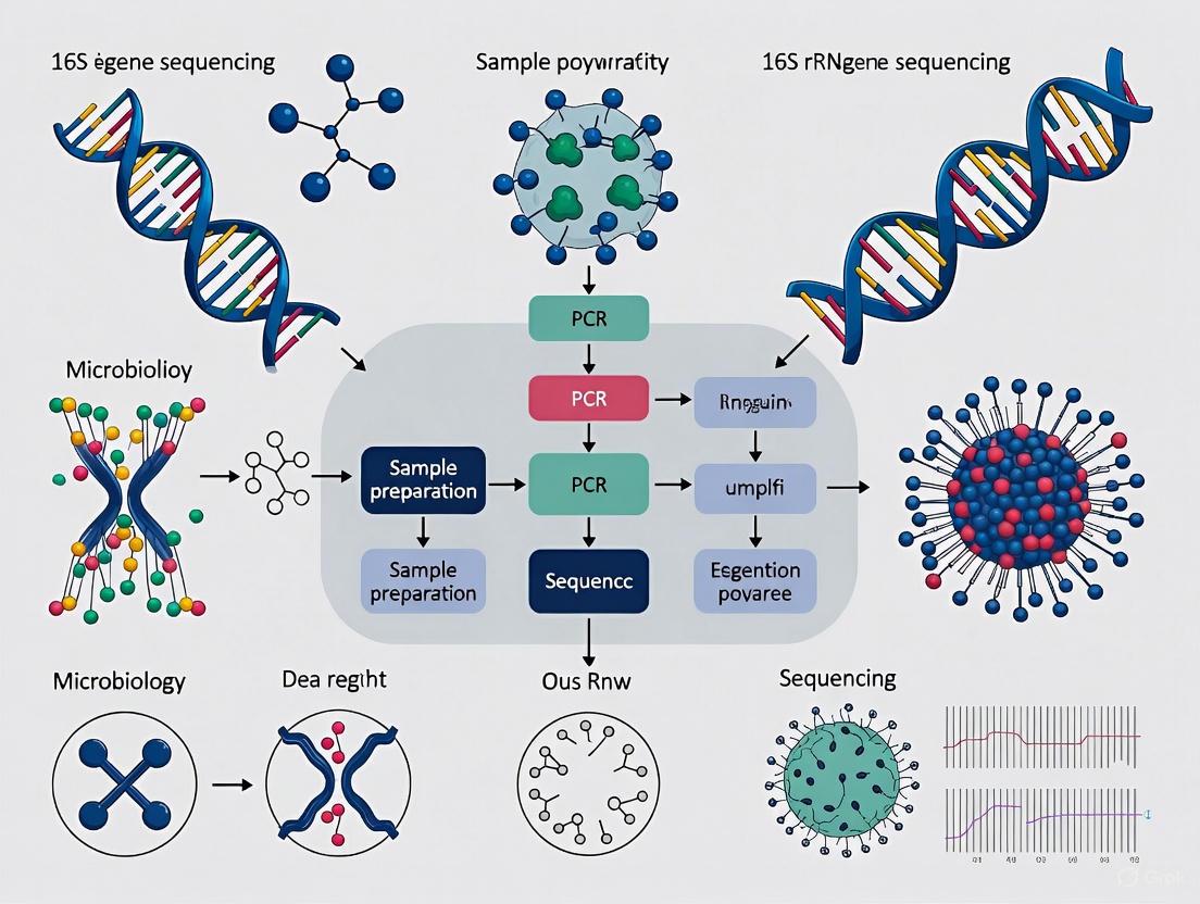
Figure 1: Experimental workflow for short-read and long-read 16S rRNA gene sequencing.
The Scientist's Toolkit: Essential Research Reagents & Materials
Table 3: Key Research Reagent Solutions for 16S rRNA Sequencing
| Reagent/Material | Function | Platform Considerations |
|---|---|---|
| DNA Extraction Kit (e.g., DNeasy PowerSoil) [29] | Isolation of high-quality genomic DNA from samples. | Long-read sequencing requires high-molecular-weight DNA [28]. |
| 16S PCR Primers (e.g., 27F/1492R) [29] [31] | Amplification of the target 16S rRNA gene region. | Illumina: Target hypervariable regions (e.g., V3-V4). Long-read: Target full-length gene (V1-V9) [29]. |
| PCR Master Mix (e.g., KAPA HiFi) [29] [31] | High-fidelity amplification of the 16S gene. | Critical for minimizing PCR errors in all platforms. |
| Library Prep Kit (Platform-Specific) | Preparation of amplicons for sequencing. | Must be selected for the specific sequencing platform (e.g., SMRTbell for PacBio [29], SQK-16S024 for Nanopore [29]). |
| Reference Database (e.g., SILVA) [29] [30] | Taxonomic classification of sequenced reads. | Database choice significantly impacts classification accuracy, especially for Nanopore [30]. |
| 8-Isoprostaglandin E2 | 8-Isoprostaglandin E2, CAS:27415-25-4, MF:C20H32O5, MW:352.5 g/mol | Chemical Reagent |
| Fura-FF pentapotassium | Fura-FF pentapotassium, MF:C28H18F2K5N3O14, MW:853.9 g/mol | Chemical Reagent |
Troubleshooting Guides & FAQs
Frequently Asked Questions
Q1: I am getting low species-level resolution with my Illumina V3-V4 data. Should I switch to a long-read platform? Yes, if species-level identification is critical for your research. Multiple studies confirm that sequencing the full-length 16S rRNA gene with PacBio or Nanopore improves species-level classification rates significantly—from about 47% with Illumina to 63-76% with long-read platforms [29] [5]. This is because the full-length gene contains more informative nucleotide variation across all variable regions, providing a stronger phylogenetic signal [25].
Q2: Are there any specific bioinformatic tools recommended for analyzing full-length 16S data from PacBio or Nanopore? Yes, the choice of tools is platform-dependent due to differing error profiles:
- PacBio HiFi Reads: The high accuracy of HiFi reads allows the use of the DADA2 pipeline to generate Amplicon Sequence Variants (ASVs), similar to the Illumina workflow [29] [5].
- Oxford Nanopore Reads: The higher error rate and lack of internal redundancy make DADA2 less suitable. Instead, specialized tools like Emu [30] or Spaghetti [29] that employ different denoising or OTU-clustering approaches are recommended.
Q3: My long-read sequencing results show many sequences classified as "uncultured_bacterium" at the species level. What does this mean? This is a common limitation, not a failure of your sequencing. It indicates that the specific bacterial species in your sample is not yet represented in the reference database used for taxonomic assignment [29]. This highlights a broader challenge in microbiology, where many environmental and host-associated microbes have not been isolated or sequenced. Using the most comprehensive and up-to-date databases can help mitigate this issue.
Q4: For a new project with a limited budget, which 16S variable region should I sequence with Illumina for the best resolution? If you are constrained to short-read sequencing, the V1-V3 region often provides a reasonable approximation of microbial diversity and has been shown to be a good compromise for skin and other microbiomes [31]. However, note that no single hypervariable region can perfectly recapitulate the resolution achieved by the full-length gene [25].
Troubleshooting Common Experimental Issues
Problem: Inconsistent taxonomic profiles between different sequencing platforms.
- Cause: This is a known issue caused by a combination of factors, including the specific 16S region targeted, PCR primer bias, and the platform's own technical artifacts [29].
- Solution:
- Wet Lab: Use the same DNA extraction for all platform comparisons to minimize batch effects.
- Bioinformatics: Apply a consistent, platform-appropriate bioinformatic pipeline. Be cautious when comparing or merging datasets generated from different platforms and primer sets [29].
- Interpretation: Focus on the overall community structure (beta-diversity) and major taxonomic groups, which should cluster by sample type rather than by sequencing platform, rather than expecting identical abundances for every taxon [5].
Problem: Low classification accuracy with Oxford Nanopore data.
- Cause: The inherent higher error rate of Nanopore sequencing can interfere with precise taxonomic assignment.
- Solution:
- Basecalling: Use the most accurate available basecalling model (e.g., "sup" or "hac" in Dorado) rather than the "fast" model, as the basecalling quality significantly influences downstream results [30].
- Database: Carefully select your reference database. The structure and composition of the database (e.g., SILVA vs. Emu's default database) can greatly influence the number and accuracy of species identifications [30].
Figure 2: A logical troubleshooting guide for diagnosing and solving low-resolution issues in 16S rRNA sequencing.
The resolution of 16S rRNA gene sequencing has long been constrained by technological limitations and bioinformatic challenges. While the full-length ~1500 bp 16S gene provides the highest taxonomic discrimination, most studies have historically sequenced only specific variable regions due to the read-length limitations of earlier sequencing platforms [25]. This represents a fundamental compromise, as different variable regions possess varying discriminatory power for distinct bacterial taxa [32] [25]. Furthermore, bioinformatic algorithms must distinguish true biological variation from sequencing errors and handle intragenomic variation between multiple 16S gene copies within a single organism [25] [13]. This technical support guide benchmarks four prominent algorithms—DADA2, DEBLUR, UNOISE3, and UPARSE—to help researchers select optimal strategies for overcoming these resolution limitations.
Algorithm Performance Benchmarking
Key Performance Metrics from Comparative Studies
Independent benchmarking studies using mock microbial communities have revealed critical differences in how algorithms resolve microbial sequences.
Table 1: Performance Comparison of 16S rRNA Analysis Algorithms
| Algorithm | Algorithm Type | Sensitivity | Specificity | Key Performance Characteristics | Computational Efficiency |
|---|---|---|---|---|---|
| DADA2 | ASV (Denoising) | Highest [33] | Lower [33] | Best recall (sensitivity); prone to over-splitting [13] [33] | Moderate [13] |
| DEBLUR | ASV (Denoising) | Moderate [13] | High [33] | Balanced performance; lower error rates [13] | Fast (runs in a single step) [13] |
| UNOISE3 | ASV (Denoising) | High [33] | Highest [33] | Best balance between resolution and specificity [33] | Moderate [13] |
| UPARSE | OTU (Clustering) | Moderate [33] | High [33] | Lower error rates; prone to over-merging [13] | Fastest [13] |
Resolution at Species and Strain Levels
Sequencing the full-length 16S rRNA gene is superior to targeting sub-regions for species-level classification. One study demonstrated that while the V4 region failed to confidently classify 56% of sequences to the correct species, the full-length gene successfully classified nearly all sequences [25]. ASV-level methods (DADA2, DEBLUR, UNOISE3) generally provide higher taxonomic resolution than OTU-level methods like UPARSE because they distinguish sequences differing by a single nucleotide [33]. Modern full-length sequencing combined with algorithms that account for intragenomic 16S copy variation can even enable strain-level discrimination [25].
Troubleshooting Guides and FAQs
Common Analysis Issues and Solutions
Table 2: Troubleshooting Common Algorithm Issues
| Problem | Possible Causes | Solutions | Preventive Measures |
|---|---|---|---|
| High rates of spurious OTUs/ASVs | Inadequate quality control; algorithm-specific error profile | For DADA2: Adjust quality filtering parameters [33] | Use synthetic mock communities to validate pipelines [34] |
| Over-splitting of biological sequences | Algorithm splitting intragenomic 16S variants into separate ASVs [13] | Use UNOISE3, which shows better specificity [33] | Select algorithms that balance sensitivity and specificity [33] |
| Over-merging of distinct taxa | OTU clustering with overly relaxed identity cutoff [13] | Use ASV methods or stricter clustering thresholds [13] | Use ASV-level methods for finer resolution [33] |
| Inconsistent results between pipelines | Different default parameters; algorithmic approaches | Re-analyze data with multiple pipelines for consensus [33] | Document all parameters and software versions used [35] |
| Low taxonomic resolution | Short read length; uninformative variable region [25] | Sequence full-length 16S gene if possible [25] | Select variable region based on target taxa [32] |
Frequently Asked Questions
Q1: Which algorithm provides the best balance between sensitivity and specificity for resolving closely related strains? A: Based on comparative studies, USEARCH-UNOISE3 generally provides the best balance, offering high sensitivity while maintaining the highest specificity among ASV methods [33].
Q2: Why does my analysis with DADA2 produce more ASVs than expected? A: DADA2 has the highest sensitivity but is prone to "over-splitting," where it may generate multiple ASVs from a single biological sequence due to intragenomic variation or minor sequencing errors [13] [33]. This can be mitigated by adjusting quality filtering parameters.
Q3: How does the choice of 16S variable region affect resolution? A: Different variable regions have varying discriminatory power for different bacterial taxa [32] [25] [34]. For instance, the V4 region performs poorly for species-level classification (failing to classify 56% of sequences in one study), while the V1-V3 region provides a reasonable approximation of diversity [25]. The full-length gene consistently provides the best resolution [25].
Q4: Should I use OTU or ASV methods for my study? A: ASV methods (DADA2, DEBLUR, UNOISE3) generally provide higher resolution and are more reproducible across studies, as they generate consistent sequence variants without clustering [33]. OTU methods (UPARSE) may be preferable for studies where computational efficiency is critical or when analyzing highly diverse communities where over-splitting is a concern [13].
Q5: How can I validate my bioinformatic pipeline's performance? A: Incorporate synthetic mock communities with known composition into your sequencing runs [34]. This allows you to quantify the error rate, sensitivity, and specificity of your chosen algorithm and parameters [13] [34].
Experimental Protocols for Benchmarking
Standardized Workflow for Method Comparison
The following workflow, based on published benchmarking studies [13] [33], provides a standardized approach for comparing algorithm performance:
Diagram 1: Algorithm Benchmarking Workflow
Protocol Steps:
- Sample Preparation: Use well-characterized synthetic mock communities (e.g., BEI Resources Mock Community [33] or HC227 complex mock [13]) alongside experimental samples.
- Library Preparation: Amplify the desired 16S rRNA variable region (e.g., V4 [33] or V3-V4 [13]) using validated primer sets.
- Sequencing: Sequence on an appropriate platform (e.g., Illumina MiSeq for short reads [33]).
- Quality Control: Process raw sequences through uniform preprocessing:
- Merge paired-end reads (if applicable)
- Quality filtering (maxEE=0.01-1.0 [13])
- Remove ambiguous bases and chimeras
- Parallel Analysis: Process the same quality-filtered dataset through each algorithm (DADA2, DEBLUR, UNOISE3, UPARSE) using default or recommended parameters.
- Performance Evaluation: Compare outputs against the known mock community composition for:
- Sensitivity: Proportion of expected taxa recovered
- Specificity: Number of spurious OTUs/ASVs generated
- Quantitative accuracy: Correlation between expected and observed abundances
Researcher's Toolkit: Essential Reagents and Materials
Table 3: Essential Research Reagents and Materials for 16S rRNA Benchmarking
| Item | Function | Example Products/Details |
|---|---|---|
| Synthetic Mock Communities | Positive control for evaluating pipeline accuracy and bias | BEI Resources HM-782D [33]; HC227 (227 strains) [13] |
| High-Fidelity DNA Polymerase | PCR amplification with minimal bias | Kapa HiFi HotStart, Q5 Polymerase [34] |
| Validated 16S Primer Sets | Amplification of target variable regions | 515F/806R (V4) [33]; 341F/785R (V3-V4) [13] |
| NGS Library Prep Kit | Preparing amplicon libraries for sequencing | Illumina MiSeq Reagent Kit [33] |
| Bioinformatic Workflow Management | Reproducible pipeline execution and error tracking | Nextflow, Snakemake [35] |
| Data Quality Control Tools | Assessing raw sequence data quality | FastQC, MultiQC [35] |
| 4-Fluoro phenibut hydrochloride | 4-Fluoro phenibut hydrochloride, CAS:3060-41-1, MF:C10H14ClNO2, MW:215.67 g/mol | Chemical Reagent |
| Ethylbenzene-d10 | Ethylbenzene-d10, CAS:25837-05-2, MF:C8H10, MW:116.23 g/mol | Chemical Reagent |
Algorithm Selection Guide
The choice of algorithm involves trade-offs between resolution, specificity, and computational efficiency. The following decision pathway can help guide researchers in selecting the most appropriate tool:
Diagram 2: Algorithm Selection Guide
To maximize resolution in 16S rRNA sequencing studies, researchers should consider a multi-faceted approach:
- Select the most informative variable region for their target taxa or, if possible, utilize full-length 16S sequencing [25].
- Choose algorithms based on their specific needs: DADA2 for maximum sensitivity, UNOISE3 for the best balance, or UPARSE for computational efficiency [33].
- Incorporate mock communities in every sequencing run to quantify pipeline performance and identify biases [34].
- Document all parameters and software versions meticulously to ensure reproducibility [35].
The ongoing development of both sequencing technologies and analysis algorithms continues to improve the resolution achievable through 16S rRNA gene sequencing, enabling more precise microbial community analysis for biomedical and environmental applications.
For decades, 16S rRNA gene sequencing has been the cornerstone of microbial ecology, enabling researchers to decipher the composition of complex bacterial communities. However, the historical compromise of sequencing short hypervariable regions (typically 300-600 bp) has imposed significant limitations on taxonomic resolution, particularly at the species and strain levels. The advent of third-generation sequencing platforms has made high-throughput sequencing of the full-length 16S rRNA gene (approximately 1500 bp, spanning regions V1-V9) a practical reality. This technical guide explores how leveraging V1-V9 sequencing resolves the pervasive challenge of low taxonomic resolution in microbiome research, providing scientists with enhanced capability for species discrimination in diagnostic and therapeutic applications.
FAQs: Understanding Full-Length 16S rRNA Gene Sequencing
What is the fundamental advantage of sequencing the full V1-V9 region over single hypervariable regions?
Sequencing the entire V1-V9 region provides significantly enhanced taxonomic resolution compared to single hypervariable regions. Short-read sequencing of individual variable regions (e.g., V4 or V3-V4) typically limits identification to the genus level, whereas full-length sequencing enables discrimination at the species and even strain level [5] [25]. This improvement occurs because the complete 1,500 bp sequence contains all nine variable regions, capturing the maximum evolutionary information available from the 16S gene for taxonomic classification [36].
Experimental evidence demonstrates that different variable regions have varying discriminatory power for specific bacterial taxa. One study found that while the V1-V2 region performed poorly for classifying Proteobacteria, and V3-V5 struggled with Actinobacteria, the complete V1-V9 region consistently produced the best results across all major phylogenetic groups [25]. The V4 region, commonly used in Illumina-based studies, performed particularly poorly, failing to confidently classify 56% of sequences to the species level in silico experiments [25].
How does full-length 16S sequencing improve species-level identification in human microbiome studies?
Full-length 16S sequencing dramatically increases the proportion of reads that can be confidently assigned to the species level. A 2024 comparative study analyzing human saliva, oral biofilm, and fecal samples found that while both Illumina (V3-V4 regions) and PacBio (V1-V9) platforms assigned a similar percentage of reads to the genus level (approximately 95%), PacBio full-length sequencing enabled a significantly higher proportion of reads to be further assigned to the species level (74.14% versus 55.23%) [5].
This enhanced resolution is particularly valuable for discriminating between closely related species with highly similar 16S sequences, such as streptococci or the Escherichia/Shigella group [5]. For example, in the analysis of oral microbiota, full-length 16S sequencing revealed a higher relative abundance of Streptococcus species compared to short-read methods (20.14% vs 14.12% in saliva), though these differences were not statistically significant after multiple testing corrections [5].
What are the technical considerations for primer selection in full-length 16S sequencing?
Primer selection critically influences the accuracy and representativeness of full-length 16S sequencing results. Different primer sets can yield strikingly different taxonomic profiles, even when sequencing the same samples [36]. A 2023 study comparing two primer sets (27F-I included in Oxford Nanopore's kit and a more degenerate 27F-II set) for human fecal microbiome analysis found significant differences in both taxonomic diversity and relative abundance across numerous taxa [36].
The conventional 27F primer (27F-I) revealed significantly lower biodiversity and showed an unusually high Firmicutes/Bacteroidetes ratio compared to the more degenerate primer set (27F-II) [36]. When evaluated against expected microbiome compositions from the American Gut Project, the more degenerate primer set (27F-II) better reflected the anticipated composition and diversity of fecal microbiomes [36]. This highlights the importance of primer optimization and selection for accurate representation of complex bacterial communities.
Can full-length 16S sequencing resolve strain-level variation?
Emerging evidence indicates that full-length 16S sequencing can detect strain-level variation through the identification of intragenomic copy variants - subtle sequence differences between multiple copies of the 16S gene within a single bacterial genome [25]. Modern sequencing platforms achieve sufficient accuracy to resolve single-nucleotide substitutions that exist between these intragenomic copies [25].
This capability is significant because many bacterial genomes contain multiple polymorphic copies of the 16S gene [25]. Appropriate bioinformatic treatment of these intragenomic variants has the potential to provide taxonomic resolution at the strain level, which is valuable for tracking clinically relevant strains or predicting phenotypic characteristics [25]. However, researchers must account for this variation in their analysis pipelines to avoid misinterpreting genuine intragenomic variation as representing distinct taxa.
Troubleshooting Guides
Low Library Yield in Full-Length 16S Amplicon Sequencing
Symptoms:
- Final library concentrations below expected values (e.g., < 10-20% of predicted yield)
- Broad or faint peaks on electropherogram traces
- Dominance of adapter-dimer peaks (~70-90 bp) in size distribution
Root Causes and Solutions:
| Cause | Diagnostic Signs | Corrective Actions |
|---|---|---|
| Poor input DNA quality | Low 260/230 ratios (<1.8), smeared electrophoregram | Re-purify input sample; ensure fresh wash buffers; use fluorometric quantification instead of UV absorbance [37] |
| Suboptimal PCR amplification | Increased small fragments (<100 bp), primer artifacts | Optimize cycling conditions; verify primer specificity; consider two-step indexing to reduce artifacts [37] |
| Inefficient bead cleanup | Unusual fragment size distribution, carryover contaminants | Adjust bead:sample ratios; avoid over-drying beads; implement rigorous washing steps [38] [37] |
| Insufficient input material | Low starting yield despite adequate concentration | Verify input DNA quality and quantity; use recommended 10 ng high molecular weight gDNA per barcode as starting point [38] |
Prevention Strategy: Implement a rigorous quality control workflow for input DNA, including fluorometric quantification and integrity assessment. Use master mixes to reduce pipetting errors, and validate each lot of purification beads with control DNA [37].
Taxonomic Representation Bias in Microbial Communities
Symptoms:
- Under-representation of Gram-positive bacteria (especially Firmicutes)
- Discrepancy between observed community structure and expected composition
- Inconsistent results between technical replicates
Root Causes and Solutions:
| Cause | Diagnostic Signs | Corrective Actions |
|---|---|---|
| Suboptimal DNA extraction | Consistent under-representation of difficult-to-lyse taxa | Implement improved lysis protocols (e.g., alkaline/heat/detergent methods) to replace gentle enzymatic lysis [39] |
| Primer bias | Systematic differences in diversity metrics between primer sets | Use degenerate primers (e.g., 27F-II instead of 27F-I); validate primer performance with mock communities [36] |
| Differential lysis efficiency | Variable recovery of Gram-positive vs. Gram-negative bacteria | Employ standardized bead-beating parameters; consider chemical lysis methods effective against tough cell walls [39] |
Experimental Protocol for Improved DNA Extraction: The "Rapid" microbial DNA extraction protocol has demonstrated improved representation of Firmicutes species compared to standard protocols [39]:
- Start with ≤10 mg of fecal sample material
- Apply alkaline lysis buffer with KOH combined with heat and detergent simultaneously
- Process samples in 96-well plate format for consistency
- Purify DNA using standard column-based or bead-based methods
- Validate extraction efficiency with mock communities containing both Gram-positive and Gram-negative bacteria
This non-enzymatic, non-mechanical approach has been shown to provide more uniform lysis across diverse bacterial populations, reducing the under-representation of Firmicutes species common with gentler lysis methods [39].
Inaccurate Species-Level Assignment
Symptoms:
- Low proportion of reads assigned to species level (<60%)
- Ambiguous taxonomic assignments for closely related species
- Inconsistent classification of streptococci or Escherichia/Shigella groups
Root Causes and Solutions:
| Cause | Diagnostic Signs | Corrective Actions |
|---|---|---|
| Insufficient sequence information | Limited discrimination between highly similar species | Switch from partial to full-length 16S gene sequencing (V1-V9) to capture all variable regions [5] [25] |
| Database limitations | High proportion of "unclassified" at species level | Use comprehensive, curated databases; regularly update reference sequences [40] |
| Platform-specific errors | Misclassification due to sequencing errors | Leverage circular consensus sequencing (CCS) to achieve high accuracy (>Q20) [5] [25] |
Experimental Protocol for Full-Length 16S Sequencing with PacBio:
- DNA Extraction: Use standardized protocol (e.g., HMP protocol or improved "Rapid" method) to ensure representative lysis [39]
- PCR Amplification: Amplify full-length 16S rRNA gene using primers 27F (5'-AGAGTTTGATCMTGGCTCAG-3') and 1492R (5'-CGGTTACCTTGTTACGACTT-3') [5]
- Library Preparation: Prepare SMRTbell libraries according to manufacturer's instructions
- Sequencing: Perform Circular Consensus Sequencing (CCS) on PacBio Sequel II system to generate high-fidelity (HiFi) reads
- Bioinformatic Analysis: Process reads using DADA2 or similar pipeline that accounts for intragenomic copy variation [25]
Performance Comparison: Full-Length vs. Partial 16S Sequencing
The table below summarizes quantitative performance differences between full-length V1-V9 sequencing and short-read approaches based on recent comparative studies:
| Metric | Illumina (V3-V4) | PacBio (V1-V9) | Improvement |
|---|---|---|---|
| Species-level assignment rate | 55.23% [5] | 74.14% [5] | +18.91% |
| Genus-level assignment rate | 94.79% [5] | 95.06% [5] | +0.27% |
| Discrimination of closely related species | Limited [25] | High [25] | Significant |
| Detection of intragenomic variation | Not feasible [25] | Possible [25] | New capability |
| Representation of Streptococcus in saliva | 14.12% [5] | 20.14% [5] | +6.02% |
Workflow Diagrams
Full-Length 16S rRNA Gene Sequencing Workflow
Research Reagent Solutions
| Reagent Category | Specific Product | Function in Experimental Protocol |
|---|---|---|
| DNA Extraction | "Rapid" alkaline/heat/detergent protocol [39] | Provides uniform lysis of diverse bacterial cells, including difficult-to-lyse Firmicutes |
| Full-Length Amplification | Degenerate primer set 27F-II [36] | Improves coverage of diverse bacterial taxa compared to conventional 27F-I primer |
| Long-read Sequencing | PacBio Sequel II with SMRT sequencing [5] | Enables high-fidelity full-length 16S sequencing through circular consensus sequencing |
| Library Preparation | Oxford Nanopore 16S Barcoding Kit [38] | Facilitates multiplexed sequencing of full-length 16S amplicons on nanopore platform |
| Quality Control | Qubit dsDNA HS Assay Kit [38] | Provides accurate quantification of input DNA and final libraries |
The transition from short-read sequencing of hypervariable regions to full-length 16S rRNA gene sequencing represents a significant advancement in microbiome research. By capturing the complete V1-V9 region, researchers can achieve substantially improved taxonomic resolution at the species level, enabling more precise microbial characterization in diagnostic, therapeutic, and ecological applications. While methodological considerations around primer selection, DNA extraction, and bioinformatic processing remain critical, the implementation of optimized protocols and troubleshooting strategies detailed in this guide will empower researchers to overcome the longstanding challenge of low resolution in 16S rRNA gene sequencing.
The introduction of Oxford Nanopore Technologies' (ONT) R10.4.1 flow cell chemistry represents a transformative advancement for clinical and research microbiology, specifically enabling high-resolution, species-level identification through full-length 16S rRNA gene sequencing. This case study evaluates the impact of R10.4.1 chemistry and subsequent basecalling improvements on taxonomic resolution within the context of 16S rRNA gene sequencing research. By comparing traditional short-read (Illumina V3V4) and long-read (ONT V1V9) approaches, recent research demonstrates that the R10.4.1 chemistry, combined with optimized bioinformatic pipelines, facilitates the discovery of more precise, disease-specific bacterial biomarkers—a crucial capability for diagnostics and therapeutic development [41]. This technical support document provides a comprehensive framework for implementing this technology, including validated experimental protocols, performance metrics, and targeted troubleshooting guides to resolve common challenges encountered during workflow establishment.
R10.4.1 Chemistry
Oxford Nanopore's R10.4.1 chemistry features an updated nanopore design that significantly improves the accuracy of base recognition, particularly in homopolymer regions. This enhancement is fundamental for sequencing the full-length ~1500 bp 16S rRNA gene (V1-V9 regions), which provides the necessary sequence diversity to discriminate between closely related bacterial species. The technology sequences any length of native DNA/RNA molecule electronically, eliminating PCR bias and enabling direct detection of epigenetic modifications [42].
Basecalling with Dorado
Basecalling, the process of translating raw electrical signals into nucleotide sequences, utilizes machine learning models within the Dorado basecaller. Accuracy is tiered through different models to balance speed and precision according to experimental needs [42]:
- Fast Basecalling: Recommended for real-time insights when computational resources are limited.
- High Accuracy (HAC): Provides highly accurate basecalling suitable for most variant analysis projects.
- Super Accuracy (SUP): The most accurate and computationally intensive model, recommended for de novo assembly and low-frequency variant analysis.
- Duplex Basecalling: Sequences both strands of a DNA molecule for maximum accuracy, recommended for hemi-methylation investigation [42].
The latest Dorado basecalling models (v5) can achieve raw read accuracies of up to 99.75% (Q26) [42]. This high single-read accuracy is critical for species-level assignment, as a quality threshold of Q20 (99% accuracy) is considered the minimum for confidently assigning an Operational Taxonomic Unit (OTU) to a specific species using full-length 16S rRNA sequencing [41].
Performance Data and Validation
Quantitative Accuracy Metrics
Table 1: Basecalling and Consensus Accuracy of R10.4.1 Chemistry
| Metric | Performance | Sequencing & Basecalling Parameters | Application Context |
|---|---|---|---|
| Single-read Accuracy | >99% (Q20) [42]; up to Q26 with Dorado v5 [42] | R10.4.1 flow cell, latest SUP models | Raw read accuracy for single DNA/RNA strand |
| Variant Calling (SNPs) | Comparable to short-read methods [42] | Q20+ chemistry | Microbial genotyping |
| Assembly Accuracy | Q50 at 10–20x coverage (bacterial genomes) [42] | MinION R10.4.1, Ligation Sequencing Kit V14, Simplex SUP | De novo assembly of mock communities |
| DNA Modification (5mC) | 99.5% accuracy in CpG context [42] | Raw read accuracy (SUP) | Epigenetic studies in bacteria |
Impact on Species-Level Identification
A 2025 study directly compared Illumina (V3V4) and ONT R10.4.1 (V1V9) for bacterial biomarker discovery in colorectal cancer (CRC), analyzing feces from 123 subjects [41].
Table 2: Species-Level Biomarkers Identified by R10.4.1 Full-Length 16S Sequencing
| Bacterial Species Identified as CRC Biomarkers | Detection Method |
|---|---|
| Parvimonas micra | ONT R10.4.1 (V1V9) |
| Fusobacterium nucleatum | ONT R10.4.1 (V1V9) & Illumina (V3V4) |
| Peptostreptococcus stomatis | ONT R10.4.1 (V1V9) |
| Peptostreptococcus anaerobius | ONT R10.4.1 (V1V9) |
| Gemella morbillorum | ONT R10.4.1 (V1V9) |
| Clostridium perfringens | ONT R10.4.1 (V1V9) |
| Bacteroides fragilis | ONT R10.4.1 (V1V9) & Illumina (V3V4) |
| Sutterella wadsworthensis | ONT R10.4.1 (V1V9) |
The study found that while basecalling models (fast, hac, sup) broadly resulted in similar taxonomic output, lower-quality basecalling led to significantly higher observed species counts and different taxonomic identifications, highlighting the importance of model selection. Furthermore, database choice greatly influenced results, with the Emu's Default database yielding higher diversity but sometimes overconfidently classifying unknown species compared to the SILVA database [41]. The ability to sequence the full-length 16S rRNA gene was critical, as Illumina's short-read approach targeting only the V3V4 regions (~400 bp) was restricted mostly to genus-level identification [41] [43].
Experimental Protocols
Full-Length 16S rRNA Gene Sequencing Workflow
Sample Preparation and DNA Extraction
- Sample Material: Use well-characterized reference materials (e.g., NML MCM2α/MCM2β, WHO WC-Gut RR) for validation alongside clinical samples [43].
- Cell Lysis: For clinical samples, subject to bead beating using Lysing Matrix E tubes on a TissueLyser (50 oscillations/second for 2 minutes) [43].
- DNA Extraction: Validate the performance of at least one DNA extraction method (e.g., AusDiagnostics MT-Prep, QIAamp DNA Micro Kit, MagMAX Microbiome Ultra Kit) with your sample type. For tissues, pre-process with Tissue Lysis Buffer and proteinase K (2 hours at 56°C) before bead-beating [43].
PCR Amplification and Library Preparation
- Target Region: Amplify the full-length ~1500 bp 16S rRNA gene (V1-V9 regions).
- PCR Conditions: Carefully optimize PCR cycle numbers to prevent non-specific over-amplification of low-abundance environmental microorganisms, which can reduce diagnostic sensitivity [43].
- Library Preparation: Use the Ligation Sequencing Kit V14 (SQK-LSK114) with R10.4.1 flow cells, following manufacturer's protocols [42].
Bioinformatic Analysis Pipeline
Basecalling and Read Processing
- Basecalling: Process POD5 files from the sequencer using Dorado.
Use the
supmodel for the highest accuracy in species-level analysis [41]. - Adapter Trimming: Trim adapter and primer sequences using Dorado's integrated trimming function.
Taxonomic Classification
For full-length 16S rRNA reads, use a taxonomy assignment tool designed for long reads, such as Emu [41]. The choice of reference database (e.g., SILVA vs. Emu's Default database) significantly influences results and should be reported and justified [41].
Basecalling Optimization Pathway
The Scientist's Toolkit: Research Reagent Solutions
Table 3: Essential Materials for R10.4.1 16S rRNA Gene Sequencing
| Item | Function/Description | Example Products/References |
|---|---|---|
| R10.4.1 Flow Cell | Core sensing device; improved homopolymer accuracy. | MinION Mk1C, PromethION [42] |
| Ligation Sequencing Kit | Library preparation for DNA sequencing. | Ligation Sequencing Kit V14 (SQK-LSK114) [42] |
| Metagenomic Control Materials | Validation and standardization of the entire workflow. | NML MCM2α/MCM2β, WHO WC-Gut RR [43] |
| Bead Beating Tubes | Mechanical lysis for efficient DNA extraction from diverse samples. | Lysing Matrix E tubes (MP Bio, 6914100) [43] |
| DNA Extraction Kit | High-yield, unbiased microbial DNA extraction. | AusDiagnostics MT-Prep, QIAamp DNA Micro Kit [43] |
| Full-Length 16S Primers | Amplification of the ~1500 bp V1-V9 region. | Standard 27F/1492R or equivalent [41] |
| Dorado Basecaller | Translates raw signals to base sequences with HAC/SUP models. | Oxford Nanopore Dorado (v5.0+) [42] [44] |
| Taxonomic Classification Tool | Assigns taxonomy to long-read 16S sequences. | Emu [41] |
| Reference Database | Curated 16S sequences for taxonomic assignment. | SILVA, Emu's Default Database [41] |
| 1-Methoxycyclopropanecarboxylic acid | 1-Methoxycyclopropanecarboxylic acid, CAS:100683-08-7, MF:C5H8O3, MW:116.11 g/mol | Chemical Reagent |
Troubleshooting Guides & FAQs
Frequently Asked Questions
Q1: Which basecalling model should I use for 16S rRNA species-level identification? For the highest species resolution, use the Super Accuracy (SUP) model in Dorado. While High Accuracy (HAC) and fast models produce broadly similar taxonomic output, the SUP model minimizes errors that can lead to over-splitting or misclassification at the species level [41].
Q2: My basecalling results show different species counts depending on the database I use. Which is correct? Database choice significantly influences results. The SILVA database may provide more conservative classifications, while Emu's Default database often identifies more species but can overconfidently assign an unknown species as the closest known match. The choice depends on your research goals: use SILVA for conservative analysis or Emu's database for maximum discovery, with caution regarding potential over-classification [41].
Q3: Can I use DADA2 for denoising my full-length 16S R10.4.1 reads? DADA2 is optimized for high-quality short reads (like Illumina) and is not currently recommended for ONT reads. Instead, use tools specifically designed for long-read 16S data, such as Emu or NanoClust [41].
Troubleshooting Common Issues
Problem: CUDA Out Of Memory Error during basecalling.
- Cause: The auto batch size algorithm selected a batch size that exceeds your GPU's available memory, especially when using SUP models or modification calling [45].
- Solution:
- Check the batch size reported in the Dorado output from a previous run.
- Manually reduce the batch size using the
--batchsizeargument (e.g., reduce by 10%). - Repeat until basecalling completes successfully [45].
Problem: Low GPU utilization, leading to slow basecalling.
- Cause: I/O bottlenecks where the system cannot supply data to the GPU fast enough [45].
- Solution:
- Use POD5 format instead of FAST5 for superior I/O performance.
- Transfer data to a local SSD before basecalling, as network disks are often too slow.
- For duplex basecalling, a local SSD is highly recommended due to heavy random data access [45].
Problem: "No supported chemistry found" error.
- Cause: Dorado cannot automatically determine the correct basecalling model, often because the data lacks recognizable flow cell or kit information [45].
- Solution: Manually specify the correct model using its file path. Download an appropriate model and run:
Problem: "Incompatible modbase models" error.
- Cause: You have selected two modification models that share a canonical base, which is an invalid configuration [45].
- Solution: Avoid using conflicting pairs of modbase models, such as
5mC_5hmC+5hmCG_5hmCGorm6A_DRACH+inosine_m6A. Check the Dorado documentation for compatible combinations [45].
Optimizing the Workflow: A Practical Guide to Minimizing Bias and Maximizing Resolution
Why is primer choice so critical for the resolution of my 16S rRNA gene sequencing results?
The selection of primers is a foundational step that directly determines the resolution and accuracy of your microbial community profile. The 16S rRNA gene contains nine variable regions (V1-V9), and the specific region you choose to amplify has a profound effect on which taxa are detected and how precisely they can be classified [46] [25].
Different variable regions possess varying degrees of discriminatory power for specific bacterial taxa. Using unsuitable primer combinations can lead to the underrepresentation or complete absence of specific, important bacterial genera from your taxonomic profile [46]. For instance, one study found that the Bacteroidetes phylum was missed when using the primers 515F-944R, and the genus Acetatifactor was not detected when using the GreenGenes database for classification [46]. Furthermore, the taxonomic resolution of short-amplicon sequencing (targeting one to three variable regions) is inherently lower than that achieved by sequencing the full-length (~1500 bp) 16S rRNA gene [25]. Conclusions drawn from comparing datasets generated with different primer pairs or V-regions can be misleading and require independent cross-validation [46].
Table 1: Impact of Selected Primer Pairs on Taxonomic Profiling
| Targeted V-Region | Example Primer Pair | Documented Impact or Limitation |
|---|---|---|
| V4 | 515F-806R | Performs worst for species-level discrimination; 56% of in-silico amplicons failed to confidently match their species of origin [25]. |
| V4-V5 | 515F-944R | Can miss entire phyla, such as Bacteroidetes [46]. |
| V1-V2 | 27F-338R | Performs poorly at classifying sequences belonging to the phylum Proteobacteria [25]. |
| V3-V5 | 341F-785R | Performs poorly at classifying sequences belonging to the phylum Actinobacteria [25]. |
How can I check if my chosen primers provide adequate coverage for my target microorganisms?
Before starting a wet-lab experiment, you can perform an in silico evaluation to estimate the theoretical coverage of your primer pair. This process assesses how well your primer sequences match the 16S rRNA gene sequences of the microorganisms you expect to find in your sample.
Experimental Protocol: In Silico Coverage Evaluation
- Define Your Target and Obtain Sequences: Identify the bacterial or archaeal species of primary interest for your study. Obtain their 16S rRNA gene sequences from a curated database. For specific environments like the oral microbiome, using a specialized database is recommended for more accurate results [47].
- Select a Primer Evaluation Tool: Utilize online resources. The TestPrime function within the SILVA database is a commonly used tool for this purpose [48] [47].
- Input Parameters: Enter your forward and reverse primer sequences into the tool. Select the appropriate reference database (e.g., SSU Ref NR) and set the maximum number of mismatches (often set to 0 for a stringent test) [48].
- Run Analysis and Interpret Results: The tool will calculate the primer's coverage, typically reported as the percentage of target sequences in the database that are perfectly matched by the primer [47]. A higher coverage percentage indicates a greater likelihood of amplifying those organisms.
What is a wet-lab experimental strategy to quantify and counteract bias in my specific workflow?
While in silico analysis is powerful, the only way to quantify the total bias introduced by your entire wet-lab workflow—from DNA extraction to data analysis—is through the use of mock microbial communities.
Experimental Protocol: Using Mock Communities to Quantify Bias
- Create a Mock Community: Obtain a defined mixture of known bacterial strains, either commercially available or created in-house. The complexity should be sufficient and adequate for your study's goals [46]. These can be mixtures of:
- Process the Mock Community: Subject the mock community to your entire standard 16S rRNA gene sequencing workflow: DNA extraction, PCR amplification with your chosen primers, and sequencing [46] [49].
- Analyze and Compare: After sequencing and bioinformatic processing, compare the observed proportions of each species in the mock community to the known, expected proportions. The discrepancy represents the total bias introduced by your protocol [49].
- Model and Correct (Optional): As demonstrated in one study, the data from mock communities can be used to build mixture effect models. These models can then predict the "true" composition of environmental samples based on the observed proportions, thereby helping to counteract the identified bias [49].
The following workflow diagram illustrates the parallel processes of using mock communities for bias quantification and in silico analysis for primer selection:
How can I improve the coverage of an existing universal primer pair?
If your in silico evaluation reveals poor coverage for a key target organism, you can modify existing primers by adding degenerate bases. These are special DNA codes (e.g., N = A/C/T/G, Y = C/T) that represent multiple nucleotides at a single position, allowing one primer to match several different gene sequences.
Experimental Protocol: Primer Improvement with the "Degenerate Primer 111" Tool
This protocol is based on a user-friendly script designed to simplify this process [48].
- Identify Mismatch: Take your universal primer sequence and the 16S rRNA gene sequence of an uncovered target microorganism. Align them to identify the specific base positions that do not match.
- Run the Tool: Input the universal primer sequence and the target gene sequence into the "Degenerate primer 111" tool. The script automates the multi-step process of reverse complementation (for reverse primers) and sequence alignment.
- Generate New Primer: The tool iteratively suggests a new primer sequence where the mismatched bases are replaced with the appropriate degenerate codes, maximizing coverage for your target microorganism without unnecessarily increasing coverage for non-targets [48].
- Validate In Silico: Test the new, degenerate primer sequence using the in silico coverage evaluation protocol (e.g., in SILVA TestPrime) to confirm the improvement in coverage.
- Validate Experimentally: As a final step, the original and improved primers should be used to amplify DNA from the same sample, followed by high-throughput sequencing to confirm that the improved primers detect more of the target microbial species [48].
Table 2: Key Research Reagent Solutions for 16S rRNA Gene Sequencing
| Reagent / Tool Category | Specific Examples | Function and Importance |
|---|---|---|
| Reference Databases | SILVA [46], GreenGenes (GG) [46], Ribosomal Database Project (RDP) [46] | Used for taxonomic classification and in silico primer evaluation. Database choice affects results due to differences in nomenclature and precision [46]. |
| Bioinformatics Pipelines | QIIME/QIIME2 [46], MOTHUR [46], DADA2 [46] | Process raw sequencing reads into Amplicon Sequence Variants (ASVs) or Operational Taxonomic Units (OTUs) for analysis. |
| Clustering Methods | OTUs (97% similarity) [46], zOTUs [46], ASVs [46] | Define taxonomic units from sequence data. ASVs are increasingly preferred for cross-study comparisons [46]. |
| Primer Design Tool | "Degenerate Primer 111" script [48] | A user-friendly tool to improve the coverage of universal primers by systematically adding degenerate bases. |
| Mock Communities | Defined mixtures of bacterial strains (e.g., 36-species community [25], 7-strain vaginal community [49]) | Essential controls for quantifying and characterizing bias introduced during the sample processing pipeline [46] [49]. |
16S rRNA gene sequencing is a cornerstone method for microbiome research, enabling the identification and characterization of bacterial and archaeal communities within diverse samples. However, the observed microbial diversity and composition can be significantly influenced by technical choices made during the experimental workflow, from sample collection to data analysis. This technical support center article addresses common challenges and provides troubleshooting guidance for issues related to low taxonomic resolution in 16S rRNA gene sequencing, helping researchers optimize their protocols for more accurate and reliable results.
Frequently Asked Questions (FAQs)
1. My 16S rRNA sequencing results show low taxonomic resolution at the species level. What are the main technical factors contributing to this? Low species-level resolution is a common limitation of 16S rRNA sequencing. The primary technical factors include:
- Choice of Variable Region: No single hypervariable region can comprehensively differentiate all species. The limited genetic information in short reads restricts discrimination between closely related species [50].
- Primer Specificity and Bias: "Universal" primers do not amplify all bacterial taxa equally. Differences in primer annealing efficiency due to sequence mismatches can lead to under-representation or complete missing of specific taxa in the results [46].
- Bioinformatics Pipeline: Clustering methods like Operational Taxonomic Units (OTUs) at a 97% similarity threshold inherently group together distinct species. While denoising methods produce Amplicon Sequence Variants (ASVs), they can over-split sequences from the same strain or struggle with higher error rates from certain sequencing platforms [13] [41].
- Reference Database: Outdated or incomplete databases lack the necessary reference sequences to accurately classify reads to the species level. Furthermore, inconsistencies in nomenclature between different databases can lead to misclassification [46].
2. How does the choice of DNA extraction method influence my observed diversity? DNA extraction methods directly impact observed diversity through lysis efficiency and co-extraction of inhibitors.
- Lysis Efficiency: Different bacterial taxa have varying cell wall structures (e.g., Gram-positive vs. Gram-negative), making some more resistant to certain lysis methods (e.g., enzymatic vs. mechanical bead-beating). Inefficient lysis of hardy cells leads to their under-representation in the final sequencing data [19].
- Inhibitors: Some extraction methods may co-purify substances that inhibit downstream PCR amplification. This can lead to reduced library complexity and an inaccurate representation of the microbial community's true abundance [19].
- Troubleshooting Tip: It is critical to use a DNA extraction kit that is appropriate for your sample type (e.g., soil, stool, water) and has been validated for microbial community analysis. Consistency in the extraction protocol across all samples in a study is essential to avoid introducing bias [19] [51].
3. I am getting inconsistent results between different runs. How can I improve reproducibility? Inconsistencies often arise from a lack of standardization. Key steps to improve reproducibility include:
- Standardize Sample Handling: Variations in sample storage time, temperature (e.g., immediate freezing at -80°C is ideal), and freeze-thaw cycles can alter the microbial profile. Use consistent preservation methods [19] [51].
- Use Controls: Include both positive controls (mock communities with known compositions) and negative controls (to detect contamination) in every sequencing run. Mock communities allow you to benchmark your entire workflow's accuracy, while negative controls allow you to identify and subtract contaminating sequences [19] [13] [46].
- Normalize Input DNA: For library preparation, ensure DNA samples are accurately quantified (e.g., using Qubit) and normalized to the same concentration to ensure equitable PCR amplification across samples [21].
4. Should I use OTU clustering or ASV denoising for my analysis? The choice between OTUs and ASVs involves a trade-off between error tolerance and resolution.
- OTUs (Operational Taxonomic Units): Cluster sequences at a fixed identity threshold (typically 97%). They are less sensitive to sequencing errors but can over-merge biologically distinct sequences into a single cluster, reducing resolution [13].
- ASVs (Amplicon Sequence Variants): Use denoising algorithms to infer biological sequences and remove errors. They offer higher resolution and are reproducible across studies. However, they can over-split non-identical 16S rRNA gene copies from the same genome into multiple ASVs [13].
- Recommendation: For most studies where species-level precision is desired, ASV-based methods (e.g., DADA2, Deblur) are generally preferred. Benchmarking with a mock community is the best way to evaluate the performance of different algorithms on your specific data [13].
5. Can switching to long-read sequencing improve my resolution? Yes, sequencing the full-length 16S rRNA gene (~1500 bp, V1-V9) with third-generation platforms like Oxford Nanopore (ONT) or PacBio can significantly improve taxonomic resolution.
- Increased Information: Full-length sequences contain all variable regions, providing substantially more phylogenetic information than a single short region (e.g., V4). This greatly improves classification confidence at the species level [41] [52].
- Platform Considerations: Recent improvements in ONT chemistry (R10.4.1) and basecalling (Dorado) have increased accuracy, making it a viable option for species-level profiling. PacBio's circular consensus sequencing (CCS) naturally provides very high accuracy (>99.9%) [41] [52]. While long-read technologies have higher per-read error rates than Illumina, analysis tools like Emu are designed to account for this and produce reliable profiles [41].
Troubleshooting Guide
| Symptom | Potential Root Cause | Recommended Solution |
|---|---|---|
| Low species-level resolution | Suboptimal variable region selected; outdated database; low sequencing depth. | Select a primer pair known for good resolution for your target taxa (see Table 1); use a curated, up-to-date database (e.g., SILVA); consider full-length 16S sequencing [46] [41]. |
| Over- or under-representation of specific taxa | Primer bias during PCR; inefficient DNA extraction for certain cell types. | Use a validated, well-established primer pair; optimize DNA extraction protocol (e.g., include bead-beating for tough cells) [46] [51]. |
| High background or unexpected taxa | Contamination during sample processing or reagent contamination. | Include negative controls (e.g., no-template PCR, extraction blanks); use UV-sterilized workspaces and filter tips; analyze negative controls and subtract contaminating sequences found in them [19]. |
| Inconsistent diversity between replicates | Inconsistent sample collection or storage; variable PCR efficiency; improper DNA normalization. | Strictly standardize sample handling protocols; use a high-fidelity PCR enzyme; accurately normalize DNA concentration before library prep (e.g., with Qubit) [19] [21]. |
| Poor correlation with metagenomic shotgun (WGS) data | Inherent technical biases of 16S amplicon sequencing vs. WGS. | For species-level abundance, consider using a calibration tool like TaxaCal, a machine learning algorithm trained on paired 16S-WGS data to correct 16S profiling biases [50]. |
Technical Data and Workflows
Primer Performance Across Variable Regions
The choice of primer pair and the variable region(s) targeted is one of the most significant sources of bias. Different regions have varying discriminatory power for different bacterial taxa. The table below summarizes findings from comparative studies [46].
Table 1: Influence of Targeted 16S rRNA Gene Region on Taxonomic Profiling
| Target Region | Common Primer Pairs | Key Strengths | Key Limitations & Biases |
|---|---|---|---|
| V1-V2 | 27F-338R | Good for certain skin and gut microbiota. | Can miss some Bacteroidetes; shorter read length on Illumina. |
| V3-V4 | 341F-785R | Widely used; good balance of length and information. | Industry standard for services like GENEWIZ's 16S-EZ [21]. |
| V4 | 515F-806R | Very popular; often used in large consortia (e.g., Earth Microbiome). | May provide less discriminative power for some species compared to multi-region targets [21]. |
| V4-V5 | 515F-944R | Captures a broader range. | Has been shown to completely miss some important phyla like Bacteroidetes in some studies [46]. |
| V6-V8 | 939F-1378R | An alternative for longer reads. | Less commonly used; database coverage may be less comprehensive. |
| Full-length (V1-V9) | Multiple | Highest possible taxonomic resolution; enables species-level identification. | Requires long-read sequencing (ONT, PacBio); higher error rates or cost [41] [52]. |
Benchmarking Bioinformatics Pipelines
The choice of bioinformatics algorithm for clustering or denoising sequences significantly impacts error rates and diversity estimates. A comprehensive benchmarking study using a complex mock community of 227 strains revealed the following performance characteristics [13]:
Table 2: Comparison of OTU Clustering and ASV Denoising Algorithms
| Algorithm | Type | Key Performance Characteristics |
|---|---|---|
| UPARSE | OTU (Greedy Clustering) | Achieved clusters with lower errors; prone to over-merging biologically distinct sequences. |
| DADA2 | ASV (Denoising) | Resulted in a consistent output; suffered from over-splitting of reference sequences. |
| Deblur | ASV (Denoising) | Similar to DADA2, uses a statistical error profile for denoising. |
| Opticlust | OTU (Distance-based) | Clusters iteratively based on a distance matrix. |
Experimental Workflow for Optimal Resolution
The following diagram outlines key decision points in the 16S rRNA sequencing workflow that influence observed diversity, highlighting steps critical for maximizing resolution.
The Scientist's Toolkit: Essential Research Reagents and Materials
Table 3: Key Reagents and Materials for 16S rRNA Gene Sequencing Studies
| Item | Function | Considerations for Use |
|---|---|---|
| DNA Extraction Kit | Lyses microbial cells and purifies genomic DNA. | Select a kit specific to your sample matrix (e.g., soil, stool, water). Kits with mechanical bead-beating provide more uniform lysis across different cell wall types [19] [52]. |
| High-Fidelity DNA Polymerase | Amplifies the target 16S rRNA gene region during PCR. | Reduces PCR errors and minimizes amplification bias, leading to a more accurate representation of the community [19]. |
| Validated Primer Panels | Specifically targets hypervariable regions of the 16S gene. | Use primers that have been benchmarked for your sample type and research question. Proprietary primer pools (e.g., from service providers) may offer enhanced performance [46] [21]. |
| Mock Community Standards | Comprises genomic DNA from a known mix of microorganisms. | Serves as a positive control to evaluate accuracy, error rate, and bias throughout the entire wet-lab and computational pipeline [13] [46]. |
| dsDNA Quantification Assay (Qubit) | Accurately measures DNA concentration. | Essential for normalizing input DNA before library preparation. More specific than spectrophotometric methods, leading to more consistent PCR amplification [21]. |
| Bioinformatics Pipelines & Databases | Process raw sequences, perform denoising/clustering, and assign taxonomy. | Use modern, supported pipelines (e.g., QIIME 2, DADA2) paired with curated, up-to-date reference databases (e.g., SILVA) for optimal classification [19] [46] [41]. |
How can I tell if my data is affected by over-splitting or over-merging, and what are the key indicators?
Key Indicators of Over-splitting and Over-merging Artifacts
| Artifact Type | Key Indicators | Common Affected Taxa/Scenarios |
|---|---|---|
| Over-splitting | - Single biological species represented by multiple ASVs/zOTUs- High alpha diversity with many rare variants- Excessive number of unique sequences from mock communities of known composition- Inflated uniqueness not reflected in expected biological diversity | - Species with multiple 16S rRNA gene copies within same genome- Common in ASV methods (DADA2, Deblur) |
| Over-merging | - Distinct biological species clustered into single OTU- Lower-than-expected alpha diversity- Reduced taxonomic resolution in mock communities- Inability to distinguish closely related species | - Common in greedy clustering algorithms (UPARSE)- Closely related species with high 16S rRNA similarity |
Both artifacts can be identified using mock communities of known composition, which serve as ground truth for validating bioinformatic outputs [8]. Over-splitting is more characteristic of denoising methods that generate amplicon sequence variants (ASVs), while over-merging typically occurs with clustering-based OTU methods using fixed similarity thresholds [8].
What are the performance differences between major clustering and denoising algorithms?
Benchmarking Analysis of Major 16S rRNA Analysis Algorithms
| Algorithm | Method | Error Rate | Tendency | Mock Community Resemblance | Best Use Cases |
|---|---|---|---|---|---|
| DADA2 | ASV (Denoising) | Low | Over-splitting | High | Studies requiring high resolution; species-level differentiation |
| UPARSE | OTU (Clustering) | Low | Over-merging | High | General community analysis; genus-level studies |
| Deblur | ASV (Denoising) | Moderate | Moderate splitting | Moderate | Rapid processing of large datasets |
| MED | ASV (Entropy-based) | Variable | Variable splitting | Variable | Complex communities with high diversity |
| UNOISE3 | ASV (Denoising) | Low | Moderate splitting | Moderate | Balanced resolution and accuracy |
| Opticlust | OTU (Clustering) | Moderate | Moderate merging | Moderate | Well-characterized microbial communities |
Performance data based on analysis using the HC227 mock community (227 bacterial strains across 197 species) [8]. ASV algorithms—particularly DADA2—produce consistent output but tend to over-split genuine biological sequences, while OTU algorithms like UPARSE achieve clusters with lower errors but with more over-merging of distinct taxa [8].
What experimental and computational strategies effectively minimize these artifacts?
Integrated Experimental and Computational Workflow to Minimize Artifacts
Key Optimization Strategies:
A. Experimental Design Considerations
- Include Mock Communities: Utilize complex mock communities (200+ strains) as internal controls [8]
- Primer Selection: Choose primers based on your target taxa; different variable regions capture different microbial groups [46]
- Sequencing Depth: Balance depth with quality; excessive sequencing can increase error detection
B. Computational Parameter Optimization
- Truncation Length: Test different truncated-length combinations for each study [46]
- Error Rate Thresholds: Adjust based on sequencing technology and read quality
- Database Selection: Use appropriate, updated reference databases (SILVA, Greengenes, RDP)
C. Algorithm Selection Guidelines
- For High Resolution: Choose ASV methods (DADA2) when species-level differentiation is critical
- For Community Ecology: OTU methods (UPARSE) may suffice for broader patterns
- Hybrid Approaches: Consider using multiple methods and comparing results
How does database selection impact taxonomic resolution and artifact formation?
Reference Database Comparison for 16S rRNA Analysis
| Database | Coverage | Update Frequency | Taxonomic Resolution | Common Artifacts |
|---|---|---|---|---|
| SILVA | Comprehensive | Regular | High to species level | Variable classification precision |
| Greengenes (GG) | Moderate | Less frequent | Mainly genus level | Missing specific taxa (e.g., Acetatifactor) |
| RDP | Moderate | Regular | Moderate | Limited resolution for rare taxa |
| GRD | Genomic-based | Regular | High | Database-specific nomenclature issues |
| LTP | Quality-focused | Regular | High | Smaller coverage |
Database choice significantly influences identified species composition. Emu's Default database obtained significantly higher diversity and identified species than SILVA, but sometimes overconfidently classified what should be an unknown species as the closest match due to its database structure [41]. Different databases use varying nomenclature (e.g., Enterorhabdus versus Adlercreutzia), making cross-database comparisons challenging [46].
Research Reagent Solutions for 16S rRNA Sequencing Studies
| Reagent/Resource | Function | Application Notes |
|---|---|---|
| HC227 Mock Community | 227 bacterial strains across 197 species for algorithm validation | Most complex mock available; essential for benchmarking [8] |
| QCMD Reference Samples | Quality control materials with known composition | Independent validation of entire workflow [53] |
| SILVA Database | Curated 16S rRNA reference database | Regular updates; comprehensive coverage [41] [46] |
| Emu Default Database | Species-specific reference database | Higher diversity recovery but potential overclassification [41] |
| Dorado Basecaller | Oxford Nanopore basecalling software | sup model recommended for highest accuracy [41] |
| DADA2 Algorithm | ASV-based denoising pipeline | Superior resolution but tendency for over-splitting [8] |
| UPARSE Algorithm | OTU-based clustering pipeline | Lower error rates but tendency for over-merging [8] |
What are the specific parameter adjustments that can reduce artifacts in DADA2 and UPARSE?
Algorithm-Specific Parameter Optimization
DADA2 Parameter Adjustments to Reduce Over-splitting:
- trimLeft: Remove 10-20 base pairs from start to eliminate primer-related errors
- truncLen: Set based on quality profiles; test different combinations for each dataset [46]
- maxEE: Use 1-2 for stringent quality filtering while retaining biological diversity
- pool=TRUE: Enable for improved detection of rare variants across samples
UPARSE Parameter Adjustments to Reduce Over-merging:
- id: Adjust similarity threshold (0.97-0.99) based on target variable region
- minsize: Implement abundance filtering to remove spurious sequences while retaining genuine rare taxa
- otutab: Use 97% identity for OTU table generation, but validate with mock communities
Cross-Algorithm Validation:
- Multiple Methods: Run both ASV and OTU methods and compare results
- Database Consistency: Use the same reference database for all comparisons
- Mock Community Integration: Include mock communities in every sequencing run [8] [46]
The optimal parameter combination is study-specific and should be determined through systematic testing with mock communities and validation samples that represent the complexity of your experimental samples [46].
A fundamental challenge in microbial research is selecting the appropriate method to characterize complex communities. While 16S rRNA gene sequencing has become a cornerstone technique for its cost-effectiveness, it often provides limited taxonomic resolution, frequently stopping at the genus level and obscuring biologically meaningful differences at the strain level. This technical support guide addresses common pitfalls and provides frameworks for integrating complementary methodologies to overcome resolution limitations in microbial studies, enabling researchers to extract more meaningful biological insights from their experiments.
FAQ: Navigating Method Selection and Troubleshooting
1. What is the primary factor limiting the resolution of my 16S rRNA sequencing results?
The resolution limitation stems from multiple factors:
- Target Region Selection: Short-read platforms typically sequence only one or two hypervariable regions (V3-V4, V4, etc.), which may lack sufficient discriminatory power for closely related species [41] [46].
- Primer Bias: Different primer pairs amplify taxa with varying efficiencies, potentially missing specific bacterial groups entirely [46].
- Reference Database Limitations: Incomplete or poorly annotated databases can prevent accurate species-level assignment [41] [46].
- Algorithm Selection: The choice between OTU clustering and ASV methods affects error correction and splitting/merging behavior of similar sequences [8].
2. When should I consider moving beyond 16S rRNA sequencing to other methods?
Consider alternative approaches when your research requires:
- Species- or strain-level discrimination for pathogens or functionally distinct variants [54].
- Functional potential assessment of the microbial community [55] [54].
- Identification of specific biomarkers for disease states where strain-level differences are clinically relevant [41].
- Studying non-bacterial community members like fungi, viruses, or archaea that require different marker genes [19].
3. How can I improve species-level detection without abandoning 16S sequencing?
Recent advancements offer several pathways:
- Full-length 16S sequencing using third-generation sequencing (Nanopore or PacBio) provides complete V1-V9 coverage, significantly improving species resolution [41] [56].
- Multi-locus approaches targeting the 16S-23S rRNA encoding region (~4.5 kb) offer more discriminatory power than 16S alone [57].
- Optimized bioinformatics with tools like Emu that leverage community-aware error correction can improve species-level calling even with error-prone long reads [41] [56].
Technical Comparison of Microbial Profiling Methods
Table 1: Key Characteristics of Major Microbial Profiling Approaches
| Method | Optimal Use Case | Taxonomic Resolution | Functional Insight | Relative Cost | Technical Considerations |
|---|---|---|---|---|---|
| 16S rRNA (Short-read) | Community composition surveys, diversity studies | Genus-level, limited species | Indirect inference only | Low | Primer selection critical; susceptible to amplification biases [55] [19] [46] |
| 16S rRNA (Full-length) | Species-level identification, biomarker discovery | Species-level possible | Indirect inference only | Medium | Higher error rate with long-read tech; requires specialized analysis [41] [56] |
| Shotgun Metagenomics | Functional potential, strain-level tracking, novel genome discovery | Species to strain-level | Comprehensive genetic potential | High | Requires deep sequencing; computationally intensive; host DNA contamination concern [55] [54] |
| Metatranscriptomics | Active community functions, gene expression dynamics | Varies with sequencing depth | Direct measurement of expression | High | RNA preservation critical; requires paired metagenome for interpretation [58] [54] |
| 16S-23S Region Sequencing | Discriminating closely related species, clinical diagnostics | High species-level discrimination | Limited | Medium | Complex analysis; less established databases [57] |
Table 2: Quantitative Performance Comparison Between 16S and Shotgun Sequencing
| Performance Metric | 16S rRNA Sequencing | Shotgun Metagenomics | Experimental Context |
|---|---|---|---|
| Genera Detection | 288 genera | Significantly higher | Chicken gut microbiome study [55] |
| Differential Abundance | 108 significant differences | 256 significant differences | Caeca vs. crop comparison [55] |
| Statistical Power | Lower detection power for rare taxa | Higher power for less abundant taxa | Sufficient sequencing depth (>500,000 reads) [55] |
| Correlation of Abundance | Good genus-level correlation (R² ≥ 0.8) | Reference method | Nanopore vs. Illumina comparison [41] |
Method Selection Workflow
Experimental Protocols for Enhanced Resolution
Protocol 1: Full-Length 16S rRNA Sequencing with Oxford Nanopore
Application: When standard 16S provides insufficient species-level resolution but metagenomic sequencing is cost-prohibitive [56].
Workflow:
- DNA Extraction: Use mechanical lysis (bead-beating) for comprehensive cell disruption (e.g., QIAamp PowerFecal Pro DNA Kit)
- Library Preparation:
- Amplify full-length 16S gene (V1-V9, ~1500 bp) with 16S Barcoding Kit
- Use LongAmp Hot Start Taq Master Mix for accurate amplification
- Sequencing:
- Load on R10.4.1 or newer flow cells
- Sequence on MinION device (>50 Mb recommended per sample)
- Bioinformatic Analysis:
Troubleshooting Tip: For low-yield samples, increase PCR cycles to 35-40 but include negative controls to monitor contamination [56].
Protocol 2: Shotgun Metagenomics for Strain-Level Resolution
Application: When researching functionally distinct strains or requiring functional genomic insights [55] [54].
Workflow:
- DNA Extraction: Use methods that preserve high-molecular-weight DNA
- Library Preparation:
- Fragment DNA to 350-800 bp
- Avoid excessive amplification cycles
- Sequencing:
- Illumina HiSeq/NovaSeq for depth (>5 million reads/sample)
- Critical: Include mock communities for quality control
- Bioinformatic Analysis:
- Strain identification via SNV calling or gene presence/absence analysis [54]
- Functional annotation through KEGG/COG databases
Quality Control: Monitor rarefaction curves; ensure >500,000 reads per sample for reliable genus-level detection [55].
Research Reagent Solutions
Table 3: Essential Research Reagents and Kits for Microbial Profiling
| Reagent/Kits | Specific Application | Key Features | Considerations for Use |
|---|---|---|---|
| QIAamp PowerFecal Pro DNA Kit | DNA extraction from complex samples | Mechanical and chemical lysis; inhibitor removal | Consistent bead-beating time critical for reproducibility [56] |
| ONT 16S Barcoding Kit | Full-length 16S amplification | Targets V1-V9; includes barcodes for multiplexing | Use R10.4.1+ flow cells for improved accuracy [41] [56] |
| ZymoBIOMICS Microbial Standards | Method benchmarking | Defined composition communities; log-distributed abundances | Essential for validating wet-lab and computational methods [55] [8] [46] |
| PureLink Genomic DNA Mini Kit | DNA for 16S-23S region sequencing | High purity; suitable for long amplicons | Alternative to DNeasy for clinical samples [57] |
Advanced Troubleshooting Guide
Issue: Inconsistent results between different 16S variable regions.
Solution:
- Validate primer selection for your specific sample type using mock communities [46]
- Test multiple variable regions (V1-V2, V3-V4, V4) to identify optimal coverage [46]
- Use consistent primer pairs throughout a study to enable cross-sample comparison [46]
Issue: Shotgun sequencing detecting significantly more taxa than 16S.
Explanation: This is expected behavior, as shotgun sequencing has greater power to detect less abundant genera, with studies showing 152 additional significant changes detected by shotgun compared to only 4 additional changes detected by 16S in gut microbiome comparisons [55].
Solution:
- For 16S studies, increase sequencing depth to improve rare taxon detection
- Apply specific truncation parameters optimized for your amplicon length [46]
- For critical applications, use shotgun sequencing as the reference method
Successful microbial community analysis requires strategic method selection based on explicit research goals, rather than defaulting to standardized protocols. When 16S rRNA gene sequencing provides insufficient resolution, researchers now have multiple validated paths forward: implementing full-length 16S sequencing, transitioning to shotgun metagenomics for functional insights, or adopting specialized multi-locus approaches for clinically relevant discrimination. By understanding the performance characteristics and limitations of each method detailed in this guide, researchers can design more robust studies and generate findings with greater biological relevance and translational potential.
Benchmarking and Validation: Establishing Confidence in Your Microbial Profiles
Frequently Asked Questions (FAQs)
Q1: What are mock communities and why are they essential for 16S rRNA gene sequencing? Mock communities, also known as mockrobiota, are synthetic samples containing a known composition of microorganisms. They serve as critical controls for validating, optimizing, and comparing bioinformatics methods in microbiome research. By providing a "ground truth," they allow researchers to objectively assess the error rates, accuracy, and limitations of wet-lab protocols and computational tools, which is a fundamental step in resolving the low resolution often observed in 16S rRNA gene sequencing studies [59].
Q2: How does the HC227 mock community improve upon earlier versions? The HC227 mock community represents a significant advance in complexity, comprising genomic DNA from 227 bacterial strains spanning 197 different species and 8 phyla [60] [61]. Earlier mock communities typically contained far fewer strains (e.g., 10 to 59). This high complexity more closely mirrors the diversity of real-world microbial samples, such as the human gut, thereby providing a more rigorous and realistic benchmark for evaluating bioinformatics algorithms [8] [61].
Q3: What is the primary purpose of the Mockrobiota resource? Mockrobiota is a public, curated resource that provides a centralized repository for mock community data sets. Its goals are to eliminate redundancy, promote standardization, and provide greater transparency and access to well-characterized mock community data for the research community. It includes data set metadata, expected composition data, and links to raw sequencing data [59].
Q4: My 16S rRNA sequencing results show unexpected taxa or miss known ones. How can mock communities help diagnose this? This is a common problem often stemming from primer bias, bioinformatics pipeline errors, or database limitations. By running a mock community with a known composition through your exact wet-lab and computational pipeline, you can identify which specific taxa are consistently overrepresented, underrepresented, or missing. For instance, one study found that the Bacteroidetes phylum was missed when using primers 515F-944R, and that specific genera were missing from certain reference databases [46]. This allows for the targeted troubleshooting of your specific protocol.
Troubleshooting Guides
Problem: Inconsistent Microbial Profiles Between Studies or Labs
Issue: When comparing 16S rRNA sequencing data from different studies or laboratories, the microbial profiles are inconsistent, making it difficult to draw reliable conclusions.
Explanation: A major source of this inconsistency is the use of different experimental and bioinformatic parameters, including the choice of the 16S variable region, primers, clustering methods, and reference databases. Each of these choices can introduce specific biases [46].
Solution:
- Standardize with Mock Communities: Use a complex mock community like HC227 as an internal control in every sequencing run.
- Cross-Validate Primers: Systematically test the primer pairs and variable regions (V-regions) relevant to your sample type. Do not assume that profiles generated with different primer pairs are directly comparable without independent validation [46].
- Unify Bioinformatics: Use the mock community results to select a single, optimal bioinformatics pipeline (including clustering method and reference database) for all comparative analyses.
Experimental Protocol for Primer & Pipeline Validation:
- Input: Use the HC227 mock community or a similarly complex standard.
- Wet-Lab Protocol: Amplify the mock community using different primer pairs targeting common variable regions (e.g., V1-V2, V3-V4, V4, V6-V8) [46].
- Bioinformatics Protocol: Process the resulting sequencing data through different clustering methods (e.g., OTU-based UPARSE, ASV-based DADA2) and classify the taxa using different reference databases (e.g., SILVA, GreenGenes) [46] [8].
- Analysis: Compare the resulting taxonomic profiles to the known composition of HC227. The optimal combination is the one that most accurately recovers the expected community with the least bias.
Table 1: Performance of Different Clustering/Denoising Algorithms on a Complex Mock Community (HC227) [8]
| Algorithm | Type | Key Strengths | Key Limitations | Best Use-Case |
|---|---|---|---|---|
| DADA2 | ASV | Consistent output; closest resemblance to intended community | Can over-split sequences from the same strain | General purpose; when high resolution is needed |
| UPARSE | OTU | Low error rates; close resemblance to intended community | Can over-merge distinct sequences into one cluster | When minimizing false diversity is a priority |
| Deblur | ASV | Statistical error correction | Suffers from over-splitting similar to DADA2 | For Illumina data with a focus on error profile |
| Opticlust | OTU | Iterative cluster quality evaluation | Performance varies with community complexity | For studies using the mothur pipeline |
Problem: Choosing Between OTU and ASV Clustering Methods
Issue: Uncertainty about whether to use Operational Taxonomic Units (OTUs) or Amplicon Sequence Variants (ASVs) for data analysis, leading to uncertainty in the biological interpretation of results.
Explanation: OTU clustering groups sequences based on a fixed similarity threshold (typically 97%), which can over-merge biologically distinct sequences. ASVs (also called zOTUs) use denoising algorithms to infer biological sequences, providing single-nucleotide resolution but sometimes over-splitting sequences from the same genome [46] [8].
Solution:
- Benchmark with Mocks: Use a mock community to evaluate how OTU and ASV methods perform in your specific experimental context.
- Understand Trade-offs: ASV methods like DADA2 produce more consistent results across studies, while OTU methods like UPARSE may achieve clusters with lower error rates but more over-merging [8].
- Select Based on Priority: Choose ASVs if high taxonomic resolution and cross-study comparison are your goals. Choose OTUs if your primary concern is minimizing the impact of sequencing errors on diversity estimates.
Visual Workflow for Algorithm Selection: The diagram below illustrates the decision-making process for selecting and validating a bioinformatics pipeline using mock communities.
Algorithm Selection Workflow
Problem: Assessing the Quality of Metagenome-Assembled Genomes (MAGs)
Issue: After performing shotgun metagenomic sequencing and binning, it is challenging to assess the completeness and contamination of reconstructed Metagenome-Assembled Genomes (MAGs) without a reference.
Explanation: Traditional tools like CheckM rely on a set of conserved single-copy marker genes (SCMGs), which may be missing in novel lineages or provide an overoptimistic quality estimate as they only cover a small part of the genome [60].
Solution:
- Use a Complex DNA Mock: Employ a genomic DNA mock community like HC227, which provides a known ground truth for a wide range of phylogenetic lineages [60] [61].
- Employ Advanced Tools: Utilize alignment-free assessment tools like MAGISTA, which was specifically developed and trained using the HC227 mock. MAGISTA uses the distribution of distances between contig fragments within a bin to estimate quality, reducing errors compared to marker-gene-based methods [60].
Research Reagent Solutions
Table 2: Key Resources for Objective Algorithm Assessment in Microbiome Research
| Resource Name | Type | Key Features | Primary Application |
|---|---|---|---|
| HC227 Mock Community | Genomic DNA Mock | 227 bacterial strains; 197 species; 8 phyla; even mixing [60] [61] | Benchmarking assemblers, binners, and 16S rRNA pipelines under high complexity |
| Mockrobiota | Public Data Repository | Curated collection of multiple mock community datasets with metadata and expected composition [59] | Accessing standardized data for method optimization, teaching, and cross-study comparison |
| SILVA Database | Reference Database | Comprehensive, curated database of ribosomal RNA sequences [46] | Taxonomic classification of 16S rRNA gene sequences |
| GreenGenes Database | Reference Database | Reference database for bacterial and archaeal 16S rRNA gene sequences [46] | Taxonomic classification (note: may lack some newer taxa) |
| DADA2 | Bioinformatics Tool | Denoising algorithm to infer true Amplicon Sequence Variants (ASVs) [8] | Resolving 16S rRNA data into high-resolution, reproducible sequence variants |
| UPARSE | Bioinformatics Tool | Clustering algorithm to generate 97% similarity OTUs [8] | Clustering 16S rRNA sequences into operational taxonomic units |
Technical Notes on Experimental Design
The Importance of Truncation Length: In 16S rRNA amplicon analysis, appropriate truncation of reads is essential for quality control. Different truncated-length combinations should be tested empirically for each study to optimize sequence quality and taxonomic assignment accuracy [46].
Visualizing the OTU vs. ASV Concept: The following diagram contrasts the core methodological concepts behind OTU clustering and ASV denoising, which is a primary source of variability in 16S rRNA analysis.
OTU vs. ASV Concepts
Troubleshooting Guides and FAQs
Frequently Asked Questions
Q1: Why do I get different taxonomic profiles when using Illumina versus full-length sequencing from PacBio or Nanopore?
A: The difference primarily stems from the resolution capability of the sequencing technology. Short-read platforms (e.g., Illumina) typically sequence only one to three variable regions of the 16S rRNA gene (e.g., V3-V4 or V4) [25] [46]. In contrast, long-read platforms (PacBio, Nanopore) can sequence the entire ~1,500 bp full-length 16S gene (V1-V9) [25] [62]. Different variable regions possess varying degrees of discriminatory power for specific bacterial taxa [25] [46]. For instance, the V4 region may miss certain taxa like Bacteroidetes or provide poor classification for Clostridium and Staphylococcus, whereas the V6-V9 region performs better for the latter [25] [46]. This inherent bias in region selection is a major source of discrepancy.
Q2: How can I improve species-level identification in my 16S sequencing data?
A: To enhance species-level resolution, consider these steps:
- Sequence the full-length 16S gene: Where feasible, use PacBio or Nanopore to sequence the entire V1-V9 region. This provides more taxonomic information and has been shown to classify nearly all sequences to the correct species in silico, unlike single variable regions [25].
- Account for intragenomic variation: Bacterial genomes often contain multiple copies of the 16S rRNA gene that may have sequence variations [25] [63]. Modern analysis pipelines for full-length sequences can resolve these subtle differences, which can improve discrimination between species and even strains [25].
- Validate with mock communities: Always include mock communities of known composition in your sequencing run. This helps validate your wet-lab and bioinformatic procedures and identifies protocol-specific biases [46].
Q3: My cross-platform results show inconsistencies in specific genera. Is this due to the reference database?
A: Yes, the choice of reference database is a critical factor. Different databases (e.g., GreenGenes, RDP, SILVA) have variations in nomenclature, taxonomy, and the comprehensiveness of their sequence collections [63] [46]. A genus may be named differently across databases (e.g., Enterorhabdus vs. Adlercreutzia), or specific taxa might be missing entirely (e.g., Acetatifactor is absent from some databases) [46]. For meaningful cross-platform comparisons, it is essential to use the same, curated reference database for all analyses.
Q4: What is the impact of bioinformatic clustering methods on my results?
A: The clustering method (e.g., OTUs vs. ASVs) significantly impacts the resolution of your data.
- OTUs (Operational Taxonomic Units): Traditionally clustered at a 97% similarity threshold, which may not be sufficient for species-level distinction and can lump together closely related taxa [46].
- ASVs (Amplicon Sequence Variants): Denoising methods that infer exact biological sequences, providing higher resolution and the potential to discriminate sequences that differ by a single nucleotide [25] [46]. ASVs are increasingly recommended for fine-scale analysis and cross-study comparisons.
Troubleshooting Common Problems
Problem: Low correlation between platforms for specific samples.
- Potential Cause: Primer bias affecting specific taxa. Certain primers do not amplify all bacterial groups equally, leading to under-representation or complete absence of some taxa in the profile [46].
- Solution: Review literature on primer universality for your sample type. If possible, target a different variable region or use full-length sequencing. The primer pair 515F-944R, for example, was shown to miss Bacteroidetes [46].
Problem: Consistent under-representation of a phylum (e.g., Actinobacteria) in one data set.
- Potential Cause: Variable region bias. Some variable regions are inherently less effective at classifying certain phyla. The V3-V5 region, for instance, performs poorly for Actinobacteria [25].
- Solution: Cross-reference your findings with studies that have validated region performance for your taxon of interest. A switch to a more appropriate region (e.g., V1-V3) may be necessary [25].
Problem: Inability to achieve strain-level differentiation.
- Potential Cause: Inadequate resolution from short-read sequencing and/or bioinformatic parameters that overlook intragenomic 16S copy variation [25].
- Solution: Utilize full-length 16S sequencing with a platform like PacBio, which can achieve high accuracy (e.g., Q30 with HiFi mode) and resolve single-nucleotide variations between gene copies [25] [64]. Ensure your analysis pipeline is configured to handle and exploit this intragenomic variation.
Experimental Protocols for Cross-Platform Validation
Protocol: Validating Taxonomic Output Across Sequencing Platforms
Objective: To systematically compare and correlate taxonomic profiles generated from the same set of samples using Illumina (short-read), PacBio (long-read), and Oxford Nanopore (long-read) technologies.
Materials:
- Sample Set: Includes both complex natural samples (e.g., human stool, soil) and a commercially available mock microbial community with known composition [46].
- DNA Extraction Kits: Bead-beating based kits suitable for Gram-positive and Gram-negative bacteria (e.g., QIAamp PowerFecal DNA Kit for stool) [62].
- PCR Reagents: High-fidelity DNA polymerase master mix (e.g., LongAmp Hot Start Taq 2X Master Mix) [65].
- Platform-Specific Library Prep Kits:
- Illumina: Kits for the V3-V4 region (e.g., primers 341F-785R) or V4 region (515F-806R).
- PacBio: 16S Barcoding Kit for full-length 16S amplification and SMRTbell library preparation.
- Oxford Nanopore: Microbial Amplicon Barcoding Kit (SQK-MAB114.24) for full-length 16S amplification and barcoding [65].
Methodology:
Step 1: DNA Extraction and Quality Control
- Extract genomic DNA from all samples and the mock community using a standardized protocol.
- Quantify DNA using a fluorescence-based method (e.g., Qubit dsDNA HS Assay Kit). Assess purity via spectrophotometry (A260/A280 ratio) and integrity by gel electrophoresis [65].
Step 2: Library Preparation
- For Illumina: Amplify the target variable region (e.g., V3-V4) using validated primer pairs. Follow manufacturer protocols for index PCR and library cleanup.
- For PacBio: Amplify the full-length 16S rRNA gene (V1-V9) using barcoded primers. Proceed with SMRTbell library construction as per the 16S Barcoding Kit protocol.
- For Oxford Nanopore: Amplify the full-length 16S rRNA gene using the provided primers. Attach barcodes and rapid sequencing adapters as described in the Nanopore protocol [65].
Step 3: Sequencing
- Illumina: Sequence on a MiSeq or similar platform with a 2x300 bp kit to cover the targeted amplicon.
- PacBio: Sequence on a Sequel II system using Circular Consensus Sequencing (CCS) to generate highly accurate long reads (HiFi reads).
- Oxford Nanopore: Load the library onto a MinION or GridION device equipped with an R10.4.1 flow cell. Perform sequencing for up to 72 hours, using the high-accuracy (HAC) basecaller in MinKNOW software [62].
Step 4: Bioinformatic Analysis
- Processing: Use a standardized pipeline (e.g., QIIME 2) where possible, with platform-specific denoising or clustering methods:
- Illumina: Use DADA2 to generate Amplicon Sequence Variants (ASVs).
- PacBio & Nanopore: Use specific tools for denoising full-length reads (e.g., DADA2 for PacBio HiFi reads; EPI2ME wf-16s or similar for Nanopore reads) [62].
- Taxonomic Assignment: Assign taxonomy to the ASVs or OTUs using the same reference database (e.g., SILVA) for all three data sets to ensure comparability [46].
- Data Truncation: For a fair comparison of full-length vs. short-read, in silico extract the V3-V4 region from the full-length PacBio and Nanopore reads and analyze them separately [25].
Step 5: Correlation and Statistical Analysis
- Compare alpha and beta diversity metrics between the three platforms.
- Correlate relative abundances at the phylum, family, genus, and species levels using Spearman or Pearson correlation.
- Assess the accuracy of each platform by comparing the mock community results to its known composition.
Data Presentation
Table 1: Comparison of Sequencing Platform Specifications for 16S rRNA Gene Analysis
| Feature | Illumina (Short-Read) | PacBio (Long-Read) | Oxford Nanopore (Long-Read) |
|---|---|---|---|
| Typical 16S Target | Single or two variable regions (e.g., V4, V3-V4) [46] | Full-length gene (V1-V9) [25] | Full-length gene (V1-V9) [62] |
| Key Technology | Sequencing-by-Synthesis (SBS) [64] | Single Molecule, Real-Time (SMRT) Sequencing with HiFi [64] | Nanopore sensing, real-time sequencing [62] |
| Reported Accuracy | Q30 (99.9%) for short reads [64] | Q30 (99.9%) for HiFi long reads [64] | Varies; improved with HAC basecaller [62] |
| Strengths | High throughput, low cost per sample, established protocols [64] | High accuracy for long reads, can resolve intragenomic variation [25] [64] | Real-time analysis, long reads, portable options [62] [65] |
| Limitations | Limited taxonomic resolution due to short read length, primer biases [25] [46] | Higher DNA input, longer prep time, historically higher cost [64] | Higher raw read error rate, requires specific basecalling [46] |
Table 2: Performance of Commonly Used 16S rRNA Gene Variable Regions
This table summarizes in-silico and experimental findings on the performance of different primer sets for species-level classification of bacterial taxa, highlighting the need for careful region selection [25] [46].
| Target Region | Common Primer Pairs | Classification Performance & Taxonomic Biases |
|---|---|---|
| V1-V2 | 27F-338R | Good for Escherichia/Shigella; Poor for Proteobacteria [25]. |
| V1-V3 | 27F-534R | Reasonable approximation of full-length diversity [25]. |
| V3-V4 | 341F-785R | Good for Klebsiella; Poor for Actinobacteria [25]. |
| V4 | 515F-806R | Lowest species-level discrimination; misses 56% of species in silico; misses Bacteroidetes with 515F-944R [25] [46]. |
| V4-V5 | 515F-944R | Misses Bacteroidetes [46]. |
| V6-V8 | 939F-1378R | Best for Clostridium and Staphylococcus [25]. |
| V7-V9 | 1115F-1492R | - |
| Full-Length (V1-V9) | - | Highest species-level classification (near 100% in silico); enables strain-level resolution [25]. |
Visualization of Workflows and Relationships
Cross-Platform Validation Workflow
Troubleshooting Low Resolution
The Scientist's Toolkit: Research Reagent Solutions
Table 3: Essential Materials for Cross-Platform 16S rRNA Gene Studies
| Item | Function | Example Products & Kits |
|---|---|---|
| Mock Community | Validates sequencing and bioinformatics pipeline; identifies technical biases. | ATCC Mock Microbial Communities, ZymoBIOMICS Microbial Community Standards [46]. |
| DNA Extraction Kit | Isolates high-quality, unbiased microbial DNA from complex samples. | ZymoBIOMICS DNA Miniprep Kit (environmental/water), QIAamp PowerFecal DNA Kit (stool), QIAGEN DNeasy PowerMax Soil Kit (soil) [62]. |
| 16S Amplification Primers | PCR amplification of targeted 16S rRNA gene regions. | Illumina: 341F-785R (V3-V4), 515F-806R (V4). PacBio/Nanopore: Full-length 16S primers from platform-specific kits [46] [65]. |
| Library Prep Kit | Prepares amplified DNA for sequencing on a specific platform. | Illumina: MiSeq Reagent Kits. PacBio: 16S Barcoding Kit. Oxford Nanopore: Microbial Amplicon Barcoding Kit (SQK-MAB114.24) [65]. |
| Reference Database | Provides reference sequences for taxonomic classification of reads. | SILVA, RDP, GreenGenes [46]. (Note: Use the same one for all analyses). |
| Bioinformatic Tools | Processes raw sequence data into analyzed taxonomic profiles. | QIIME2, MOTHUR, DADA2 for Illumina/PacBio; EPI2ME wf-16s for Nanopore [62] [46]. |
FAQs: Overcoming Low Taxonomic Resolution in 16S rRNA Gene Sequencing
Why can't I distinguish between closely related bacterial species in my 16S data?
A primary reason is the choice of a sub-optimal variable region of the 16S rRNA gene for sequencing. The discriminatory power of variable regions is taxon-dependent, meaning no single region is best for all bacteria [4]. Furthermore, reliance on short-read sequencing of a single variable region provides insufficient genetic information to resolve subtle nucleotide differences between closely related species [25]. For instance, while the commonly used V4 region is a poor performer, combining multiple regions can significantly improve resolution [25] [66].
Resolution of Common Variable Regions for Selected Genera [4]:
| Genus Example | Best Performing Region(s) | Poor Performing Region(s) |
|---|---|---|
Cupriavidus, Pseudomonas |
V1-V3 | V6-V8 |
Massilia, Xylella |
V6-V9, V6-V8 | V1-V3 |
Actinoplanes |
V3-V4 | V4 |
Bacillus, Streptomyces |
V1-V3 | V4 |
What wet-lab strategies can I use to improve taxonomic resolution?
- Sequence the Full-Length 16S rRNA Gene: Third-generation sequencing platforms (e.g., PacBio) can sequence the entire ~1500 bp gene. This provides the maximum phylogenetic information and is demonstrably more accurate for species-level classification than any single variable region [25] [67].
- Adopt a Multi-Region Amplification Approach: Use a framework like SMURF (Short MUltiple Regions Framework), which involves independent PCR amplification and sequencing of several variable regions. These are then computationally combined, effectively creating a long, high-resolution "virtual amplicon" without the technical challenges of wet-lab long-amplicon preparation [66].
- Select Taxon-Specific Variable Regions: If full-length sequencing is not feasible, research the most discriminating variable region for your genera of interest. In silico analyses show that, for many important genera, the V1-V3 region provides better resolution than the widely used V3-V4 region [4].
How do bioinformatic choices impact the resolution of ambiguous taxa?
Bioinformatic decisions that may seem minor can radically alter biological interpretations [68].
- Reference Database and Algorithm Selection: The same sequence can be assigned to different taxa by different classifiers (e.g., QIIME 1 vs. QIIME 2) or different search algorithms (e.g., uclust vs. vsearch) due to heuristic matching methods [68]. A sequence with a perfect match to a database entry might be misidentified if the algorithm finds another "good enough" match first.
- Treatment of Intragenomic Copy Variation: Many bacterial genomes contain multiple, slightly different copies of the 16S rRNA gene. Modern, accurate full-length sequencing can resolve these subtle variations. Instead of clustering them, treating them as distinct "sub-species" signals can provide strain-level insights [25].
- Read Processing and Denoising: The direction of reads (forward, reverse, or merged) and the denoising method (e.g., Deblur) can affect the final sequence variant and its subsequent taxonomic assignment, especially when a perfect reference match is absent [68].
Decision Workflow for Resolving Taxonomic Ambiguity
What are the key reagents and tools for high-resolution 16S analysis?
Research Reagent & Computational Toolkit
| Category | Item | Function / Key Detail |
|---|---|---|
| Wet-Lab Reagents | Universal PCR Primers (e.g., for V1-V3, V3-V4, V4) | Amplify specific 16S variable regions for short-read sequencing. [67] |
| Full-Length 16S Primers (e.g., 8F/1492R) | Amplify the entire ~1500 bp gene for long-read sequencing. [66] | |
| High-Fidelity Polymerase | Minimizes PCR errors during amplification, crucial for resolving true sequence variation. | |
| Computational Tools | QIIME 2 / Mothur | Integrated bioinformatics pipelines for processing and analyzing 16S sequencing data. [69] |
| SMURF | Computational framework for combining sequencing data from multiple, independent 16S regions. [66] | |
| Greengenes / RDP / SILVA | Curated 16S rRNA reference databases for taxonomic assignment. [70] | |
| Sequencing Platforms | PacBio (Sequel II) | Third-generation platform for highly accurate full-length 16S sequencing (Circular Consensus Sequencing). [25] [67] |
| Illumina (MiSeq) | Second-generation platform for high-throughput sequencing of single or paired variable regions. [67] |
FAQs: Overcoming Low Resolution in 16S rRNA Gene Sequencing
Q1: Our 16S rRNA amplicon sequencing consistently fails to achieve species-level resolution for our microbial samples. What is the primary factor we should change? The most significant factor is the sequencing region and technology. Short-read sequencing of hypervariable regions (e.g., V3V4) typically provides genus-level resolution. For species-level identification, you must switch to full-length 16S rRNA gene sequencing (V1-V9 regions) using long-read technologies like Oxford Nanopore Technologies (ONT) with R10.4.1 chemistry. This approach allows for more precise differentiation between species, as demonstrated in a 2025 study where ONT-V1V9 sequencing identified specific bacterial biomarkers for colorectal cancer that Illumina-V3V4 could not resolve [41].
Q2: We are using the correct full-length 16S protocol but our taxonomic assignment is inconsistent. How can we improve the fidelity of our database assignments? Database choice and clustering thresholds are critical. Under the Genome Taxonomy Database (GTDB), the required clustering thresholds for taxonomic resolution vary significantly. For species-level resolution, a divergence threshold of ~0.01 (99% identity) is needed. For genus-level resolution, thresholds of 0.04–0.08 (92–96% identity) are optimal. Using a single, fixed threshold across all branches is a common pittage; a more adaptive approach tailored to your sample's diversity is recommended for improved classification [3]. Furthermore, ensure you are using an appropriate and modern database, as this greatly influences the identified species [41].
Q3: What is the practical difference between ASV and OTU clustering methods, and which should we choose to minimize errors? A 2025 benchmarking analysis clarifies the strengths and weaknesses of each approach. Your choice depends on whether your priority is consistent output or minimizing errors from over-splitting.
- Amplicon Sequence Variants (ASVs): Methods like DADA2 produce a consistent output but can suffer from over-splitting (generating multiple ASVs for a single biological strain). They provide single-nucleotide resolution [8].
- Operational Taxonomic Units (OTUs): Methods like UPARSE achieve clusters with lower errors but can lead to over-merging (lumping distinct biological sequences into one cluster) [8].
The study concluded that UPARSE and DADA2 showed the closest resemblance to the intended microbial community composition [8].
Q4: How can we trace the source of insoluble particulate contamination in a parenteral drug formulation? This is a classic pharmaceutical trace analysis problem. A systematic methodology is required [71]:
- Detection & Isolation: Use membrane filtration to isolate particles from the solution. Gold-coated membranes are recommended if subsequent surface-analysis techniques will be used.
- Microscopy & Spectroscopy: Begin with light microscopy to assess particle heterogeneity. Then, apply a combination of techniques:
- SEM/XRF for elemental analysis.
- Infrared or Raman spectroscopy for molecular identity and functional groups.
- Mass spectrometry for precise molecular identification.
- Identification & Elimination: Correlate all findings to identify the chemical identity and then trace the source (e.g., packaging, raw materials, manufacturing equipment) to eliminate the root cause [71].
Troubleshooting Guides
Guide 1: Troubleshooting Low Taxonomic Resolution in 16S rRNA Studies
| Symptom | Possible Cause | Solution | Key References |
|---|---|---|---|
| Inability to distinguish between closely related species. | Sequencing of a short, non-informative hypervariable region (e.g., V4 alone). | Implement full-length 16S rRNA gene sequencing (V1-V9) using long-read sequencers (ONT, PacBio). | [41] |
| High rates of false positives or misclassification in species assignments. | Suboptimal basecalling quality or using an outdated/inappropriate reference database. | Use the most accurate basecalling model available (e.g., Dorado 'sup' model for ONT) and validate findings with a curated, modern database like GTDB. | [3] [41] |
| Inconsistent clustering results and inflated diversity metrics. | Using a fixed clustering identity threshold where a dynamic, branch-specific threshold is needed. | For GTDB taxonomy, use a 99% identity threshold for species-level clustering and 92-96% for genus-level. Avoid a universal 97% OTU cutoff. | [3] |
| Denoising process creates an unnaturally high number of unique sequence variants. | Over-splitting by ASV algorithms, where non-identical 16S gene copies from the same strain are called as separate ASVs. | Consider applying a post-denoising clustering step or using an OTU-based algorithm (e.g., UPARSE) if over-splitting is a primary concern. | [8] |
Workflow for Achieving High-Resolution 16S rRNA Analysis: The following diagram outlines the critical decision points for moving from low-resolution to high-resolution 16S rRNA analysis.
Guide 2: Troubleshooting Particulate Contamination in Pharmaceutical Solutions
| Symptom | Possible Cause | Solution | Key References |
|---|---|---|---|
| Visible particles or haze in a parenteral solution. | Leachables from packaging (e.g., rubber stoppers), insolubles from raw materials, or reaction products. | Isolate particles via filtration. Use microscopy (SEM) and spectroscopy (IR, Raman) for identification. Trace source via material analysis. | [71] |
| Sub-visible particles exceeding compendial limits. | Interaction of formulation components with trace metal impurities (e.g., Cu) from raw materials. | Isolate and analyze particles. Use elemental analysis (XRF, AA) to detect metals. Investigate raw material quality and drug-container interactions. | [71] |
| Inability to identify the chemical nature of isolated particles. | Relying on a single analytical technique that does not provide molecular or elemental information. | Employ a complementary analytical suite: Microscopy for physical properties, SEM/XRF for elements, IR/Raman for molecular structure, MS for exact mass. | [71] |
Analytical Workflow for Particulate Identification: The following workflow details the step-by-step protocol for identifying the source of particulate contamination.
Experimental Protocols
Protocol 1: Full-Length 16S rRNA Sequencing for Species-Level Biomarker Discovery
This protocol is adapted from a 2025 study that successfully identified bacterial biomarkers for colorectal cancer using Oxford Nanopore Technology [41].
1. Sample Preparation and DNA Extraction:
- Collect samples (e.g., fecal material) under approved ethical guidelines.
- Extract genomic DNA using a kit designed for Gram-positive and Gram-negative bacteria to ensure broad lysis efficiency.
- Quantify DNA using fluorometric methods.
2. Library Preparation for ONT Sequencing:
- Amplify the full-length 16S rRNA gene (~1500 bp) using primers targeting the V1-V9 regions.
- Use a high-fidelity PCR polymerase to minimize amplification errors.
- Purify the amplicons using magnetic beads.
- Prepare the sequencing library using the ONT native barcoding kit to enable multiplexing.
3. Sequencing:
- Load the library onto a MinION or GridION sequencer equipped with an R10.4.1 (or newer) flow cell.
- Perform sequencing for approximately 48 hours, or until sufficient coverage is achieved.
4. Bioinformatic Analysis:
- Basecalling: Use the Dorado basecaller with the "super-accurate" (
sup) model for the highest quality sequence data [41]. - Demultiplexing: Assign reads to samples based on their barcodes.
- Taxonomic Classification: Use a tool like Emu for taxonomic assignment. The study found that database choice significantly influences results; therefore, using a comprehensive and well-curated database is critical [41].
- Differential Abundance Analysis: Use statistical methods to identify taxa that are significantly enriched in case groups compared to controls.
Protocol 2: Identification of Trace Particulate Matter in Pharmaceutical Solutions
This protocol is derived from established practices in pharmaceutical trace analysis for identifying insoluble particles [71].
1. Problem Definition and Isolation:
- Define: Characterize the particles as visible (>100 µm) or sub-visible. Note if they appear as haze, flakes, or individual particles.
- Isolate: Filter the solution through a membrane filter (e.g., 0.2-0.45 µm pore size). For critical samples, use a gold-coated polycarbonate membrane to facilitate subsequent surface analysis. Alternatively, for large particles, use a micropipette under a microscope for single-particle isolation.
2. Microscopic Examination:
- Use light microscopy to determine particle morphology, size distribution, crystallinity, and refractive index. This initial assessment guides the choice of further analytical techniques.
3. Spectroscopic and Spectrometric Analysis:
- Elemental Analysis: Use Scanning Electron Microscopy coupled with X-ray Fluorescence (SEM/XRF) to determine the elemental composition of the particles. This can detect metals like copper (Cu) or sulfur (S) which are key to identifying catalysts or leachables [71].
- Molecular Spectroscopy:
- Infrared (IR) Spectroscopy: Analyze particles directly on the filter using FTIR microscopy to identify organic functional groups (e.g., esters, carbonyls).
- Raman Spectroscopy: Use as a complementary technique to IR, particularly useful for aromatic compounds and inorganic species.
- Mass Spectrometry (MS): For definitive molecular identification, use techniques like MALDI-TOF or LC-MS to determine the exact molecular weight and structure of the contaminant. This is essential for identifying polymers or specific organic complexes like copper mercaptobenzothiazole [71].
4. Source Identification and Elimination:
- Correlate all analytical findings. For example, the identification of silicone oil (by IR), copper (by XRF), and 2-mercaptobenzothiazole (by MS) points to an interaction between a lubricated stopper and a copper impurity [71].
- Test this hypothesis by conducting controlled experiments with different raw material lots and packaging components.
The Scientist's Toolkit: Essential Research Reagent Solutions
| Item | Function / Application | Context of Use |
|---|---|---|
| ONT R10.4.1 Flow Cell | Provides improved basecalling accuracy for long-read sequencing, enabling reliable full-length 16S rRNA analysis. | Essential for species-level resolution in bacterial biomarker discovery using Nanopore sequencing [41]. |
Dorado Basecaller (sup model) |
A super-accurate basecalling model that reduces errors in raw sequencing data, leading to more faithful taxonomic assignment. | Used in the bioinformatic processing step after full-length 16S sequencing [41]. |
| Gold-Coated Membrane Filters | Serve as a substrate for filtering and collecting insoluble particles. The gold coating prevents interference during surface-sensitive spectroscopic analysis. | Used in the isolation step for pharmaceutical trace analysis of particulate matter [71]. |
| SEM/XRF (Scanning Electron Microscope/X-Ray Fluorescence) | Provides high-resolution imaging and simultaneous elemental composition analysis of isolated particles. | Critical for detecting trace metal impurities (e.g., Cu) in particulate contaminants [71]. |
| GTDB (Genome Taxonomy Database) | A modern, genome-based taxonomy database that provides a standardized framework for prokaryotic classification. | Used for accurate taxonomic assignment of 16S sequences; requires understanding of its specific clustering thresholds [3]. |
Conclusion
Resolving the low-resolution problem in 16S rRNA gene sequencing is not a matter of a single solution but requires a holistic, optimized workflow. The convergence of full-length sequencing technologies, refined bioinformatic algorithms like DADA2 and UNOISE3, and careful primer and database selection provides a clear path to robust, species-level identification. For researchers in drug development and clinical diagnostics, this enhanced resolution is paramount—it enables the discovery of specific bacterial biomarkers for diseases like colorectal cancer and ensures accurate traceability of contamination in pharmaceutical manufacturing. Future progress hinges on continued improvements in sequencing accuracy, expansion of curated reference databases, and the development of standardized, end-to-end protocols that minimize bias and maximize reproducibility across studies.
References
- 1. Advantages and Limitations of 16S rRNA Next-Generation ... [https://pmc.ncbi.nlm.nih.gov/articles/PMC7602188/]
- 2. Impact of 16S rRNA Gene Sequence Analysis for ... [https://pmc.ncbi.nlm.nih.gov/articles/PMC523561/]
- 3. Creating unity: linking 16S rRNA gene sequence ... [https://pmc.ncbi.nlm.nih.gov/articles/PMC12570451/]
- 4. Taxonomic resolution of different 16S rRNA variable ... [https://pmc.ncbi.nlm.nih.gov/articles/PMC10980831/]
- 5. Full-length 16S rRNA gene sequencing by PacBio improves ... [https://bmcgenomics.biomedcentral.com/articles/10.1186/s12864-024-10213-5]
- 6. Improved resolution of microbial diversity in deep-sea ... [https://pubmed.ncbi.nlm.nih.gov/39530673/]
- 7. Evaluating the efficiency of 16S-ITS-23S operon ... [https://www.nature.com/articles/s41598-024-83410-7]
- 8. The unresolved struggle of 16S rRNA amplicon sequencing [https://environmentalmicrobiome.biomedcentral.com/articles/10.1186/s40793-025-00705-6]
- 9. Taxonomy annotation and guide tree errors in 16S rRNA ... [https://pmc.ncbi.nlm.nih.gov/articles/PMC6003391/]
- 10. Reducing the Effects of PCR Amplification and Sequencing ... [https://journals.plos.org/plosone/article?id=10.1371/journal.pone.0027310]
- 11. PCR-Induced Sequence Artifacts and Bias [https://pmc.ncbi.nlm.nih.gov/articles/PMC1317340/]
- 12. Handling of spurious sequences affects the outcome ... [https://www.nature.com/articles/s43705-021-00033-z]
- 13. The unresolved struggle of 16S rRNA amplicon sequencing [https://pmc.ncbi.nlm.nih.gov/articles/PMC12076876/]
- 14. Chimeric 16S rRNA sequence formation and detection in ... [https://pmc.ncbi.nlm.nih.gov/articles/PMC3044863/]
- 15. Evaluation of long-read 16S rRNA next-generation ... [https://pmc.ncbi.nlm.nih.gov/articles/PMC12077174/]
- 16. OTU vs. ASV in Metagenomic Analysis [https://www.seqme.eu/en/blog/article/otu-vs-asv-in-metagenomic-analysis]
- 17. Impact of DNA Sequencing and Analysis Methods on 16S ... [https://pmc.ncbi.nlm.nih.gov/articles/PMC6193606/]
- 18. The choice of OTUs vs. ASVs in 16S rRNA amplicon data ... [https://journals.plos.org/plosone/article?id=10.1371/journal.pone.0264443]
- 19. 16S rRNA Sequencing Guide [https://blog.microbiomeinsights.com/16s-rrna-sequencing-guide]
- 20. Ion 16Sâ„¢ Metagenomics Kit - FAQs [https://www.thermofisher.com/order/catalog/product/A26216/faqs]
- 21. 16S-EZ Frequently Asked Questions [https://web.genewiz.com/faqs/16s-ez]
- 22. Ten common issues with reference sequence databases ... [https://pmc.ncbi.nlm.nih.gov/articles/PMC10978663/]
- 23. Ten common issues with reference sequence databases ... [https://www.frontiersin.org/journals/bioinformatics/articles/10.3389/fbinf.2024.1278228/full]
- 24. MIMt: a curated 16S rRNA reference database with less ... [https://environmentalmicrobiome.biomedcentral.com/articles/10.1186/s40793-024-00634-w]
- 25. Evaluation of 16S rRNA gene sequencing for species and ... [https://www.nature.com/articles/s41467-019-13036-1]
- 26. Opportunities and challenges in long-read sequencing data ... [https://genomebiology.biomedcentral.com/articles/10.1186/s13059-020-1935-5]
- 27. Bioinformatic strategies to address limitations of 16rRNA ... [https://www.sciencedirect.com/science/article/abs/pii/S0167701219307845]
- 28. Long-read vs short-read sequencing in microbiome research [https://blog.microbiomeinsights.com/long-read-vs-short-read-sequencing-in-microbiome-research]
- 29. Frontiers | Comparative analysis of Illumina, PacBio , and nanopore ... [https://www.frontiersin.org/journals/microbiomes/articles/10.3389/frmbi.2025.1587712/full]
- 30. full length Nanopore gene 16 increases species... S rRNA sequencing [https://www.nature.com/articles/s41598-025-10999-8?error=cookies_not_supported&code=6aa04ff4-2b29-4028-ae37-917fac2b544d]
- 31. Comparison of the full-length sequence and sub-regions ... [https://pmc.ncbi.nlm.nih.gov/articles/PMC11264597/]
- 32. Benchmarking taxonomic assignments based on 16S rRNA ... [https://pmc.ncbi.nlm.nih.gov/articles/PMC5967554/]
- 33. Comparing bioinformatic pipelines for microbial 16S rRNA ... [https://journals.plos.org/plosone/article?id=10.1371/journal.pone.0227434]
- 34. A comprehensive benchmarking study of protocols and ... [https://bmcgenomics.biomedcentral.com/articles/10.1186/s12864-015-2194-9]
- 35. Bioinformatics Pipeline Troubleshooting [https://www.meegle.com/en_us/topics/bioinformatics-pipeline/bioinformatics-pipeline-troubleshooting]
- 36. Comparative analysis of full-length 16s ribosomal RNA ... [https://pmc.ncbi.nlm.nih.gov/articles/PMC10411958/]
- 37. How to Troubleshoot Preparation Errors (NGS Sequencing ) Guide [https://www.cd-genomics.com/resource-sequencing-preparation-troubleshooting.html]
- 38. Rapid sequencing DNA - 16 Barcoding Kit 24 V14 (SQK- S 114.24) 16 S [https://nanoporetech.com/document/rapid-sequencing-DNA-16s-barcoding-kit-v14-sqk-16114-24]
- 39. Improved DNA Extraction and Amplification Strategy for ... [https://www.mdpi.com/1422-0067/25/5/2966]
- 40. rRNA Gene 16 for Bacterial Identification in the... S Sequencing [https://pmc.ncbi.nlm.nih.gov/articles/PMC2045242/]
- 41. Nanopore full length 16S rRNA gene sequencing ... [https://www.nature.com/articles/s41598-025-10999-8]
- 42. Oxford Nanopore sequencing accuracy [https://nanoporetech.com/platform/accuracy]
- 43. Standardization of 16S rRNA gene sequencing using ... [https://www.frontiersin.org/journals/cellular-and-infection-microbiology/articles/10.3389/fcimb.2025.1517208/full]
- 44. nanoporetech/dorado: Oxford Nanopore's Basecaller [https://github.com/nanoporetech/dorado]
- 45. Troubleshooting - Dorado Documentation [https://software-docs.nanoporetech.com/dorado/latest/troubleshooting/troubleshooting/]
- 46. Primer, Pipelines, Parameters: Issues in 16S rRNA Gene ... [https://pmc.ncbi.nlm.nih.gov/articles/PMC8544895/]
- 47. In silico evaluation and selection of the best 16S rRNA gene ... [https://microbiomejournal.biomedcentral.com/articles/10.1186/s40168-023-01481-6]
- 48. Developing the script “degenerate primer 111†to enhance ... [https://www.frontiersin.org/journals/microbiology/articles/10.3389/fmicb.2024.1394303/full]
- 49. quantifying and counteracting bias in 16S rRNA studies | BMC ... [https://bmcmicrobiol.biomedcentral.com/articles/10.1186/s12866-015-0351-6]
- 50. enhancing species-level profiling accuracy of 16S amplicon data [https://bmcbioinformatics.biomedcentral.com/articles/10.1186/s12859-025-06156-7]
- 51. Do we need a standardized 16S rRNA gene amplicon ... [https://www.sciencedirect.com/science/article/pii/S0032579125004845]
- 52. Comparative evaluation of sequencing platforms: Pacific ... [https://www.frontiersin.org/journals/microbiology/articles/10.3389/fmicb.2025.1633360/full]
- 53. Optimization of 16S RNA Sequencing and Evaluation ... [https://pmc.ncbi.nlm.nih.gov/articles/PMC12428380/]
- 54. Experimental design and quantitative analysis of microbial ... [https://genomebiology.biomedcentral.com/articles/10.1186/s13059-017-1359-z]
- 55. Comparison between 16S rRNA and shotgun sequencing ... [https://www.nature.com/articles/s41598-021-82726-y]
- 56. Microbial community profiling protocol with full-length 16S ... [https://pmc.ncbi.nlm.nih.gov/articles/PMC10963033/]
- 57. A Comparison of Three Different Bioinformatics Analyses ... [https://www.frontiersin.org/journals/microbiology/articles/10.3389/fmicb.2019.00620/full]
- 58. Metagenomics versus total RNA sequencing - PubMed Central [https://pmc.ncbi.nlm.nih.gov/articles/PMC9458450/]
- 59. mockrobiota: a Public Resource for Microbiome Bioinformatics ... [https://pubmed.ncbi.nlm.nih.gov/27822553/]
- 60. Accurate prediction of metagenome-assembled genome ... [https://pmc.ncbi.nlm.nih.gov/articles/PMC8898458/]
- 61. Benchmarking short-, long- and hybrid-read assemblers for ... [https://pmc.ncbi.nlm.nih.gov/articles/PMC11261854/]
- 62. Workflow overview: 16S sequencing [https://nanoporetech.com/resource-centre/workflow-16s-sequencing]
- 63. Performance and Application of 16S rRNA Gene Cycle ... [https://pmc.ncbi.nlm.nih.gov/articles/PMC7484979/]
- 64. PacBio vs. Illumina: Which DNA sequencing technology ... [https://mynucleus.com/blog/pacbio-vs-illumina]
- 65. Microbial Amplicon Barcoding Sequencing for 16S and ITS ... [https://nanoporetech.com/document/microbial-amplicon-barcoding-sequencing-for-16s-and-its-sqk-mab114-24]
- 66. Combining 16S rRNA gene variable regions enables high ... [https://microbiomejournal.biomedcentral.com/articles/10.1186/s40168-017-0396-x]
- 67. Introduce to 16S rRNA and 16S rRNA Sequencing [https://www.cd-genomics.com/blog/introduce-to-16s-rrna-and-16s-rrna-sequencing/]
- 68. 16S rRNA sequencing analysis: the devil is in the details [https://pmc.ncbi.nlm.nih.gov/articles/PMC7524321/]
- 69. 16S rRNA seq software [https://www.biostars.org/p/230668/]
- 70. Accurate taxonomy assignments from 16S rRNA ... [https://pmc.ncbi.nlm.nih.gov/articles/PMC2566877/]
- 71. Pharmaceutical Trace Analysis - PMC [https://pmc.ncbi.nlm.nih.gov/articles/PMC5181874/]